Article

Selective Inhibition on Growth and Photosynthesis of Harmful Cyanobacteria (Microcystis aeruginosa) by Water Soluble Substances of Dendranthema indicum Flowers

Engineering Lab of Henan Province for Aquatic Animal Disease Control, Engineering Technology Research Center of Henan Province for Aquatic Animal Cultivation, College of Fisheries, Henan Normal University, Xinxiang 453007, China
*
Author to whom correspondence should be addressed.
Water 2020, 12(7), 2014; https://doi.org/10.3390/w12072014
Submission received: 19 May 2020 / Revised: 28 June 2020 / Accepted: 13 July 2020 / Published: 15 July 2020
(This article belongs to the Section Water Quality and Contamination)

Abstract

:
Harmful cyanobacterial blooms become serious environmental issues in various waterbodies, including aquaculture ponds, which inherently need a high biomass and healthy composition of phytoplankton to sustain their high productivity. Indoor bioassays were conducted to investigate the effects of an aqueous extract of Dendranthema indicum flowers on cyanobacteria and green algae species. The thermal stability and polarity of the water-soluble active substances of D. indicum flowers were also assessed based on the growth and photosynthesis responses of Microcystis aeruginosa. There was obvious growth promotion of green algae, including Chlorella vulgaris, Kirchneriella sp. and Haematococcus pluvialis and strong growth inhibition of toxigenic and non-toxigenic M. aeruginosa by aqueous extracts of D. indicum flowers at concentrations ranging from 0.5 to 2.0 g·DW·L−1. The cell concentrations of M. aeruginosa and C. vulgaris were 46.5% and 242.2% of those in the corresponding controls after a 10-day exposure to aqueous extracts of D. indicum flowers at 1.0 g·DW·L−1. There must be some water-soluble active allelochemicals released from D. indicum flowers that are responsible for the selective inhibition of M. aeruginosa rather than green algae. The inhibition ratio of the growth and photosynthesis of M. aeruginosa by aqueous extracts of D. indicum flowers at 2.0 g·DW·L−1 prepared with water at 25 °C and 100 °C were above 90% and 80% compared to the cell density and performance index on absorption basis (PIABS) value of M. aeruginosa in the control on day 6, without significant differences. It indicates that the active substances of D. indicum flowers were thermally stable. The methanol fraction eluted from solid phase extraction (SPE)-enriched aqueous extracts of D. indicum flowers showed the strongest inhibition of the growth and photosynthesis of M. aeruginosa compared to the other four fractions. It indicates that the most polar substances of D. indicum flowers were responsible for the selective inhibition of M. aeruginosa. More experiments are required to identify the responsible active substances and reveal the underlying mechanisms of aqueous extracts of D. indicum flowers that selectively inhibit cyanobacteria and regulate the phytoplankton community structure.

1. Introduction

Harmful cyanobacterial blooms have increasingly become a global environmental and public issue due to their damage to water ecosystems and threat to human health [1]. The frequency, magnitude, and duration of harmful cyanobacterial blooms have been found to have increased globally [2]. Microcystis aeruginosa is the most widely distributed species causing harmful cyanobacterial blooms in freshwater worldwide [3]. It is also the most frequently found cyanobacterial taxon in aquaculture water [4]. Some M. aeruginosa strains naturally produce microcystins, e.g., microcystin-YR, microcystin-LR, and microcystin-RR, or off-flavor compounds, e.g., geosmin and 2-methylisoborneol [5], which have substantial adverse impacts on drinking water and aquatic food security and consequently present health risks to humans [6].
Multiple countermeasures, including nutrient management [7,8], hydrodynamic regulation [9,10], chemical treatments [11,12], and biological control [13,14], have been developed and used to control cyanobacterial blooms. However, it is still a challenge to control harmful cyanobacterial blooms effectively and protect harmless aquatic organisms, especially for aquaculture ponds. There are 2.53 × 10⁶ ha of pond-based freshwater aquaculture in China [15], which accounts for more than half of the total aquaculture area in China and contributes to the global aquatic food supply [16]. The most urgent need is for green technology to selectively inhibit cyanobacteria but promote the growth of beneficial algae to ensure pond fishery productivity.
It has been reported that some cyanobacteria species are more susceptible than green algae to active substances derived from aquatic plants, such as Myriophyllum spicatum [17,18], and terrestrial plants, such as Salix atrocinerea [19]. This provides a possibility to explore plant-derived chemical resources to regulate and improve phytoplankton community structure in aquaculture ponds. Compared to aquatic plants, terrestrial plants contain more abundant active compounds, which have been widely used in the fields of traditional Chinese medicine and biopesticides [20,21]. They have been also used as feed additives and immunostimulants to improve aquaculture environments and aquatic product quality [22]. Compositae is the largest family of angiosperms, it is widely distributed throughout the world and is famous for its bioactivity and various active secondary metabolites, including terpenoids, polyacetylenes, phenolics, and organic acids [23]. Some of these compounds have been isolated and identified to be antimicrobial agents [24], anti-inflammatory agents [25], and antimalarial drugs [26]. The inhibition effects of Compositae plants on M. aeruginosa were found for a few species, such as Artemisia lavandulifolia [27], however, whether they can selectively control harmful cyanobacteria rather than beneficial algae, so as to regulate freshwater phytoplankton composition, is not clear.
Dendranthema indicum is one available Compositae species that is widely distributed in most areas of China. Its dried buds and flowers have been used as a popular tea, as an antipyretic to clear the eye and the mind, and as a remedy for common symptoms including headache, dizziness, and red eye [28]. It has been suggested to be a valuable source of antimicrobial ingredients in the food industry [29]. To assess its potential to control harmful cyanobacteria, our preliminary experiments indicated that D. indicum flowers could effectively inhibit the growth of one M. aeruginosa strain. As for its selectivity, we proposed a hypothesis that D. indicum flowers may selectively inhibit M. aeruginosa rather than green algae. To test this hypothesis, the concentration-dependent effects of water extracts of D. indicum flowers were investigated using the green algae Chlorella vulgaris and the cyanobacteria M. aeruginosa. Its species-specific inhibition effects were further tested using two other green algae species and two M. aeruginosa strains. To explore the responsible substances, the basic chemical properties, including the thermal stability and polarity of the selective inhibition substances from D. indicum flowers, were also investigated in two separate experiments, where chlorophyll fluorescence rise kinetics were observed to reveal their effects on the status and performance of photosynthetic system II (PSII) in M. aeruginosa cells.

2. Materials and Methods

2.1. Culture Condition

Two harmful cyanobacterial strains, i.e., one toxigenic (FACHB 905) and one non-toxigenic M. aeruginosa (FACHB 526), and three beneficial green alga strains, i.e., C. vulgaris (FACHB 8), Kirchneriella sp. (FACHB 1433) and Haematococcus pluvialis (FACHB 1164), were purchased from the Freshwater Algae Culture Collection at the Institute of Hydrobiology. All the cultures were grown in 250 mL conical flasks containing 100 mL of autoclaved BG 11 medium under 25 μmol·m−2·s−1 of light intensity with a 12:12 h light: dark cycle at 25 °C. The cultures were manually shaken twice each day and cells in exponential growth phase were prepared for the experiments.

2.2. Experimental Design for Selective Effects of Aqueous Extract from D. indicum Flowers

The dry flowers of D. indicum used in the study were collected from Dabie Mountain, Anhui Province, China. They were added into glass beakers, immersed in distilled water, and then they stood at room temperature for 8 h to prepare the aqueous extracts of D. indicum flowers. The aqueous extracts were filtered by sterile gauze and a 0.22 μm filter membrane in turn, and then they were ready for the experiments.
To analyze the concentration-dependent effects of aqueous extracts of D. indicum flowers on phytoplankton species, the cells of C. vulgaris and toxigenic M. aeruginosa were used for bioassays. The final concentration of the aqueous extracts of D. indicum flowers in the bioassays were equivalent to adding 0.5, 1.0, 2.0, 4.0 and 8.0 g·DW D. indicum flowers per liter, respectively. The initial cell density for both strains was between 5.0–5.5 × 10⁵ cells·mL−1. The control for each strain without the addition of plant aqueous extracts was set simultaneously. Three replicates were prepared for both the treatments and controls. The bioassays lasted 10 days. Sub-samples were collected at 9 a.m. on days 0, 2, 4, 6, 8, and 10 for cell counts.
To further testify the species-specific inhibition by aqueous extracts of D. indicum flowers, the responses of Kirchneriella sp. and H. pluvialis, toxigenic and non-toxigenic M. aeruginosa, to aqueous extracts of 1.0 g·DW·L−1 D. indicum flowers, were compared. The initial OD680 for each strain was 0.03, and the corresponding cell density was 6.7 × 10⁴ cells·mL−1 for Kirchneriella sp., 9.8 × 10³ cells·mL−1 for H. pluvialis, 5.3 × 10⁵ cells·mL−1, and 5.2 × 10⁵ cells·mL−1 for non-toxigenic and toxigenic M. aeruginosa. The control for each strain without the addition of plant aqueous extracts was set simultaneously. Three replicates were prepared for both the treatments and controls. The bioassays lasted 3 days. Sub-samples were collected at the beginning and the end for cell counts and photosynthesis pigments analysis.

2.3. Characterization of Its Selective Inhibition Substances

To test the thermal stability of selective inhibition substances from D. indicum flowers, pure water at 25 °C and 100 °C was used to prepare aqueous extracts of D. indicum flowers. Pure water in conical flasks was placed in an incubator at 25 °C for 12 h to obtain the extraction water at 25 °C. Pure water in conical flasks was boiled at 100 °C to obtain the extraction water at 100 °C. Two kinds of aqueous extracts from D. indicum flowers were prepared as per the protocols mentioned in Section 2.2, and cooled to room temperature. The toxigenic M. aeruginosa cells were used to compare the inhibition effects of the two kinds of aqueous extracts of 2.0 g·DW·L−1 D. indicum flowers in a 9-day bioassay. The control without plant aqueous extracts was set simultaneously. Three replicates were prepared for both the treatments and controls. The initial cell density of M. aeruginosa was 4.6 × 10⁵ cells·mL−1. Sub-samples were collected on day 0, day 3, day 6, and day 9 to measure the cell density, chl a concentration, and chlorophyll fluorescence parameters of M. aeruginosa.
To test the polarity of selective inhibition substances from D. indicum flowers, the aqueous extracts from D. indicum flowers were prepared as per the protocols mentioned in Section 2.2. The prepared aqueous extracts were enriched using solid phase extraction (SPE) with activated Oasis HLB cartridges (500 mg, 6cc. Waters, Milford, MA, USA). The five enriched cartridges were eluted with hexane, dichloromethane, ethyl acetate, acetone, and methanol, respectively. The eluted solvents were dried with nitrogen, and the eluted fractions were re-dissolved in DMSO for bioassays with toxigenic M. aeruginosa in a 9-day bioassay. The initial cell density of M. aeruginosa was 4.5 × 10⁵ cells·mL−1, and the final concentration of each eluting fraction in the bioassay was equivalent to 2.0 g·DW·L−1 D. indicum flowers. The control without eluting fractions was set simultaneously. Three replicates were prepared for both the treatments and controls. The final volume of the cosolvent DMSO in the control and the treatments accounted for 0.1%, which showed no significant inhibition of M. aeruginosa in our preliminary experiment. Sub-samples were collected on day 0, day 3, day 6, and day 9 to measure the cell density, chl a concentration, and chlorophyll fluorescence parameters of M. aeruginosa.

2.4. Determination of Growth Parameters

Five-milliliter sub-samples were collected every time and fixed with Lugol’s solution. A cell count was conducted for each sample with a Sedgewick Rafter counter under light microscopy (BA210, MOTIC, Xiamen, China) at ×400 magnification. The daily growth rate of each strain was calculated according to the following equation
Daily growth rate (day−1) = ln (Nt2/Nt1)/(t2 − t1)
where Nt2 and Nt1 are the counted cell density of one strain in the control or treatments on the days of t2 and t1, respectively.

2.5. Measurement of Photosynthesis Pigments

A 5-mL subsample of each culture was centrifuged at 7100× g for 10 min and the pellets were harvested and extracted with 95% ethanol for 24 h in the dark. The extracts were centrifuged again at 7100× g for a further 10 min. The absorbance of the supernatant was measured at 665, 649, and 470 nm. The concentrations of chl a and carotenoids in the extracts were calculated as described previously [30].

2.6. Measurement of Chlorophyll Fluorescence Parameters

Chlorophyll fluorescence transient O-J-I-P and JIP tests were used to assess the effects of aqueous extracts of D. indicum flowers on the photosynthetic activity of M. aeruginosa. A 5-mL subsample of M. aeruginosa was collected in a tube, dark-adapted for 20 min, and the chlorophyll fluorescence rise kinetics (OJIP curves) were measured with an AquaPen-C AP-C 100 fluorometer (Photon Systems Instruments, Drasov, Czech Republic) according to the manufacturer’s instructions. The fluorescence intensity at 2 ms (J-step), 30 ms (I-step), and the maximum fluorescence (P-step) were denoted as Fj, Fi, and Fp, respectively. Selected JIP test parameters quantifying PSII behavior were automatically calculated from the above time-resolved fluorescence and the formulae listed in Table 1 [31].

2.7. Data Analysis

Data were analyzed with Microsoft Excel 2010 and SPSS 23 (IBM, Armonk, NY, USA). The inhibition ratio of plant aqueous extracts or the eluting fractions on the targeted organisms was calculated according to the following equation:
Inhibition ratio (%) = 100 × (Pc − Pt)/Pc
where Pt is the value of each parameter for the specific tested strains in the treatments and Pc is that in the control. ANOVA and a subsequent Tukey’s test were used to analyze the differences between treatments. Statements of significance refer to a less than 5% level of statistical confidence.

3. Results

3.1. Concentration-Dependent Effects of Aqueous Extracts from D. indicum Flowers

The cell concentrations of C. vulgaris increased from 5.5 × 10⁵ cells·mL−1 to 7.9 × 10⁶ cells·mL−1 in the control over 10 days. Aqueous extracts of D. indicum flowers at 0.5–2.0 g·DW·L−1 significantly promoted the growth of C. vulgaris (p < 0.05). When the concentrations of D. indicum flowers in the treatments were 0.5 g·DW·L−1, 1.0 g·DW·L−1, and 2.0 g·DW·L−1, the growth of C. vulgaris was 235.5%, 384.2%, and 303.4% of that in the control on day 4, respectively, and 184.5%, 242.2%, and 262.7% on day 10, respectively. When the concentrations of D. indicum flowers were 4.0 g·DW·L−1 and 8.0 g·DW·L−1, the growth of C. vulgaris was 17.0% and 12.9% relative to the control on day 4, and 29.3% and 2.0% on day 10 (Figure 1A). The growth of C. vulgaris was significantly inhibited by aqueous extracts of D. indicum flowers at a concentration higher than 4.0 g·DW·L−1 (p < 0.05).
The cell concentrations of M. aeruginosa increased from 5.0 × 10⁵ cells·mL−1 to 2.1 × 10⁷ cells·mL−1 in the control over 10 days. A concentration-dependent inhibition of aqueous extracts of D. indicum flowers at 0.5–8.0 g·DW·L−1 on the growth of M. aeruginosa was recorded (Figure 1B). In the treatments with aqueous extracts of D. indicum flowers at 0.5 g·DW·L−1, the growth of M. aeruginosa from day 4 to day 10 ranged from 89.0% to 57.2% of that in the control. When the concentration of D. indicum flowers was 1.0 g·DW·L−1, the growth of M. aeruginosa from day 4 to day 10 ranged from 49.0% to 46.5% of that in the control. When the concentration of D. indicum flowers was 2.0 g·DW·L−1 or higher, cell concentrations of M. aeruginosa were even lower than those on day 0, and lower than 10% of those in the control on each sampling day, indicating thorough inhibition by aqueous extracts of D. indicum flowers.

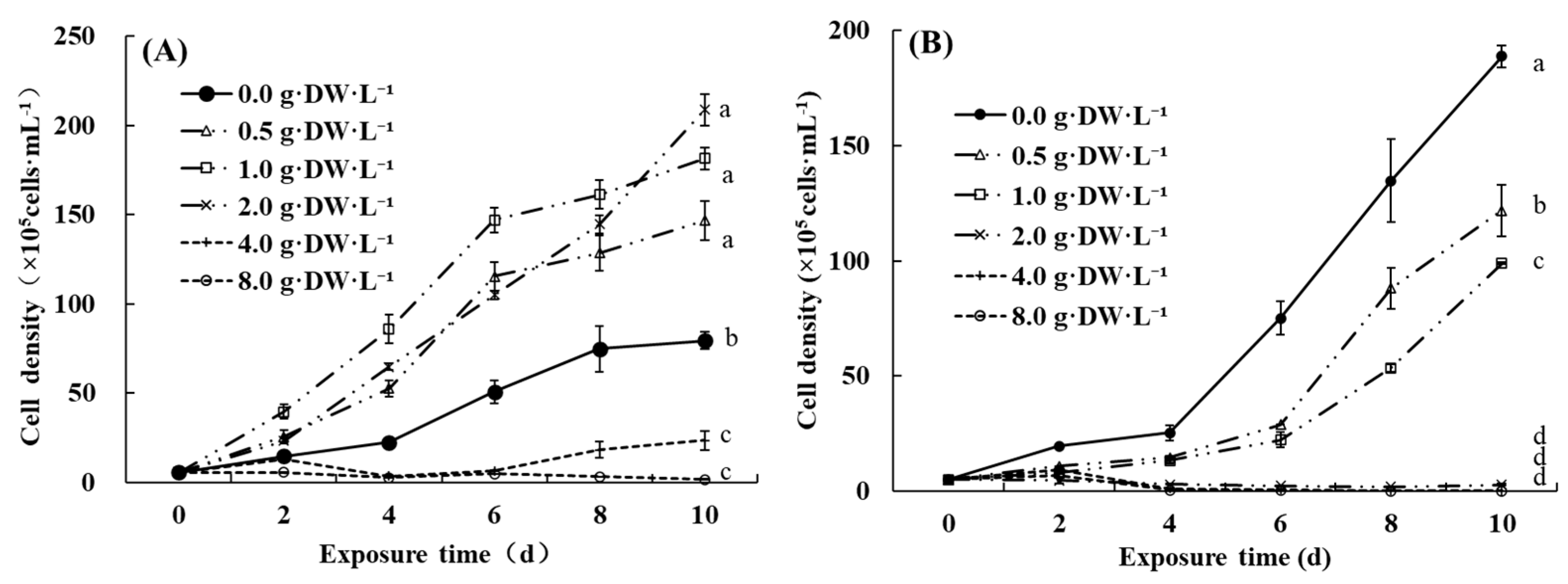
3.2. Species-Specific Effects of Aqueous Extracts from D. indicum Flowers

During the 3-day bioassays, the responses of two green algae strains and two cyanobacterial strains to aqueous extracts from D. indicum flowers differed significantly (p < 0.05). The daily growth rate, chl a, and carotenoid contents of Kirchneriella sp. and H. pluvialis were promoted significantly compared to the control, whereas those of toxigenic and non-toxigenic M. aeruginosa were inhibited significantly by the aqueous extracts from D. indicum flowers (Figure 2A–C). The promotion ratio of Kirchneriella sp. and H. pluvialis by the plant extracts was 126.4% and 58.6% based on the daily growth rate, 40.4% and 72.9% based on chl a content, and 35.5% and 84.5% based on carotenoid contents. The inhibition ratio of toxigenic and non-toxigenic M. aeruginosa by the plant extracts was 33.3% and 31.6% based on daily growth rate, 20.7% and 13.8% based on chl a content, and 23.5% and 16.7% based on carotenoid contents (Figure 2D).

3.3. Effects of Water Extraction Temperature on Cyanoabcterial Inhibition by Aqueous Extracts of D. indicum Flowers

There was no significant difference in the responses of M. aeruginosa to water-soluble substances extracted from D. indicum flowers in normal temperature water (25 °C) or boiling water (100 °C) during a 9-day bioassay (p > 0.05, Figure 3). The cell density and chl a concentration of M. aeruginosa in the control increased 27-fold and 14-fold from day 0 to day 9, respectively. However, the cell density and chl a concentration of M. aeruginosa treated with both kinds of plant aqueous extracts increased only twofold (Figure 3A,B). The corresponding inhibition ratios increased over time for both treatments. The inhibition ratios of the plant extracts prepared with water at 25 °C and 100 °C reached up to 93.1% and 93.6% compared to the cell density in the control, and 86.1% and 89.1% relative to the chl a concentration of M. aeruginosa in the control on day 9 (Figure 3C,D).
The fast chlorophyll fluorescence induction curves of M. aeruginosa treated with aqueous extracts of D. indicum flowers using water at 25 °C and 100 °C showed no significant difference (p > 0.05), but were significantly lower than that in the control (p < 0.05, Figure 4). The fluorescence intensity at 2 ms (Fj) and 30 ms (Fi) and the maximum fluorescence intensity (Fp) of M. aeruginosa cells in the control increased 5.1, 5.3, and 5.8-fold from day 3 to day 9. However, the extent of the increase of the corresponding values was lower than 2.0-fold in both treatments. The gap between the control and the treatments increased over time. Compared to the control, the Fp values decreased by 79.65% and 84.22% in the treatments with normal temperature water (25 °C) and boiling water (100 °C) on day 9, respectively.
The parameters extracted from OJIP curves using JIP test analysis, including Fv/Fm, ψo, φEo, and PIABS of M. aeruginosa cells in both treatments, showed no obvious differences (p > 0.05), but were significantly lower than those in the control (p < 0.05) after 9 days. The ETo/RC of M. aeruginosa cells in both treatments was also significantly lower than that in the control (p < 0.05). The values of ABS/RC and DIo/RC of M. aeruginosa in both treatments were higher than those in the control, and the differences between the control and the treatments increased over time. However, there was no significant difference in TRo/RC between the control and the treatments (Table 2).

3.4. Effects of Eluting Solvents on Cyanoabcterial Inhibition by SPE-Enriched Aqueous Extracts of D. indicum Flowers

SPE-enriched aqueous extracts of D. indicum flowers were eluted with five solvents with increasing polarities, i.e., hexane, dichloromethane, ethyl acetate, acetone, and methanol, in sequence. The obtained five eluting fractions showed polarity-dependent inhibition effects on M. aeruginosa (Figure 5). The cell density of M. aeruginosa in the control increased 33-fold over 9 days, 43-fold when treated with the hexane fraction, eightfold with the dichloromethane fraction, fivefold with the ethyl acetate fraction, threefold with the acetone fraction, and twofold with the methanol fraction (Figure 5A). The chl a concentration of M. aeruginosa in the control increased 10-fold for 9 days, 12-fold for the hexane fraction, eightfold for the dichloromethane fraction, sevenfold for the ethyl acetate fraction, threefold for the acetone fraction, and twofold for the methanol fraction (Figure 5B). The hexane fraction showed significant promotion effects on M. aeruginosa, and the maximum promotion ratio was 28.9% and 38.6% in terms of cell density and chl a content, respectively, on day 9. However, the inhibition ratio of the other four eluting fractions was above 50% from day 3 and increased over time. The inhibition ratio of the methanol fraction on the cell density and chl a content of M. aeruginosa was the highest, 94.9% and 85.6%, on day 9 (Figure 5C,D).
The polarity-dependent inhibition effects of aqueous extracts from D. indicum flowers on the fast chlorophyll fluorescence induction curves of M. aeruginosa were also observed (Figure 6). The Fj, Fi, and Fp values of the typical OJIP curves of M. aeruginosa in the control increased 5.2, 5.2, and 4.8-fold from day 3 to day 9. The addition of the hexane fraction promoted the chlorophyll fluorescence intensity of M. aeruginosa compared to the control. However, the other four fractions showed strong inhibition effects, and the inhibition degree enhanced with the polarity of the solvents. The differences increased with time. The Fp value increased by 5.04% for the hexane fraction, and decreased by 60.51% for the dichloromethane fraction, 71.97% for the ethyl acetate fraction, 85.62% for the acetone fraction, and 92.32% for the methanol fraction by day 9.
The parameters extracted from the OJIP curves of M. aeruginosa cells were affected by eluting fractions of D. indicum flowers (Table 3). There were no significant differences in Fv/Fm values between the control and the hexane fraction (p > 0.05). The Fv/Fm values of M. aeruginosa cells treated with the other four eluting fractions showed obvious polarity-dependent decreases on day 6 and day 9 and were significantly lower than that in the control (p < 0.05). The Fv/Fm value of M. aeruginosa cells treated with the methanol fraction was the lowest, as it decreased by 28.50% on day 3, 29.11% on day 6, and 32.01% on day 9. The ψo value of M. aeruginosa for the control and hexane fraction was significantly higher than that for the other four fractions (p < 0.05). It increased slightly for all the treatments from day 3 to day 9. The φEo value of M. aeruginosa cells in the control and hexane fraction showed no significant differences (p > 0.05) but were significantly higher than those in the other four fractions (p < 0.05). It increased slightly for the four eluting fractions with higher polarity from day 3 to day 9, and the polarity-dependent inhibition was obvious on day 6 and day 9. The ABS/RC and DIo/RC values did not show strict polarity-dependent tendencies, but they were the highest in the methanol fraction. The values of TRo/RC and ETo/RC of M. aeruginosa for the dichloromethane fraction and ethyl acetate fraction were the lowest, but they were not significantly different from those for the acetone and methanol fractions in most cases (p > 0.05). The PIABS values of M. aeruginosa showed decreasing trends, along with an increase in solvent polarity, with the lowest for the methanol fraction on day 6 and day 9. The inhibition ratio of PIABS values in M. aeruginosa cells by the methanol fraction was 76.84% on day 3, 81.24% on day 6, and 72.02% on day 9 compared to that in the control.

4. Discussion

4.1. Selective Growth Inhibition of Cyanobacteria and the Promotion of Green Algae by Aqueous Extracts of D. indicum Flowers

A high phytoplankton abundance with a healthy community structure is necessary to improve fishery productivity and the water environment of aquaculture ponds [32,33]. Mechanical removal, water change, engineering measures, and investment in chemical algicides are normally used to control harmful cyanobacteria blooms occurring in the middle and late stages of pond culture [15,34,35]. Nutrient enrichment and the addition of beneficial algae are traditionally used to improve the phytoplankton abundance and structure during the early stage of aquaculture [36,37]. The present study provides a new way to promote green algae and inhibit M. aeruginosa simultaneously by aqueous extracts of D. indicum flowers. The anti-inflammatory and antimicrobial activities of D. indicum extract have been reported [38,39], and our findings reveal its potential to selectively control harmful cyanobacteria and regulate phytoplankton composition to improve fishery productivity in aquaculture ponds.
The concentration-dependent inhibition effects of the aqueous extracts of D. indicum flowers on M. aeruginosa were found in our bioassays. It is consistent with previous studies showing the concentration-dependent allelopathic effects of aquatic plants on cyanobacteria [40]. Their promotion effects on the green algae C. vulgaris were obvious with a slightly enhanced trend when the flower concentration increased from 0.5 to 2.0 g·DW·L−1, whereas their concentration-dependent inhibition effects on C. vulgaris were obvious when the flower concentration ranged from 4.0 to 8.0 g·DW·L−1. These results indicated that the added dose played a critical role in the selective inhibition of harmful cyanobacteria by the aqueous extracts of D. indicum flowers. The concentration range of the plant extracts to exhibit the selective inhibition of harmful cyanobacteria and the promotion of beneficial eukaryotic algae needs to be ascertained at larger and longer timescales to ensure their successful application in the restoration of healthy phytoplankton communities in aquaculture ponds.

4.2. Chemical Characteristics of the Selective Inhibition Substances of D. indicum Flowers

The significant inhibition of M. aeruginosa and the promotion of green algae by aqueous extracts of D. indicum flowers were observed under identical culture conditions, excluding nutrients and light limitation or the impacts of any coexisting organisms. It indicated that there must be some water-soluble active substances derived from D. indicum flowers contributing to the selective inhibition effects on M. aeruginosa. There was no significant difference in inhibition effects on M. aeruginosa by aqueous extracts prepared with water at 25 °C and 100 °C. It indicated that the selective inhibition substances of D. indicum flowers were thermally stable. The methanol fraction eluted from SPE-enriched aqueous extracts of D. indicum flowers showed the strongest inhibition on M. aeruginosa compared to the other four fractions. It indicated that the most polar substances of D. indicum flowers were responsible for the selective inhibition of M. aeruginosa. These results together provided clues to reveal the basic chemical characteristics of selective inhibition substances of D. indicum flowers for M. aeruginosa. It was very helpful for us to identify the specific substances and their application in the ecological control of harmful cyanobacteria and the regulation of phytoplankton composition. Duddleoside, luteolin, chlorogenic acid, caffeic acid, and 3,5-dicaffeoylquinic acid were typical active substances of D. indicum flowers [41]. Flavonoids, including linatin, luteolin, quercetin, and apigenin, were detected in D. indicum flowers from various locations in China [42]. Luteolin showed the strongest inhibition effects on the growth and photosynthesis activity of M. aeruginosa from among the selected three flavonoids [43]. Whether these co-existing substances contribute to the selective inhibition of cyanobacteria needs further research.

4.3. Photosynthetic Activity of M. aeruginosa Treated with Water-Soluble Substances of D. indicum Flowers

The chlorophyll fluorescence transients and subsequent JIP test have been widely used to reflect the status and behavior of PSII in phototrophs, including plants and phytoplankton [31,44]. The obvious decrease in the fluorescence transients of M. aeruginosa treated with aqueous extracts of D. indicum flowers indicated that the successive reduction in the electron acceptor pools of PSII, including QA, QB, and PQ pool, was seriously affected by water soluble substances extracted from D. indicum flowers [31]. The parameter Fv/Fm, the maximum quantum yield of primary photochemistry, has been widely used to reflect the potential photosynthetic capacity of PSII in phototrophs [31,43]. The inhibition ratios of the Fv/Fm values of M. aeruginosa by aqueous extracts of D. indicum flowers and the methanol eluting fractions were higher than 20% in most cases. The parameter PIABS reflects the performance index on an absorption basis, which is the combination of three independent parameters, including RC/ABS, Fv/Fm, and ψo [31]. Its values decreased by more than 50% in the treatments with aqueous extracts of D. indicum flowers and the methanol eluting fractions. The extent of the change in PIABS was more consistent with the extent of the change in cell density and chl a of M. aeruginosa than Fv/Fm, which was also found in previous literature [44]. The significant decrease in ψo, φEo, and ETo/RC indicated that the performance of electron transfer from QA to QB was seriously inhibited by polar active substances of D. indicum flowers, and the acceptor side of the electron transport of PSII was a target site. The parameter ABS/RC reflects absorption flux per reaction center [45]. The increase in ABS/RC for M. aeruginosa treated with aqueous extracts of D. indicum flowers and the methanol fraction indicated the changes in the RC number. Since the trapped energy flux per reaction center (TRo/RC) did not change significantly, the decrease in the electron transport flux per reaction center (ETo/RC) corresponded to the increase in the dissipated energy flux per reaction center (DIo/RC) [31].

4.4. The Application of Water-Soluble Substances of D. indicum Flowers in Aquaculture Ponds

The present study provided the possibility to use water-soluble substances of D. indicum flowers to selectively control harmful M. aeruginosa proliferation and regulate phytoplankton community structure in aquaculture ponds. However, verifying this possibility fully and revealing the underlying mechanisms are necessary before its final application. The cyanobacteria proportion in total phytoplankton density treated with aqueous extracts of D. indicum flowers was obviously reduced in our outdoor mesocosms, but the dissolved carbon, nitrogen, and phosphorus concentrations were increased compared to the control (unpublished data). It indicated that a more ecological approach to regulate phytoplankton composition using D. indicum flowers was to input the specific responsible substances in aqueous extracts of D. indicum flowers in aquaculture ponds. Hence, identifying specific active substances was a crucial step to promote its application.
The well-known benefits to human health and antimicrobial activity also provided a possibility of D. indicum flowers to prevent and cure fish disease and promote the quality of aquatic products [28,29], which needs to be verified experimentally using specific substances isolated from D. indicum flowers. Medicinal plant derivatives have been widely used as immunostimulants [46]. Once the responsible substances isolated from D. indicum flowers were proved to be eco-friendly cyanocides, enough substance sources for wide application are necessary. In addition to extraction from D. indicum flowers, more plant sources need to be screened, and synthetic analogs are also feasible. Some plant-derived active compounds for medical, agronomic, and environmental applications have been successfully synthesized and commercially obtained [12,31].

5. Conclusions

The present study demonstrated the strong growth inhibition of toxigenic and non-toxigenic M. aeruginosa and the obvious growth promotion of green algae, including C. vulgaris, Kirchneriella sp., and H. pluvialis, by aqueous extracts of D. indicum flowers. The responsible substances of D. indicum flowers were thermally stable with high polarity. The acceptor side of the electron transport of PSII was a target site of M. aeruginosa cells in response to water-soluble substances of D. indicum flowers. More experiments are required to identify the responsible active substances and reveal the underlying mechanisms of aqueous extracts of D. indicum flowers to selectively inhibit cyanobacteria and regulate the phytoplankton community structure.

Author Contributions

Conceptualization, Y.G.; methodology, F.Z., J.W. and H.Y.; software, F.Z.; validation, J.D. and M.Z.; formal analysis, Y.G. and F.Z.; writing, Y.G., F.Z. and X.L.; supervision and funding acquisition, Y.G. and X.L. All authors have read and agreed to the published version of the manuscript.

Funding

This research was funded by the National Natural Science Foundation of China (grant number 31700405), the Major Science and Technology Program in Henan Province (grant number 202102310272 and 202102310028), and the Breeding Project of Henan Normal University (grant number HNU20180073).

Conflicts of Interest

The authors declare no conflict of interest.

References

  1. Paerl, H.W.; Otten, T.G. Harmful Cyanobacterial Blooms: Causes, Consequences, and Controls. Microb. Ecol. 2013, 65, 995–1010. [Google Scholar] [CrossRef]
  2. Huisman, J.; Codd, G.A.; Paerl, H.W.; Ibelings, B.W.; Verspagen, J.M.H.; Visser, P.M. Cyanobacterial blooms. Nat. Rev. Microbiol. 2018, 16, 471–483. [Google Scholar] [CrossRef] [PubMed]
  3. Xiao, M.; Li, M.; Reynolds, C.S. Colony formation in the cyanobacterium Microcystis. Biol. Rev. 2018, 93, 1399–1420. [Google Scholar] [CrossRef] [PubMed] [Green Version]
  4. Sinden, A.; Sinang, S.C. Cyanobacteria in aquaculture systems: Linking the occurrence, abundance and toxicity with rising temperatures. Int. J. Environ. Sci. Technol. 2016, 13, 2855–2862. [Google Scholar] [CrossRef]
  5. Rimando, A.M.; Schrader, K.K. Off-Flavors in Aquaculture; American Chemical Society: Washington, DC, USA, 2003. [Google Scholar]
  6. Dai, R.; Wang, P.; Jia, P.; Zhang, Y.; Chu, X.; Wang, Y. A review on factors affecting microcystins production by algae in aquatic environments. World J. Microbiol. Biotechnol. 2016, 32, 1–7. [Google Scholar] [CrossRef]
  7. Gobler, C.J.; Burkholder, J.A.M.; Davis, T.W.; Harke, M.J.; Johengen, T.; Stow, C.A.; Van de Waal, D.B. The dual role of nitrogen supply in controlling the growth and toxicity of cyanobacterial blooms. Harmful Algae 2016, 54, 87–97. [Google Scholar] [CrossRef]
  8. Paerl, H.W.; Xu, H.; McCarthy, M.J.; Zhu, G.; Qin, B.; Li, Y.; Gardner, W.S. Controlling harmful cyanobacterial blooms in a hyper-eutrophic lake (Lake Taihu, China): The need for a dual nutrient (N & P) management strategy. Water Res. 2011, 45, 1973–1983. [Google Scholar]
  9. Mitrovic, S.M.; Hardwick, L.; Dorani, F. Use of flow management to mitigate cyanobacterial blooms in the Lower Darling River, Australia. J. Plankton Res. 2011, 33, 229–241. [Google Scholar] [CrossRef]
  10. Visser, P.M.; Ibelings, B.W.; Bormans, M.; Huisman, J. Artificial mixing to control cyanobacterial blooms: A review. Aquat. Ecol. 2016, 50, 423–441. [Google Scholar] [CrossRef] [Green Version]
  11. Matthijs, H.C.P.; Visser, P.M.; Reeze, B.; Meeuse, J.; Slot, P.C.; Wijn, G.; Talens, R.; Huisman, J. Selective suppression of harmful cyanobacteria in an entire lake with hydrogen peroxide. Water Res. 2012, 46, 1460–1472. [Google Scholar] [CrossRef]
  12. Matthijs, H.C.P.; Jančula, D.; Visser, P.M.; Maršálek, B. Existing and emerging cyanocidal compounds: New perspectives for cyanobacterial bloom mitigation. Aquat. Ecol. 2016, 50, 443–460. [Google Scholar] [CrossRef] [Green Version]
  13. Van Wichelen, J.; Vanormelingen, P.; Codd, G.A.; Vyverman, W. The common bloom-forming cyanobacterium Microcystis is prone to a wide array of microbial antagonists. Harmful Algae 2016, 55, 97–111. [Google Scholar] [CrossRef] [PubMed]
  14. Xie, P.; Liu, J. Practical success of biomanipulation using filter-feeding Fish to control cyanobacteria blooms: A synthesis of decades of research and application in a subtropical hypereutrophic lake. Sci. World J. 2001, 1, 337–356. [Google Scholar] [CrossRef] [PubMed] [Green Version]
  15. Zhong, F.; Gao, Y.; Yu, T.; Zhang, Y.; Xu, D.; Xiao, E.; He, F.; Zhou, Q.; Wu, Z. The management of undesirable cyanobacteria blooms in channel catfish ponds using a constructed wetland: Contribution to the control of off-flavor occurrences. Water Res. 2011, 45, 6479–6488. [Google Scholar] [CrossRef]
  16. Blanchard, J.L.; Watson, R.A.; Fulton, E.A.; Cottrell, R.S.; Nash, K.L.; Bryndum-Buchholz, A.; Büchner, M.; Carozza, D.A.; Cheung, W.W.L.; Elliott, J.; et al. Linked sustainability challenges and trade-offs among fisheries, aquaculture and agriculture. Nat. Ecol. Evol. 2017, 1, 1240–1249. [Google Scholar] [CrossRef] [PubMed]
  17. Zhu, J.; Liu, B.; Wang, J.; Gao, Y.; Wu, Z. Study on the mechanism of allelopathic influence on cyanobacteria and chlorophytes by submerged macrophyte (Myriophyllum spicatum) and its secretion. Aquat. Toxicol. 2010, 98, 196–203. [Google Scholar] [CrossRef] [PubMed]
  18. Švanys, A.; Paškauskas, R.; Hilt, S. Effects of the allelopathically active macrophyte Myriophyllum spicatum on a natural phytoplankton community: A mesocosm study. Hydrobiologia 2013, 737, 57–66. [Google Scholar] [CrossRef]
  19. Le Rouzic, B.; Thiébaut, G.; Brient, L. Selective growth inhibition of cyanobacteria species (Planktothrix agardhii) by a riparian tree leaf extract. Ecol. Eng. 2016, 97, 74–78. [Google Scholar] [CrossRef]
  20. Tanuja, P.; Venugopal, N.; Sashidhar, R.B. Development and evaluation of thin-layer chromatography-digital image-based analysis for the quantitation of the botanical pesticide azadirachtin in agricultural matrixes and commercial formulations: Comparison with ELISA. J. AOAC Int. 2007, 90, 857–863. [Google Scholar] [CrossRef] [Green Version]
  21. Tu, Y.Y. Artemisia Annua and Artemisinin-Based Drugs; Chemical Industry Press: Beijing, China, 2009. [Google Scholar]
  22. Van Hai, N. The use of medicinal plants as immunostimulants in aquaculture: A review. Aquaculture 2015, 446, 88–96. [Google Scholar] [CrossRef]
  23. Zhou, K.; Guo, W.; Xu, Y. Advances of research on allelopathic potential in Compositae. Acta Ecol. Sin. 2004, 24, 1776–1784. [Google Scholar]
  24. Rasdi, N.H.M.; Samah, O.A.; Sule, A.; Ahmed, Q.U. Antimicrobial studies of Cosmos caudatus Kunth. (Compositae). J. Med. Plants Res. 2010, 4, 669–673. [Google Scholar]
  25. Bukhari, I.A. The central analgesic and anti-inflammatory activities of the methanolic extract of Carthamus oxycantha. J. Physiol. Pharmacol. 2013, 64, 369–375. [Google Scholar] [PubMed]
  26. Gao, Y.N.; Liu, B.Y.; Ge, F.J.; He, Y.; Lu, Z.Y.; Zhou, Q.H.; Zhang, Y.Y.; Wu, Z.B. Joint effects of allelochemical nonanoic acid, n-phenyl-1-naphtylamine and caffeic acid on the growth of Microcystis aeruginosa. Allelopath. J. 2015, 35, 249–258. [Google Scholar]
  27. Xu, F.; He, W.; Zheng, X.; Zhang, W.; Cai, W.; Tang, Q. Inhibitive effects on Microcystis aeruginosa by Artemisia lavandulaefoli and its three organic solvents extracts. Acta Ecol. Sin. 2010, 30, 745–750. [Google Scholar]
  28. Fang, H.; Guo, Q.; Shen, H. Study on antioxidation ability of essential oil from Dendranthema indicum dry flower in five edible oils. J. Plant Resour. Environ. 2010, 19, 54–59. [Google Scholar]
  29. Liu, X.Q.; Zhang, X.F.; Lee, K.S. Antimicrobial activity of the extracts of Forsythia suspensa and Dendranthema indicum. J. Appl. Biol. Chem. 2005, 48, 29–31. [Google Scholar]
  30. Lichtenthaler, H.K.; Buschmann, C. Chlorophylls and carotenoids: Measurement and characterization by UV-VIS spectroscopy. Curr. Protoc. Food Anal. Chem. 2001, 1, F4.3.1–F4.3.8. [Google Scholar] [CrossRef]
  31. Ni, L.X.; Acharya, K.; Hao, X.Y.; Li, S.Y.; Li, Y.; Li, Y.P. Effects of artemisinin on photosystem II performance of Microcystis aeruginosa by in vivo chlorophyll fluorescence. Bull. Environ. Contam. Toxicol. 2012, 89, 1165–1169. [Google Scholar] [CrossRef]
  32. Fan, L.; Xu, P.; Wu, W.; Qu, J.; Qiu, L.; Chen, J. Regulation of micro-ecological environment in freshwater aquaculture pond: A review. Chin. J. Ecol. 2013, 32, 3094–3100. [Google Scholar]
  33. Daume, S. The roles of bacteria and micro and macro algae in abalone aquaculture: A review. J. Shellfish Res. 2006, 25, 151–157. [Google Scholar] [CrossRef]
  34. Cong, H.; Sun, F.; Wu, J.; Zhou, Y.; Yan, Q.; Ren, A.; Xu, H. Study on method and mechanism of deep well circulation for the growth control of Microcystis in aquaculture pond. Water Sci. Technol. 2017, 75, 2692–2701. [Google Scholar] [CrossRef] [PubMed]
  35. Smith, J.L.; Boyer, G.L.; Zimba, P.V. A review of cyanobacterial odorous and bioactive metabolites: Impacts and management alternatives in aquaculture. Aquaculture 2008, 280, 5–20. [Google Scholar] [CrossRef]
  36. Qin, J.; Culver, D.A.; Yu, N. Effect of organic fertilizer on heterotrophs and autotrophs: Implications for water quality management. Aquac. Res. 1995, 26, 911–920. [Google Scholar] [CrossRef]
  37. Terziyski, D.; Grozev, G.; Kalchev, R.; Stoeva, A. Effect of organic fertilizer on plankton primary productivity in fish ponds. Aquac. Int. 2007, 15, 181–190. [Google Scholar] [CrossRef]
  38. Lee, D.Y.; Choi, G.; Yoon, T.; Cheon, M.S.; Choo, B.K.; Kim, H.K. Anti-inflammatory activity of Chrysanthemum indicum extract in acute and chronic cutaneous inflammation. J. Ethnopharmacol. 2009, 123, 149–154. [Google Scholar] [CrossRef]
  39. Shunying, Z.; Yang, Y.; Huaidong, Y.; Yue, Y.; Guolin, Z. Chemical composition and antimicrobial activity of the essential oils of Chrysanthemum indicum. J. Ethnopharmacol. 2005, 96, 151–158. [Google Scholar] [CrossRef]
  40. Chen, X.; Huang, G.; Fu, H.; An, C.; Yao, Y.; Cheng, G.; Suo, M. Allelopathy inhibitory effects of Hydrodictyon reticulatum on Chlorella pyrenoidosa under co-culture and liquor-cultured conditions. Water 2017, 9, 416. [Google Scholar] [CrossRef] [Green Version]
  41. Zhang, L.; Dai, Q. Determination of active ingredients in flos Chrysanthemi Indici. Guangdong Chem. Ind. 2019, 46, 195–196. [Google Scholar]
  42. He, D.; Zhang, W.; Qin, M. Content analysis of flavonoids in Dendranthema indicum flower from different locations by HPLC. J. Plant Resour. Environ. 2009, 18, 91–93. [Google Scholar]
  43. Huang, H.; Xiao, X.; Ghadouani, A.; Wu, J.; Nie, Z.; Peng, C.; Xu, X.; Shi, J. Effects of natural flavonoids on photosynthetic activity and cell integrity in Microcystis aeruginosa. Toxins 2015, 7, 66–80. [Google Scholar] [CrossRef] [PubMed] [Green Version]
  44. Zhang, L.; Peng, X.; Liu, B.; Zhang, Y.; Zhou, Q.; Wu, Z. Effects of the decomposing liquid of Cladophora oligoclona on Hydrilla verticillata turion germination and seedling growth. Ecotoxicol. Environ. Saf. 2018, 157, 81–88. [Google Scholar] [CrossRef] [PubMed]
  45. Appenroth, K.J.; Stöckel, J.; Srivastava, A.; Strasser, R.J. Multiple effects of chromate on the photosynthetic apparatus of Spirodela polyrhiza as probed by OJIP chlorophyll a fluorescence measurements. Environ. Pollut. 2001, 115, 49–64. [Google Scholar] [CrossRef]
  46. Vaseeharan, B.; Thaya, R. Medicinal plant derivatives as immunostimulants: An alternative to chemotherapeutics and antibiotics in aquaculture. Aquac. Int. 2014, 22, 1079–1091. [Google Scholar] [CrossRef]
Figure 1. The cell density changes of C. vulgaris (A) and M. aeruginosa (B) exposed to varying concentrations of aqueous extracts of D. indicum flowers (g·DW·L−1) in mono-culture bioassays. Different letters indicate significant differences among varying groups at p < 0.05 level.
Water 12 02014 g001
Figure 2. Effects of water extracts from D. indicum flowers at 1.0 g·DW·L−1 on daily growth rate (A), chlorophyll a content (B), and carotenoid content (C) of Kirchneriella sp. (Kir.), H. pluvialis (Hae.), and non-toxigenic (N-Mic.) and toxigenic M. aeruginosa (T-Mic.), and the inhibition ratios according to the three parameters (D) during 3-day bioassays. * indicates significant differences between the control and the treatment at p < 0.05 level.
Water 12 02014 g002
Figure 3. Cell density (A) and chl a content (B) of M. aeruginosa in response to water-soluble substances extracted from D. indicum flowers at 2.0 g·DW·L−1 with normal temperature water (25 °C) and boiling water (100 °C), and the corresponding inhibition ratio in terms of cell density (C) and chl a content (D) in a 9-day bioassay. Different letters indicate significant differences among different groups at p < 0.05 level.
Water 12 02014 g003
Figure 4. The fast chlorophyll fluorescence induction curves of M. aeruginosa in response to water-soluble substances extracted from D. indicum flowers at 2.0 g·DW·L−1 with normal temperature water (25 °C) and boiling water (100 °C) during a 9-day bioassay. (A) day 0; (B) day 3; (C) day 6; (D) day 9.
Water 12 02014 g004
Figure 5. Cell density (A) and chl a content (B) of M. aeruginosa treated with the eluting fractions of SPE-enriched aqueous extracts from D. indicum flowers at 2.0 g·DW·L−1 and the corresponding inhibition ratio based on cell density (C) and chl a content (D) in a 9-day bioassay. Different letters indicate significant differences among different groups at p < 0.05 level.
Water 12 02014 g005
Figure 6. The fast chlorophyll fluorescence curves of M. aeruginosa treated with the eluting fractions of SPE-enriched aqueous extracts from D. indicum flowers at 2.0 g·DW·L−1. (A) Day 0; (B) day 3; (C) day 6; (D) day 9.
Water 12 02014 g006
Table 1. The formulae and illustrations of the selected JIP test parameters in the study.
ParametersFormulae and TermsIllustrations
VjVj = (Fj − Fo)/(Fm − Fo)Relative variable fluorescence intensity at the J-step
MoMo = 4(F300μs − Fo)/(Fm − Fo)Approximated initial slope of the fluorescence transient
Quantum efficiencies or flux ratios
Fv/FmFv/Fm = TRo/ABS= 1 − Fo/Fm = φPo Maximum quantum yield for primary photochemistry
ΨoΨo = ETo/TRo = (Fm − Fj)/(Fm − Fo)Probability that a trapped exciton moves an electron into the electron transport chain beyond QA (at t = 0)
φEoφEo = ETo/ABS= (Fm − Fj)/FmQuantum yield for electron transport (at t = 0)
Specific activities or specific fluxes
ABS/RCABS/RC = Mo·(1/Vj)·(1/φPo)Absorption flux per reaction center
TRo/RCTRo/RC = Mo·(1/Vj)Trapped energy flux per reaction center (at t = 0)
ETo/RCETo/RC = Mo·(1/Vj)·ΨoElectron transport flux per reaction center (at t = 0)
DIo/RCDIo/RC = (ABS/RC) − (TRo/RC)Dissipated energy flux per reaction center (at t = 0)
Performance indexes
PIABSPIABS = (RC/ABS)·[φPo/(1 − φPo)]·[Ψo/(1 − Ψo)]Performance index on absorption basis
Table 2. The JIP test parameters of M. aeruginosa in response to water-soluble substances extracted from D. indicum flowers at 2.0 g·DW·L−1 with normal temperature water (25 °C) and boiling water (100 °C) in a 9-day bioassay. Different letters indicate significant differences among the control and the treatments on the same day at p < 0.05 level.
ParametersTimeTreatments
Control25 °C100 °C
Fv/Fmday 30.44 ± 0.01 a 0.36 ± 0.01 b 0.37 ± 0.01 b
day 60.55 ± 0.01 a 0.36 ± 0.08 b 0.41 ± 0.04 b
day 90.54 ± 0.01 a 0.41 ± 0.03 b 0.41 ± 0.04 b
ψoday 30.61 ± 0.01 a0.55 ± 0.03 b 0.55 ± 0.02 b
day 60.67 ± 0.01 a0.49 ± 0.03 b0.47 ± 0.02 b
day 90.66 ± 0.02 a 0.58 ± 0.01 ab 0.53 ± 0.07 b
φEoday 30.27 ± 0.01 a 0.19 ± 0.01 b 0.21 ± 0.01 b
day 60.37 ± 0.01 a 0.18 ± 0.03 b 0.19 ± 0.02 b
day 90.35 ± 0.02 a 0.24 ± 0.02 b 0.21 ± 0.02 b
ABS/RCday 32.30 ± 0.17 a 2.86 ± 0.32 a 2.65 ± 0.15 a
day 61.72 ± 0.18 a2.72 ± 0.52 b 2.47 ± 0.25 ab
day 91.69 ± 0.06 a 2.26 ± 0.30 b 2.10 ± 0.19 ab
TRo/RCday 31.02 ± 0.09 a 1.02 ± 0.10 a 0.99 ± 0.05 a
day 60.94 ± 0.09 a 0.95 ± 0.07 a 1.00 ± 0.03 a
day 90.91 ± 0.02 a 0.92 ± 0.07 a 0.85 ± 0.11 a
ETo/RCday 30.62 ±0.05 a 0.55 ± 0.06 a 0.55 ± 0.04 a
day 60.63 ± 0.06 a0.47 ± 0.02 b0.47 ± 0.01 b
day 90.59 ± 0.01 a0.53 ± 0.03 b 0.45 ± 0.00 c
DIo/RCday 31.28 ±0.08 a 1.85 ± 0.22 b 1.66 ± 0.12 ab
day 60.78 ± 0.09 a 1.77 ± 0.56 b 1.47 ± 0.24 b
day 90.78 ± 0.04 a 1.34 ± 0.23 b 1.25 ± 0.14 b
PIABSday 30.54 ± 0.05 a 0.23 ± 0.04 b 0.28 ± 0.03 b
day 61.43 ± 0.22 a 0.21 ± 0.09 b 0.25 ± 0.06 b
day 91.32 ± 0.20 a 0.44 ± 0.13 b 0.38 ± 0.10 b
Table 3. The JIP test parameters of M. aeruginosa treated with the eluting fractions of SPE-enriched aqueous extracts from D. indicum flowers at 2.0 g·DW·L−1 in a 9-day bioassay. Different letters indicate significant differences among the control and the treatments on the same day at p < 0.05 level.
ParametersTimeTreatments
ControlHexaneDichloromethaneEthyl AcetateAcetoneMethanol
Fv/Fmday 30.53 ± 0.01 a0.52 ± 0.01 a0.38 ± 0.02 b0.37 ± 0.01 b0.40 ± 0.01 b0.38 ± 0.01 b
day 60.53 ± 0.01 a0.51 ± 0.01 a0.44 ± 0.00 b0.42 ± 0.01 b0.38 ± 0.01 c0.37 ± 0.01 c
day 90.53 ± 0.02 a0.51 ± 0.01 a0.50 ± 0.00 ab0.48 ± 0.01 b0.44 ± 0.01 c0.36 ± 0.01 d
ψoday 30.79 ± 0.01 a 0.77 ± 0.03 a 0.61 ± 0.01 c 0.66 ± 0.01 b 0.70 ± 0.01 b 0.68 ± 0.02 b
day 60.80 ±0.01 a0.81 ± 0.00 a 0.69 ± 0.01 cd 0.74 ± 0.01 b 0.71 ± 0.01 bc 0.66 ± 0.03 d
day 90.79 ± 0.03 a 0.81 ± 0.01 a 0.71 ± 0.01 b 0.75 ± 0.02 b 0.74 ± 0.00 b 0.74 ± 0.01 b
φEoday 30.42 ± 0.01 a 0.40 ± 0.02 a 0.23 ± 0.02 b 0.24 ± 0.01 b 0.28 ± 0.01 b 0.25 ± 0.00 b
day 60.42 ± 0.01 a 0.41 ± 0.01 a 0.31 ± 0.01 b 0.31 ± 0.01 b 0.27 ± 0.01 c 0.25 ± 0.01 d
day 90.37 ± 0.03 a 0.36 ± 0.01 a 0.36 ± 0.01 a 0.36 ± 0.01 a 0.32 ± 0.01 b 0.27 ± 0.00 c
ABS/RCday 32.43 ± 0.23 a 2.51 ± 0.25 a 3.03 ± 0.44 a 3.69 ± 0.74 a 2.88 ± 0.20 a 3.16 ± 0.21 a
day 62.36 ± 0.07 b 2.51 ± 0.09 b 2.40 ± 0.06 b 2.96 ± 0.04 ab 2.88 ± 0.34 ab 3.44 ± 0.54 a
day 92.43 ± 0.06 b 2.93 ± 0.12 b 2.16 ± 0.12 c 2.26 ±0.07 c2.71 ± 0.20 b 3.31 ± 0.08 a
TRo/RCday 31.28 ± 0.11 a 1.30 ± 0.13 a 1.16 ± 0.11 a 1.37 ± 0.28 a 1.16 ± 0.08 a 1.19 ± 0.10 a
day 61.24 ± 0.03 a 1.29 ± 0.04 a 1.06 ± 0.02 b 1.24 ± 0.05 a 1.10 ± 0.17 b 1.28 ± 0.22 a
day 91.24 ± 0.06 a 1.30 ± 0.02 a 1.08 ± 0.06 b 1.09 ± 0.03 b 1.19 ± 0.06 ab 1.19 ± 0.03 ab
ETo/RCday 31.01 ± 0.09 a 1.00 ± 0.13 a 0.71 ± 0.06 b 0.90 ± 0.18 a 0.81 ± 0.06 ab 0.80 ± 0.05 ab
day 60.99 ± 0.0 a 1.04 ± 0.03 a 0.73 ± 0.03 b 0.92 ± 0.04 ab 0.78 ± 0.11 b 0.84 ± 0.11 a
day 90.98 ± 0.08 a 1.05 ± 0.01 a 0.77 ± 0.04 b 0.82 ± 0.04 b 0.88 ± 0.04 ab 0.88 ± 0.02 ab
DIo/RCday 31.15 ± 0.12 a 1.21 ± 0.12 ab 1.87 ± 0.33 b 2.33 ± 0.46 b 1.71 ± 0.12 b 1.97 ± 0.11 b
day 61.12 ± 0.06 a 1.22 ± 0.07 a 1.34 ± 0.04 ab 1.72 ± 0.03 b 1.79 ± 0.17 bc 2.15 ± 0.32 c
day 91.42 ± 0.05 b 1.63 ± 0.09 b 1.08 ± 0.07 c 1.16 ± 0.04 c 1.52 ± 0.14 b 2.12 ± 0.06 a
PIABSday 31.73 ± 0.23 a1.43 ± 0.24 a0.33 ± 0.09 b0.32 ± 0.08 b0.55 ± 0.06 b0.40 ± 0.04 b
day 61.86 ± 0.14 a1.75 ± 0.12 a0.74 ± 0.04 b0.70 ± 0.05 b0.54 ± 0.06 b0.35 ± 0.10 c
day 91.73 ± 0.29 a1.13 ± 0.13 a1.13 ± 0.09 a1.08 ± 0.00 a0.82 ± 0.10 ab0.48 ± 0.01 b

