Article

Enhancing Biocontrol of Harmful Algae Blooms: Seasonal Variation in Allelopathic Capacity of Myriophyllum aquaticum

by
Rafael Shinji Akiyama Kitamura
1,*,
Ana Roberta Soares da Silva
2,
Thomaz Aurelio Pagioro
3 and
Lúcia Regina Rocha Martins
3,*
1
Department of Genetics, Federal University of Paraná, Curitiba 81530-980, Brazil
2
Management and Supervision Monitoring Division, Water and Earth Institute (IAT), Curitiba 80215-100, Brazil
3
Department of Chemistry and Biology, Federal University Technology of Parana, Curitiba 81280-340, Brazil
*
Authors to whom correspondence should be addressed.
Water 2023, 15(13), 2344; https://doi.org/10.3390/w15132344
Submission received: 25 May 2023 / Revised: 19 June 2023 / Accepted: 23 June 2023 / Published: 24 June 2023

Abstract

:
Myriophyllum aquaticum has shown potential allelopathic effects for the biocontrol of cyanobacteria and cyanotoxins. However, the composition of allelochemicals and their biological effects may be influenced by seasonal changes. In this study, we investigated the impact of aqueous extracts of M. aquaticum collected in different seasons on the growth of Microcystis aeruginosa and the concentration of microcystin-LR. Plant samples were extracted using ultrasound cycles in aqueous solutions, and extracts at varying concentrations (0.1, 10, and 100 mg/L) and a control treatment were inoculated with M. aeruginosa, and cell growth was analyzed using a Neubauer chamber. Photosynthetic pigment quantification was used to measure physiological effects and liquid chromatography was used to evaluate the microcystin-LR concentrations. The extracts of plants collected during autumn and winter exhibited higher inhibition of M. aeruginosa growth and a reduction in photosynthetic pigments compared to those collected during spring and summer. These results can be explained by the higher presence of phenolic compounds in the composition of extracts from autumn and winter. Microcystin-LR concentrations were decreased at 10 and 100 mg/L, with the highest efficiency observed in autumn, while spring showed lower efficiency. Our findings suggest that M. aquaticum extracts have inhibitory potential on M. aeruginosa, particularly during the autumn season, making them a promising nature-based solution for the biocontrol of harmful algal blooms.

1. Introduction

Cyanobacterial blooms can affect human health and cause economic and environmental problems [1]. This phenomenon promotes alterations in the trophic structure and local functionality of the ecosystem due to the deoxygenation of the water column and the production of cyanotoxins. These alterations decrease biodiversity and generate mortality of organisms, as well as other adverse effects [2,3,4]. Among the most common cyanobacteria that establish harmful algae blooms worldwide is the species Microcystis aeruginosa Kutzing. This species is mainly responsible for producing microcystins [2,5]. Microcystins are hepatotoxins that can be associated with the occurrence of tumors, histopathological changes, genotoxicity, biochemical alterations, and episodes of acute toxicity in different organisms such as fish, rats, and microcrustaceans [6,7,8].
Different strategies have been developed, and some adopted, for the control of cyanobacteria blooms, mainly in public supply reservoirs [4,9,10]. However, cyanotoxins are not completely removed by physical and chemical treatments. Moreover, these treatments can result in the production of other toxic products and the release of even more toxic metabolites [11]. Thus, the use of biological treatments becomes relevant because they present a low cost and cause lower impacts on ecosystems [11,12,13]. Among these treatments, the Nature-Based Solution (NbS) employing submerged macrophytes can contribute to the control of cyanobacteria blooms [14]. This solution is based on studies using the allelopathic potential of aquatic macrophytes on phytoplankton, mainly for the inhibition of cyanobacteria [12,15,16], which can be considered an important ecosystems service to water purification [17]. Allelopathy consists of the positive or negative effect that an organism exerts on others by the release of compounds in the environment, called allelochemicals [9,18,19]. The application of this technique has advantages, mainly in lowering toxicity for non-target organisms [19].
In this context, submerged aquatic macrophytes contribute to the maintenance of water bodies, mainly through local oxygenation, and the control of phytoplankton by releasing allelochemicals into the aquatic environment to reduce competition for light, carbon, and nutrients [9,15,17,18]. Among the species of submerged aquatic macrophytes, the genus Myriophyllum has been studied due to its allelopathic capacity in cyanobacteria, mainly through the production of allelochemicals [20,21,22,23,24]. The major class with an inhibitory potential on cyanobacteria is phenolic compounds [9,25,26]. In addition, it is known that the production of allelochemicals, mainly phenolic compounds, varies according to the seasons in the species of the genus Myriophyllum [26].
The use of aqueous extract can be an alternative to evaluate the allelopathic potential due to the extraction of more bioactive, water-soluble compounds of aquatic macrophytes [27], which can be more easily released into the aquatic environment. Thus, the aim of this work was to evaluate the effect of the aqueous extract of Myriophyllum aquaticum (Vell.) Verdc. on the growth and physiology of Microcystis aeruginosa, as well as its interference in the production capacity of microcystin-LR by comparing plants collected in different seasons. In this case, we hypothesized that plants collected in different seasons may show differences in total phenolic compound concentration and, consequently, in their allelopathic capacity to biocontrol cyanobacteria and cyanotoxins.

2. Materials and Methods

2.1. Culture of Microcystis aeruginosa

The Microcystis aeruginosa strain (code BB005—provided by the Botany Department of the Federal University of São Carlos, Brazil) was cultivated in the ASM-1 medium (Gorham et al. [28] with adaptations by De Almeida et al. [29]). For the initial inoculum a concentration of 1.6 × 10⁵ cell mL−1 was used. The cultures were maintained at controlled conditions of temperature (25 ± 2 °C), luminosity (36.81 ± 2.58 µmol of photons, m²·s−1), and photoperiod (12:12 h/light:dark).

2.2. Collection of Macrophytes

Specimens of Myriophyllum aquaticum were collected in the Rio Verde reservoir (25°90.81′ S 49°28.03′ W), located in the municipality of Araucaria, Paraná, Brazil. The samples were collected in different seasons: spring, autumn, winter and summer. In the laboratory, the fresh plant material was washed in running water and dried in a stove (30 °C) for 15 days. Dry biomass was crushed and granulometry was standardized (0.5 mm).

2.3. Preparation and Obtaining of Aqueous Extracts

The extract was prepared with distilled water as an extractor solution. After the granulometry was standardized, the material was weighed (20 g of powder) and diluted in 100 mL of extracting solution. Afterward, the solution was submitted to three extraction cycles in an ultrasonic bath, for a duration of 45 min each cycle, followed by filtration and renewal of the extractor liquid. The liquid extract was frozen at −80 °C and lyophilized (Liotop, Brazil). The lyophilized extract was stored at −20 °C, until the preparation of the solutions for the experiments to evaluate allelopathic activity to control the cyanobacteria [20].

2.4. Analysis of Total Phenolic Compounds of Plant Extracts

The determination of phenolic compounds was performed by the Folin-Ciocalteau method [30]. For the analysis, stock solutions of 100 mg·L−1 of the lyophilized extracts of M. aquaticum were prepared for each season. In a volumetric balloon (capacity of 10 mL), 5 mL of osmosis water, 150 μL of M. aquaticum extracts and 0.5 mL of Folin-Ciocalteu reagent (SigmaAldrich®, Sao Paulo, Brazil) were added, and the material was maintained for 3 min. Subsequently, 2 mL of sodium carbonate solution (Na2CO3) and osmosis water were added until the volume of 10 mL was completed. After preparation, all balloons were stored in the absence of light (to avoid oxidation of the reagents) for two hours. The analysis was performed by reading in a UV-Vis spectrophotometer (UV-1800®, SHIMADZU, Barueri, Brazil) at a wavelength of 765 nm. For the quantification of total phenolic compounds, the calibration curve (y = 0.4942x − 0.0106) was used from solutions of salicylic acid at concentrations of 0.05; 0.1; 0.2; 0.4; 0.6; 0.8 and 1 μg·mL−1 according to linear regression obtained from R² = 0.999.

2.5. Bioassay

The study used aqueous extracts of Myriophyllum aquaticum to test their allelopathic effects on Microcystis aeruginosa. The extracts were solubilized in ASM-1 medium, and the test solutions were prepared in three concentrations: 0.1, 10 and 100 mg·L−1. The bioassay was carried out in Erlenmeyer flasks containing 100 mL of the test solution or control (ASM-1 medium only), and inoculated with M. aeruginosa at an initial concentration of 106 cells·mL−1. The flasks were kept under controlled conditions of temperature, luminosity, and photoperiod. The M. aeruginosa growth was monitored by counting cells in a Neubauer chamber every 48 h for a period of 9 days. At the end of the experiment, samples were collected for cell viability tests, photosynthetic pigment analysis, and microcystin-LR concentration measurement.

2.6. Cyanobacteria Growth

Every 48 h, aliquots (10 µL) were collected to evaluate the M. aeruginosa growth by cellular counting in a Neubauer chamber. The inhibition rate (IR) was calculated according to Cheng et al. [31].
I R = 1 N 0 × N × 100
where N = density cells in exposure to extracts and N0 = density cells in the control group.

2.7. Evaluation of Cellular Viability

On the last day of the experiment, 1 mL samples were taken from the experimental flasks and transferred to new test tubes containing 9 mL of ASM-1 medium (n = 3). The cultures were maintained under controlled conditions of temperature, light, and photoperiod as described previously. After 10 days of incubation, the cell concentration was measured to determine if the extracts had an algicidal (growth inhibition) or algistatic effect (growth after treatment in a new culture medium without allelopathic compounds) [20].

2.8. Determination of Photosynthetic Pigments

To analyze chlorophyll-a, 6 mL aliquots were transferred to plastic tubes (15 mL capacity) and centrifuged at 10,500× g for four minutes at 20 °C using an ultra-centrifuge. The supernatant was discarded, and the pellet was resuspended in 2 mL of 80% (v/v) acetone after homogenization. The sample was kept refrigerated (4 °C) and in the absence of light for 24 h to extract pigments. Afterward, the sample was homogenized and centrifuged under the same conditions as before. The supernatant was analyzed using a UV-Vis spectrophotometer (UV-1800®, SHIMADZU) at wavelengths 646 and 663 nm. The concentration of chlorophyll-a was calculated using Equation (2) [32].
C h l o r o p h y l l a m g · L 1 = 12.21 × A 633 2.81 × ( A 646 )
In which: A633 = Absorbance value at 633 nm; A646 = Absorbance value at 646 nm.
The accessory pigments were analyzed from 10 mL aliquots collected on the last experimental day and stored in plastic tubes (15 mL capacity). Subsequently, the samples underwent 2 freezing/defrosting cycles to release the pigments into the supernatant [33]. Methodology adaptations were made to reduce the processing time, and centrifugation was performed at 10,500× g for 4 min in an ultracentrifuge.
The supernatant was collected, and its absorbance was measured at specific wavelengths for each accessory pigment using a UV-Vis spectrophotometer (UV-1800®, SHIMADZU): 565 nm for Phycoerythrin (PE), 620 nm for Phycocyanin (PC), and 650 nm for Allophycocyanin (APC). Additionally, the wavelength of 750 nm was used to check for the presence of cell fragment residues. The ASM-1 medium was used as a standard control for the analysis. Chromatic equations were used to estimate the concentrations of phycobiliproteins [34]. The values obtained from the wavelengths were subtracted from the measurement at 750 nm to eliminate any interference from cellular residues in the readings of the accessory pigments.
P C m g · m L 1 = At 620 0.72 × At 650 6.20
A P C   ( m g · m L 1 ) = At 650 0.191 × At 620 5.79
P E   ( m g · m L 1 ) = At 565 2.41 × P C ( 1.41 × A P C ) 13.02
In which: At620 = absorbance obtained at 620 nm wavelength; At650 = absorbance obtained at 650 nm wavelength; At565 = absorbance obtained at wavelength 565 nm.

2.9. Intracellular Microcystin-LR Quantification (MCLR)

The analysis was performed using a high-performance liquid chromatograph with a diode array detector (Prominence, Shimadzu®) equipped with a hybrid C18 column (XTerra®, Waters, Brazil) with dimensions of 150 × 3 mm and internal diameter of 3.4 μm particle size, maintained at 35 °C. The mobile phase consisted of solvent A (0.05% trifluoroacetic acid v/v) and solvent B (HPLC grade methanol, J.T Baker). The flow rate was 0.3 mL·min−1, and the temperature was maintained at 35 °C. The separation was performed under isocratic conditions with 50% solvent B for 50 min, followed by a cleaning gradient (50 to 100% solvent B in 2 min, maintained at 100% for 5 min), with a return to the initial condition for 2 min) [21,25]. The quantification method previously validated in the research group [35] analyzed the concentrations of MCLR from area values at 238 nm using linear regression (Equation (6)) with a regression coefficient of 0.9998.
y = 322.48 x 3.2831
In which: y = area of the peak; x = MCLR concentration (mg·L −1).
To estimate the intracellular concentration of MCLR, we standardized the measurement by the number of cells present at the end of each treatment and the cyanotoxin levels in the medium [25].

2.10. Data Analysis

The data were tested for normality (Shapiro–Wilk) and homoscedasticity (Levene) and evaluated using two-way ANOVA (cell growth, inhibition rate, photosynthetic pigments, cellular viability and microcystin-LR concentration). To compare the differences of total phenolic compounds, one-way ANOVA was used to evaluate differences between seasons. Interactions between extract concentrations (Control, 0.1, 10 and 100 mg·L−1) and seasons (spring, autumn, winter and summer) were included in the model and when differences were detected by ANOVA, the means were compared using the Tukey test, at a 0.05% level of significance for parametric data. The data were statistically analyzed using GraphPad PRISM, version 7.01 software.

3. Results

3.1. Content Total of Phenolic Compounds on Aqueous Extract

The total content concentrations of phenolic compounds in the extracts differed among seasons, with autumn and winter showing the highest concentrations when compared to summer and spring (p < 0.001; Table 1).

3.2. Inhibitory Effects of Aqueous Extract of M. aquaticum on M. aeruginosa Growth

The aqueous extracts influenced the growth of M. aeruginosa from the first day of the experiment, regardless of the tested extract concentration and the season (p < 0.001; Figure 1; Table S1). However, from the third experimental day, extracts obtained from plants collected in autumn showed significant reductions in M. aeruginosa cell growth when compared to other seasons, particularly at concentrations of 10 and 100 mg·L−1 (p < 0.001; Figure 1B; Table S1). Regardless of the extract concentration, the autumn and winter seasons had lower cellular concentrations on the final experimental day, compared to the summer and spring seasons (p < 0.001; Figure 1; Table S1).
These results are supported by the inhibition rates observed over time, which ranged from 17% to 96% on the final experimental day for the different seasons (Figure 2; Table S2). Starting from the 3rd experimental day, autumn had a significantly higher inhibition rate than the other seasons for the concentration of 10 mg·L−1 (p < 0.001, Figure 2B, Table S2). The time required for the extracts to cause the death of 50% of the cells of M. aeruginosa (LC50) was reached by the concentration of 100 mg·L−1 for all seasons from the 3rd experimental day. However, for the concentration of 10 mg·L−1, only the autumn season achieved this level of inhibition from the 5th experimental day until the last experimental day (p < 0.001, Figure 2, Table S2). Therefore, although the extracts of all seasons have high inhibiting potential, autumn stands out, particularly in inhibitions at the concentration of 10 mg·L−1, and spring is the season with the lowest efficiency compared to the other seasons.

3.3. Cellular Viability of M. aeruginosa after Exposure to Aqueous Extracts

There were significant effects on cell viability among the different extract concentrations and seasons (p < 0.0001; Table 2). The autumn season exhibited the highest algicide and algistatic effects in comparison to other seasons, particularly at concentrations of 10 and 100 mg·L−1 (p < 0.0001; Table 2). Furthermore, algistatic effects were observed at extract concentrations of 0.1 and 10 mg·L−1, while algicidal effects were observed at a concentration of 100 mg·L−1, regardless of the season assessed (Table 2).

3.4. Effects on Photosynthetic Pigments from M. aeruginosa

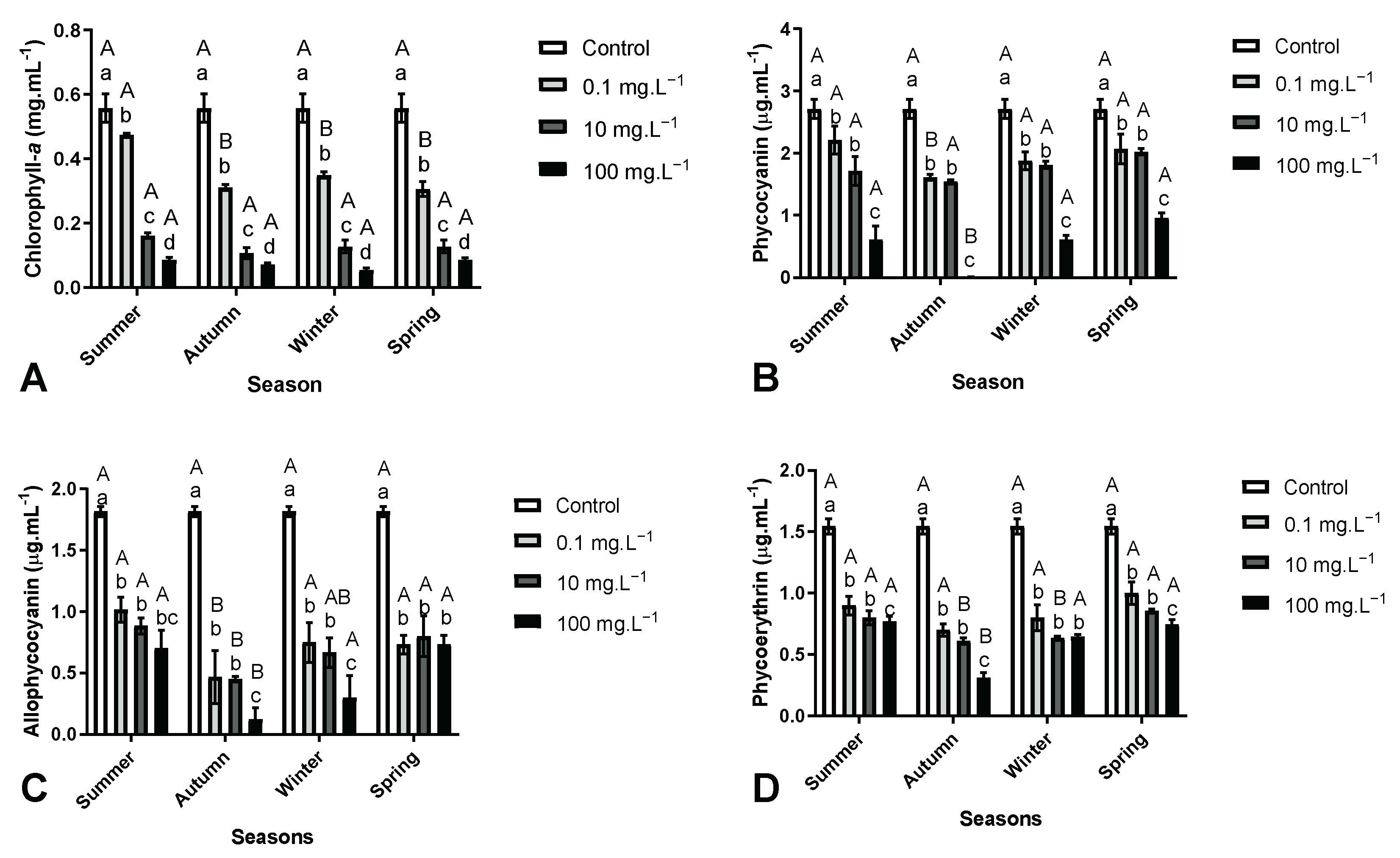
The concentrations of chlorophyll-a, allophycocyanin, and phycoerythrin were significantly affected by both the extract concentration and the season (p < 0.0001; Figure 3, Table S3). In all seasons, lower concentrations of these photosynthetic pigments were observed regardless of the extract concentration, when compared to the control group (p < 0.001; Figure 3, Table S3). Specifically, in the summer season, the chlorophyll-a content was lower for the concentration of 0.1 mg·L−1 when compared to other seasons (p < 0.001; Figure 3A, Table S3). Moreover, for the accessory pigments (phycocyanin, allophycocyanin, and phycoerythrin), lower concentrations were observed in the autumn and winter seasons, when compared to summer and spring (p < 0.001; Figure 3B–D, Table S3).

3.5. Concentration and Removal Efficiency of Intracellular MCLR

The concentration of intracellular MCLR decreased as the extract concentration increased, regardless of the season evaluated (p < 0.001, Table 3). The extracts from all seasons were efficient in removing MCLR, achieving 89% efficiency (p < 0.0001, Table 3). Autumn exhibited the best efficiency in removing microcystin-LR, especially at concentrations of 10 and 100 mg·L−1, compared to other seasons (p < 0.01, Table 3), while spring showed the lowest efficiency (p < 0.001, Table 3).

4. Discussion

The use of allelopathic applications from submerged aquatic macrophytes can be considered a promising strategy for controlling cyanobacteria. This technique is inspired by natural phenomena and offers a nature-based solution [15,18,24]. In this context, the use of the Myriophyllum genus is reported to show a higher presence of phenolic compounds in their phytochemical composition [22,23,24,26,36,37]. It has already been identified that phenolic compounds can be produced by macrophytes of this genus and may act as allelochemicals. Among the molecules that have shown allelopathic activities, the following can be evidenced: (+)-Catechin, caffeic acid, ellagic acid, gallic acid, nonanoic acid, pyrogallol, tellimagrandin II, phenylpropanoid glucosides, and different hydrolysable tannins [15]. Nakai et al. [21] observed that M. spicatum was responsible for inhibiting the growth of M. aeruginosa cells by releasing five polyphenols: catechin, eugeniin, and gallic, pyrogallic, and ellagic acids, and fatty acids in the culture medium. In in vivo experiments, the allelopathic potential of M. aquaticum on cyanobacteria has already been proven, evidencing its allelopathic capacity and its relationship with phenolic compounds. [22,23,24,36]. However, the phytochemical composition of macrophytes may vary depending on spatial and temporal factors, which can affect their allelopathic capacity to inhibit cyanobacteria [26,27,37] demanding a better comprehension of this dynamic. In this context, these findings are supported by the present work, which demonstrated significant differences in the content of total phenolic compounds in different seasons, with higher concentrations observed in winter and autumn (Table 1). Therefore, it was important to evaluate the allelopathic capacity of M. aquaticum in different seasons and understand the physiological effects it has against cyanobacteria and cyanotoxins.
It has been proven that there is variation in the content of total phenolic compounds during the seasonal dynamics of the life cycle of Myriophyllum verticillatum [26]. Bauer et al. [26] observed that in the colder months (dry seasons), there is a greater release of phenolic compounds in the apical parts of Myriophyllum spicatum, which may be associated with the inhibition of the cyanobacterium Anabaena flos-aquae. This information is consistent with the results obtained in the present study, as the dry season period (autumn) showed the highest inhibition rates, mainly at lower extract concentrations from the fifth experimental day (Figure 2; Table S2). The highest concentration of phenolic compounds can be associated with plant defense mechanisms due to the availability of nutrients and light in the environment, as well as defense mechanisms against predators and competitors for resources [18,26,37,38]. Thus, the allelochemicals produced by M. aquaticum can be related to inhibiting the cyanobacteria against competition for nutrients, light and space, mainly in periods of lower resources in the aquatic environment [15,23,27,39], as in the autumn and winter seasons.
The presence of allelochemicals, such as pyrogallic acid, extracted by an aqueous solution, can generate oxidative stress by increasing the content of hydrogen peroxide, which in turn can lead to lipid peroxidation and inhibit cell growth [15,22,40]. These possible effects can explain the observed reduction in cell growth of M. aeruginosa when exposed to aqueous extracts from different seasons (Figure 1, Table S1). Li et al. [40] observed that the use of aqueous extracts from the macrophytes Sagittaria trifolia caused oxidative stress, despite the increase in the antioxidant system of cyanobacteria. The author explains that a possible explanation for the death of cyanobacteria cells could be caused by oxidative stress.
Another possible explanation for the effects on cell growth and function is through interference with the photosynthetic apparatus, which can be related to the decrease in photosynthetic pigments as observed in the present study (Table 1). Tellimagrandin II is an allelochemical identified in the phytochemical composition of the Myriophyllum genus, and studies have demonstrated its effects on cyanobacterial growth, possibly due to its effects on the photosynthetic apparatus and pigments, such as chlorophyll-a and accessory pigments (phycocyanin, allophycocyanin, and phycoerythrin) [40,41]. The phycobilisomes are the core complexes that facilitate the conversion of solar energy into high-energy electrons in the photosystem II of cyanobacteria [42]. The pigment chlorophyll-a and accessory pigments such as phycoerythrin, phycocyanin, and allophycocyanin are essential for the reactions in photosynthesis [42]. When interferences occur on these pigments, they can generate inefficiencies and reductions in the photosynthetic potential of cyanobacteria, which highlights the importance of analyzing these pigments [24,42]. In this context, it has been proven that allelochemicals can cause effects on the photosynthetic apparatus. This has been shown in previous studies using a medium culture of Myriophyllum spicatum [36] and co-exposure to M. aquaticum and the cyanobacteria M. aeruginosa [24].
Phenolic compounds isolated from M. spicatum, such as tellimagrandin II, have been shown to affect photosystem II and degrade accessory pigments, causing interferences in metabolism and cell death by inhibiting enzymes such as alkaline phosphatase [43]. Other phenolic compounds, such as catechins, gallic acid, and ellagic acid extracted from M. spicatum, can also affect photosystem II in M. aeruginosa, promoting a reduction in electron transduction [44]. These phenolic compounds, when applied alone or in synergistic mechanisms, can have a greater impact on cyanobacteria, especially in co-exposure with pyrogallic and gallic acid [15,44]. The synergistic effects can explain the efficiency of aqueous extracts of M. aquaticum in inhibiting cyanobacteria growth and reducing photosynthetic pigment concentrations, as observed in the present study and in tests using co-exposure with plants [22,24]. In studies testing allelopathic potential, photosynthetic pigments and cell growth are considered efficient methods to screen the effects of allelochemicals in inhibiting cyanobacteria, either in isolation or in combination with other compounds [19,24].
The use of algicides can effectively inhibit the growth of cyanobacteria, but it may also induce cell lysis, resulting in the release of higher concentrations of intracellular microcystins [45]. However, the use of macrophyte extracts, as reported by Wu et al. [46], can effectively inhibit the growth of cyanobacteria and reduce the concentration of cyanotoxins. Kitamura et al. [20] demonstrated that methanolic extracts of M. aquaticum caused a reduction in microcystin-LR concentration and inhibited cell growth and photosynthetic pigments. Accessory pigments, such as phycocyanin, can act as photoprotectors of microcystins, and when their concentration is reduced or degraded [47], as observed in this study, it can increase the sensitivity of microcystins and increase the chances of degradation by photocatalysis [47,48]. Considering the observed physiological effects (growth and photosynthetic pigments) and the variation in phenolic compound concentration across different seasons, this may provide a possible explanation for the mechanism of microcystin degradation following the application of aqueous extracts of M. aquaticum, particularly during the autumn season, as indicated in Table 3.
The knowledge about the contribution of allelopathy activity in combating cyanobacteria using submerged macrophytes “in vivo” is attributed to the synergistic effects resulting from the combined action mechanisms of various phenolic compounds [24,26,36]. While our study only examined the total phenolic compounds, it is recommended that future research focuses on analyzing the phytochemical composition of plant extracts. This will provide valuable insights into how seasonality can affect the presence and quantity of different compounds which may possess bioactivity against cyanobacteria and influence the production of cyanotoxins. It is important to consider the seasonal variation in the allelopathic capacity of M. aquaticum when employing it for water remediation treatments aimed at mitigating harmful Microcystis blooms. Nevertheless, this study has demonstrated that allelopathy can be an effective technique for reducing microcystin-LR concentrations and controlling the growth and proliferation of harmful cyanobacteria, particularly those collected during the autumn season. Therefore, these findings encourage the use of M. aquaticum in water remediation treatments, as it has the potential to inhibit the growth of harmful cyanobacteria and mitigate their toxin production.

5. Conclusions

Plants collected in different seasons may exhibit changes in their allelopathic capacity. M. aquaticum collected during the autumn and winter seasons exhibited higher concentrations of phenolic compounds, which may be associated with greater inhibition of cyanobacterial cell growth and impact on photosynthetic pigments when compared to exposure to extracts from plants collected during the spring and summer seasons. These effects on accessory photosynthetic pigments may explain the effectiveness of microcystin-LR reduction following exposure. Thus, it can be inferred that the allelopathic capacity of M. aquaticum can inhibit M. aeruginosa cell growth and remove microcystin-LR from the medium, making it a nature-based solution for addressing harmful algae blooms, particularly those collected and produced during the autumn season. These significant findings underscore the imperative of comprehending the biological fluctuations across seasons, thus amplifying our capacity to achieve an optimally efficient and satisfactory approach.

Supplementary Materials

The following supporting information can be downloaded at: https://www.mdpi.com/article/10.3390/w15132344/s1. Table S1, Table S2, Table S3.

Author Contributions

R.S.A.K.: Conceptualization, methodology, validation, formal analysis, investigation, writing—original draft preparation. A.R.S.d.S.: Conceptualization; methodology, validation (Chemical analysis); T.A.P. and L.R.R.M.: Supervision, conceptualization, methodology, writing—original draft preparation and review, funding acquisition. All authors have read and agreed to the published version of the manuscript.

Funding

This research was financed in part by the Coordenação de Aperfeiçoamento de Pessoal de Nível Superior—Brazil (CAPES)—Finance Code 001. The authors thank the Laboratory of Equipment and Environmental Analyses of the Federal University of Technology (LAMEAA—UTFPR) for chromatographic analysis.

Data Availability Statement

Not applicable.

Conflicts of Interest

The authors declare no conflict of interest.

References

  1. Tanvir, R.U.; Hu, Z.; Zhang, Y.; Lu, J. Cyanobacterial Community Succession and Associated Cyanotoxin Production in Hypereutrophic and Eutrophic Freshwaters. Environ. Pollut. 2021, 290, 118056. [Google Scholar] [CrossRef]
  2. Ramya, M.; Umamaheswari, A.; Elumalai, S. Global Health Concern of Cyanotoxins in Surface Water and Its Various Detection Methods. Curr. Bot. 2020, 11, 65–74. [Google Scholar] [CrossRef]
  3. O’Neil, J.M.; Davis, T.W.; Burford, M.A.; Gobler, C.J. The Rise of Harmful Cyanobacteria Blooms: The Potential Roles of Eutrophication and Climate Change. Harmful Algae 2012, 14, 313–334. [Google Scholar] [CrossRef]
  4. Li, B.; Yin, Y.; Kang, L.; Feng, L.; Liu, Y.; Du, Z.; Tian, Y.; Zhang, L. A Review: Application of Allelochemicals in Water Ecological Restoration—Algal Inhibition. Chemosphere 2021, 267, 128869. [Google Scholar] [CrossRef] [PubMed]
  5. Mishra, S.; Kumari, N.; Häder, D.-P.; Sinha, R.P. Cyanobacterial Blooms and Their Implications in the Changing Environment. Adv. Environ. Eng. Res. 2022, 3, 1–41. [Google Scholar] [CrossRef]
  6. Calado, S.L.d.M.; Vicentini, M.; Santos, G.S.; Pelanda, A.; Santos, H.; Coral, L.A.; Magalhães, V.d.F.; Mela, M.; Cestari, M.M.; Silva de Assis, H.C. Sublethal Effects of Microcystin-LR in the Exposure and Depuration Time in a Neotropical Fish: Multibiomarker Approach. Ecotoxicol. Environ. Saf. 2019, 183, 109527. [Google Scholar] [CrossRef]
  7. Takahashi, T.; Umehara, A.; Tsutsumi, H. Diffusion of Microcystins (Cyanobacteria Hepatotoxins) from the Reservoir of Isahaya Bay, Japan, into the Marine and Surrounding Ecosystems as a Result of Large-Scale Drainage. Mar. Pollut. Bull. 2014, 89, 250–258. [Google Scholar] [CrossRef]
  8. Rodrigues, N.B.; Pitol, D.L.; Tocchini de Figueiredo, F.A.; Tenfen das Chagas Lima, A.C.; Burdick Henry, T.; Mardegan Issa, J.P.; de Aragão Umbuzeiro, G.; Pereira, B.F. Microcystin-LR at Sublethal Concentrations Induce Rapid Morphology of Liver and Muscle Tissues in the Fish Species Astyanax altiparanae (Lambari). Toxicon 2022, 211, 70–78. [Google Scholar] [CrossRef]
  9. Tazart, Z.; Douma, M.; Tebaa, L.; Loudiki, M. Use of Macrophytes Allelopathy in the Biocontrol of Harmful Microcystis aeruginosa Blooms. Water Sci. Technol. Water Supply 2019, 19, 245–253. [Google Scholar] [CrossRef]
  10. Mathur, P.; Sanyal, D.; Callahan, D.L.; Conlan, X.A.; Pfeffer, F.M. Treatment Technologies to Mitigate the Harmful Effects of Recalcitrant Fluoroquinolone Antibiotics on the Environment and Human Health. Environ. Pollut. 2021, 291, 118233. [Google Scholar] [CrossRef]
  11. Tazart, Z.; Manganelli, M.; Scardala, S.; Buratti, F.M.; Di Gregorio, F.N.; Douma, M.; Mouhri, K.; Testai, E.; Loudiki, M. Remediation Strategies to Control Toxic Cyanobacterial Blooms: Effects of Macrophyte Aqueous Extracts on Microcystis aeruginosa (Growth, Toxin Production and Oxidative Stress Response) and on Bacterial Ectoenzymatic Activities. Microorganisms 2021, 9, 1782. [Google Scholar] [CrossRef]
  12. Swe, T.; Lombardo, P.; Ballot, A.; Thrane, J.E.; Sample, J.; Eriksen, T.E.; Mjelde, M. The Importance of Aquatic Macrophytes in a Eutrophic Tropical Shallow Lake. Limnologica 2021, 90, 125910. [Google Scholar] [CrossRef]
  13. Kakade, A.; Salama, E.S.; Han, H.; Zheng, Y.; Kulshrestha, S.; Jalalah, M.; Harraz, F.A.; Alsareii, S.A.; Li, X. World Eutrophic Pollution of Lake and River: Biotreatment Potential and Future Perspectives. Environ. Technol. Innov. 2021, 23, 101604. [Google Scholar] [CrossRef]
  14. Triest, L.; Stiers, I.; Van Onsem, S. Biomanipulation as a Nature-Based Solution to Reduce Cyanobacterial Blooms. Aquat. Ecol. 2016, 50, 461–483. [Google Scholar] [CrossRef]
  15. Zhu, X.; Dao, G.; Tao, Y.; Zhan, X.; Hu, H. A Review on Control of Harmful Algal Blooms by Plant-Derived Allelochemicals. J. Hazard. Mater. 2021, 401, 123403. [Google Scholar] [CrossRef] [PubMed]
  16. Chen, J.; Zhang, H.; Han, Z.; Ye, J.; Liu, Z. The Influence of Aquatic Macrophytes on Microcystis aeruginosa Growth. Ecol. Eng. 2012, 42, 130–133. [Google Scholar] [CrossRef]
  17. Thomaz, S.M. Ecosystem Services Provided by Freshwater Macrophytes. Hydrobiologia 2023, 850, 2757–2777. [Google Scholar] [CrossRef]
  18. Zhang, T.; Liu, L.; Yang, X.; Zhang, S.; Xia, W.; Li, C. Allelopathic Control of Freshwater Phytoplankton by the Submerged Macrophyte Najas Minor All. Acta Ecol. Sin. 2014, 34, 351–355. [Google Scholar] [CrossRef]
  19. Huang, H.; Xiao, X.; Lin, F.; Grossart, H.-P.; Nie, Z.; Sun, L.; Xu, C.; Shi, J. Continuous-Release Beads of Natural Allelochemicals for the Long-Term Control of Cyanobacterial Growth: Preparation, Release Dynamics and Inhibitory Effects. Water Res. 2016, 95, 113–123. [Google Scholar] [CrossRef]
  20. Kitamura, R.; Silva, A.; Pagioro, T.; Martins, L. Bioatividade alelopática de extratos metanólicos de Myriophyllum aquaticum (Vell.) Verdc. sobre Microcystis aeruginosa Kutzing. Encicl. Biosf. 2021, 18, 530–543. [Google Scholar] [CrossRef]
  21. Nakai, S.; Zou, G.; Okuda, T.; Nishijima, W.; Hosomi, M.; Okada, M. Polyphenols and Fatty Acids Responsible for Anti-Cyanobacterial Allelopathic Effects of Submerged Macrophyte Myriophyllum spicatum. Water Sci. Technol. 2012, 66, 993–999. [Google Scholar] [CrossRef] [PubMed]
  22. Wang, H.; Liu, F.; Luo, P.; Li, Z.; Zheng, L.; Wang, H.; Zou, D.; Wu, J. Allelopathic Effects of Myriophyllum aquaticum on Two Cyanobacteria of Anabaena Flos-Aquae and Microcystis aeruginosa. Bull. Environ. Contam. Toxicol. 2017, 98, 556–561. [Google Scholar] [CrossRef]
  23. Nakai, S. Myriophyllum spicatum-Released Allelopathic Polyphenols Inhibiting Growth of Blue-Green Algae Microcystis aeruginosa. Water Res. 2000, 34, 3026–3032. [Google Scholar] [CrossRef]
  24. Kitamura, R.S.A.; da Silva, A.R.S.; Pagioro, T.A.; Martins, L.R.R. Use of Myriophyllum aquaticum to Inhibit Microcystis aeruginosa Growth and Remove Microcystin-LR. Rev. Bras.Ciênc. Ambient. 2022, 57, 434–441. [Google Scholar] [CrossRef]
  25. He, Y.; Zhou, Q.H.; Liu, B.Y.; Cheng, L.; Tian, Y.; Zhang, Y.Y.; Wu, Z. Bin Programmed Cell Death in the Cyanobacterium Microcystis aeruginosa Induced by Allelopathic Effect of Submerged Macrophyte Myriophyllum spicatum in Co-Culture System. J. Appl. Phycol. 2016, 28, 2805–2814. [Google Scholar] [CrossRef]
  26. Bauer, N.; Blaschke, U.; Beutler, E.; Gross, E.M.; Jenett-Siems, K.; Siems, K.; Hilt, S. Seasonal and Interannual Dynamics of Polyphenols in Myriophyllum verticillatum and Their Allelopathic Activity on Anabaena variabilis. Aquat. Bot. 2009, 91, 110–116. [Google Scholar] [CrossRef] [Green Version]
  27. Santonja, M.; le Rouzic, B.; Thiébaut, G. Seasonal Dependence and Functional Implications of Macrophyte–Phytoplankton Allelopathic Interactions. Freshw. Biol. 2018, 63, 1161–1172. [Google Scholar] [CrossRef]
  28. Gorham, P.R.; McLachlan, J.; Hammer, U.T.; Kim, W.K. Isolation and Culture of Toxic Strains of Anabaena Flos-Aquae (Lyngb.) de Bréb. SIL Proc. 1922–2010 1964, 15, 796–804. [Google Scholar] [CrossRef]
  29. De Almeida, A.R.; Passig, F.H.; Pagioro, T.A.; Do Nascimento, P.T.H.; De Carvalho, K.Q. Remoção de Microcistina-LR Da Microcystis aeruginosa Utilizando Bagaço de Cana-de-Açúcar in Natura e Carvão Ativado. Rev. Ambiente Água 2016, 11, 188–197. [Google Scholar] [CrossRef]
  30. Amerine, M.A.; Ough, C.S. Análisis de Vinos y Mostos; Acribia: Zaragoza, Spain, 1976. [Google Scholar]
  31. Cheng, W.; Xuexiu, C.; Hongjuan, D.; Difu, L.; Junyan, L. Allelopathic Inhibitory Effect of Myriophyllum aquaticum (Vell.) Verdc. on Microcystis aeruginosa and Its Physiological Mechanism. Acta Ecol. Sin. 2008, 28, 2595–2603. [Google Scholar] [CrossRef]
  32. Lichtenthaler, H.K.; Wellburn, A.R. Determinations of Total Carotenoids and Chlorophylls a and b of Leaf Extracts in Different Solvents. Biochem. Soc. Trans. 1983, 603, 591–592. [Google Scholar] [CrossRef] [Green Version]
  33. Rizzo, R.F.; dos Santos, B.d.N.C.; de Castro, G.F.P.d.S.; Passos, T.S.; Nascimento, M.d.A.; Guerra, H.D.; da Silva, C.G.; Dias, D.d.S.; Domingues, J.R.; de Lima-Araújo, K.G. Production of Phycobiliproteins by Arthrospira Platensis under Different Lightconditions for Application in Food Products. Food Sci. Technol. 2015, 35, 247–252. [Google Scholar] [CrossRef] [Green Version]
  34. Chapman, D.J.; Kremer, B.P. Experimental Phycology: A Laboratory Manual; CUP Archiv: Cambridge, UK, 1988. [Google Scholar]
  35. Torres, M.d.A.; Micheletto, J.; de Liz, M.V.; Pagioro, T.A.; Rocha Martins, L.R.; Martins de Freitas, A. Microcystis aeruginosa Inactivation and Microcystin-LR Degradation by the Photo-Fenton Process at the Initial near-Neutral PH. Photochem. Photobiol. Sci. 2020, 19, 1470–1477. [Google Scholar] [CrossRef] [PubMed]
  36. Jeong, S.; Yang, D.; Joo, S.; Park, S. Allelopathic Inhibition Effects of Myriophyllum spicatum on Growths of Bloom-Forming Cyanobacteria and Other Phytoplankton Species in Coexistence Experiments. J. Plant Biol. 2021, 64, 501–510. [Google Scholar] [CrossRef]
  37. Riedl, J.; Kluender, C.; Sans-Piché, F.; Heilmeier, H.; Altenburger, R.; Schmitt-Jansen, M. Spatial and Temporal Variation in Metabolic Fingerprints of Field-Growing Myriophyllum spicatum. Aquat. Bot. 2012, 102, 34–43. [Google Scholar] [CrossRef]
  38. Wersal, R.M.; Cheshier, J.C.; Madsen, J.D.; Gerard, P.D. Phenology, Starch Allocation, and Environmental Effects on Myriophyllum aquaticum. Aquat. Bot. 2011, 95, 194–199. [Google Scholar] [CrossRef]
  39. Hilt, S.; Gross, E.M. Can Allelopathically Active Submerged Macrophytes Stabilise Clear-Water States in Shallow Lakes? Basic Appl. Ecol. 2008, 9, 422–432. [Google Scholar] [CrossRef] [Green Version]
  40. Li, J.; Liu, Y.; Zhang, P.; Zeng, G.; Cai, X.; Liu, S.; Yin, Y.; Hu, X.; Hu, X.; Tan, X. Growth Inhibition and Oxidative Damage of Microcystis aeruginosa Induced by Crude Extract of Sagittaria trifolia Tubers. J. Environ. Sci. 2016, 43, 40–47. [Google Scholar] [CrossRef]
  41. Gross, E.M.; Meyer, H.; Schilling, G. Release and ecological impact of algicidal hydrolysable polyphenols in Myriophyllum spicatum. Phytochemistry 1996, 41, 133–138. [Google Scholar] [CrossRef] [Green Version]
  42. MacColl, R.; Guard-Friar, D. Phycobiliproteins, 1st ed.; CRC Press: New York, NY, USA, 2018; Volume 1. [Google Scholar]
  43. Leu, E.; Krieger-Liszkay, A.; Goussias, C.; Gross, E.M. Polyphenolic Allelochemicals from the Aquatic Angiosperm Myriophyllum spicatum Inhibit Photosystem II. Plant Physiol. 2002, 130, 2011–2018. [Google Scholar] [CrossRef] [Green Version]
  44. Zhu, J.; Liu, B.; Wang, J.; Gao, Y.; Wu, Z. Study on the Mechanism of Allelopathic Influence on Cyanobacteria and Chlorophytes by Submerged Macrophyte (Myriophyllum spicatum) and Its Secretion. Aquat. Toxicol. 2010, 98, 196–203. [Google Scholar] [CrossRef] [PubMed]
  45. Wang, J.; Xie, P.; Guo, N. Effects of Nonylphenol on the Growth and Microcystin Production of Microcystis Strains. Environ. Res. 2007, 103, 70–78. [Google Scholar] [CrossRef] [PubMed]
  46. Wu, X.; Wu, H.; Chen, J.; Ye, J. Effects of Allelochemical Extracted from Water Lettuce (Pistia stratiotes Linn.) on the Growth, Microcystin Production and Release of Microcystis aeruginosa. Environ. Sci. Pollut. Res. 2013, 20, 8192–8201. [Google Scholar] [CrossRef]
  47. Xie, G.; Hu, X.; Du, Y.; Jin, Q.; Liu, Y.; Tang, C.; Hu, X.; Li, G.; Chen, Z.; Zhou, D.; et al. Light-Driven Breakdown of Microcystin-LR in Water: A Critical Review. Chem. Eng. J. 2021, 417, 129244. [Google Scholar] [CrossRef]
  48. Robertson, P.K.J.; Lawton, L.A.; Cornish, B.J.P.A. The Involvement of Phycocyanin Pigment in the Photodecomposition of the Cyanobacterial Toxin, Microcystin-LR. J. Porphyr. Phthalocyanines 1999, 3, 544–551. [Google Scholar] [CrossRef]
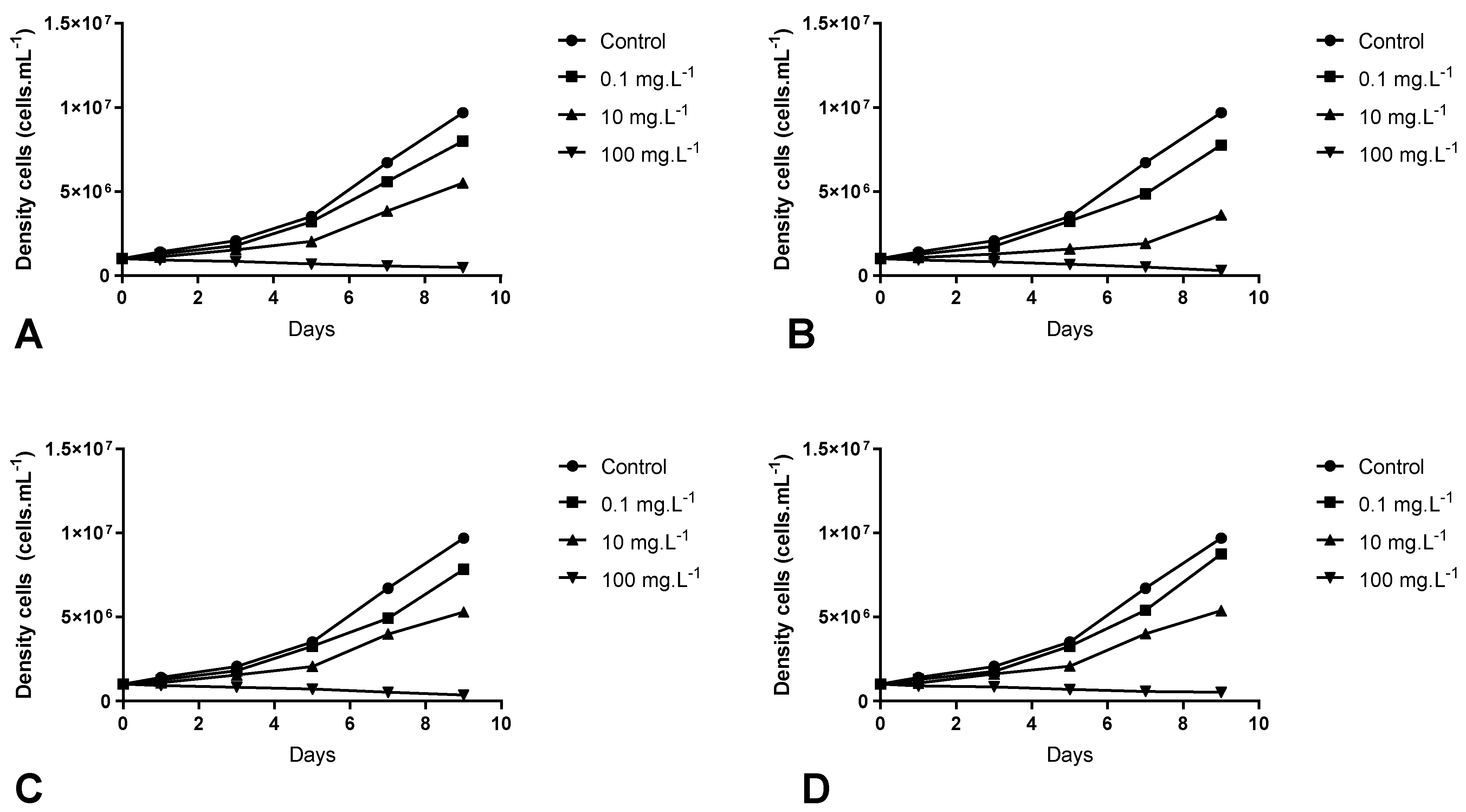
Figure 1. Effects of different concentrations of Myriophyllum aquaticum aqueous extract (Control, 0.1, 10 and 100 mg·L−1) of plants collected in different seasons under the cell growth of Microcystis aeruginosa: (A) Summer; (B) Autumn; (C) Winter; (D) Spring.
Water 15 02344 g001
Figure 2. Inhibition rate (means ± standard error) of different concentrations of Myriophyllum aquaticum aqueous extract (0.1, 10 and 100 mg·L−1)of plants collected in different seasons (summer, autumn, winter and spring) under the cell growth of Microcystis aeruginosa at different exposure times: (A) 1 day; (B) 3 Days; (C) 5 Days; (D) 7 Days; (E) 9 Days. Treatment means from two-way ANOVA followed by Tukey test. Different lowercase letters indicate significant differences between different extract concentrations. Different uppercase letters indicate significant differences between the same extract concentration in different seasons (p ≤ 0.05).
Water 15 02344 g002
Figure 3. Effects on photosynthetic pigments of Microcystis aeruginosa after exposure to different concentrations of aqueous extracts of Myriophyllum aquaticum (Control, 0.1, 10 and 100) collected in different seasons: summer, autumn, winter and spring. (A) Chlorophyll-a; (B) Phycocyanin; (C) Allophycocyanin; (D) Phycoerythrin. Values of means ± standard error. Treatment means from two-way ANOVA followed by the Tukey test. Different lowercase letters indicate significant differences between different extract concentrations. Different uppercase letters indicate significant differences between the same extract concentration in different seasons (p ≤ 0.05).
Water 15 02344 g003
Table 1. Means ± Standard Error of the content of total phenolic compounds concentration (mg·mL−1) of aqueous extracts of Myriophyllum aquaticum collected at different seasons: Summer, Autumn, Winter and Spring.
SeasonTotal Phenolic Compounds
(mg·mL−1)
One-Way ANOVA
D.FFp
Summer47.40 ± 2.31 a343.09<0.0001
Autumn97.40 ± 2.31 b
Winter84.73 ± 4.81 b
Spring62.73 ± 3.52 a
Note(s): Treatment means from one-way ANOVA followed by Tukey test (p ≤ 0.05). Different lowercase letters indicate significant differences between different seasons.
Table 2. Values (means ± standard error) of cell viability of Microcystis aeruginosa after exposure to different concentrations of aqueous extracts of Myriophyllum aquaticum (Control, 0.1, 10 and 100 mg·L−1) collected in different seasons (summer, autumn, winter and spring).
D.FCells Growth (Cells·mL−1)Extract Effects
Two-way ANOVA F-values
Extracts248.18 ***-
Season359.77 ***-
Extracts vs. season926.92 ***-
SeasonsExtracts (mg·L−1) -
SummerControl (0)1.06 × 10⁷ ± 2.30 × 10⁵ aA-
0.17.85 × 106 ± 9.77 × 10⁴ bAAlgistatic
105.59 × 10⁶ ± 1.76 × 10⁵ cAAlgistatic
1008.00 × 103 ± 2.00 × 10³ dAAlgicide
AutumnControl (0)1.06 × 10⁷ ± 2.30 × 10⁵ aA-
0.16.63 × 106 ± 1.29 × 10⁵ bBAlgistatic
104.63 × 10⁶ ± 1.33 × 10⁵ cBAlgistatic
1001.33 × 103 ± 6.66 × 10² dBAlgicide
WinterControl (0)1.06 × 10⁷ ± 2.30 × 10⁵ aA-
0.17.23 × 106 ± 5.88 × 10⁵ bAAlgistatic
105.88 × 10⁶ ± 1.66 × 10⁵ cAAlgistatic
1006.00 × 103 ± 3.46 × 10² dAAlgicide
SpringControl (0)1.06 × 10⁷ ± 2.30 × 10⁵ aA-
0.17.84 × 106 ± 6.66 × 10⁴ bAAlgistatic
105.81 × 10⁶ ± 1.11 × 10⁵ cAAlgistatic
1002.53 × 106 ± 7.42 × 10⁴ dCAlgicide
Comparison of means (p ≤ 0.05)
SeasonsSummer6.01 × 106 ± 2.25 × 10⁶ A-
Autumn5.47 × 106 ± 2.20 × 10⁶ B-
Winter5.94 × 106 ± 2.22 × 10⁶ B-
Spring6.13 × 106 ± 2.19 × 10⁶ A-
Note(s): *** Significant p ≤ 0.001. Treatment means from two-way ANOVA followed by the Tukey test. Different lowercase letters indicate significant differences between different extract concentrations. Different uppercase letters indicate significant differences between the same extract concentration in different seasons (p ≤ 0.05).
Table 3. Values (means ± standard error) of intracellular microcystin-LR concentration (µg·L−1) and removal rate (%) after exposure of Microcystis aeruginosa to different concentrations of aqueous extract of Myriophyllum aquaticum (0.1, 10 and 100 mg·L−1) collected in different seasons (summer, autumn, winter and spring).
D.FMicrocystin-LR Concentration (ng·L−1)Intracellular Microcystin-LR
Removal (%)
Two-way ANOVA F-values
Extracts21700 ***2130 ***
Season3263.3 ***517.9 ***
Extracts vs. season9122.3 ***132.6 ***
SeasonsExtracts (mg·L−1)
SummerControl (0)30.93± 0.69 aA-
0.130.98 ± 0.76 aA0.18 ± 0.05 aA
107.58 ± 0.31 bA75.51 ± 1.44 bAB
1004.21 ± 0.68 cA85.98 ± 3.90 dA
AutumnControl (0)30.93 ± 0.69 aA-
0.128.19 ± 1.00 bB6.68 ± 1.23 aB
106.42 ± 0.30 cB79.28 ± 0.54 bA
1004.93 ± 0.83 dA89.03 ± 0.97 cA
WinterControl (0)30.93± 0.69 aA-
0.130.86 ± 1.18 aA0.40 ± 0.27 aA
106.58 ± 0.18 bAB78.75 ± 0.12 bAB
1004.47 ± 0.27 cA85.54 ± 1.03 cA
SpringControl (0)30.93± 0.69 aA-
0.129.21 ± 1.41 aA2.89 ± 0.43 aA
107.29 ± 0.48 bA75.10 ± 1.65 bB
1006.78± 0.04 bB78.11 ± 0.36 bB
Comparison of means (p ≤ 0.05)
SeasonsSummer18.44± 7.27 A53.83± 17.80 AC
Autumn17.82± 7.02 A58.33± 18.76 B
Winter18.23± 7.34 A54.90 ± 18.46 AB
Spring18.07 ± 6.41 A52.02 ± 20.46 C
Note(s): *** Significant p ≤ 0.001. Treatment means from two-way ANOVA followed by the Tukey test. Different lowercase letters indicate significant differences between different extract concentrations. Different uppercase letters indicate significant differences between the same extract concentration in different seasons (p ≤ 0.05).
Disclaimer/Publisher’s Note: The statements, opinions and data contained in all publications are solely those of the individual author(s) and contributor(s) and not of MDPI and/or the editor(s). MDPI and/or the editor(s) disclaim responsibility for any injury to people or property resulting from any ideas, methods, instructions or products referred to in the content.