Article

An Assessment of the Effects of Light Intensities and Temperature Changes on Cyanobacteria’s Oxidative Stress via the Use of Hydrogen Peroxide as an Indicator

by
Mizanur Rahman
1,*,
Takashi Asaeda
1,2,3,*,
Helayaye Damitha Lakmali Abeynayaka
1 and
Kiyotaka Fukahori
1
1
Graduate School of Science and Engineering, Saitama University, Saitama 338-8570, Japan
2
Hydro Technology Institute, Shimo-meguro, Tokyo 153-0064, Japan
3
Research and Development Center, Nippon Koei, Tsukuba 300-1259, Japan
*
Authors to whom correspondence should be addressed.
Water 2023, 15(13), 2429; https://doi.org/10.3390/w15132429
Submission received: 10 June 2023 / Revised: 27 June 2023 / Accepted: 28 June 2023 / Published: 30 June 2023

Abstract

:
Humans and other organisms are adversely affected by cyanobacterial blooming. This study aims to investigate the long-term effects of light intensities and different temperatures on Phormidium ambiguum and Pseudanabaena foetida. Enough P. ambiguum and P. foetida cells were acclimated for 24 days at 30 °C, 20 °C, and 10 °C in separate incubators. The starting day sample was collected after 24 days of acclimatization, and a second sample was collected seven days later at light intensities of 10, 30, 50, 200, and 600 µmol m−2 s−1 for each temperature. The optical density (OD730), hydrogen peroxide (H2O2) concentration, protein content, chlorophyll-a (Chl-a) concentration, and catalase (CAT) activity were measured. Light intensity changes soon after collection resulted in nearly identical starting day samples at each individual temperature. The H2O2 concentration and algal biomass increased until a light intensity of 200 µmol m−2 s−1 was reached and decreased afterward in each temperature for both species after seven days. In association with an increasing H2O2 concentration, the Chl-a concentration decreased after 50 µmol m−2s−1 of light intensity, affecting the protein content. The algal biomass was significantly lower at 10 °C compared to 30 °C. The CAT activity increased proportionately with the H2O2 concentration and algal biomass. Therefore, water bodies in the field can be illuminated with long-term high light intensities in different temperatures to reduce algal biomass.

1. Introduction

There are frequent outbreaks of cyanobacteria blooms in eutrophic lakes and reservoirs which can cause severe environmental and economic problems [1,2]. Cyanobacteria produce toxins that have adverse effects on aquatic organisms and human health [3]. Cyanobacteria species can cause allergies, skin irritation, and respiratory problems [4,5,6]. Climate change and nutrient availability have contributed to the spread of cyanobacteria. The production of cyanotoxins by cyanobacteria can cause instability in water bodies [7,8]. Cyanobacteria produce toxic secondary metabolites that harm ecosystems [9] and volatile organic compounds that deteriorate water quality by increasing unpleasant odors [10]. Light intensities and temperatures greatly affect cyanobacterial habitat preferences and growth proliferation conditions [11,12].
Cyanobacteria are photoautotrophs in nature. The presence of light directly affects the growth of cyanobacteria, and even moderate light changes are stressful [13]. The ability of cyanobacteria to respond to varying light intensities provides insights into photosynthesis and biotechnology applications [14]. The physiological metabolism of cyanobacteria depends on light intensity, so the effects of different light intensities must be investigated [12,15]. Due to extreme light conditions (very low or very high), photoinhibition can occur [16]. In photoinhibition, the electron transport in photosystem II is reversibly reduced to protect the photosynthesis apparatus [17] and other photoprotective mechanisms, macroalgae, and phytoplankton [18,19,20]. Changes in chlorophyll fluorescence characteristics in response to light result from photoinhibition [21].
Reactive oxygen species (ROS) cause photoinhibition. In the presence of excessive oxidative stress, ROS will become imbalanced in comparison to the antioxidant’s capacity to scavenge them [22,23]. The electron transport process, as well as the metabolism of substances and energy, create ROS as byproducts within chloroplasts, mitochondria, peroxisomes, and the cytoplasm [24,25]. Among ROS, H2O2 is the most common. Superoxide radicals are also converted into H2O2 by superoxide dismutase, which is then detoxified into water by antioxidants [26,27]. ROS accumulation harms cyanobacterial cells as it can destroy the cytoplasmic membrane, Chl-a concentration, photosynthetic apparatus, protein content, and DNA [28,29].
Phormidium ambiguum Gomont and Pseudanabaena foetida Niiyama, Tuji et Ichise are two cyanobacterial species that have been linked to water quality and safety problems around the world [30,31]. Eutrophication and global warming have led to increases in their worldwide occurrences [32,33]. Eutrophication is an alarming problem in developing countries with limited financial resources for waste treatment [34]. P. ambiguum is a filamentous benthic cyanobacterial species [35]. Benthic cyanobacteria are capable of forming biofilms in water bodies. As cyanobacteria form mats or clusters close to banks, animals consume high levels of toxins, often resulting in their death [36]. As high concentrations of these toxins can be found in floating mats, they can also pose a health risk to humans via ingestion and direct skin exposure. Benthic cyanobacteria have also been found to contain hepatotoxins [37,38]. P. foetida is a non-blooming odorous compound containing cyanobacterial species. P. foetida produces 2-methylisoborneol (2-MIB), which causes operational issues with water supplies [39]. Both cyanobacterial species are known to release harmful cytotoxins and grow in tropical and subtropical waters [35,40].
During the growth of cyanobacteria, higher temperatures promote metabolic acceleration. At the same time, at the ecosystem level, they contribute to the formation of thermal stratification, which promotes biomass accumulation at the water’s surface [41]. P. ambiguum and P. foetida grow best at 25 °C to 33 °C [42,43,44]. Subtropical and tropical regions can experience cyanobacterial blooms all year round due to their ability to proliferate at high temperatures [45,46]. Aside from the higher temperatures of their preferential growth conditions, little is known about how fluctuating temperatures affect the growth of cyanobacteria.
Considering the environmental factors that influence cyanobacterial growth and metabolism is essential to solving real-life problems associated with cyanobacteria. The growth of cyanobacteria is dependent on water temperature, light intensity, the availability of nitrogen (N) and phosphorus (P), salinity, and turbidity [47,48,49]. Light intensity and temperature are the major factors that affect the growth of cyanobacteria [47]. It is important for freshwater resource management to understand how light intensity and temperature affect changes in the growth of cyanobacteria.
Cyanobacterial blooms have been eradicated using a variety of chemical and physical methods globally [50,51]. However, it is discouraging to apply chemical methods to reduce cyanobacteria due to limitations in their application and their peripheral impact on the environment [52]. There is a huge possibility of using abiotic stresses like light intensities and temperatures instead of chemical methods [53,54,55]. Therefore, this study aims to (1) find the oxidative stress threshold level of cyanobacteria, using H2O2 as an indicator, and (2) determine the long-term effects of different light intensities at different temperatures.

2. Materials and Methods

2.1. P. ambiguum and P. foetida Cell Cultures

Strains of P. ambiguum (NIES-2119) and P. foetida (NIES-512) were provided by the National Institute for Environmental Studies, Japan. Throughout the experiment, an autoclaved BG 11 medium [56] was provided to culture P. ambiguum and P. foetida. The strains were cultured at 30 °C (±0.3 °C) with a white, fluorescent lamp that provided a 12 h:12 h cycle of light and darkness in an incubator (KMH-259, AS ONE Corporation, Osaka, Japan). The light intensity in the incubator ranged between 10 and 20 µmol m−2 s−1 for P. ambiguum and between 20 and 30 µmol m−2 s−1 for P. foetida. When the cultures were incubated under light, they were manually shaken three times a day. They were subcultured until enough P. ambiguum and P. foetida were produced [57,58].

2.2. Experimental Procedure

We transferred sufficient amounts of P. ambiguum and P. foetida into three incubators at 30 °C (±0.3 °C), 20 °C (±0.3 °C), and 10 °C (±0.3 °C) for 24 days at a light intensity of 10–20 µmol m−2 s−1 for P. ambiguum and a light intensity of 20–30 µmol m−2 s−1 for P. foetida. The effects of light exposure on P. ambiguum and P. foetida at each temperature were examined by introducing white, fluorescent light at intensities of 10, 30, 50, 200, and 600 µmol m−2 s−1. In order to maintain 12 h of light and 12 h of darkness, an automatic timer was set (REVEX PT7, Kawaguchi city, Saitama, Japan). A quantum sensor (Apogee, MQ-200, Logan, UT, USA) was used to measure the light intensities, which were then uniformly adjusted in the medium. The culture flasks were gently shaken three times per day for homogeneous exposure to light. Samples were taken twice for analysis. The first samples were taken at 12:00 noon on the 24th day of acclimatization, considered the starting day. Following the first sampling, a second sampling was conducted after an interval of seven days at the same time of day. The starting day collection samples were identical because the preliminary light intensities were changed soon after the collection. Using 1.5 mL tubes, 1 mL of P. ambiguum and P. foetida cells were collected separately. The cells were centrifuged at 10,000 rotations per minute (rpm) for 10 min at 4 °C. The supernatant was removed and kept at −80 °C until biochemical analysis. The buffer (specified for different bioassays) cannot break down hard cyanobacterial cell walls. To homogenize the cyanobacterial cells, five to eight 3 mm beads (Bio Medical Science Inc., Tokyo, Japan) were added to the buffer, and the mixture was vigorously shaken. The mixture was centrifuged at 10,000 rpm for 10 min at 4 °C, and the supernatant was used as an extract for further analysis. Three replicates of each culture group were conducted.

2.3. Estimation of Total Soluble Protein Content

The total soluble protein concentrations were measured following Abeynayaka’s [57] method with minor changes. The cyanobacterial cells were homogenized using 500 mM of NaOH solution. The supernatant was collected after centrifugation at 4 °C for 15 min at 10,000 rpm. Then, 51 µL of supernatant extract was added to 969 µL of Coomassie Bradford protein assay for each sample. The mixture was kept at room temperature for 10 min, and the absorbance was recorded at 595 nm using a UVmini−1240. A series of known albumin concentration standard curves were used to measure the known protein concentrations.

2.4. Chlorophyll-a Concentration Measurements

The Chl-a concentrations were quantified using the method described in [59,60]. First, 1.5 mL of 90% ethanol was added to the unfrozen cyanobacteria cells. The mixture was homogenized with 3 mm beads via vertexing and kept at room temperature (25 ± 2 °C) for 24 h in darkness by wrapping it with aluminum foil. Each sample was centrifuged at 12,000 rpm, and the supernatant was measured with a UVmini−1240 at absorption wavelengths of 750 nm, 665 nm, and 649 nm. The 750 nm reading was obtained to confirm whether the extract interfered with organic matters or not. Each value lay between −0.003 and 0.003, indicating the purity of the extract. The formula for calculating the Chl-a content is as follows:
Chl-a (µg/mL) = (13.95 ∗ A665 − 6.88 ∗ A649) ∗ 1.5

2.5. Identification of Cell Growth

OD730 measurements were carried out with the help of a UV-Vis spectrophotometer (UVmini-1240, Shimadzu, Japan) in order to quantify the growth of the cyanobacteria. The OD730 was measured using a previously proposed methodology [61] with a minor modification. An amount of 1 mL of cyanobacterial cells was homogenized with 3 mm beads (Bio Medical Science Inc., Tokyo, Japan) and measured at a wavelength of 730 nm.

2.6. H2O2 Concentration Measurement Assay

The modified ferrous oxidation xylenol orange (commonly known as eFOX) assay was adapted to quantify the H2O2 concentration [62,63]. First, 100 µL of supernatant was mixed with 1 mL of the assay solution, which contained 0.250 mM of ferrous ammonium sulfate, 0.1 mM of sorbitol, 0.1 mM of xylenol orange, and 25 mM of H2SO4 (sulfuric acid), and 1% ethanol was added to increase the sensitivity of the reaction with H2O2 by 50% (Fujifilm wako pure chemical corporation, Osaka, Japan). The reaction mixtures were incubated for a period of 15 min at room temperature. Spectrophotometric measurements were carried out at 560 nm (UVmini-1240, Shimadzu, Japan). A H2O2 standard curve was developed via dilution solutions of commercially available 9.8 M H2O2 (30%) (w/w) [55,64]. Trials were conducted to confirm the H2O2 concentrations inside and outside of the cells. The cyanobacterial cells were separated with filter paper (0.22 μm pore size). The separated water was analyzed to quantify the H2O2 concentration. No H2O2 was detected from the separated water. It was confirmed that the H2O2 concentration obtained via the methodology described above indicated the sum of the H2O2 contained by the cells and the intercellular H2O2.

2.7. CAT Assay

The CAT activity was measured spectrophotometrically at room temperature by monitoring the decrease in absorbance at 240 nm resulting from the decomposition of H2O2. The CAT activity was measured via the method of Aebi [65]. The enzyme was extracted using 3 mm beads with 1 mL of potassium phosphate buffer (0.05 M, pH 7.0) containing 0.1 mM of EDTA. The reaction mixture contained 15 μL of 0.75 M H2O2, 920 μL of potassium phosphate buffer, and 65 μL of enzyme extract. Measurements were obtained every 10 s for 3 min, and calculations were conducted using the 39.4 mM/cm extinction coefficient.

2.8. Statistical Analysis
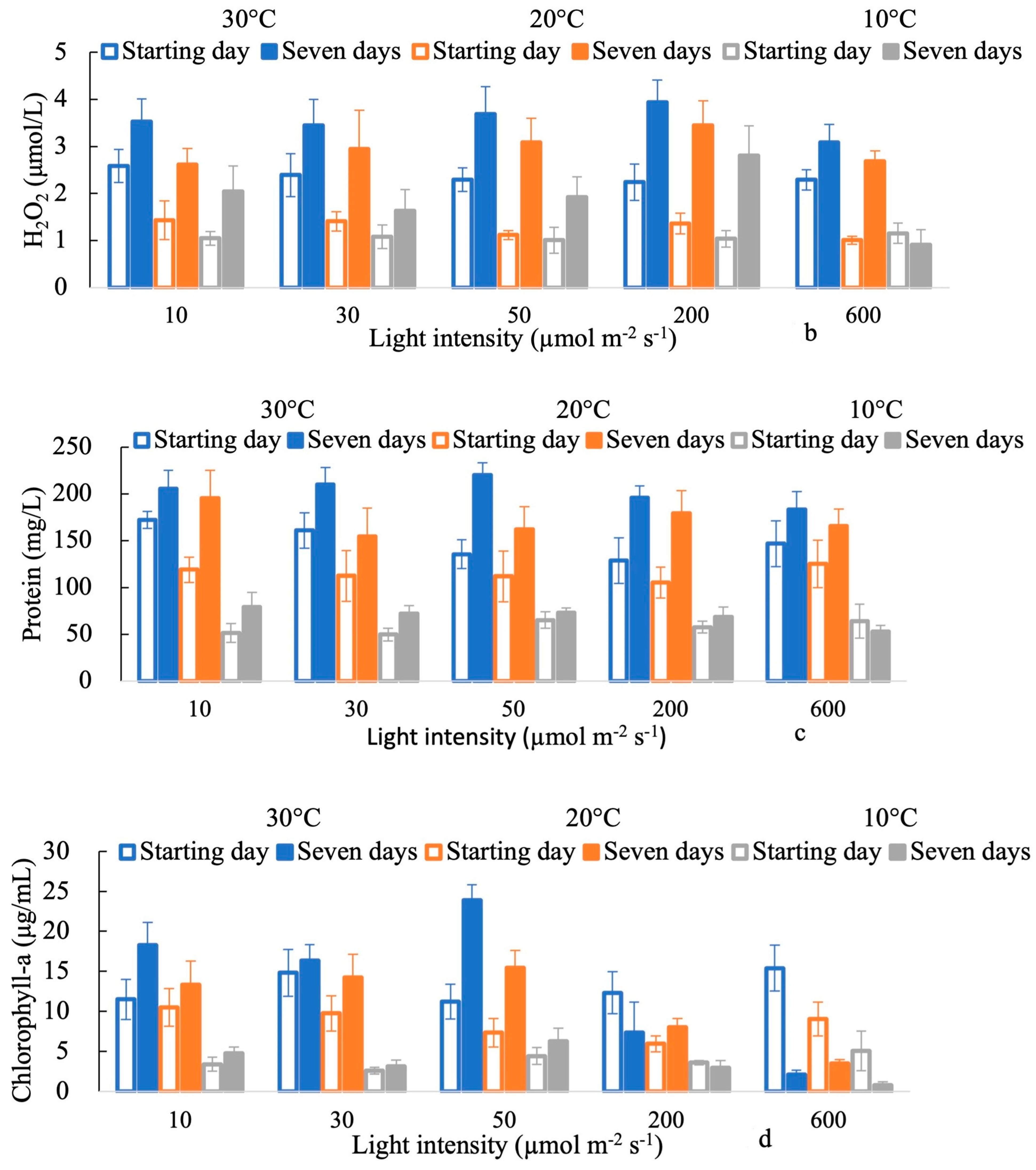
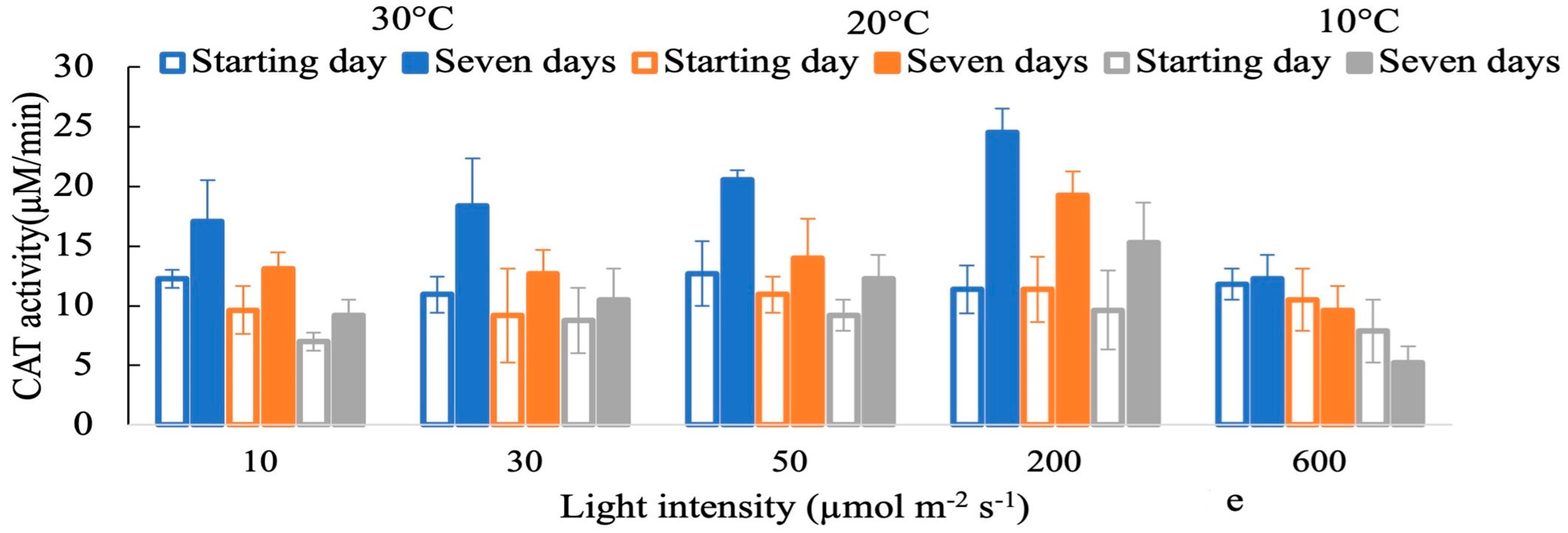
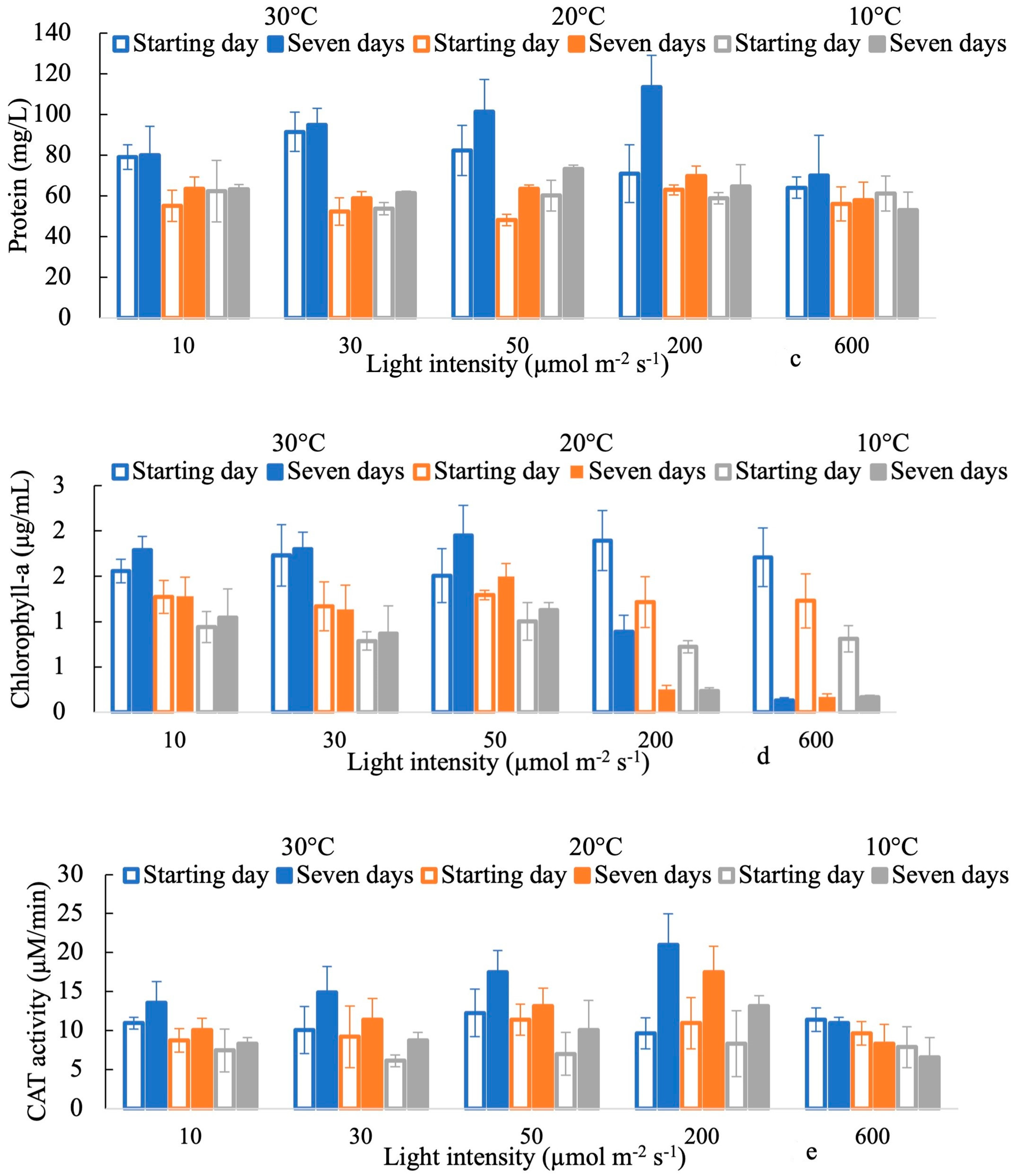
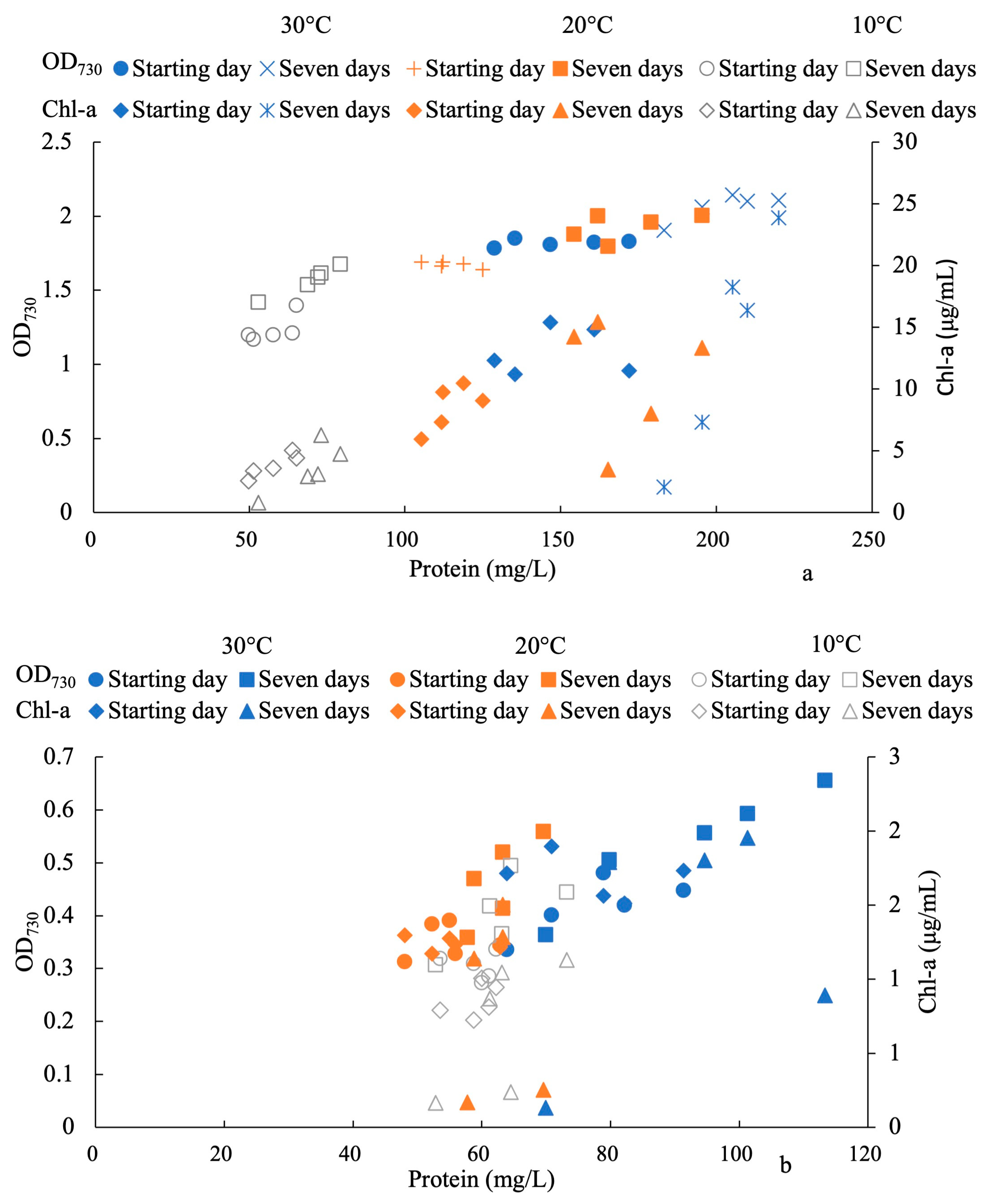
A one-way analysis of variance (ANOVA), followed by Tukey’s test, was performed to check the statistical significance of the variations among different PAR groups. To determine how the light intensities and temperatures interacted, we used a two-way ANOVA. Pearson’s correlation analysis was conducted to evaluate the correlations among parameters. IBM SPSS Statistics (Version 28.0 IBM Corporation, Chicago, IL, USA) software was used to execute the statistical analyses.

3. Results

OD730, H2O2 concentration, protein concentration, Chl-a content, and CAT activity were monitored in P. ambiguum and P. foetida at 30 °C, 20 °C, and 10 °C based on each light intensity for the starting day and after the seven-day period. Due to changes in the preliminary light intensities soon after the samples were collected, the starting day collection for each temperature did not show much fluctuation. Over the seven days of exposure, the H2O2 concentration steadily increased with increases in the light intensity until a light intensity of 200 µmol m−2 s−1, while it declined at a light intensity of 600 µmol m−2 s−1 at each temperature in both species (Figure 1b and Figure 2b). In both species, the OD730 and protein concentration, which had been lower at the starting day at 20 °C, increased up to the 30 °C level in seven days (Figure 1a,c and Figure 2a,c). The OD730 values were higher in P. ambiguum than P. foetida at each temperature after seven days of exposure. For example, at 30 °C, the OD730 value for P. ambiguum was ~2, while the OD730 value for P. foetida was ~0.6. Over the seven days of treatment, the protein concentrations of P. ambiguum and P. foetida decreased as the temperature decreased from 30 °C to 10 °C. The Chl-a concentrations of P. ambiguum and P. foetida under different light intensities at each temperature are shown in Figure 1d and Figure 2d. The Chl-a concentration substantially decreased as the light intensity increased from 50 µmol m−2 s−1 to 200 µmol m−2 s−1 (p < 0.001 for both species) and thereafter at 30 °C, 20 °C, and 10 °C in both species over seven days of light exposure (Figure 1d and Figure 2d). After seven days, P. ambiguum had higher Chl-a contents than P. foetida at 30 °C, 20 °C, and 10 °C. The antioxidant activity of CAT increased until a light intensity of 200 µmol m−2 s−1 was reached in response to oxidative damage due to the H2O2 concentration and decreased with higher light intensities at 30 °C, 20 °C, and 10 °C in both species (Figure 1e and Figure 2e). In Figure 3a,b, OD730 is presented with respect to the protein and Chl-a contents, respectively. There is a significantly high proportionate correlation observed between OD730 and protein for all temperatures (at 30 °C for P. ambiguum r = 0.991, p < 0.001 and at 30 °C for P. foetida r = 0.952, p < 0.001; at 20 °C for P. ambiguum r = 0.978, p < 0.001 and at 20 °C for P. foetida r = 0.990, p < 0.001; at 10 °C for P. ambiguum r = 0.829, p < 0.001 and at 10 °C for P. foetida r = 0.715, p < 0.001) after the seven days of exposure. On the starting day, both Chl-a and OD730 were significantly proportionate with respect to protein concentration, whereas after seven days, the Chl-a content was significantly smaller compared to the starting level (p < 0.001 for both species). For both species, the concentration of H2O2 per biomass was expressed by the change in H2O2/OD730 after seven days, which is presented in Figure 4. In every case, the H2O2/OD730 increased following the light intensity until 200 µmol m−2 s−1, while it declined at a light intensity of 600 µmol m−2 s−1. The H2O2/OD730 values were lower at 10 °C compared to 30 °C in both species. There was a positive correlation observed between H2O2/OD730 and different light intensities at temperatures ranging from 30 °C to 10 °C in P. ambiguum (r = 0.390, p = 0.176) and P. foetida (r = 0.282, p = 0.307).

4. Discussion

4.1. Effects of Light Intensities on the Response of H2O2

Excess levels of ROS can be generated through abiotic stress [66,67,68]. The accumulation of ROS in each cyanobacteria species under high light (higher than 200 µmol m−2 s−1) and alterations in temperature may result in oxidative damage, which may be one reason for the inhibition of cell growth (Figure 1a and Figure 2a). Cells exposed to high levels of environmental stress produce and accumulate H2O2. As cyanobacteria are exposed to various abiotic stresses in natural water, they are more likely to undergo oxidative stress, producing H2O2 in the process, which may deteriorate cyanobacterial biomass by producing hydroxyl radicals. The production of H2O2 may not necessarily be cumulative in abiotic stresses [69,70]. The H2O2 concentration was enhanced until a light intensity of 200 µmol m−2 s−1 and decreased afterward at each temperature (Figure 1b and Figure 2b). The trend was more prominent at 30 °C in the seven days of exposure. In the present study, photoinhibition was seen even with a light intensity of 200 µmol m−2 s−1, whereas a light intensity of 1000 µmol m−2 s−1 has been reported in field observations [71,72]. In field monitoring, light intensity in water decreases relatively quickly with depth, despite surface colonies receiving high-intensity solar radiation. As a result, the colony of cyanobacteria does not receive strong solar radiation directly, especially P. ambiguum and P. foetida, since they stay in relatively deep water (~2 m) [73]. In addition, they avoid the highest level of solar radiation during the day by migrating to deeper zones [74,75], where they are likely to avoid the high level of solar radiation and oxidative stress before the recovery of homeostasis via increases in antioxidant activities [74].
In the past, laboratory incubations under different H2O2 concentrations provided the lethal H2O2 dosage for cyanobacteria [76,77,78], which implies that cyanobacterial biomass is degraded at higher H2O2 concentrations. The lethal H2O2 dosage for the suppression of cyanobacteria in vitro under different H2O2 concentrations ranges from 1 to 1000 μmol L−1 [76,79,80]. This study suggests that if the H2O2/OD730 exceeded ~2 to 6 μmol/L (depending on temperatures) (Figure 4) after seven days of treatment, it significantly declined afterward, even against higher temperatures or light intensities. The high H2O2 concentration deteriorates the physiological condition without generating further H2O2 [55,81]. In contrast, the Chl-a concentration was affected by even lower levels of light intensities (after 50 µmol m−2 s−1). A low H2O2 concentration is generated with sufficiently low PAR (50 µmol m−2 s−1), and the Chl-a concentration fluctuates positively with the PAR (data are not shown). This may be due to an adaptation to increase productivity. Chl-a concentration increases due to the breakdown of photosynthesis components, such as photosystems I and II [82] and phycobilisomes [16]. In contrast, a high Chl-a concentration increases the photosynthesis rate, generating more H2O2 and deteriorating the photosynthesis apparatus. Thus, an increased H2O2 concentration negatively alters the Chl-a concentration under highly stressful conditions [55]. The H2O2 concentration also becomes a useful indicator for demonstrating the effects of environmental stress on other plants and their productivities [55].
In the present study, we used the protein content as an indicator of biomass rather than the Chl-a content for both cyanobacterial species [83]. One-third to one-half of the biomass of cyanobacteria cells is protein [84]. There was a high correlation between OD730 and protein content (Figure 3a,b), indicating that the protein content is a proper biomass reference. Compared with the OD730 values and protein contents, the Chl-a contents were affected by lower light intensities. A suitable parameter should be selected to identify the cyanobacterial biomass. H2O2/OD730 could be a suitable parameter for evaluating the H2O2 amount in a cell. In the field treatment for artificially inducing H2O2, the light intensity is the primary component in increasing the H2O2 concentration in nature and accumulating it in a cell. The required amount of H2O2 to be induced should be identified using a suitable factor, such as H2O2/OD730, for the suppression of algal blooms.
The seasonal dynamics of phytoplankton are largely determined by temperature [85]. When temperatures are relatively high, cyanobacteria grow the fastest [86]. Depending on the species, they respond differently to low temperatures. In the present study, the growth of P. ambiguum was significantly decreased by decreasing the temperature from 30 °C to 10 °C (p < 0.001 for both species), similar to other studies [26,87,88] (Figure 1a and Figure 2a). Decreasing temperatures (from 30 °C to 10 °C) also significantly affected the H2O2, Chl-a, and protein concentrations. The amount of H2O2 in a cell (H2O2/OD730) was also lower at 10 °C compared to 30 °C (Figure 4). These results indicate that P. ambiguum and P. foetida maintain higher metabolisms with higher temperatures, bringing about high growth rates with high temperatures.

4.2. Antioxidant Activity in Response to Oxidative Stress

The tissue H2O2 concentration at a particular time is determined as the balance between the generation rate and the scavenging by antioxidant activities. This concentration works as a signal to activate antioxidant behavior. Antioxidant activity increases in response to oxidative stress to prevent cell damage [89]. Oxidative stress and antioxidative enzymes are triggered by abiotic stress [26,90]. In the present study, high light intensities (200 to 600 µmol m−2 s−1) decreased the H2O2 concentration, and low light intensities (10 to 200 µmol m−2 s−1) increased it. CAT activity proportionally increased with H2O2 concentration until 200 µmol m−2 s−1 at each temperature and decreased afterward, following the reduction in H2O2 concentration with high light intensities (Figure 1e and Figure 2e).

4.3. Management of Cyanobacterial Blooms

Long-term exposure to light intensities at different temperatures is highly appreciated as a non-chemical approach to controlling P. ambiguum and P. foetida. This result opposes the hypothesis that low-light exposure suppresses cyanobacterial growth. According to Visser et al. [73], artificial water mixing in lakes and reservoirs is only sometimes efficient. During artificial mixing, oxygen levels increase in the water, the temperatures of the deep layers are increased, and the temperature on the surface is lowered. For instance, destratification (the development of vertical mixing) decreases the surface temperature from 28.9 °C to 26.4 °C, whereas deep-water aeration increases the temperature from 8 °C to 23.7 °C [91]. The mixing occurs deep enough to limit light availability, and the mixing devices are well distributed horizontally over the lake. A reduction in light exposure will lead to decreases in the growth of cyanobacteria and algae. Among the drawbacks of artificial mixing are the cost of installation and operation and the energy required during the entire growing season. Using intermittent mixing might be beneficial in reducing energy costs and reducing the flotation velocities of cyanobacteria. The availability of light under water can influence cyanobacterial growth composition [92]. When the wind is mixed with the unsustainable vertical heterogeneity of an algal biomass, light cannot enter the deeper zone of water. On the other hand, under calm conditions (a stable water column, vertical migration, and a sustained surface biomass maximum) in a lake, light may enter the deeper zone [92]. For example, in comparison with the euphotic depth, the Secchi depth is roughly half. Water transparency is measured via the Secchi depth, where the Secchi depth increases with increasing transparency. A lake’s bottom is illuminated when the Secchi depth is roughly half its euphotic depth [92]. Phytoplankton may experience light limitations when the Secchi depth/total euphotic depth ratio is below 0.5. It is important to consider both species’ low-light and high-light vulnerabilities when introducing the system. A destratification process must be designed beforehand based on the intensity and distribution of light and temperature [93,94]. Other practical methods can be introduced to control cyanobacterial species, such as exposing deep water to high intensities of light without raising the temperature. There should be a variety of methods developed to illuminate the water column with a greater light intensity than what is tolerated by these species.

5. Conclusions

Cyanobacterial species exhibit photoinhibition even after seven days of exposure to a light intensity of 200 µmolm−2s−1 at 30 °C, 20 °C, and 10 °C. High light intensities (higher than 200 µmol m−2 s−1) significantly affect H2O2 concentrations, protein concentrations, and Chl-a contents at each temperature. CAT activity increased until a light intensity of 200 µmolm−2s−1 in response to the H2O2 concentration and the ability of the algal biomass to scavenge oxidative stress, and it decreased afterward with higher light intensities. The algal biomass decreased from 30 °C to 10 °C due to the effect of temperature. The opportunity of developing control mechanisms based on high light intensities that result in slower growth could be explored, as could the further development of existing methods that rely on low light intensities, which could possibly result in reductions in growth rates. Water bodies could be effectively controlled via illumination with a greater light intensity than the intensities tolerated by these species.

Author Contributions

Conceptualization, T.A. and M.R.; methodology, T.A.; software, M.R.; validation, T.A., H.D.L.A., K.F. and M.R.; formal analysis, M.R. and H.D.L.A.; investigation, M.R.; resources, T.A.; data curation, M.R.; writing—original draft preparation, M.R.; writing—review and editing, T.A. and K.F.; visualization, T.A. and M.R.; supervision, T.A. and K.F.; project administration, T.A.; funding acquisition, T.A. All authors have read and agreed to the published version of the manuscript.

Funding

The Grant-in-Aid for Scientific Research (B) (19H02245), and (C) (20K04714) provided financial support for this work, and the Japanese Society for the Promotion of Science (JSPS) Fund for Joint International Research (18KK0116) also provided support.

Data Availability Statement

The authors state that we will make all data available to anyone who requests the data without undue preservation. This manuscript contains information supporting this study’s findings in the Materials and Methods section in the form of a preprint at “Research square” with the identifier (https://doi.org/10.21203/rs.3.rs-2326349/v1) (accessed on 11 June 2023); (https://doi.org/10.21203/rs.3.rs-2276741/v1) (accessed on 11 June 2023); and (https://doi.org/10.21203/rs.3.rs-2232330/v1) (accessed on 11 June 2023). The authors declare that previous DOIs will be updated with the new DOI upon acceptance.

Conflicts of Interest

The authors declare no conflict of interest.

References

  1. Dokulil, M.T.; Teubner, K. Cyanobacterial dominance in lakes. Hydrobiologia 2000, 438, 1–12. [Google Scholar] [CrossRef]
  2. Chen, Y.; Weng, Y.; Zhou, M.; Meng, Y.; Liu, J.; Yang, L.; Zuo, Z. Linalool-and α-terpineol-induced programmed cell death in Chlamydomona sreinhardtii. Ecotoxicol. Environ. Saf. 2019, 167, 435–440. [Google Scholar] [CrossRef]
  3. Carmichael, W.W. Freshwater Blue-Green Algae (Cyanobacteria) ToxinsA Review. Water Environ. 1981, 405, 1–13. [Google Scholar] [CrossRef]
  4. Fleming, L.E.; Kirkpatrick, B.; Backer, L.C.; Bean, J.A.; Wanner, A.; Reich, A.; Zaias, J.; Cheng, Y.S.; Pierce, R.; Naar, J.; et al. Aerosolized Red-Tide Toxins (Brevetoxins) and Asthma. Chest 2007, 131, 187–194. [Google Scholar] [CrossRef] [Green Version]
  5. Sharma, N.K.; Rai, A.K. Allergenicity of air-borne cyanobacteria Phormidium fragile and Nostoc muscorum. Ecotoxicol. Environ. Saf. 2008, 69, 158–162. [Google Scholar] [CrossRef]
  6. Moustaka-Gouni, M.; Genitsaris, S.; Kormas, K.A. Airborne Algae and Cyanobacteria Occurrence and Related Health Effects. Front. Biosci. 2011, E3, 772–787. [Google Scholar] [CrossRef] [Green Version]
  7. Hudnell, H.K. Cyanobacterial harmful algal blooms: State of the science and research needs. In Advances in Experimental Medicine and Biology; Springer: New York, NY, USA, 2008; Volume 619. [Google Scholar]
  8. Butakova, E.A. Specific features of odor-causing compounds (geosmin and 2-methylisoborneol) as secondary metabolites of cyanobacteria. Russ. J. Plant Physiol. 2013, 60, 507–510. [Google Scholar] [CrossRef]
  9. Pflugmacher, S. Promotion of oxidative stress in the aquatic macrophyte Ceratophyllum demersum during biotransformation of the cyanobacterial toxin microcystin-LR. Aquat. Toxicol. 2004, 70, 169–178. [Google Scholar] [CrossRef]
  10. Zhang, C.; Chen, J.; Ding, A.; Hou, Y.; Li, Z.; Niu, X.; Su, Y.; Xu, E. Laws The 2007 water crisis in Wuxi, China: Analysis of the origin. J. Hazard. Mater. 2010, 182, 130–135. [Google Scholar] [CrossRef]
  11. Cao, H.-S.; Tao, Y.; Kong, F.-X.; Yang, Z. Relationship between Temperature and Cyanobacterial Recruitment from Sediments in Laboratory and Field Studies. J. Freshw. Ecol. 2008, 23, 405–412. [Google Scholar] [CrossRef] [Green Version]
  12. Asaeda, T.; Rahman, M.; Abeynayaka, H.D.L. Hydrogen peroxide can be a plausible biomarker in cyanobacterial bloom treatment. Sci. Rep. 2022, 12, 1–11. [Google Scholar] [CrossRef]
  13. Welkie, D.; Rubin, B.E.; Diamond, S.; Hood, R.D.; Savage, D.F.; Golden, S.S. A Hard Day’s Night: Cyanobacteria in Diel Cycles. Trends Microbiol. 2019, 27, 231–242. [Google Scholar] [CrossRef] [PubMed]
  14. Flombaum, P.; Gallegos, J.L.; Gordillo, R.A.; Rincón, J.; Zabala, L.L.; Jiao, N.; Karl, D.M.; Li, W.K.W.; Lomas, M.W.; Veneziano, D.; et al. Present and future global distributions of the marine Cyanobacteria Prochlorococcus and Synechococcus. Proc. Natl. Acad. Sci. USA 2013, 110, 9824–9829. [Google Scholar] [CrossRef] [Green Version]
  15. Barnuevo, A.; Asaeda, T. Integrating the ecophysiology and biochemical stress indicators into the paradigm of mangrove ecology and a rehabilitation blueprint. PLoS ONE 2018, 13, e0202227. [Google Scholar] [CrossRef] [Green Version]
  16. Walsh, K.; Jones, G.J.; Dunstan, R.H. Effect of irradiance on fatty acid, carotenoid, total protein composition and growth of Microcystis aeruginosa. Phytochemistry 1997, 44, 817–824. [Google Scholar] [CrossRef]
  17. Hanelt, D.P. Photoinhibition of photosynthesis in marine macroalgae. Sci. Mar. 1996, 60, 243–248. [Google Scholar]
  18. Hortonn, P.; Ruban, A. Molecular design of the photosystem II light-harvesting antenna: Photosynthesis and photoprotection. J. Exp. Bot. 2005, 56, 365–373. [Google Scholar] [CrossRef]
  19. Kirilovsky, D. Photoprotection in cyanobacteria: The orange carotenoid protein (OCP)-related non-photochemical-quenching mechanism. Photosynth. Res. 2007, 93, 7–16. [Google Scholar] [CrossRef]
  20. Zhang, Y.; Hu, M.; Zhong, L.; Wiedensohler, A.; Liu, S.; Andreae, M.; Wang, W.; Fan, S. Regional Integrated Experiments on Air Quality over Pearl River Delta 2004 (PRIDE-PRD2004): Overview. Atmos. Environ. 2008, 42, 6157–6173. [Google Scholar] [CrossRef]
  21. Jones, R.J.; Hoegh-Guldberg, O. Diurnal changes in the photochemical efficiency of the symbiotic dinoflagellates (Dinophyceae) of corals: Photoprotection, photoinactivation and the relationship to coral bleaching. Plant Cell Environ. 2001, 24, 89–99. [Google Scholar] [CrossRef]
  22. Marino, D.; Dunand, C.; Puppo, A.; Pauly, N. A burst of plant NADPH oxidases. Trends Plant Sci. 2012, 17, 9–15. [Google Scholar] [CrossRef] [PubMed]
  23. Ugya, A.Y.; Imam, T.S.; Li, A.; Ma, J.; Hua, X. Antioxidant response mechanism of freshwater microalgae species to reactive oxygen species production: A mini review. Chem. Ecol. 2020, 36, 174–193. [Google Scholar] [CrossRef]
  24. Darehshouri, A.; Lütz-Meindl, U. H2O2 localization in the green alga Micrasterias after salt and osmotic stress by TEM-coupled electron energy loss spectroscopy. Protoplasma 2010, 239, 49–56. [Google Scholar] [CrossRef] [Green Version]
  25. Zuo, Z.; Peng, K.; Shi, M.; Chen, Z.; Zhu, Y.; Bai, Y.; Gao, Y.; Wang, Y. ROS production is associated with glycolate metabolism in Chlamydomonas reinhardtii (Chlorophyceae) under salt stress. Phycologia 2014, 53, 502–507. [Google Scholar] [CrossRef]
  26. Rastogi, R.P.; Singh, S.P.; Häder, D.P.; Sinha, R.P. Detection of reactive oxygen species (ROS) by the oxidant-sensing probe 2′, 7′-dichlorodihydrofluorescein diacetate in the cyanobacterium Anabaena variabilis PCC 7937. Biochem. Biophys. Res. Commun. 2010, 397, 603–607. [Google Scholar] [CrossRef] [PubMed]
  27. Sharma, P.; Jha, A.B.; Dubey, R.S.; Pessarakli, M. Reactive Oxygen Species, Oxidative Damage, and Antioxidative Defense Mechanism in Plants under Stressful Conditions. J. Bot. 2012, 2012, 217037. [Google Scholar] [CrossRef] [Green Version]
  28. Tartoura, K.A.; Youssef, S.A. Stimulation of ROS-scavenging systems in squash (Cucurbita pepo L.) plants by compost supplementation under normal and low temperature conditions. Sci. Hortic. 2011, 130, 862–868. [Google Scholar] [CrossRef]
  29. He, L.-L.; Wang, X.; Wu, X.-X.; Wang, Y.-X.; Kong, Y.-M.; Liu, B.-M.; Liu, B. Protein damage and reactive oxygen species generation induced by the synergistic effects of ultrasound and methylene blue. Spectrochim. Acta Part A Mol. Biomol. Spectrosc. 2015, 134, 361–366. [Google Scholar] [CrossRef]
  30. Niiyama, Y.; Tuji, A.; Takemoto, K.; Ichise, S. Pseudanabaena foetida sp. nov. and P. subfoetida sp. nov.(Cyanophyta/Cyanobacteria) producing 2-methylisoborneol from Japan. Fottea 2016, 16, 1–11. [Google Scholar] [CrossRef]
  31. Tavakoli, Y.; Mohammadipanah, F.; Te, S.H.; You, L.; Gin, K.Y.-H. Biodiversity, phylogeny and toxin production profile of cyanobacterial strains isolated from lake Latyan in Iran. Harmful Algae 2021, 106, 102054. [Google Scholar] [CrossRef]
  32. Abbas, M.; Dia, S.; Deutsch, E.S.; Alameddine, I. Analyzing eutrophication and harmful algal bloom dynamics in a deep Mediterranean hypereutrophic reservoir. Environ. Sci. Pollut. Res. 2023, 30, 37607–37621. [Google Scholar] [CrossRef]
  33. Zepernick, B.N.; Wilhelm, S.W.; Bullerjahn, G.S.; Paerl, H.W. Climate change and the aquatic continuum: A cyanobacterial comeback story. Environ. Microbiol. Rep. 2023, 15, 3–12. [Google Scholar] [CrossRef]
  34. Bijekar, S.; Padariya, H.D.; Yadav, V.K.; Gacem, A.; Hasan, M.A.; Awwad, N.S.; Yadav, K.K.; Islam, S.; Park, S.; Jeon, B.-H. The State of the Art and Emerging Trends in the Wastewater Treatment in Developing Nations. Water 2022, 14, 2537. [Google Scholar] [CrossRef]
  35. Gaget, V.; Humpage, A.R.; Huang, Q.; Monis, P.; Brookes, J.D. Benthic cyanobacteria: A source of cylindrospermopsin and microcystin in Australian drinking water reservoirs. Water Res. 2017, 124, 454–464. [Google Scholar] [CrossRef]
  36. Gugger, M.; Lenoir, S.; Berger, C.; Ledreux, A.; Druart, J.-C.; Humbert, J.-F.; Guette, C.; Bernard, C. First report in a river in France of the benthic cyanobacterium Phormidium favosum producing anatoxin-a associated with dog neurotoxicosis. Toxicon 2005, 45, 919–928. [Google Scholar] [CrossRef] [PubMed]
  37. Bormans, M.; Lengronne, M.; Brient, L.; Duval, C. Cylindrospermopsin Accumulation and Release by the Benthic Cyanobacterium Oscillatoria sp. PCC 6506 under Different Light Conditions and Growth Phases. Bull. Environ. Contam. Toxicol. 2014, 92, 243–247. [Google Scholar] [CrossRef] [Green Version]
  38. Fetscher, A.E.; Howard, M.D.; Stancheva, R.; Kudela, R.M.; Stein, E.D.; Sutula, M.A.; Busse, L.B.; Sheath, R.G. Wadeable streams as widespread sources of benthic cyanotoxins in California, USA. Harmful Algae 2015, 49, 105–116. [Google Scholar] [CrossRef]
  39. Kakimoto, M.; Ishikawa, T.; Miyagi, A.; Saito, K.; Miyazaki, M.; Asaeda, T.; Yamaguchi, M.; Uchimiya, H.; Kawai-Yamada, M. Culture temperature affects gene expression and metabolic pathways in the 2-methylisoborneol-producing cyanobacterium Pseudanabaena galeata. J. Plant Physiol. 2014, 171, 292–300. [Google Scholar] [CrossRef]
  40. Wang, Z.; Li, R. Effects of light and temperature on the odor production of 2-methylisoborneol-producing Pseudanabaena sp. and geosmin-producing Anabaena ucrainica (cyanobacteria). Biochem. Syst. Ecol. 2015, 58, 219–226. [Google Scholar] [CrossRef]
  41. O’Neil, J.M.; Davis, T.W.; Burford, M.A.; Gobler, C.J. The rise of harmful cyanobacteria blooms: The potential roles of eutrophication and climate change. Harmful Algae 2012, 14, 313–334. [Google Scholar] [CrossRef]
  42. Ganf, G.G. Rates of oxygen uptake by the planktonic community of a shallow equatorial lake (Lake George, Uganda). Oecologia 1974, 15, 17–32. [Google Scholar] [CrossRef]
  43. Bryanskaya, A.V.; Orleanskii, V.K.; Dagurova, O.P. A laboratory model of the cyanobacterial mat from the Kotel’nikovskii hot spring (Baikal Region). Microbiology 2008, 77, 490–496. [Google Scholar] [CrossRef]
  44. Chamizo, S.; Adessi, A.; Certini, G.; De Philippis, R. Cyanobacteria inoculation as a potential tool for stabilization of burned soils. Restor. Ecol. 2020, 28, S106–S114. [Google Scholar] [CrossRef]
  45. Rangel, L.M.; Soares, M.C.S.; Paiva, R.; Silva, L.H.S. Morphology-based functional groups as effective indicators of phytoplankton dynamics in a tropical cyanobacteria-dominated transitional river–reservoir system. Ecol. Indic. 2016, 64, 217–227. [Google Scholar] [CrossRef]
  46. De la Escalera, G.M.; Kruk, C.; Segura, A.M.; Nogueira, L.; Alcántara, I.; Piccini, C. Dynamics of toxic genotypes of Microcystis aeruginosa complex (MAC) through a wide freshwater to marine environmental gradient. Harmful Algae 2017, 62, 73–83. [Google Scholar] [CrossRef] [PubMed]
  47. Mohanty, B.; Majedi, S.M.; Pavagadhi, S.; Te, S.H.; Boo, C.Y.; Gin, K.Y.-H.; Swarup, S. Effects of Light and Temperature on the Metabolic Profiling of Two Habitat-Dependent Bloom-Forming Cyanobacteria. Metabolites 2022, 12, 406. [Google Scholar] [CrossRef] [PubMed]
  48. Zou, W.; Zhu, G.; Xu, H.; Zhu, M.; Zhang, Y.; Qin, B. Temporal dependence of chlorophyll a–nutrient relationships in Lake Taihu: Drivers and management implications. J. Environ. Manag. 2022, 306, 114476. [Google Scholar] [CrossRef] [PubMed]
  49. Tran, T.H.Y.; Tran, T.T.; Nguyen, T.M.Y.; Ngo, X.Q.; Nguyen, X.D.; Pham, T.L. Seasonal changes in phytoplankton assemblages and environmental variables in highly turbid tropical estuaries of the Mekong River, Vietnam. Environ. Monit. Assess. 2022, 194 (Suppl. S2), 776. [Google Scholar] [CrossRef]
  50. Rajasekhar, P.; Fan, L.; Nguyen, T.; Roddick, F.A. A review of the use of sonication to control cyanobacterial blooms. Water Res. 2012, 46, 4319–4329. [Google Scholar] [CrossRef]
  51. Rodriguez-Molares, A.; Dickson, S.; Hobson, P.; Howard, C.; Zander, A.; Burch, M. Quantification of the ultrasound induced sedimentation of Microcystis aeruginosa. Ultrason. Sonochem. 2014, 21, 1299–1304. [Google Scholar] [CrossRef] [PubMed]
  52. Jančula, D.; Maršálek, B. Critical review of actually available chemical compounds for prevention and management of cyanobacterial blooms. Chemosphere 2011, 85, 1415–1422. [Google Scholar] [CrossRef] [PubMed]
  53. Asaeda, T.; Senavirathna, M.J.; Vamsi Krishna, L. Evaluation of habitat preferences of invasive macrophyte Egeria densa in different channel slopes using hydrogen peroxide as an indicator. Front. Plant Sci. 2020, 11, 422. [Google Scholar] [CrossRef] [PubMed]
  54. Asaeda, T.; Rashid, H.; Schoelynck, J. Tissue Hydrogen Peroxide Concentration Can Explain the Invasiveness of Aquatic Macrophytes: A Modeling Perspective. Front. Environ. Sci. 2021, 8, 516301. [Google Scholar] [CrossRef]
  55. Asaeda, T.; Rahman, M.; Liping, X.; Schoelynck, J. Hydrogen Peroxide Variation Patterns as Abiotic Stress Responses of Egeria densa. Front. Plant Sci. 2022, 13, 855477. [Google Scholar] [CrossRef]
  56. Rippka, R.; Deruelles, J.; Waterbury, J.B.; Herdman, M.; Stanier, R.Y. Generic Assignments, Strain Histories and Properties of Pure Cultures of Cyanobacteria. Microbiology 1979, 111, 1–61. [Google Scholar] [CrossRef] [Green Version]
  57. Abeynayaka, H.D.L.; Asaeda, T.; Kaneko, Y. Buoyancy Limitation of Filamentous Cyanobacteria under Prolonged Pressure due to the Gas Vesicles Collapse. Environ. Manag. 2017, 60, 293–303. [Google Scholar] [CrossRef]
  58. Abeynayaka, H.D.L.; Asaeda, T.; Rashid, M.H. Effects of elevated pressure on Pseudanabaena galeata Böcher in varying light and dark environments. Environ. Sci. Pollut. Res. 2018, 25, 21224–21232. [Google Scholar] [CrossRef] [PubMed]
  59. ISO 10260:1992; Water Quality—Measurement of Biochemical Parameters—Spectrometric Determination of the Chlorophyll—A Concentration. International Organization for Standardization: Geneva, Switzerland, 1992.
  60. Gregor, J.; Maršálek, B. Freshwater phytoplankton quantification by chlorophyll a: A comparative study of in vitro, in vivo and in situ methods. Water Res. 2004, 38, 517–522. [Google Scholar] [CrossRef] [PubMed]
  61. Axler, R.P.; Owen, C.J. Measuring chlorophyll and phaeophytin: Whom should you believe? Lake Reserv. Manag. 1994, 8, 143–151. [Google Scholar] [CrossRef]
  62. Cheeseman, J.M. Hydrogen peroxide concentrations in leaves under natural conditions. J. Exp. Bot. 2006, 57, 2435–2444. [Google Scholar] [CrossRef] [Green Version]
  63. Queval, G.; Hager, J.; Gakière, B.; Noctor, G. Why are literature data for H2O2 contents so variable? A discussion of potential difficulties in the quantitative assay of leaf extracts. J. Exp. Bot. 2008, 59, 135–146. [Google Scholar] [CrossRef] [Green Version]
  64. Dautania, G.K.; Singh, G.P. Role of Light and Dark Cycle and Different Temperatures in the Regulation of Growth and Protein Expression in Oscillatoria agardhii Strain. Braz. Arch. Biol. Technol. 2014, 57, 933–940. [Google Scholar] [CrossRef] [Green Version]
  65. Aebi, H. Catalase in vitro. In Methods in Enzymology; Academic Press: Cambridge, MA, USA, 1984; Volume 105, pp. 121–126. [Google Scholar] [CrossRef]
  66. Vesterkvist, P.S.M.; Misiorek, J.O.; Spoof, L.E.M.; Toivola, D.M.; Meriluoto, J.A.O. Comparative Cellular Toxicity of Hydrophilic and Hydrophobic Microcystins on Caco-2 Cells. Toxins 2012, 4, 1008–1023. [Google Scholar] [CrossRef] [PubMed] [Green Version]
  67. Preece, E.P.; Hardy, F.J.; Moore, B.C.; Bryan, M. A review of microcystin detections in Estuarine and Marine waters: Environmental implications and human health risk. Harmful Algae 2017, 61, 31–45. [Google Scholar] [CrossRef] [Green Version]
  68. Pham, T.L.; Utsumi, M. An overview of the accumulation of microcystins in aquatic ecosystems. J. Environ. Manag. 2018, 213, 520–529. [Google Scholar] [CrossRef]
  69. Mittler, R. Abiotic stress, the field environment and stress combination. Trends Plant Sci. 2006, 11, 15–19. [Google Scholar] [CrossRef]
  70. Sainz, M.; Díaz, P.; Monza, J.; Borsani, O. Heat stress results in loss of chloroplast Cu/Zn superoxide dismutase and increased damage to Photosystem II in combined drought-heat stressed Lotus japonicus. Physiol. Plant. 2010, 140, 46–56. [Google Scholar] [CrossRef]
  71. Machová, K.; Elster, J.; Adamec, L. Xanthophyceaen assemblages during winter–spring flood: Autecology and ecophysiology of Tribonema fonticolum and T. monochloron. Hydrobiologia 2008, 600, 155–168. [Google Scholar] [CrossRef]
  72. Harel, Y.; Ohad, I.; Kaplan, A. Activation of Photosynthesis and Resistance to Photoinhibition in Cyanobacteria within Biological Desert Crust. Plant Physiol. 2004, 136, 3070–3079. [Google Scholar] [CrossRef] [Green Version]
  73. Visser, P.M.; Ibelings, B.W.; Bormans, M.; Huisman, J. Artificial mixing to control cyanobacterial blooms: A review. Aquat. Ecol. 2016, 50, 423–441. [Google Scholar] [CrossRef] [Green Version]
  74. Athukorala, D.A.; Amarasinghe, U.S. Status of the fisheries in two reservoirs of the Walawe river basin, Sri Lanka: A case of participation of fishers in management. Asian Fish. Sci. 2010, 23, 284–300. [Google Scholar] [CrossRef]
  75. McLellan, N.L.; Manderville, R.A. Toxic mechanisms of microcystins in mammals. Toxicol. Res. 2017, 6, 391–405. [Google Scholar] [CrossRef] [PubMed] [Green Version]
  76. Leunert, F.; Eckert, W.; Paul, A.; Gerhardt, V.; Grossart, H.-P. Phytoplankton response to UV-generated hydrogen peroxide from natural organic matter. J. Plankton Res. 2014, 36, 185–197. [Google Scholar] [CrossRef]
  77. Derakhshan, Z.; Mahvi, A.H.; Ehrampoush, M.H.; Ghaneian, M.T.; Yousefinejad, S.; Faramarzian, M.; Mazloomi, S.M.; Dehghani, M.; Fallahzadeh, H. Evaluation of kenaf fibers as moving bed biofilm carriers in algal membrane photobioreactor. Ecotoxicol. Environ. Saf. 2018, 152, 1–7. [Google Scholar] [CrossRef]
  78. Foo, S.C.; Chapman, I.J.; Hartnell, D.M.; Turner, A.D.; Franklin, D.J. Effects of H2O2 on growth, metabolic activity and membrane integrity in three strains of Microcystis aeruginosa. Environ. Sci. Pollut. Res. 2020, 27, 38916–38927. [Google Scholar] [CrossRef] [PubMed]
  79. Bouchard, J.N.; Purdie, D.A. Effect of elevated temperature, darkness and hydrogen peroxide treatment on oxidative stress and cell death in the bloom-forming toxic cyanobacterium Microcystis aeruginosa. J. Phycol. 2011, 47, 1316–1325. [Google Scholar] [CrossRef]
  80. Drábková, M.; Admiraal, W.; Maršálek, B. Combined exposure to hydrogen peroxide and light selective effects on cyanobacteria, green algae, and diatoms. Environ. Sci. Technol. 2007, 41, 309–314. [Google Scholar] [CrossRef] [PubMed]
  81. De Souza, T.C.; Souza, E.D.S.; Dousseau, S.; de Castro, E.M.; Magalhães, P.C. Seedlings of Garcinia brasiliensis (Clusiaceae) subjected to root flooding: Physiological, morphoanatomical, and antioxidant responses to the stress. Aquat. Bot. 2013, 111, 43–49. [Google Scholar] [CrossRef] [Green Version]
  82. Kura-Hotta, M.; Satoh, K.; Katoh, S. Relationship between Photosynthesis and Chlorophyll Content during Leaf Senescence of Rice Seedlings. Plant Cell Physiol. 1987, 28, 1321–1329. [Google Scholar] [CrossRef]
  83. Tandeau de Marsac, N. Occurrence and nature of chromatic adaptation in cyanobacteria. J. Bacteriol. 1977, 130, 82–91. [Google Scholar] [CrossRef] [Green Version]
  84. López, C.V.G.; García, M.D.C.C.; Fernández, F.G.A.; Bustos, C.S.; Chisti, Y.; Sevilla, J.M.F. Protein measurements of microalgal and cyanobacterial biomass. Bioresour. Technol. 2010, 101, 7587–7591. [Google Scholar] [CrossRef] [PubMed]
  85. Zhang, H.; Yang, Y.; Liu, X.; Huang, T.; Ma, B.; Li, N.; Yang, W.; Li, H.; Zhao, K. Novel insights in seasonal dynamics and co-existence patterns of phytoplankton and micro-eukaryotes in drinking water reservoir, Northwest China: DNA data and ecological model. Sci. Total. Environ. 2023, 857, 159160. [Google Scholar] [CrossRef]
  86. Zhang, Z.; Fan, X.; Peijnenburg, W.; Zhang, M.; Sun, L.; Zhai, Y.; Yu, Q.; Wu, J.; Lu, T.; Qian, H. Alteration of dominant cyanobacteria in different bloom periods caused by abiotic factors and species interactions. J. Environ. Sci. 2021, 99, 1–9. [Google Scholar] [CrossRef] [PubMed]
  87. Imai, H.; Chang, K.-H.; Kusaba, M.; Nakano, S.-I. Temperature-dependent dominance of Microcystis (Cyanophyceae) species: M. aeruginosa and M. wesenbergii. J. Plankton Res. 2009, 31, 171–178. [Google Scholar] [CrossRef] [Green Version]
  88. You, J.; Mallery, K.; Hong, J.; Hondzo, M. Temperature effects on growth and buoyancy of Microcystis aeruginosa. J. Plankton Res. 2018, 40, 16–28. [Google Scholar] [CrossRef] [Green Version]
  89. Liu, M.; Shi, X.; Chen, C.; Yu, L.; Sun, C. Responses of Microcystis Colonies of Different Sizes to Hydrogen Peroxide Stress. Toxins 2017, 9, 306. [Google Scholar] [CrossRef] [Green Version]
  90. Chalanika De Silva, H.C.; Asaeda, T. Effects of heat stress on growth, photosynthetic pigments, oxidative damage and competitive capacity of three submerged macrophytes. J. Plant Interact. 2017, 12, 228–236. [Google Scholar] [CrossRef] [Green Version]
  91. Heo, W.M.; Kim, B. The effect of artificial destratification on phytoplankton in a reservoir. Hydrobiologia 2004, 524, 229–239. [Google Scholar] [CrossRef]
  92. Havens, K.E.; Phlips, E.J.; Cichra, M.F.; Li, B.-L. Light availability as a possible regulator of cyanobacteria species composition in a shallow subtropical lake. Freshw. Biol. 1998, 39, 547–556. [Google Scholar] [CrossRef]
  93. Asaeda, T.; Imberger, J. Structure of bubble plumes in linearly stratified environments. J. Fluid Mech. 1993, 249, 35–57. [Google Scholar] [CrossRef]
  94. Imteaz, M.A.; Asaeda, T. Artificial mixing of lake water by bubble plume and effects of bubbling operations on algal bloom. Water Res. 2000, 34, 1919–1929. [Google Scholar] [CrossRef]
Figure 1. Light intensities at 30 °C, 20 °C, and 10 °C affected OD730 (a), H2O2 concentration (b), protein concentration (c), Chl-a content (d), and CAT activity (e). Solid quadrate indicates seven-day treatment, whereas blank quadrate indicates starting day. The error bars indicate standard deviations.
Water 15 02429 g001aWater 15 02429 g001bWater 15 02429 g001c
Figure 2. Changes in OD730 (a), H2O2 concentration (b), protein concentration (c), Chl-a content (d), and CAT activity (e) with respect to light intensities at the start and after seven days of P. foetida at 30 °C, 20 °C, and 10 °C. Solid quadrate indicates seven-day treatment, whereas blank quadrate indicates starting day. The error bars indicate standard deviation.
Water 15 02429 g002aWater 15 02429 g002b
Figure 3. The relationship between protein and OD730, as well as the relationship between protein and Chl-a in P. ambiguum (a) and P. foetida (b) at 30 °C, 20 °C, and 10 °C.
Water 15 02429 g003
Figure 4. Changes in H2O2/OD730 after seven days of exposure to different light intensities in P. ambiguum and P. foetida at 30 °C, 20 °C, and 10 °C.
Water 15 02429 g004
Disclaimer/Publisher’s Note: The statements, opinions and data contained in all publications are solely those of the individual author(s) and contributor(s) and not of MDPI and/or the editor(s). MDPI and/or the editor(s) disclaim responsibility for any injury to people or property resulting from any ideas, methods, instructions or products referred to in the content.

