Article

Occurrence of Antibiotic Resistance Genes, Antibiotics-Resistant and Multi-Resistant Bacteria and Their Correlations in One River in Central-Western Brazil

by
Raylane Pereira Gomes
1,
Thais Reis Oliveira
2,
Ariadne Bernardes Rodrigues
3,
Leandro Martins Ferreira
3,
José Daniel Gonçalves Vieira
1 and
Lilian Carla Carneiro
1,*
1
Institute of Tropical Pathology and Public Health, Federal University of Goiás, Rua 235—s/n—University Sector, Goiânia 74605-050, Brazil
2
Faculty of Medicine, Federal University of Goiás, Goiânia 74605-050, Brazil
3
Institute of Biological Sciences, Federal University of Goiás, Goiânia 74045-155, Brazil
*
Author to whom correspondence should be addressed.
Water 2023, 15(4), 747; https://doi.org/10.3390/w15040747
Submission received: 15 December 2022 / Revised: 1 February 2023 / Accepted: 6 February 2023 / Published: 14 February 2023
(This article belongs to the Special Issue Ecotoxicity of Wastewater in Urban and Industrial Areas)

Abstract

:
(1) Background: The uncontrolled increase in pollutants in the aquatic environment results in antibiotic-resistant bacteria and antibiotic resistance genes (ARBs and ARGs). The overuse and misuse of antibiotics is also a crucial factor for public health. (2) Methods: In this study, the presence of ARBs and the presence of 24 resistance genes from eight different classes of antibiotics were evaluated in addition to performing statistical correlations and intercorrelations. Samples of water and sediment were collected from a river in central-western Brazil, responsible for supplying water to more than 3 million people. Physicochemical analyses were performed on the water samples, as well as methodological approaches based on culture and molecular biology, such as real-time polymerase chain reaction (qPCR). (3) Results: The results of the analysis of apparent color, turbidity, thermotolerant coliforms and E. coli were not in accordance with Brazilian legislation. A total of 203 bacterial strains were isolated, of which 30.54% were from the Entero-bacteriaceae family and 29.06% from the Staphylococcaceae family. For the ARBs found, a higher prevalence of resistance to lyconsamides and β-lactams was detected. Among all isolated strains, a multi-drug resistance profile of 59.37% was found. The presence of ARGs was detected in all water and sediment samples; of the 24 genes searched, the presence of 22 was found, and the sul2 and ermC genes were detected in all samples. According to the statistical analysis, the Meia Ponte River is suffering a great anthropogenic impact, and the current Brazilian legislation is not sufficient to prevent it. This water environment is serving as a reservoir of resistance genes, and measures such as monitoring, depollution, management and preservation must be taken, so that the population does not suffer great damage. (4) Conclusions: This is the first study in the State of Goiás, Brazil, to indicate the existence of ARGs in samples of raw water and river sediments, supporting the worldwide investigation of ARBs and ARGs in a water environment. In addition, few studies address the correlations between the ARBs and ARGs groups, which is an important factor in the field of antimicrobial resistance.

1. Introduction

Water is essential for the continuation of living beings on earth, being an indispensable work of art. With the increase in population and the development of civilizations (economic, technological and demographic development), the quality of water had deteriorated, and its scarcity had increased, mainly in surface water [1,2,3]. Water quality is among the main concerns around the world; in 2019, water crisis was among the greatest global risks. In 2021, water crisis added to the crises of natural resources and environmental damage. To improve water quality for future generations, more research and monitoring of the world’s watersheds is essential [4,5].
Emerging pollutants or contaminants of emerging concern degrade water quality, which is not included in routine monitoring programs; they cause adverse effects on human and ecological health [6,7]. The NORMAN network (Reference laboratory network, research centers and related organizations for monitoring emerging environmental substances) cites approximately 900 compounds as emerging pollutants [8]. Among these substances are antibiotic-resistant bacteria (ARBs) and antibiotic resistance genes (ARGs), which are considered as the emerging contaminants in the aquatic environment, making it not only an environmental problem but also a public health problem [9,10,11,12,13].
Antibiotics are used in human and animal health, serving as growth factors and in food producers. Their extensive use contributes to the emergence and spread of ARBs and ARGs. Poorly metabolized antibiotics are released into the aquatic environment by human and animal excreta. Antibiotics can be released into the aquatic environment through sewage treatment plants, untreated sewage, hospital waste, wastewater, agricultural and hospital effluents, surface runoff or atmospheric deposition, causing selective pressures on the aquatic bacterial community when they are at sub-lethal or residual dosages in the aquatic environment. Antibiotics can also cause spontaneous mutations (intrinsic resistance) promoting ARBs and ARGs. When ARBs are present in water, they can undergo horizontal gene transfer (acquired resistance to antimicrobials) to other bacteria and vice versa. Another cause of the increase in ARBs and ARGs is cross-resistance caused by selective agents, such as disinfectants, detergents or metals, which can also affect antibiotic resistance [9,12,13,14,15].
The ARBs and ARGs can come into contact again with the entire environment that surrounds them, inserting themselves in the food chains and human beings who use the water source for drinking, or recreation, swimming or water sports [16]. In this way, the aquatic environment serves as a reservoir and spread of antibiotic resistance [15,17].
Compared to the hospital and clinical areas, the investigation and research of occurrences and types of resistance to antimicrobials in freshwater is limited [18], and this topic has been forgotten in the environmental sphere [15]. Monitoring and surveillance approach, along with future research, can be based on culture-dependent (ARBs) and culture-independent methods (antibiotics, ARGs and genetic materials), which are considered a good strategy to identify antimicrobial resistance in the aquatic environment. It is necessary to analyze the amount and type of resistance found to assess the risks to human health [19,20].
In Brazil, 82% of municipalities discharge their sewage and wastewater into rivers, increasing the risks of the presence of antibiotics, ARBs and ARGs, and there are few studies that assess these in their aquatic matrices [21]. Given the above, the objectives of this study were: (1) to determine the status, prevalence and abundance of ARB, ARG and multi-resistance in samples from a river in central-western Brazil; (2) to verify the physicochemical and microbiological parameters related to quality; and (3) to evaluate the correlation and inter-relationships among the parameters analyzed in this study. Thus, this study will contribute to indicating whether this aquatic environment serves as a reservoir for ARGs. This contribution will aid in new public policies being implemented in relation to the presence of these emerging contaminants, since the State of Goiás (the object of the study) does not have related legislation.

2. Materials and Methods

2.1. Study Area

The Meia Ponte River is located in the State of Goiás, central-western Brazil. Its area corresponds to approximately 4% of the total area of the state. It is estimated that in 2019, the population that used the basin numbered 3,356,708 inhabitants, and approximately 48% of this population used this system for public water supply. The drainage area of this basin is 14,521.8 km2. Its source is located in Serra dos Brandões in the municipality of Itauçu, and it flows into the Paranaíba river in the municipality of Cachoeira Dourada. This basin belongs to the Cerrado biome, characteristic of tropical savannah, where the seasons are quite distinct, i.e., a dry season (May–September) and a rainy season (October–April). The use of land belonging to the Meia Ponte River is divided into 51.90% for agriculture and pasture, 22.9% for natural vegetation (preservation), 20.60% for crops, 3.40% for urban areas, 0.8% for water bodies and 0.40% for forestry [22,23,24,25,26,27].
The Meia Ponte River is classified in accordance with the CONAMA Resolution (National Council for the Environment), the current Brazilian legislation, as a class 2 freshwater river, which is intended for the protection of aquatic communities, the supply for human consumption, after conventional or advanced treatment, for irrigation of vegetables, fruit plants and parks, gardens, sports and leisure fields, with which the public may come into direct contact, for primary contact recreation and for aquaculture and fishing activity [28,29].
Figure 1 illustrates the four water and sediment sampling sites located in the Meia Ponte River. Figure S1 shows the photographs of the sampling points. The sites have the characteristics of MP01 located in the municipality of Bela vista de Goiás, near the source of the river (mixed composition area, rural and urban), MP02 located in the municipality of Senador Canedo (urban composition area), and MP03 and MP04 located in state capital, Goiânia (urban area). These sampling points were chosen to mainly characterize the metropolitan macro-region of Goiânia (downstream of the river), where the four sampling points represent the aquatic environment under study. It was thus possible to cover the entire basin and its area of greater degradation.

2.2. Sample Collection

The protocol for collecting and preserving water and sediment samples until the time of analysis was in accordance with the National Guide for the collection and preservation of samples. Water, sediment, aquatic communities and liquid effluents [30] were also collected from three liters of raw surface water (separated into specific bottles, each bottle being designated for analysis according to the guide). Collection was carried out up to 30–50 cm from the margin at a depth of 0–30 cm from the water depth. The sediments collected consisted of approximately 500 g of surface sediment (composed of ½ the right bank and ½ the left bank) up to 0–10 cm in depth. The samples were refrigerated at 4 °C until processing, which occurred within 24 h after collection. The collections were carried out in two rounds: December 2018 (rainy season) and August 2019 (dry season).

2.3. Physicochemical Analysis of Water

The physicochemical analyses (pH, turbidity, apparent color, electrical conductivity, nitrate, chloride) of the surface water samples were carried out according to the standard methods for the examination of water and wastewater [31]. The dissolved oxygen analyses followed the recommendations of the Brazilian Association of Technical Standards [32]. All analyses were conducted in triplicate. The pHs were measured on a bench pH meter (microprocessed) MPA-210 (analytical method 4500-H+). Turbidities were measured using the Ap200 turbidimeter device—PoliControl Analytical Instruments (analytical method 2130B). The apparent color of the samples was measured using the Aquacolor Multi I Colorimeter (analytical method 2120C).
The electrical conductivities were measured on a microprocessed bench-top conductivity meter—HMCDB-150 (2510B). Nitrate analyses were performed on the DR6000 UV-VIS Spectrophotometer (analytical method 4500-NO3-)–Hach Be RightTM, Colorado, EUA. Chloride analyses were performed by iodometry (analytical method 450-Cl-). The measurement of hardness in the samples was carried out using the titrimetric EDTA method (analytical method 2340C). The dissolved oxygen of surface water samples was measured by permanganometry in an acid medium (analytical method NBR 10.739).

2.4. Culture-Dependent Method

2.4.1. Thermotolerant Coliforms and Escherichia Coli Count

The most probable number (MPN) method or multiple-tube technique was applied to quantify thermotolerant coliforms and Escherichia coli in surface water samples from the Meia Ponte River, according to the analytical method 9010 APHA, 2018, and Ref [33] with modifications.
Water samples were analyzed in three steps: a presumptive test (presence/quantitation of total coliforms), a confirmatory test (presence/quantification of thermotolerant coliforms) and a conclusive test (presence/quantitation of E. coli). Five series with five tubes containing double-strength lactose broth (2× concentration) and inverted Durham tubes (10 mL each) were inoculated with 10 mL of the surface water sample. They were incubated at 37 °C for 48 h. After the incubation time, the positive samples in the presumptive test were those that were turbid, with positive growth and gas formation.
The positive tubes in the presumptive test were inoculated into tubes containing bright green broth with inverted Durham tubes and incubated again at 44 °C for 48 h for the confirmatory test. Afterward, the positivity was verified through the production of bacterial growth again, quantifying the number of positive tubes per series. The same positive tubes in the presumptive test were inoculated in Escherichia coli broth and incubated at 44 °C for 48 h. Positivity in the conclusive test was ascertained in the same manner as in the confirmatory test. The number of positive and negative tubes was interpreted according to the MPN index with a 95% confidence limit for possible combinations, where the result was expressed in MPN/100 mL.

2.4.2. Standard Plate Count

Plate counting for the water samples was carried out with the spread plate method (analytical method 9215C) [31] to ascertain the total abundance of bacteria. A volume of 200 µL of the homogenized surface water samples was spread on the surface of the agar R2A (incubated for 24 h at 30 °C), mannitol salt agar (incubated for 48 h at 37 °C), MacConkey agar and Violet Red agar (incubated for 48 h at 30 °C) (Kasvi©). After the incubation time, the colony-forming units were counted and calculated as CFUs/mL, with a detection limit of 2500 CFUs/mL.
Plate counting for the sediment samples was carried out following the same method, culture media, temperature, incubation time and interpretation of the result as for the water samples. However, pre-processing and detachment of the bacteria adsorbed in the sediment was carried out, which occurred according to the methodology in reference [34] with small modifications. Quantities of 10 g of the left margin and 10 g of the right margin were weighed (performing one sediment sample pool with 20 g). This sediment sample pool was added to 200 mL of a 0.5% Tween 80 solution and placed in a shaker for 2 h, rotating at 130 rpm at 30 °C. After incubation, 200 µL of the substance was inoculated into the culture media, as performed for the water samples. The limit of detection was 25,000 CFUs/g.

2.4.3. Bacterial Isolation

The plates used in the CFU/mL counts were used for isolation. One to five colonies were isolated following the striped plate method with five repetitions until pure colonies were obtained. The choice of CFUs for isolation occurred due to morpho-colonial characteristics, selecting distinct characteristics among colonies on the same plate. The bioprospected strains were stored in a freezer at −20 °C in Brain Heart Infusion Broth medium supplemented with 20% glycerol. These were stored for further analysis of susceptibility to antimicrobials and were reactivated in the same culture medium—however, without supplementation — at 30 °C for 48 h.

2.4.4. Bacterial Identification

To identify the isolated bacterial strains, phenotypic tests of morpho-tinctorial characterization were performed using the Gram technique, the production of the enzymes oxidase, catalase, lysine decarboxylase, deoxyribonuclease, phenylalanine deaminase, arginine dihydrolase, the production of acids from glucose, lactose and sucrose, gas production due to a fermentation process, hemolysis, tolerance to 6.5% NaCl, hydrogen sulfide production, indole production, motility, use of citrate, hydrolysis of esculin, gelatin and urea. The phenotypic tests for performance, interpretation and identification of isolated strains followed the norms in the “Manual of Clinical Microbiology for the Control of Infection Related to Health Care, Module 6: Detection and identification of medically important bacteria” [35] and the Abis online software (https://www.tgw1916.net/bacteria_logare_desktop.html) accessed on 14 December 2022.

2.4.5. Antibiogram

The disc diffusion method was performed according to the established standards of the Clinical and Laboratory Standards Institute (CLSI) [36]. Strains were reactivated, standardized according to the McFarland scale and spread on Mueller Hinton agar with the aid of a swab. After antibiotic discs, Gram-negative and Gram-positive Polisensidiscs DME®, according to each bacterial strain, were placed in contact with the samples and incubated at 37 °C for 16–24 h. After the incubation period, the inhibition halos around the discs were measured and compared with the CLSI normative.
The antibiotics tested were of the aminoglycoside class (amikacin 30 µg (AMI), gentamicin 10 µg (GEN)), amphenicols (chloramphenicol 30 µg (CLO)), carbapenems (meropenem 10 µg (MPM)), cephalosporins (cefazolin 30 µg (CFZ), cefepime 30 µg (CPM), cefoxitin 30 µg (CFO), ceftazidime 30 µg (CAZ), ceftriaxone 30 µg (CRO)), glycopeptides (vancomycin 30 µg (VAN)), folate pathway inhibitors (sulfazotrim sulfametaxazol/trimetroprim 25 µg (SUT)), lincosamines (clindamycin 2 µg (CLI)), macrolides (azithromycin 15 µg (AZI), erythromycin 15 µg (ERI), rifampicin 5 µg (RIF)), monobactans (aztreonam 30 µg (ATM)), oxazolidinone (linezolid 30 µg (LNZ)), penicillins (amoxicillin/clavulanic acid 20/10 µg (AMC), ampicillin 10 µg (AMP), oxacillin 1 µg (OXA), penicillin G 10 µg (PEN)), tetracyclines (tetracycline 30 µg (TET)) and quinolones (ciprofloxacin 5 µg (CIP)), and they were tested through Gram-positive and Gram-negative Polisensidiscs DME®.
For strains identified as Gram-positive bacilli (with spores (Bacillus spp.) and without spores) and from the Pasteurellaceae family (Actinobacillus spp.) specifically, the CLSI does not have a standardization for the fusion disc test, so it was established that inhibition halos >10 mm would be considered sensitive and <10 mm would be considered resistant. This value was considered after a study by the CLSI revealed that the smallest zone of inhibition considering a bacterium resistant to a certain antibiotic was 10 mm, so this pattern was used.
Antibiotic multi-resistance was analyzed, whereby the isolate was considered multi-resistant to antibiotics if it was not susceptible to at least 1 of the agents in ≥3 different antibiotic categories/classes [37]. In this way, the ARBs were evaluated.

2.5. Culture-Independent Method

2.5.1. DNA Extraction from Environmental Samples

To assess the ARGs present in the water and sediment samples, DNA extraction was first performed using the ZymoBIOMICS™ DNA Miniprep Kit — Zymo Research®, California, EUA, following the manufacturer’s guidelines. The 500 mL water samples were vacuum filtered on cellulose ester membrane filters (Merck Millipore®, Darmstadt, Germany.) with a pore size of 0.22 μm. The filters were used for extraction. For the sediment samples, 1 g was used for direct extraction.
Extractions occurred in triplicate for water and sediment samples. The amount and concentration of the extracted DNA was verified by spectrophotometric analysis on the NanoVue Plus™ spectrophotometer (GE Healthcare®, Chicago, IL, USA). Quality was assessed by 1% agarose gel electrophoresis. The DNA was stored in a freezer at −20 °C until polymerase chain reaction (PCR) analysis.

2.5.2. Real-Time PCR (qPCR)

Twenty-four ARGs were found resistant to the β-lactam classes (blaOXA, blaCTX-M, blaTEM, blaKPC, blaCMY and blaSHV) [38], macrolides (ermB and ermC) [39], quinolones (qnrA, qnrB and qnrS) [40], fluoroquinolone [aac(6’)-ib] [41], Integron class 1 integrase (IntI1) [42], tetracyclines [tet(A), tet(B), tet(M) and tet(O)] [43], sulfonamides (suf1, suf2 and suf3) [44,45] and amphenicols (floR, cfr, cmlA, fexB) [46]. Forward and reverse primers are described in Table S1.
The qPCR reactions were performed on the Rotor-Gene Q (QIAGEN®) using the Real-Time PCR MasterMix—SYBR Green/ROX (quatroG Biotechnology®, Porto alegre, Brazil). The reactions were conducted according to the manufacturer’s instructions, with a final volume of 25 µL (12.5 µL of MasterMix PCR + 2.5 µL of DNA extracted from each sample (concentration of 20–45 ng/µL) + 1 µL of forward primer + 1 µL of reverse primer (10 pmol/µL) + 8 µL RNAse and DNAse free ultrapure water).
Amplification for each set of primers was carried out by performing the cycling steps in the qPCR at 95 °C for 2 min and 40 cycles with repetitions of 95 °C for 15 s and 60 °C for 30 s, followed by a denaturation cycle between 60 and 95 °C, with 0.5 °C increments every 30 s, to obtain the dissociation curve and verify the specificity of the product in case of amplification. In all reactions, negative controls were used, which were performed with sterile water to eliminate possible contamination.

2.6. Statistical Analysis of Data

Data normality was verified with the Shapiro–Wilk test. According to data distribution, the Mann–Whitney test for non-normal samples was used. To evaluate the corrections of the pooled data, Spearman’s correlation and principal component analysis (PCA) were used for the inter-relationships. Data without variance were excluded from the analyses. For the Spearman correlations, the strength of the linear correlation was classified as follows: above 0.80—very strong; 0.60–0.80—moderately strong; 0.30–0.50—fair; and under 0.30—poor [47]. Descriptive analyses (mean, standard deviation and percentage) were also used. For acceptability of the hypotheses, a significance limit of 5% (p-value ≤ 0.05) was considered. The analyses and graphs generated were performed with Statistica® 7.0 software.

3. Results and Discussion

This study demonstrated the seasonal variation and the total average of water quality related to the physicochemical parameters, as shown in Table 1. The other parameters, i.e., chloride, hardness, nitrate, dissolved oxygen and pH, were in accordance with the current legislation, demonstrating good water quality. There was no statistically significant difference among the collection periods (rainy and dry) in the physicochemical parameters.
The analyses of color and turbidity are the parameters that were not in accordance with the current Brazilian legislation, in accordance with the criteria for the purpose and quality of river water, especially in the rainy season. The color was not in accordance with the legislation in all samples from the rainy season and sampling points MP01 and MP02 from the dry period, with the total mean (172.89 ± 107.75 mg Pt/l) higher than that allowed in the legislation.
Turbidity was higher than the maximum values that the Brazilian legislation recommends in the rainy season for sampling points MP01, MP03 and MP04. The high values of color and turbidity are related to the rainy season and are associated with agricultural activities close to the basin due to the preparation of land for planting, with little vegetation cover, leaving the soil uncovered, facilitating surface runoff, carrying particles and fertilizers to the riverbed [48]. In a doce river study, the parameters of color and turbidity were related to seasonality (rainy season). Color and turbidity are closely associated, with color indicating a high concentration of suspended particles. However, humic acids and the presence of biofilms can change the color of the water (Santana et al., 2021). Turbidity above 50 NTU indicates very turbid water, which can be observed in the Meia Ponte River (Azis et al., 2015).
Electrical conductivity was high at all sampling points during the rainy season and may indicate transport by surface runoff, mainly related to fertilizers and discharge by leaching. This causes greater salinity and the ability to conduct electrical current due to the large amount of dissolved ions [49,50,51]. Points MP01 and MP02 during the dry period had very high electrical conductivity values compared to the other points. The higher conductivity dosage at these two points may be related to punctual pollution.
Dissolved oxygen was not in accordance with the Brazilian legislation for point MP04 of the dry period and could have been influenced in a specific way, as the other parameters were in agreement. Dissolved oxygen concentration depends on many factors of water quality parameters, such as photosynthesis (oxygen production), oxygen consumption, temperature, salinity, among others [52]. The low concentration of dissolved oxygen can be related to a time of drought, where the flow of water is smaller, and there is an increase in the decomposition of aquatic plants and organic matter, which causes great harm to the survival and preservation of several aquatic species [53,54].
For the microbiological standards of water quality, using the thermotolerant coliform indicators and E. coli, all samples from the two periods analyzed reached the detection limit, which was >1600 MPN/100 mL, with the exception of point MP02 (rainy season), which was 1600 MPN/100 mL. The plate count was verified in the water and sediment samples from Meia Ponte River, demonstrating bacterial quantification in the water samples (Table 2)—another parameter of microbiological quality of the water and sediment.
The culture media with the highest bacterial count followed the trend of R2A > MacConkey > Violet Red > Salt Mannitol, with heterotrophic bacteria > Gram-negative bacteria and Enterobacteriaceae > total coliforms >, respectively, indicated by the isolation of staphylococcus. The sampling points with the highest bacterial count were MP02, MP03 and MP04 for the sediment samples in the rainy season. There was no statistically significant difference between the rainy and dry periods in the microbiological analysis.
According to the 357 CONAMA regulation (Brazil, 2005) for class 2 of freshwater rivers, the limit of thermotolerant coliforms or E. coli should not exceed 1000 per 100 milliliters in 80% in six samples. What was verified, therefore, according to the legislation criteria, based on the microbiological parameter, was that the Meia Ponte River does not conform to the standards, presenting a larger amount of thermotolerant coliforms and E. coli. Moreover, according to legislation 274 CONAMA Resolution outlining the criteria for bathing in Brazilian waters [55], the Meia Ponte River can be suggested as unsuitable for primary contact recreation. According to the microbiological criterion, other parameters can be applied with 2000 E. coli/mL, since the detection limit of the technique is close to the value.
High concentrations of total coliforms and thermotolerant coliforms in surface water are linked to the impact of urban contamination and its development, wastewater, sewage, population growth, runoff (urban and agricultural) and domestic animals, degrading the microbiological quality of water and being associated with the presence of enteric pathogens [56,57]. In addition, the high thermotolerant coliform index has negative extensions in the use of water and increases the risk of its use, harming the environmental services that the river provides to man and to the ecosystem as a whole [58]. A study verified the concentration of thermotolerant coliforms in 26 Brazilian rivers and found median loads of 3.7 × 10³ to 6.8 × 10⁸ MPN/100 mL [56].
The microbiological quality of the Meia Ponte River is not different from other water environments in Brazil. It may be being degraded by anthropogenic influence, causing all the aforementioned harms, and this contamination may be associated with human development in the region, considering that the human development index (0.706) of the studied region is medium–high [59], since a high count of coliforms was verified in the water samples and sediment samples.
The enumeration of E. coli also had a high concentration, indicating a source of fecal contamination in the raw water of the Meia Ponte River, influencing the treatment that had to be carried out in the water for potability (ingestion). If not properly treated, it can cause peak events of gastrointestinal bacterial infections in addition to parasitic or viral diseases, since E. coli is the indicator used. The raw water of the Meia Ponte River must also be taken into account, which is used for irrigation of food, which, if not properly sanitized, can also lead to these gastrointestinal diseases [60,61].
Verifying the presence and distribution of heterotrophic bacteria is essential to establish bacterial density in the environment. Heterotrophic bacteria were found in high concentrations in all samples and periods studied (Table 2). The high concentration of heterotrophic bacteria in the Meia Ponte River may be related to the many pollutants present, considering that they are bacteria responsible for decomposition, transfer of organic matter and energy. They also participate in the food chain and can inhibit the emergence of large concentrations of pathogenic bacteria present in the river [62]. The other bacterial counts verify the bacterial diversity in this water environment.
The correlations and inter-correlations among the physicochemical and microbiological parameters were verified. The PCA is shown in Figure 2 and in Tables S2 and S3. In Figure 2, the PCA1 and PCA2 analyses explained 57.07% of the total variance of the data. The inter-correlations were verified among multi-tube analyses, apparent color, turbidity, hardness and bacteria count. For water samples sown on Violet Red medium, PCA1 analysis showed a positive influence among conductivity and bacterial count. For the water samples sown in salted mannitol medium, PCA1 analysis showed a negative influence (Figure 2a). PCA2 positively influenced the water temperature and bacterial count in MacConkey medium. The sampling points and their respective collections were grouped mainly among PM03, PM01 during the dry period and PM01, PM02 and PM03 during the rainy season (Figure 2b).
The two-by-two correlations were considered very strong among: chlorides with conductivity and dissolved oxygen—positive correlation; among conductivity and nitrate—negative correlation; apparent color with hardness and turbidity—positive correlation; hardness and turbidity, dissolved oxygen with bacterial count in the salty mannitol medium in sediment samples—positive correlation; pH with bacterial counts with water samples in MacConkey medium—positive correlation and Mannitol salty medium—negative correlation; turbidity and count in Violet Red medium—positive correlation; and finally, among counts in MacConkey medium and Mannitol medium—negative correlation. All correlations were statistically significant (Table S2).
Rain increases the concentration of suspended solids in water samples and consequently increases the apparent color, turbidity and hardness of the water. Thus, correlations among these parameters and bacterial concentrations can be positively related [63]. The authors’ data corroborate those observed in the Meia Ponte River.
In the Meia Ponte River, it was possible to bioprospect a total of 203 bacterial isolates to verify the phenotypic resistance and susceptibility to antibiotics. Of the bacterial strains isolated, 4.93% (10/203) belonged to the Aeromonadaceae families, 30.54% (62/203) to Enterobacteriaceae, 2.96% (6/203) to Enterococcaceae, 1.97% (4/203) to Moraxellaceae, 8.87% (18/203) to Pasteurellaceae, 29.06% (59/203) to Staphylococcaceae, and those classified as Gram-positive rod/bacillus amounted to 21.67% (44/203). The bioprospecting data by gender and period analyzed are detailed in Table S2.
The prevalence of ARBs was verified, and the total data are described in Table 3. The data are broken down into periods classified by the families, as described in Table S5 for the rainy period, Table S6 for the dry period and Table S7 for the total. In descending order, the highest antibiotic resistance ratio for all samples was CLI > CFZ > AMC > AMP > OXA > CFO > PEN > RIF > LNZ > ERI > AZI > SUT > CLO > TET > ATM > CIP > VAN > CRO > CAZ > GEN > COM > MPM > AMI.
The prevalence of resistance among the isolates was very high, ranging in general from 10.64% (10/94) to 88.89% (96/108) for AMI and CLI, respectively. When separated into dry and rainy seasons, the four highest resistance prevalences were found for the rainy season CFZ with 95.65% (44/46), AMC with 91.30% (42/46), CLI with 90.57% (48/53) and AMP with 61.84% (47/76), and for the dry period CLI with 87.27% (48/55), AMC with 84.21% (32/38), CFZ with 78.95% (30/38) and AMP with 70.83% (51/72).
The greatest resistances were found for the lincosamide and beta-lactam classes. ARBs isolated from surface water samples from the Ganga River, India, have also identified the highest resistance to these two classes of antibiotics [64]. Resistance to the lincosamide class is influenced by the Staphylococcaceae family at the CLI of 93.18% (41/44) (Table S7). A study [65] in the same aquatic environment in India found a resistance to CLI of 75.90% for Streptococcus spp., 77.80% for Staphylococcus spp. and 85.7% for Bacillus spp., corroborating the resistance found in this work.
However, resistance to β-lactams is associated with Gram-negative bacteria. A study in the Amazon Lake found a percentage of ARBs in β-lactams of 88.00%, suggesting a high spread of extended-spectrum beta-lactamase-producing bacteria [66]. This could be occurring in the Meia Ponte River due to the high resistance found in the beta-lactam class.
Non-pathogenic ARBs can serve as a reservoir for transferring their ARGs to pathogens [17]. This demonstrates the importance of screening for Gram-positive bacilli (Bacillus spp., for example) resistant to antibiotics in the aquatic environment. In the Meia Ponte River, resistance was verified and found at 83.05% (49/58) for CLI, 40.68% (24/58) for SUT, 35.59% (21/58), 28.81% for PEN and AMP (Table S7).
Antibiotic multi-resistance was observed in bacterial isolates, and 56.57% of multi-resistance (142/203) was identified. In a study using E. coli isolates from aquatic environments, multi-resistance was found in 26% of the isolates, indicating that multi-resistance is associated with ARG reservoirs [67]. In a study of ARBs in riparian systems, multi-resistance of 71.27% was found, indicating that the existence of multi-resistance implies that this environment is anthropogenically affected [66,68]. The above studies corroborate our study.
In sewage sludge samples, 60% of multi-resistance profile was found among bacterial isolates. Untreated sewage released into receiving rivers may be a risk factor for antibiotic resistance [69,70]. The high prevalence of multi-resistance found in the Meia Ponte River suggests that this river is contaminated with ARBs, justified by the release of wastewater and sewage, and that it is also being used as an ARG reservoir.
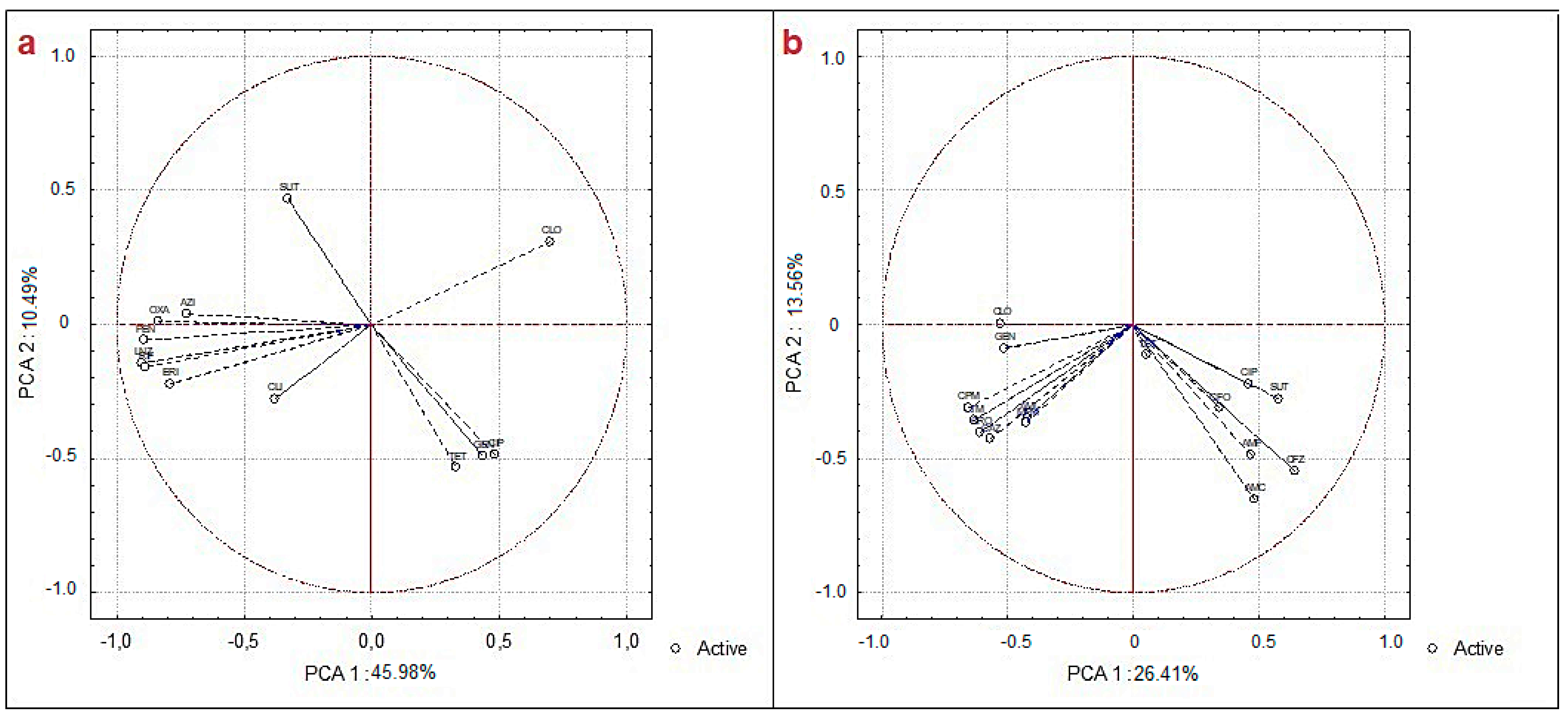
Correlation analyses were applied for antimicrobial susceptibility and resistance of bacterial isolates among Enterobacteriaceae and Pasteurellaceae and among Gram-positive rod/bacillus and Staphylococcaceae. The gaps were eliminated in order to perform the PCA. The PCA is illustrated in Figure 3, and its loading matrix is shown in Tables S8 and S9. The Spearman correlation is shown in Tables S10 and S11.
For the Gram-positive rod/bacillus and Staphylococcaceae data, two groups with inter-correlations were formed—a group medially in the fourth quadrant of the PCA and another group of data influenced by the third quadrant of the PCA—both being negatively influenced by PCA1. For the multiple analyses of Enterobacteriaceae and Pasteurellaceae regarding resistance, two large groups were grouped: one negatively influenced by PCA1 and PCA2 and the other moderately positively influenced by PCA1 and negatively influenced by PCA2. The total variance of the data was 39.96% (Figure 3b).
From the analysis of correlations performed for each two antimicrobials, only the correlation between CFZ with AMC and AMP was positively moderately strong and statistically significant for the resistance of Gram-negatives. The other correlations found were reasonable and negative (Table S10). For Gram-positive bacteria (Table S11), the very strong, statistically significant correlations were found between LNZ with ERI, PEN and RIF, and between PEN and OXA.
The correlations and inter-correlations found may be related to antibiotic classes. The correlations between lincosamides and β-lactams may be related to the mechanism of action of both, and they may act on cell wall synthesis. Another point to be discussed is that many ARGs are often found in the same plasmid or in mobile genetic elements, increasing the associations between antibiotic resistance and susceptibility [71].
The presence of several ARGs was identified in the water and sediment samples from the Meia Ponte River (Table 4). The most prevalent ARGs in the water and sediment samples were e sul2 e ermC, with 100% (16/16), and sul1, qnrB and aac(‘6)-ib with 93.75% (15/16). All genes for the eight classes of antibiotics tested were amplified. The sampling points with the highest prevalence of ARGs were point MP03, a water sample collected during the rainy season, and points MP02 and MP03, sediment samples collected during the dry season, with 83.33% (20/24). The total prevalence of ARGs in the sediment and water samples of the Meia Ponte River was 68.49% (263/384), of which 39.58% (152/384) were isolated in the rainy season and 28.91% (111/384) in the dry season. On the other hand, 29.17% (112/384) were isolated from water samples and 39.32% (151/384) from sediment samples (Table S12).
The presence of ARGs is reported in the southeast and south of Brazil; however, in the midwest, there are very few studies [72], highlighting the importance of this study, as the identification of ARBs and ARGs in the environment of a river is indicative of a strong anthropogenic impact, with monitoring being the first complex task [73]. In Brazil, the main potential sources of ARGs in the environment are domestic and hospital sewage, as well as wastewater from agriculture. The high prevalence of ARBs and ARGs in the environment will affect the treatment of bacterial infections in the future in Latin America [72]. The state of the Meia Ponte River in relation to ARGs and ARBs is critical, requiring intervention and creation of measures to block the increase in this contamination, as in the future, it could be disastrous for the environment and for the public health of central-western Brazil.
PCA was performed and included in Figure 4, detailing the presence and absence of ARGs. For the PCA of resistance genes, two groups were represented with the total variance of 70.42%. The first group was strongly negatively influenced by PCA1 in quadrant III, and the second group was strongly positively influenced by PCA2 (flower genes, tet(O), aac(‘6)-ib) (Figure 4a). IntI1 had an eigenvalue for PCA1 of −0.85, being associated with other ARGs (Figure 4a and Table S13), and may suggest that this gene may affect the formation of other ARGs [74].
The points and periods were grouped into PM02 and PM03 for water collection during the dry period; points PM01 and PM04 for water collection during the dry period were far from the other points. This association among the points of the water samples is directly linked, as they had the highest values of absence of ARGs in relation to the other sampling periods and were probably influenced by the dry season, in this case, the flow of the river decreased. The other sample points and remaining periods were all grouped into a set with high correlation. In urban rivers, it is difficult to distinguish the factors that affect the relationship of ARGs, as they are influenced by several parameters, pollutants and components [75].
Among the two-by-two correlations, most of the statistically significant analyses were considered very strong or moderately strong, indicating that this group of genes studied had a strong relationship. In the PCA shown in Figure S2, temperature is strongly correlated with crf, tet(A), tet(O) and floR. This association was proven, indicating temperature as a limiting factor in the concentration of ARGs [76].
PCA, as a multi-variate method, was applied to data from the Meia Ponte River, managing to explain strong and significant inter-correlations. PCA is a method commonly applied to verify the correlations among water quality constituents and in watersheds, reducing the number of variables [77]. In addition, it also constitutes an appropriate method for portraying the anthropogenic impact and grouping according to seasonality and groups that share similar data [78]. It was observed that the Meia Ponte River suffers from a great impact caused by human activity. The data for the three groups (Figure 2, Figure 3 and Figure 4) indicated the association among the sampling points and periods.

4. Conclusions

When a water body does not meet the quality criteria, it harms and poses a threat to human health and ecological integrity. The Meia Ponte River is a water body in a critical state, which needs great attention. Seasonal variation had no statistically significant influence among the analyzed parameters. Among the physicochemical parameters analyzed, some were not in accordance with the Brazilian legislation. With regard to microbiological parameters, the Meia Ponte River is highly contaminated, with a high prevalence of ARBs and ARGs. A high percentage of multi-drug resistance was found.
The literature indicates that this is the first study verifying the abundance of resistance genes in water and sediment samples in an aquatic system in the Brazilian midwest region. ARGs were found for eight classes of antibiotics: β-lactams, quinolones, fluoroquinolones, sulfonamides, tetracyclines, macrolides, amphenicols and integron. Several significant correlations (p-value < 0.05) were verified, demonstrating multi-variate and two-by-two correlation analyses, which are important for explaining resistance data and indicate their possible anthropogenic influence on the aquatic environment. The data indicated that the Meia Ponte River serves as a reservoir of antibiotic resistance genes in central-western Brazil. This represents a warning sign for public health.
Alternatives to reduce the impact on the Meia Ponte River need to be studied, in addition to reducing or interrupting the disposal of untreated wastewater, increasing the points of sewage treatment plants. Another point is to improve the identification of point/diffuse or direct/indirect sources that increase the concentration of ARGs, ARBs and pollutants, such as antibiotics, metals, personal care products and pharmaceuticals. Finally, better communication between researchers, politicians, government and the population is also suggested in order to clarify the harm and possible consequences if this water environment is not preserved.
Finally, it is recommended that more in-depth microbiological parameters, such as analyses of antibiotics, ARBs and ARGs, are carried out and monitored in central-western Brazil and around the world. This is because the problem of resistance is global, and actions must be taken.

Supplementary Materials

The following supporting information can be downloaded at: https://www.mdpi.com/article/10.3390/w15040747/s1, Figure S1: Photograph demonstrating the sampling points of the Meia Ponte River. a. MP01 sampling point; b. MP02 sampling point; c. MP03 sampling point; d. MP04 sampling point; Table S1: Primers used in this study; Table S2: Percentage of bioprospecting of bacterial isolates from water and sediment samples from the Meia Ponte River; Table S3: Spearman correlation of physicochemical and microbiological parameters of water and sediment samples from the Meia Ponte River; Table S4: Loading matrix showing the main components of the physicochemical and microbiological dataset of water and sediment samples from the Meia Ponte River; Table S5: Percentage of sensitivity and resistance of bacterial isolates bioprospected from the João Leite stream, categorized by rainy season; Table S6: Percentage of sensitivity and resistance of bacterial isolates bioprospected from the João Leite stream, categorized by dry period; Table S7: Percentage of sensitivity and resistance of bacterial isolates bioprospected from the João Leite stream, total; Table S8: Loading matrix showing the main components of the Gram-positive rod/bacillus and Staphylococcaceae resistance and susceptibility dataset isolated from the Meia Ponte River sediment and water samples; Table S9: Loading matrix showing the main components of the resistance and susceptibility dataset of Enterobacteriaceae and Pasteurellaceae isolated from water and sediment samples from the Meia Ponte River; Table S10: Spearman correlation of the resistance and susceptibility dataset of Gram-positive rod/bacillus and Staphylococcaceae isolated from water and sediment samples from the Meia Ponte River; Table S11: Spearman correlation of the resistance and susceptibility dataset from the resistance and susceptibility dataset of Enterobacteriaceae and Pasteurellaceae isolated from water and sediment samples from the Meia Ponte River.

Author Contributions

R.P.G.: conceptualization, methodology, data curation, writing—review, writing—original draft preparation, formal analysis and editing; T.R.O.: methodology; A.B.R.: methodology; L.M.F.: methodology; J.D.G.V.: data curation and supervision; L.C.C.: conceptualization, supervision, data curation and writing—review. All authors have read and agreed to the published version of the manuscript.

Funding

This research did not have financial support.

Institutional Review Board Statement

Not applicable.

Informed Consent Statement

Not applicable.

Data Availability Statement

Not applicable.

Acknowledgments

My thanks also go to the Coordination for the Improvement of Higher Education Personnel (CAPES), the State of Goiás Research Support Foundation (FAPEG) and the National Council for Scientific and Technological Development (CNPq).

Conflicts of Interest

The author declares that they have no competing interest.

References

  1. Uddin, M.G.; Nash, S.; Olbert, A.I. A review of water quality index models and their use for assessing surface water quality. Ecol. Indic. 2021, 122, 107218. [Google Scholar] [CrossRef]
  2. Tiyasha; Tung, T.M.; Yaseen, Z.M. A survey on river water quality modelling using artificial intelligence models: 2000–2020. J. Hydrol. 2020, 585, 124670. [Google Scholar] [CrossRef]
  3. Kachroud, M.; Trolard, F.; Kefi, M.; Jebari, S.; Bourrié, G. Water quality indices: Challenges and application limits in the literature. Water 2019, 11, 361. [Google Scholar] [CrossRef]
  4. Giri, S. Water quality prospective in Twenty First Century: Status of water quality in major river basins, contemporary strategies and impediments: A review. Environ. Pollut. 2021, 271, 116332. [Google Scholar] [CrossRef] [PubMed]
  5. World Economic Forum. The Global Risks Report 2021, 16th ed.; World Economic Forum: Geneva, Switzerland, 2021; ISBN 9782940631247. [Google Scholar]
  6. Geissen, V.; Mol, H.; Klumpp, E.; Umlauf, G.; Nadal, M.; van der Ploeg, M.; van de Zee, S.E.A.T.M.; Ritsema, C.J. Emerging pollutants in the environment: A challenge for water resource management. Int. Soil Water Conserv. Res. 2015, 3, 57–65. [Google Scholar] [CrossRef]
  7. Kumar, M.; Borah, P.; Devi, P. Priority and emerging pollutants in water. In Inorganic Pollutants in Water; Elsevier: Amsterdam, The Netherlands, 2020; pp. 33–49. [Google Scholar] [CrossRef]
  8. Dulio, V.; van Bavel, B.; Brorström-Lundén, E.; Harmsen, J.; Hollender, J.; Schlabach, M.; Slobodnik, J.; Thomas, K.; Koschorreck, J. Emerging pollutants in the EU: 10 years of NORMAN in support of environmental policies and regulations. Environ. Sci. Eur. 2018, 30, 5. [Google Scholar] [CrossRef] [PubMed]
  9. García, J.; García-Galán, M.J.; Day, J.W.; Boopathy, R.; White, J.R.; Wallace, S.; Hunter, R.G. A review of emerging organic contaminants (EOCs), antibiotic resistant bacteria (ARB), and antibiotic resistance genes (ARGs) in the environment: Increasing removal with wetlands and reducing environmental impacts. Bioresour. Technol. 2020, 307, 123228. [Google Scholar] [CrossRef] [PubMed]
  10. Li, S.; Zhang, C.; Li, F.; Hua, T.; Zhou, Q.; Ho, S.H. Technologies towards antibiotic resistance genes (ARGs) removal from aquatic environment: A critical review. J. Hazard. Mater. 2021, 411, 125148. [Google Scholar] [CrossRef]
  11. Sanderson, H.; Fricker, C.; Brown, R.S.; Majury, A.; Liss, S.N. Antibiotic Resistance Genes as an Emerging Environmental Contaminant Haley. Environ. Rev. 2016, 24, 205–218. [Google Scholar] [CrossRef]
  12. Shao, S.; Hu, Y.; Cheng, J.; Chen, Y. Research progress on distribution, migration, transformation of antibiotics and antibiotic resistance genes (ARGs) in aquatic environment. Crit. Rev. Biotechnol. 2018, 38, 1195–1208. [Google Scholar] [CrossRef]
  13. Sharma, V.K.; Johnson, N.; Cizmas, L.; McDonald, T.J.; Kim, H. A review of the influence of treatment strategies on antibiotic resistant bacteria and antibiotic resistance genes. Chemosphere 2016, 150, 702–714. [Google Scholar] [CrossRef] [PubMed]
  14. Li, R.; Jay, J.A.; Stenstrom, M.K. Fate of antibiotic resistance genes and antibiotic-resistant bacteria in water resource recovery facilities. Water Environ. Res. 2019, 91, 5–20. [Google Scholar] [CrossRef]
  15. Marti, E.; Variatza, E.; Balcazar, J.L. The role of aquatic ecosystems as reservoirs of antibiotic resistance. Trends Microbiol. 2014, 22, 36–41. [Google Scholar] [CrossRef]
  16. Caniça, M.; Manageiro, V.; Abriouel, H.; Moran-Gilad, J.; Franz, C.M.A.P. Antibiotic resistance in foodborne bacteria. Trends Food Sci. Technol. 2019, 84, 41–44. [Google Scholar] [CrossRef]
  17. Suzuki, S.; Pruden, A.; Virta, M.; Zhang, T. Antibiotic resistance in aquatic systems. Front. Microbiol. 2017, 8, 14. [Google Scholar] [CrossRef]
  18. Nnadozie, C.F.; Odume, O.N. Freshwater environments as reservoirs of antibiotic resistant bacteria and their role in the dissemination of antibiotic resistance genes. Environ. Pollut. 2019, 254, 113067. [Google Scholar] [CrossRef]
  19. Sanganyado, E.; Gwenzi, W. Antibiotic resistance in drinking water systems: Occurrence, removal, and human health risks. Sci. Total Environ. 2019, 669, 785–797. [Google Scholar] [CrossRef]
  20. Bürgmann, H.; Frigon, D.; Gaze, W.H.; Manaia, C.M.; Pruden, A.; Singer, A.C.; Smets, B.F.; Zhang, T. Water and sanitation: An essential battlefront in the war on antimicrobial resistance. FEMS Microbiol. Ecol. 2018, 94, fiy101. [Google Scholar] [CrossRef]
  21. Böger, B.; Surek, M.; Vilhena, R.D.O.; Fachi, M.M.; Junkert, A.M.; Santos, J.M.; Domingos, E.L.; Cobre, A.d.F.; Momade, D.R.; Pontarolo, R. Occurrence of antibiotics and antibiotic resistant bacteria in subtropical urban rivers in Brazil. J. Hazard. Mater. 2021, 402, 123448. [Google Scholar] [CrossRef]
  22. Amaral, A.K.N.; Formiga, K.T.M. Análise da distribuição granulométrica ao longo da Bacia Hidrográfica do Rio Meia Ponte—Goiás. Rev. Gestão Água América Lat. 2019, 16, 1–13. [Google Scholar] [CrossRef]
  23. Costa, H.S.; Tejerina-Garro, F.L.; Rocha, C. Trace elements: Water-sediment interactions in tropical rivers. Environ. Sci. Pollut. Res. 2017, 24, 22018–22025. [Google Scholar] [CrossRef] [PubMed]
  24. De Paula, L.G.F.; Zeringóta, V.; Sampaio, A.L.N.; Bezerra, G.P.; Barreto, A.L.G.; dos Santos, A.A.; Miranda, V.C.; Paula, W.V.D.F.; Neves, L.C.; Secchis, M.V.; et al. Seasonal dynamics of Amblyomma sculptum in two areas of the Cerrado biome midwestern Brazil, where human cases of rickettsiosis have been reported. Exp. Appl. Acarol. 2021, 84, 215–225. [Google Scholar] [CrossRef] [PubMed]
  25. Gomes, R.P.; de Paula Silva, J.A.; Carvalho Junior, M.C.; Alburquerque, W.C.A.; Scalize, P.S.; Galvão Filho, A.R.; de Jesus Pires, D.; Vieira, J.D.G.; Carneiro, L.C. Evaluation of the raw water quality: Physicochemical and toxicological approaches. Environ. Geochem. Health. 2019, 41, 2425–2442. [Google Scholar] [CrossRef]
  26. Soares, A.K.; Salgado, A.D.A.; Scaratti, D.; Milograna, J.; Hora, K.E.R.; Formiga, K.T.M.; Pinho, L.; Mauro, R.A.; Duarte, L.V.; Casaroli, L.; et al. Plano de Ação da Upgrh do Rio Meia Ponte 2020, pp. 1–199. Available online: http://pbapgo.meioambiente.go.gov.br/wp-content/uploads/2020/12/RT-04-Plano-de-Acoes-UPGRH-Meia-Ponte-V01.pdf (accessed on 9 February 2023).
  27. Veiga, A.M.; Santos, C.C.P.; Cardoso, M.R.D.; Lino, N.C. Caracterização hidromorfológica da bacia do rio Meia Ponte. Caminhoas Geogr. 2013, 14, 126–138. [Google Scholar]
  28. CONAMA. Brasil Resolução CONAMA N° 357, de 17 de Março de 2005; Retificada; CONAMA: Brasilia, Brazil, 2005. Available online: https://www.icmbio.gov.br/cepsul/images/stories/legislacao/Resolucao/2005/res_conama_357_2005_classificacao_corpos_agua_rtfcda_altrd_res_393_2007_397_2008_410_2009_430_2011.pdf (accessed on 9 February 2023).
  29. Bailão, E.F.L.C.; Zago, L.D.M.S.; Silva, N.C.; Machado, K.B.; D’Abadia, P.L.; de Oliveira, P.H.F.; Nabout, J.C.; de Almeida, L.M. Urban occupation increases water toxicity of an important river in central Brazil. Fronteiras 2020, 9, 73–86. [Google Scholar] [CrossRef]
  30. Cetesb Guia Nacional de Coleta e Preservação de Amostras. Água, Sedimento, Comunidades Aquáticas e Efluentes Líquidos; Copmhania Ambiental do Estado do São Paulo: Sao Paulo, Brazil, 2011; p. 326. Available online: https://cetesb.sp.gov.br/wp-content/uploads/2021/10/Guia-nacional-de-coleta-e-preservacao-de-amostras-2012.pdf (accessed on 9 February 2023).
  31. American Public Health Association (APHA). Standard Methodds for The Examination of Water and Wastewater; American Public Health Association (APHA): Washington, DC, USA, 2018; Volume 23, ISBN 9788578110796. Available online: https://www.wef.org/resources/publications/books/StandardMethods/ (accessed on 9 February 2023).
  32. Associação Brasileira de Normas Técnicas (ABNT). Água—Determinação de Oxigênio Consumido—Método do Permanganato de Potássio; SET 1989; Associação Brasileira de Normas Técnicas (ABNT): Sao Paulo, Brazil, 1989; Available online: https://www.target.com.br/produtos/normas-tecnicas/39198/nbr10739-agua-determinacao-de-oxigenio-consumido-metodo-do-permanganato-de-potassio-metodo-de-ensaio (accessed on 9 February 2023).
  33. Shariq, M.; Singh, S.; Farooq, U.; Dhariyal, K.K.; Singh, K.; Kaur, N. Presumptive Coliform Count in Water Sample Collected from Different Sites of. Int. J. Sci. Study 2016, 3, 12. [Google Scholar] [CrossRef]
  34. Da Cunha, F.V.; Batista, S.B. Análise fenotípica e genotípica de bactérias heterotróficas e fixadoras de nitrogênio em sedimento na bacia do Rio Cuiabá-MT. Rev. Ambient. Agua. 2014, 9, 68–80. [Google Scholar] [CrossRef]
  35. Agência Nacional de Vigilância Sanitária. Módulo 6: Detecção e identificação e bactérias de importância médica. In Manual de Microbiologia Clínica Para o Controle Assistência à Saúde; Agência Nacional de Vigilância Sanitária: Brasilia, Brazil, 2013; Volume 9. Available online: https://www.gov.br/anvisa/pt-br/centraisdeconteudo/publicacoes/servicosdesaude/publicacoes/modulo-10_manual-de-microbiologia.pdf (accessed on 9 February 2023).
  36. Clinical and Laboratory Standards Institute. Performance Standards for Antimicrobial Susceptibility Testing; 29th informational supplement; CLSI document M100-S29; 29th suppl.; Clinical and Laboratory Standards Institute: Wayne, PA, USA, 2019; ISBN 978-1-68440-034. Available online: https://clsi.org/media/2663/m100ed29_sample.pdf (accessed on 9 February 2023).
  37. Magiorakos, A.P.; Srinivasan, A.; Carey, R.B.; Carmeli, Y.; Falagas, M.E.; Giske, C.G.; Harbarth, S.; Hindler, J.F.; Kahlmeter, G.; Olsson-Liljequist, B.; et al. Multidrug-resistant, extensively drug-resistant and pandrug-resistant bacteria: An international expert proposal for interim standard definitions for acquired resistance. Clin. Microbiol. Infect. 2012, 18, 268–281. [Google Scholar] [CrossRef]
  38. Santos, A.L.; Dos Santos, A.P.; Ito, C.R.M.; de Queiroz, P.H.P.; de Almeida, J.A.; Júnior, M.A.B.D.C.; de Oliveira, C.Z.; Avelino, M.A.G.; Wastowski, I.J.; Gomes, G.P.L.A.; et al. Profile of enterobacteria resistant to beta-lactams. Antibiotics 2020, 9, 410. [Google Scholar] [CrossRef]
  39. Knapp, C.W.; Zhang, W.; Sturm, B.S.M.; Graham, D.W. Differential fate of erythromycin and beta-lactam resistance genes from swine lagoon waste under different aquatic conditions. Environ. Pollut. 2010, 158, 1506–1512. [Google Scholar] [CrossRef]
  40. Robicsek, A.; Strahilevitz, J.; Sahm, D.F.; Jacoby, G.A.; Hooper, D.C. qnr prevalence in ceftazidime-resistant Enterobacteriaceae isolates from the United States. Antimicrob. Agents Chemother. 2006, 50, 2872–2874. [Google Scholar] [CrossRef]
  41. Park, C.H.; Robicsek, A.; Jacoby, G.A.; Sahm, D.; Hooper, D.C. Prevalence in the United States of aac(6′)-Ib-cr encoding a ciprofloxacin-modifying enzyme. Antimicrob. Agents Chemother. 2006, 50, 3953–3955. [Google Scholar] [CrossRef]
  42. Waldron, L.S.; Gillings, M.R. Screening foodstuffs for class 1 integrons and gene cassettes. J. Vis. Exp. 2015, 2015, e52889. [Google Scholar] [CrossRef]
  43. Ng, L.K.; Martin, I.; Alfa, M.; Mulvey, M. Multiplex PCR for the detection of tetracycline resistant genes. Mol. Cell. Probes 2001, 15, 209–215. [Google Scholar] [CrossRef]
  44. Heuer, H.; Smalla, K. Manure and sulfadiazine synergistically increased bacterial antibiotic resistance in soil over at least two months. Environ. Microbiol. 2007, 9, 657–666. [Google Scholar] [CrossRef]
  45. Lu, Z.; Na, G.; Gao, H.; Wang, L.; Bao, C.; Yao, Z. Fate of sulfonamide resistance genes in estuary environment and effect of anthropogenic activities. Sci. Total Environ. 2015, 527–528, 429–438. [Google Scholar] [CrossRef]
  46. Li, J.; Shao, B.; Shen, J.; Wang, S.; Wu, Y. Occurrence of chloramphenicol-resistance genes as environmental pollutants from swine feedlots. Environ. Sci. Technol. 2013, 47, 2892–2897. [Google Scholar] [CrossRef]
  47. Chan, Y.H. Biostatistics 304: Cluster analysis. Singapore Med. J. 2003, 44, 614–619. [Google Scholar]
  48. Sória, M.; Tavares, V.E.Q.; Pinto, M.A.B.; Stumpf, L.; Zarnott5, D.; Bubolz, J.; Nörenberg, B.G. Evaluation of physicochemical water parameters in watersheds of southern southern Brazil. Rev. Ambient. Agua 2020, 15, e2596. [Google Scholar] [CrossRef]
  49. Mustapha, A.; Aris, A.Z.; Juahir, H.; Ramli, M.F.; Kura, N.U. River water quality assessment using environmentric techniques: Case study of Jakara River Basin. Environ. Sci. Pollut. Res. 2013, 20, 5630–5644. [Google Scholar] [CrossRef]
  50. Rusydi, A.F. Correlation between conductivity and total dissolved solid in various type of water: A review. IOP Conf. Ser. Earth Environ. Sci. 2018, 118, 012019. [Google Scholar] [CrossRef]
  51. Wu, H.; Yang, W.; Yao, R.; Zhao, Y.; Zhao, Y.; Zhang, Y.; Yuan, Q.; Lin, A. Evaluating surface water quality using water quality index in Beiyun River, China. Environ. Sci. Pollut. Res. 2020, 27, 35449–35458. [Google Scholar] [CrossRef]
  52. Ahmed, A.A.M. Prediction of dissolved oxygen in Surma River by biochemical oxygen demand and chemical oxygen demand using the artificial neural networks (ANNs). J. King Saud Univ. Eng. Sci. 2017, 29, 151–158. [Google Scholar] [CrossRef]
  53. Bayram, A.; Uzlu, E.; Kankal, M.; Dede, T. Modeling stream dissolved oxygen concentration using teaching–learning based optimization algorithm. Environ. Earth Sci. 2015, 73, 6565–6576. [Google Scholar] [CrossRef]
  54. Harvey, R.; Lye, L.; Khan, A.; Paterson, R. The influence of air temperature on water temperature and the concentration of dissolved oxygen in Newfoundland Rivers. Can. Water Resour. J. 2011, 36, 171–192. [Google Scholar] [CrossRef]
  55. CONAMA. Brasil Resolução CONAMA no 274, de 29 de Novembro de 2000; CONAMA: Brasilia, Brazil, 2000. Available online: http://pnqa.ana.gov.br/Publicacao/Resolu%C3%A7%C3%A3o_Conama_274_Balneabilidade.pdf (accessed on 9 February 2023).
  56. Garbossa, L.H.P.; Souza, R.V.; Campos, C.J.A.; Vanz, A.; Vianna, L.F.N.; Rupp, G.S. Thermotolerant coliform loadings to coastal areas of Santa Catarina (Brazil) evidence the effect of growing urbanisation and insufficient provision of sewerage infrastructure. Environ. Monit. Assess. 2017, 189, 27. [Google Scholar] [CrossRef]
  57. Poma, V.; Mamani, N.; Iñiguez, V. Impact of urban contamination of the La Paz River basin on thermotolerant coliform density and occurrence of multiple antibiotic resistant enteric pathogens in river water, irrigated soil and fresh vegetables. Springerplus 2016, 5, 499. [Google Scholar] [CrossRef]
  58. Marques, L.O.D.A.; Taffarello, D.; Calijuri, M.D.C.; Mendiondo, E.M.; Ferreira, M.D.S.; Cunha, D.G.F. Phosphorus and thermotolerant coliforms’ loads in brazilian watersheds with limited data: Considerations on the integrated analysis of water quality and quantity. Rev. Bras. Recur. Hidricos 2019, 24, 1–13. [Google Scholar] [CrossRef]
  59. Da Cunha, D.F.; da Costa, N.M.; Barreira, C.C.M.A. Integração E Cooperação Territorial Na Região Metropolitana De Goiânia / Territorial Integration and Cooperation in the Goiania Metropolitan Region. Geo UERJ 2017, 30, 76–98. [Google Scholar] [CrossRef]
  60. Sylvestre, É.; Burnet, J.B.; Smeets, P.; Medema, G.; Prévost, M.; Dorner, S. Can routine monitoring of E. coli fully account for peak event concentrations at drinking water intakes in agricultural and urban rivers? Water Res. 2020, 170, 115369. [Google Scholar] [CrossRef]
  61. Mena-Rivera, L.; Vásquez-Bolaños, O.; Gómez-Castro, C.; Fonseca-Sánchez, A.; Rodríguez-Rodríguez, A.; Sánchez-Gutiérrez, R. Ecosystemic assessment of surface water quality in the Virilla River: Towards sanitation processes in Costa Rica. Water 2018, 10, 845. [Google Scholar] [CrossRef] [Green Version]
  62. De Oliveira, A.J.F.C.; Hollnagel, H.C.; Lima Mesquita, H.D.S.; Fontes, R.F.C. Physical, chemical and microbiological characterization of the intertidal sediments of Pereque Beach, Guarujá (SP), Brazil. Mar. Pollut. Bull. 2007, 54, 921–927. [Google Scholar] [CrossRef] [PubMed]
  63. Seo, M.; Lee, H.; Kim, Y. Relationship between Coliform bacteria and water quality factors at weir stations in the Nakdong River, South Korea. Water 2019, 11, 1171. [Google Scholar] [CrossRef]
  64. Chaturvedi, P.; Chowdhary, P.; Singh, A.; Chaurasia, D.; Pandey, A.; Chandra, R.; Gupta, P. Dissemination of antibiotic resistance genes, mobile genetic elements, and efflux genes in anthropogenically impacted riverine environments. Chemosphere 2021, 273, 129693. [Google Scholar] [CrossRef] [PubMed]
  65. Oliveira, T.R.; Gomes, R.P.; Rodrigues, A.B.; Ferreira, L.M.; Gama, A.R.; Vieira, J.D.G.; Fernandes, M.R.; Carneiro, L.C. Identification and resistance profile of gram positive bacteria from aquatic environment. Res. Soc. Dev. 2021, 10, e226101321182. [Google Scholar] [CrossRef]
  66. Freitas, D.Y.; Araújo, S.; Folador, A.R.C.; Ramos, R.T.J.; Azevedo, J.S.N.; Tacão, M.; Silva, A.; Henriques, I.; Baraúna, R.A. Extended spectrum beta-lactamase-producing gram-negative bacteria recovered from an amazonian lake near the city of Belém, Brazil. Front. Microbiol. 2019, 10, 364. [Google Scholar] [CrossRef]
  67. Rebello, R.C.D.L.; Regua-Mangia, A.H. Potential enterovirulence and antimicrobial resistance in Escherichia coli isolates from aquatic environments in Rio de Janeiro, Brazil. Sci. Total Environ. 2014, 490, 19–27. [Google Scholar] [CrossRef]
  68. Chaturvedi, P.; Singh, A.; Chowdhary, P.; Pandey, A.; Gupta, P. Occurrence of emerging sulfonamide resistance (sul1 and sul2) associated with mobile integrons-integrase (intI1 and intI2) in riverine systems. Sci. Total Environ. 2021, 751, 142217. [Google Scholar] [CrossRef]
  69. Heck, K.; de Marco, É.G.; Duarte, M.W.; Salamoni, S.P.; van der Sand, S. Pattern of multiresistant to antimicrobials and heavy metal tolerance in bacteria isolated from sewage sludge samples from a composting process at a recycling plant in southern Brazil. Environ. Monit. Assess. 2015, 187, 328. [Google Scholar] [CrossRef]
  70. Lee, J.; Beck, K.; Bürgmann, H. Wastewater bypass is a major temporary point-source of antibiotic resistance genes and multi-resistance risk factors in a Swiss river. Water Res. 2021, 208, 117827. [Google Scholar] [CrossRef]
  71. Chen, Z.; Yu, D.; He, S.; Ye, H.; Zhang, L.; Wen, Y.; Zhang, W.; Shu, L.; Chen, S. Prevalence of antibiotic-resistant Escherichia coli in drinking water sources in Hangzhou City. Front. Microbiol. 2017, 8, 1133. [Google Scholar] [CrossRef] [PubMed] [Green Version]
  72. Reichert, G.; Hilgert, S.; Fuchs, S.; Azevedo, J.C.R. Emerging contaminants and antibiotic resistance in the different environmental matrices of Latin America. Environ. Pollut. 2019, 255, 113140. [Google Scholar] [CrossRef] [PubMed]
  73. Arsand, J.B.; Hoff, R.B.; Jank, L.; Bussamara, R.; Dallegrave, A.; Bento, F.M.; Kmetzsch, L.; Falção, D.A.; do Carmo Ruaro Peralba, M.; de Araujo Gomes, A.; et al. Presence of antibiotic resistance genes and its association with antibiotic occurrence in Dilúvio River in southern Brazil. Sci. Total Environ. 2020, 738, 139781. [Google Scholar] [CrossRef] [PubMed]
  74. Zheng, J.; Zhou, Z.; Wei, Y.; Chen, T.; Feng, W.; Chen, H. High-throughput profiling of seasonal variations of antibiotic resistance gene transport in a peri-urban river. Environ. Int. 2018, 114, 87–94. [Google Scholar] [CrossRef] [PubMed]
  75. Xu, Y.; Guo, C.; Luo, Y.; Lv, J.; Zhang, Y.; Lin, H.; Wang, L.; Xu, J. Occurrence and distribution of antibiotics, antibiotic resistance genes in the urban rivers in Beijing, China. Environ. Pollut. 2016, 213, 833–840. [Google Scholar] [CrossRef] [PubMed]
  76. Yang, J.; Wang, H.; Roberts, D.J.; Du, H.N.; Yu, X.F.; Zhu, N.Z.; Meng, X.Z. Persistence of antibiotic resistance genes from river water to tap water in the Yangtze River Delta. Sci. Total Environ. 2020, 742, 140592. [Google Scholar] [CrossRef]
  77. Olsen, R.L.; Chappell, R.W.; Loftis, J.C. Water quality sample collection, data treatment and results presentation for principal components analysis—Literature review and Illinois River watershed case study. Water Res. 2012, 46, 3110–3122. [Google Scholar] [CrossRef]
  78. Menció, A.; Mas-Pla, J. Assessment by multivariate analysis of groundwater-surface water interactions in urbanized Mediterranean streams. J. Hydrol. 2008, 352, 355–366. [Google Scholar] [CrossRef]
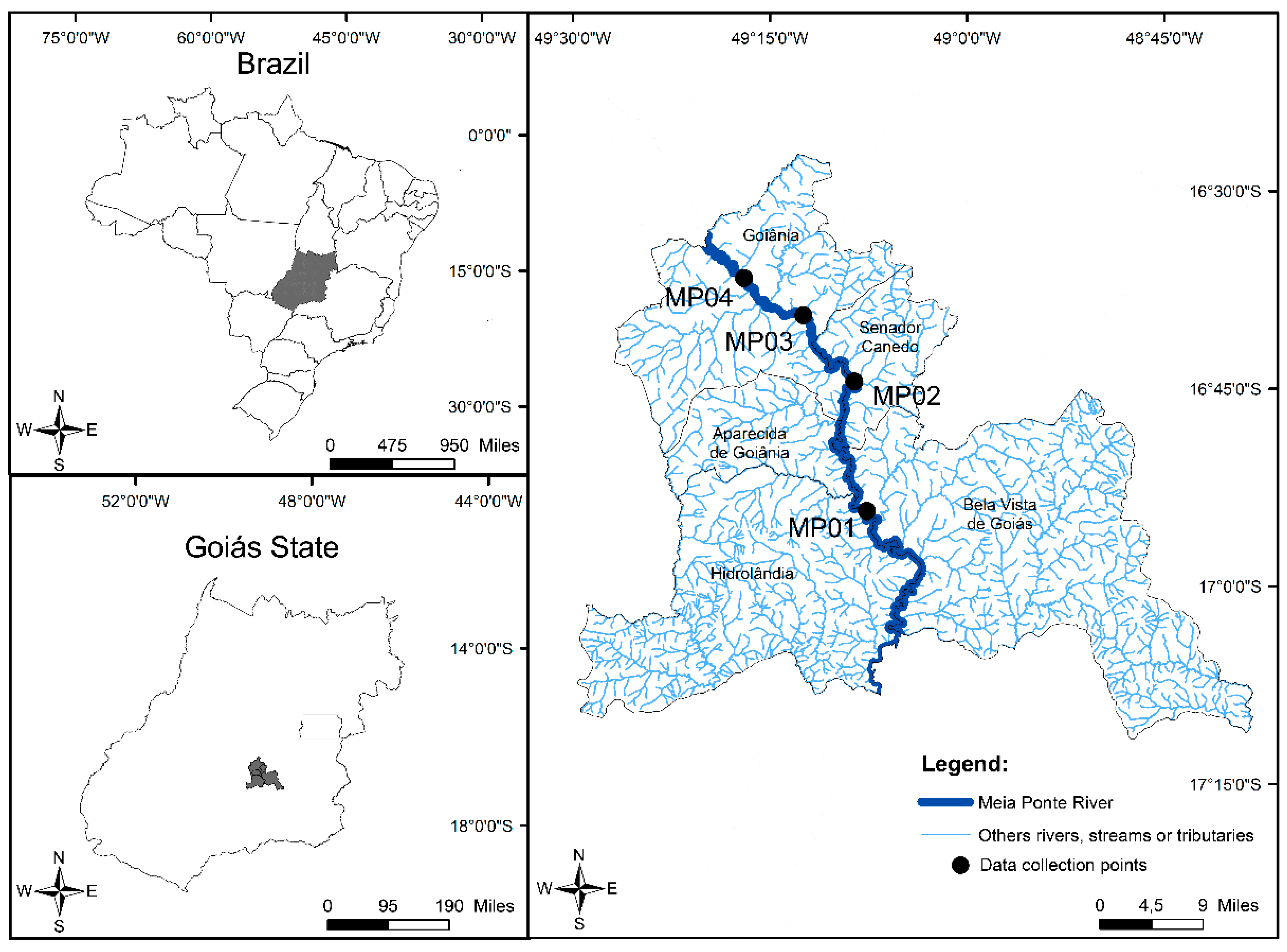
Figure 1. Map showing the sampling locations of water and sediment samples from the Meia Ponte River. The referred geographic coordinates of the sampling points are MP01 16°54′16.3″ S; 49°07′37.8″ W, MP02 16°44′27.7″ S; 49°08′35.3″ W, MP03 16°39′26.0″ S; 49°12′27.1″ W and MP04 16°36′36.7″ S; 49°16′58.8″ W.
Water 15 00747 g001
Figure 2. PCA of physicochemical and microbiological parameters regarding the quality of water and sediments of Meia Ponte River. (a) Dot and vector graph of physicochemical and microbiological parameters. (b) Loads graph of the influence of physicochemical and microbiological parameters in the sampling points and their distribution based on this influence.
Water 15 00747 g002
Figure 3. PCA of resistance and susceptibility data of bacterial strains isolated from water and sediment samples from Meia Ponte River. (a) Dot and vector plot of Gram-positive rod/bacillus and Staphylococcaceae data. (b) Dot and vector plot of Enterobacteriaceae and Pasteurellaceae data.
Water 15 00747 g003
Figure 4. PCA of the presence and absence of ARGs detected in the water and sediments of Meia Ponte River. (a) Dot and vector graphics for the ARGs. (b) ARBs influence on loads graph at sampling points and their distribution based on this influence.
Water 15 00747 g004
Table 1. Physicochemical parameters of the water quality of Meia Ponte River.
ParameterChloride (mg/L)Electrical Conductivity (µS/cm)Apparent Color (mg Pt/L)Hardness (mg/L)Nitrate (mg/L)Oxygen Dissolved (mg/L)pHWater Temperature (°C)Turbidity (NTU)
Rainy periodMP011.77180.00150.00 *60.001.726.887.3722.50218.00 *
MP021.76202.00251.00 *106.711.345.797.2723.0097.70
MP031.40179.70321.00 *38.701.385.897.2225.00137.00 *
MP040.53131.50314.00 *25.301.405.867.4225.00100.00 *
average ± SD1.365 ± 0.58173.30 ± 29.76259.00 * ± 79.1957.68 ± 35.671.46 ± 0.0186.10 ± 0.527.32 ± 0.0923.87 ± 1.31138.18 * ± 56.18
Dry periodMP012.51383.00104.00 *14.670.306.597.3023.5010.30
MP022.82431.00123.00 *17.340.0010.127.1124.5016.30
MP031.78291.0066.900.000.207.027.0426.0013.00
MP041.09187.0053.200.001.300.73 *7.6026.005.81
average ± SD2.05 ±0.77323.00 ± 107.6886.78 ± 32.318.00 ± 9.300.45 ± 0.586.12 ± 3.927.26 ± 0.2525.00 ± 1.2211.35 ± 4.44
Maximum recommended values for class 2 of CONAMA Regulation No. 357 *250.00NR75.00NR10.00Not inferior 5.006.0–9.0NR100.00
media ± DP total1.71 ± 0.73248.15 ± 108.40172.89 ± 107.7532.84 ± 35.880.95 ± 0.676.11 ± 2.597.29 ± 0.1824.44 ± 1.3274.76 ± 77.18
NR: Not regulated; SD: Standard deviation. * Not in accordance with current legislation, for class 2.
Table 2. Bacterial quantification of water and sediment samples from Meia Ponte River using the plate counting technique.
SamplePeriodSample PointCulture Media
R2AMacConkeySalt ManitolViolet Red
Water (CFUs/mL)RainyMP01>25004551051360
MP02>2500>2500240>2500
MP03>2500>2500395940
MP041565555135220
Sediment (CFUs/g)RainyMP01>25,000>25,00015,25010,800
MP02>25,000>25,000>25,000>25,000
MP03>25,000>25,000>25,000>25,000
MP04>25,000>25,000>25,000>25,000
Water (CFUs/mL)DryMP01>250010053595
MP02>2500>25002390>2500
MP03>2500>25002325>2500
MP04>25001540545
Sediment (CFUs/g)DryMP01>25,000610068501000
MP02>25,000>25,00011,850>25,000
MP03>25,000>25,000115011,400
MP04>25,000>25,000>25,0005900
Table 3. Percentage of sensitivity and total resistance of bacterial isolates bioprospected from Meia Ponte River.
AntibioticRainy PeriodDry PeriodTotal
RISRISRIS
AMI13.04% (6/46)6.52% (3/46)80.43% (37/46)8.33% (4/48)6.25% (3/48)85.42% (41/48)10.64% (10/94)6.38% (6/94)82.98% (78/94)
AMC91.30% (42/46)0% (0/46)8.7% (4/46)84.21% (32/38)7.89% (3/38)7.89% (3/38)88.10% (74/84)3.57% (3/84)8.33% (7/84)
AMP61.84% (47/76)1.32% (1/76)36.84% (28/76)70.83% (51/72)1.39% (1/72)27.78% (20/72)66.22% (98/148)1.35% (2/148)32.43% (48/148)
ATM36.96% (17/46)2.17% (1/46)60.87% (28/46)29.55% (13/44)0% (0/44)70.45% (31/44)33.33% (30/90)1.11% (1/90)65.56% (59/90)
CFZ95.65% (44/46)0% (0/46)4.35% (2/46)78.95% (30/38)0% (0/38)21.05% (8/38)88.10% (74/84)0% (0/84)11.90% (10/84)
CPM32.61% (15/46)0% (0/46)67.39% (31/46)2.08% (1/48)16.67% (8/48)81.25% (39/48)17.02% (16/94)8.51% (8/94)74.47% (70/94)
CFO57.89% (44/76)2.63% (2/76)39.47% (30/76)60.53% (46/76)1.32% (1/76)38.16% (29/76)59.21% (90/152)1.97% (3/152)38.82% (59/152)
CAZ30.43% (14/46)4.35% (2/46)65.22% (30/46)6.25% (3/48)22.92% (11/48)70.83% (34/48)18.09% (17/94)13.83% (13/94)68.09% (64/94)
CRO26.09% (12/46)6.52% (3/46)67.39% (31/46)25.00% (12/48)12.5% (6/48)62.50% (30/48)25.53% (24/94)9.57% (9/94)64.89% (61/94)
CIP35.35% (35/99)5.05% (5/99)59.60% (59/99)21.36% (22/103)9.71% (10/103)68.93% (71/103)28.22% (57/202)7.43% (15/202)64.36% (130/202)
CLO41.41% (41/99)8.08% (8/99)50.51% (50/99)39.39% (39/99)8.08% (8/99)52.53% (52/99)40.40% (80/198)8.08% (16/198)51.52% (102/198)
GEN14.14% (14/99)4.04% (4/99)81.82% (81/99)21.65% (21/97)2.06% (2/97)76.29% (74/97)17.86% (35/196)3.06% (6/196)79.08% (155/196)
MPM17.39% (8/46)2.17% (1/46)80.43% (37/46)14.58% (7/48)4.17% (2/48)81.25% (39/48)15.96% (15/94)3.19% (3/94)80.85% (76/94)
SUT58.59% (58/99)1.01% (1/99)40.40% (40/99)40.21% (39/97)4.12% (4/97)55.67% (54/97)49.49% (97/196)2.55% (5/196)47.96% (94/196)
TET44.44% (44/99)5.05% (5/99)50.51% (50/99)31.07% (32/103)9.71% (10/103)59.22% (61/103)37.62% (76/202)7.43% (15/202)54.95% (111/202)
AZI43.40% (23/53)1.89% (1/53)54.72% (29/53)59.18% (29/49)4.08% (2/49)36.73% (18/49)50.98% (52/102)2.94% (3/102)46.08% (47/102)
CLI90.57% (48/53)0% (0/53)9.43% (5/53)87.27% (48/55)0% (0/55)12.73% (7/55)88.89% (96/108)0% (0/108)11.11% (12/108)
ERI49.06% (26/53)0% (0/53)50.94% (27/53)54.55% (30/55)5.45% (3/55)40.00% (22/55)51.85% (56/108)2.78% (3/108)45.37% (49/108)
OXA52.83% (28/53)0% (0/53)47.17% (25/53)69.39% (34/49)0% (0/49)30.61% (15/49)60.78% (62/102)0% (0/102)39.22% (40/102)
LNZ49.06% (26/53)0% (0/53)50.94% (27/53)56.36% (31/55)0% (0/55)43.64% (24/55)52.78% (57/108)0% (0/108)47.22% (51/108)
PEN49.06% (26/53)0% (0/53)50.94% (27/53)67.27% (37/55)0% (0/55)32.73% (18/55)58.33% (63/108)0% (0/108)41.67% (45/108)
RIF47.17% (25/53)0% (0/53)52.83% (28/53)58.18% (32/55)0% (0/55)41.82% (23/55)52.78% (57/108)0% (0/108)47.22% (51/108)
VAN6.67% (2/30)0% (0/30)93.33% (28/30)47.06% (16/34)0% (0/34)52.94% (18/34)28.13% (18/64)0% (0/64)71.88% (46/64)
AMC: amoxicillin/clavulanic acid 20/10 µg; AMI: amikacin 30 µg; AMP: ampicillin 10 µg; ATM: aztreonam 30 µg; AZI: azithromycin 15 µg; CAZ: ceftazidime 30 µg; CFO: cefoxitin 30 µg; CFZ: cefazolin 30 µg; CIP: ciprofloxacin 5 µg; CLI: clindamycin 2 µg; CLO: chloramphenicol 30 µg; CPM: cefepime 30 µg; ORC: ceftriaxone 30 µg; ERI: erythromycin 15 µg; GEN: 10 µg gentamicin; LNZ: linezolid 30 µg; MPM: meropenem 10 µg; OXA: oxacillin 1 µg; PEN: penicillin G 10 µg; RIF: rifampicin 5 µg; SUT: sulfazotrim sulfametaxozole/trimethoprim 25 µg; TET: tetracycline 30 µg; VAN: vancomycin 30 µg; A: resistant; I: intermediate resistance; S: sensitive.
Table 4. Presence or absence of resistance genes in water and sediment samples from Meia Ponte River.
Antibiotic ClassTarget GeneWaterSedimentTotal
Rainy PeriodDry PeriodRainy PeriodDry Period
MP01MP02MP03MP04MP01MP02MP03MP04MP01MP02MP03MP04MP01MP02MP03MP04p [% (Amount)]a [% (Amount)]
β-lactamsblaKPCppppaaappppppppp81.25%
(13/16)
18.75%
(3/16)
blaCTX-Maaaaaaaaaaaaaaaa0%
(0/16)
100%
(16/16)
blaSHVappapaaaaaaaappp37.50%
(6/16)
62.50%
(10/16)
blaOXAppppaaappppppppa75.00%
(12/16)
25.00%
(4/16)
blaCMYppppaaappppppppp81.25%
(13/16)
18.75%
(3/16)
blaTEMaaaaaaaaaaaaaaaa0%
(0/16)
100%
(16/16)
QuinolonesqnrApppppaappppppppp87.50%
(14/16)
12.50%
(3/16)
qnrBpppppppppppapppp93.75%
(15/16)
6.25%
(1/16)
qnrSppppaaaapppppppp75.00%
(12/16)
25.00%
(4/16)
Fluoroquinoloneaac(‘6)-ibppppappppppppppp93.75%
(15/16)
6.25%
(1/16)
Sulfonamidessul1ppppappppppppppp93.75%
(15/16)
6.25%
(1/16)
sul2pppppppppppppppp100%
(16/16)
0%
(0/16)
sul3pppppaappppppppa81.25%
(13/16)
18.75%
(3/16)
Tetracyclinestet(A)ppppppaapppppppp87.50%
(14/16)
12.50%
(2/16)
tet(B)ppppaaaapppppppp75.00%
(12/16)
25.00%
(4/16)
tet(M)pppppaaapppppppp81.25%
(13/16)
18.75%
(3/16)
tet(O)ppppappapppppppa81.25%
(13/16)
18.75%
(3/16)
MacrolidesermBppppaaappppppppp81.25%
(13/16)
18.75%
(3/16)
ermCpppppppppppppppp100%
(16/16)
0%
(0/16)
Integron integrase class 1IntI1pappaaaapppppppp68.75%
(11/16)
31.25%
(5/16)
AmphenicolsfloRppppappapppppppp87.50%
(14/16)
1.50%
(3/16)
cfrppppapaapppppppp81.25%
(13/16)
18.755%
(3/16)
cmlAaaaaaaaaaaaaaaaa0%
(0/16)
100%
(16/16)
fexBaaaaaaaaaaaaaaaa0%
(0/16)
100%
(16/16)
Totalp [% (amount)]79.17%
(19/24)
79.17%
(19/24)
83.33%
(20/24)
79.17%
(19/24)
33.33%
(8/24)
37.50%
(9/24)
29.17%
(7/24)
45.83%
(11/24)
79.17%
(19/24)
79.17%
(19/24)
79.17%
(19/24)
75.00%
(18/24)
79.17%
(19/24)
83.33%
(20/24)
83.33%
(20/24)
70.83%
(17/24)
68.49%
(263/384)
-
a [% (amount)]20.83%
(5/24)
20.83%
(5/24)
16.67%
(4/24)
20.83%
(5/24)
66.67%
(16/24)
62.50%
(15/24)
70.83%
(17/24)
54.17%
(13/24)
20.83%
(5/24)
20.83%
(5/24)
20.83%
(5/24)
25.00%
(6/24)
20.83%
(5/24)
16.67%
(4/24)
16.67%
(4/24)
29.17%
(7/24)
-31.51%
(121/384)
a: Absence; p: Presence. MP__: Meia Ponte River followed by the sample point number.
Disclaimer/Publisher’s Note: The statements, opinions and data contained in all publications are solely those of the individual author(s) and contributor(s) and not of MDPI and/or the editor(s). MDPI and/or the editor(s) disclaim responsibility for any injury to people or property resulting from any ideas, methods, instructions or products referred to in the content.