Article

Confocal Laser Scanning Microscopy Applied to a New Species Helps Understand the Functioning of the Reproductive Apparatus in Stylet-Bearing Urodasys (Gastrotricha: Macrodasyida) †

by
Agata Cesaretti
1,
Francesca Leasi
2 and
M. Antonio Todaro
1,3,*
1
Department of Life Sciences, University of Modena and Reggio Emilia, 41125 Modena, Italy
2
Department of Biology, Geology and Environmental Science, University of Tennessee at Chattanooga, Chattanooga, TN 37403, USA
3
National Biodiversity Future Center (NBFC), 90133 Palermo, Italy
*
Author to whom correspondence should be addressed.
urn:lsid:zoobank.org:pub:BEB0E157-DE10-44D9-8E62-9E7B64928123; urn:lsid:zoobank.org:act:F6DE1286-082E-441B-8608-34F7CBDCF47A.
Water 2023, 15(6), 1106; https://doi.org/10.3390/w15061106
Submission received: 3 February 2023 / Revised: 2 March 2023 / Accepted: 11 March 2023 / Published: 14 March 2023
(This article belongs to the Special Issue Research on the Aquatic Species Biodiversity and Morphology)

Abstract

:
Gastrotrichs are highly diverse and abundant in all aquatic ecosystems; however, they are often overlooked. During a biodiversity survey in Sardinia (Italy), a new species of gastrotrich herein described was discovered. Specimens of Urodasys bifidostylis sp. nov. were found in sandy sediments from two submarine caves. Using an integrative approach of traditional light (DIC) and high-resolution (CLSM) microscopies, we herein reveal, for the first time, the fine structure and function of the reproductive organ in an Urodasys representative. This is particularly relevant considering the complex reproductive organs and strategies of this group. Results allow comparisons between the reproductive apparatus and sperm transfer modalities in Urodasys and the closely related genus Macrodasys. One similarity is that both groups transfer male gametes in packets, suggesting the production of spermatophores to be a common phenomenon in Gastrotricha. Unique to Urodasys is the ability of multiple and consecutive copulations and sperm transfers and, differently than Macrodasys, the transfer of sperms unlikely occurs simultaneously between the two hermaphroditic partners. These findings provide new insights into the reproductive strategies of Urodasys and are expected to advance future studies on the evolution of reproductive strategies and the rise of interspecific reproductive barriers in interstitial meiofauna.

1. Introduction

The phylum Gastrotricha is composed of small-sized (0.08–3.5 mm in length) meiobenthic, worm-like invertebrates, commonly found in both marine and freshwater environments. The clade is highly diversified, and, to date, it includes more than 860 species in 60 genera, 18 families and 2 orders: Macrodasyida and Chaetonotida [1,2]. The phylum is cosmopolitan, and its representatives are numerically abundant in all the aquatic systems, especially in the marine interstitial environments where they typically rank among the top three meiobenthic taxa [3,4].
Despite their abundance and variety, gastrotrichs are still understudied, and many questions regarding the structure and functions of their internal organs are yet to find satisfactory answers. Traditional light microscopy, while informative on general external morphology, is unable to uncover many internal characteristics in animals of such small size as gastrotrichs; for this reason, the application of high-resolution microscopy techniques is becoming more commonplace in the study of this phylum in line with the more modern integrated approaches in the field of biodiversity research [5].
One of these techniques is confocal laser scanning microscopy (CLSM), which, in combination with appropriate fluorochromes, allows for the reconstruction of three-dimensional structures by capturing multiple two-dimensional images on a vertical z-axis at different depths [6]. For example, CLSM combined with fluorescent phalloidin has unveiled the full muscular organisation of numerous microinvertebrates [7,8,9], particularly gastrotrichs [10,11,12,13]. Based on these studies, the general muscle arrangement appears to be relatively conserved among Gastrotricha at a higher taxonomic ranking, while the diversification of the selected muscular characteristics seems connected to the reproductive and ecological strategies of the diverse taxa [14,15]; for these reasons, the muscular system also bears potential phylogenetic significance as well as insights on the ecological adaptations [11]. This work focuses on the iconic genus Urodasys (Macrodasyida), whose representatives are easily identifiable by the presence of a long contractile tail. Among Gastrotricha, the genus Urodasys is of particular interest regarding their reproduction, as its members present a particularly complex variety of reproductive organs and strategies, whose origin and evolution are still unclear. Based on the reproductive characteristics, species of the genus can be allocated into three main groups: (i) hermaphroditic species lacking a copulatory sclerotised stylet; (ii) hermaphroditic species possessing a copulatory sclerotised stylet; (iii) Urodasys viviparus, a parthenogenetic, ovoviviparous species lacking testicles and accessory reproductive structures altogether [16,17].
The sclerotised stylet is an accessory structure of the male reproductive apparatus, and its shape also represents an essential feature for identifying the species, as it presents an astounding variety of species-specific forms (e.g., [18,19]). The stylet is part of the caudal organ, which as a whole collects autosperm and transfers them to the partner during copulation. The allosperm are received and stored in the frontal organ, which is structurally simpler and often described as “sack-like” [20,21].
The caudal organ is primarily muscular, but its complex structure and function are not fully understood. In this work, we describe a new stylet-bearing species of Urodasys examined through more traditional light microscopy (e.g., DIC) and high-resolution confocal microscopy (CLSM), with the primarily goal to shed light on the fine structure and function this complex reproductive apparatus.

2. Materials and Methods

2.1. Sampling and Sample Processing

Specimens of the new species were found in sandy sediments collected from two submarine caves near Capo Caccia (Sardinia, Italy) during a meiofauna biodiversity survey focused on the Italian marine protected areas in 2005 [22,23]. Being microscopic and not pathogenic invertebrates, the collection, handling and use of meiofaunal organisms and particularly Gastrotricha is not regulated/prohibited; moreover, their collection does not damage the environment. Sampling was performed by scuba divers who filled up, by hand, two 500 mL plastic jars by scraping the top 5 cm sediment layer from each cave. Further information on sampling sites and characteristics of the microhabitats is provided below (see type material). After collection, samples were transported to the field laboratory in Fertilia near Alghero (Sassari) and processed within one week. Fauna were extracted daily by the narcotisation–decantation technique, using a 7% MgCl2 solution, and by pouring aliquots of the supernatant straight into a 5/3 cm diameter Petri dish.

2.2. Microscopical Study

2.2.1. Differential Interference Contrast (DIC)

Animals were searched for using a Wild M3 stereomicroscope, and when located, single, narcotised gastrotrichs were transferred by a micropipette to a glass slide and studied alive under a Leitz Dialux 20 microscope, fitted for Nomarski observation (DIC, differential interference contrasts optics) and a Nikon 995 digital camera for vouchering.

2.2.2. Confocal Laser Scanning Microscopy (CLSM)

Two identified specimens were fixed at 4 °C for 1 h in 4% paraformaldehyde in 0.1 M phosphate-buffered saline solution and stored in PBS for later use. At the Modena laboratory, the fixed specimens were repeatedly rinsed with freshly made 0.1 M PBS solution, permeabilised for 1 h in a 0.2% Triton X-100 solution, stained for 1.5 h with TRITC-phalloidin (Sigma, Schnelldorf, Germany), washed again in PBS and embedded Citifluor (Plano, Wetzlar, Germany) on microscope slides, and surveyed using a Leica DM IRE 2 Confocal Laser Scanning Microscope (see [11]). Image stacks of optical sections were projected in one maximum-projection (MPJ) image or visualised as a simulated fluorescence projection (SFPJ) for a three-dimensional appearance.

2.3. General Conventions

The convention used in the description of the new species, the logic for the derivation of its ecological characteristics and the procedure to obtain the granulometric parameters of the sediment are the same as in Todaro et al. [24].

3. Results

Taxonomic Account

Order Macrodasyida Remane, 1925 [25] [Rao and Clausen, 1970] [26]
Family Macrodasyidae Remane, 1924 [27]
Genus Urodasys Remane, 1926 [28]
Urodasys bifidostylis sp. nov. (Figure 1, Figure 2, Figure 3 and Figure 4)
Diagnosis: Body worm-like, up to 493 µm in total length (LT), vaulted dorsally, flattened ventrally and with numerous epidermal glands along the sides; cuticle smooth; body width mostly uniform but presenting an evident constriction in the posterior third of the trunk region; head blunt, narrowing towards the mouth, with sparse sensorial cilia but deprived of pestle organs or eyespots; other sensory hairs organised in columns on the lateral and dorsolateral sides of the body; locomotory ciliature in form of a continuous field under the cephalic and pharyngeal region, then forming two paired bands spanning to the end of the body. TbA, five to six per side, arranged in a lateral (four to five tubes) and a medial column (one tube); TbVL, seven per side, broadly even spaced from the pharyngeal pores to the end of the body region; TbL, two per side, one along the pharyngeal region and one in the posterior trunk region; TbDL, three per side, one anterior to the trunk constriction and two past it. TbD and TbV apparently absent. Mouth narrow and terminal; buccal cavity weakly cuticolarised; pharynx up to 202 µm in length; pharyngeal pores, sub-basal, with ventrolateral openings; pharyngo-intestinal junction (PhIJ) at about U45; intestine simple and apparently blind; testis single, on the right side; male pore ventral; mature sperm, filiform (48 µm in length), showing a slightly spiralled anterior portion; female gonads paired, oocytes maturing in a caudocephalic direction; largest egg dorsal to the mid-intestine; frontal organ dorsal to the intestine, centred at U74; sac-like, with slightly muscularised wall (56 µm in length and 16–18 µm in width), external pore dorsal; caudal organ in the posterior body region; strongly muscularised and furnished of a sclerotised stylet; stylet compose of narrow funnel-shaped anterior portion and a characteristic distally-forked posterior portion; one end is corkscrew-shaped while the other is in the form of a short hook.
Etymology:Bifidostylis (from the Latin bifidus = bifurcated) referring to the bifurcated terminal portion of the sclerotised stylet in the caudal organ.
Examined material: The description of Urodasys bifidostylis sp. nov. is derived from five adult specimens, three observed alive, under DIC optics, and two studied with confocal microscopy. The microscopically examined specimens were destroyed during observation. The holotype, LT 493 µm, is the adult shown in Figure 2 (International Code of Zoological Nomenclature, Articles 73.1.1, 73.1.4 see also recommendation 73G–J of Declaration 45—Addition of Recommendations to Article 73, 19) [29,30].
Distribution and ecology: Sardinia (Italy)—Type locality: Grotta il Porticato (submarine cave il Porticato, latitude 40°34′17.3″ N; longitude 08°09′39.6″ E); common in frequency and numerous in abundance at 20 m depth in coarse (Medium grain size = 0.51 phi = 0.7 mm) moderately sorted (sorting value = 0.84) sand. Other locations: Nereo’s cave (latitude 40°33′70.5″ N, longitude 08°09′62.9″ E); common in frequency of occurrence and scarce in abundance in medium (medium grain size = 1.28 phi = 0.41 mm), moderately sorted (sorting value = 0.88) sand collected at a depth of 30.7 m. Two additional specimens of this species were found in 2010 at a nearby location (Costa Paradiso, 41°3′8.84″ N, 8°56′15.71″ E, see Curini et al. [23], Urodasys sp.3 it, Table 6S). These two specimens were stored in alcohol and are kept in the senior author’s collection for future DNA analysis. At the three sites reported above, values of water temperature and salinity at the time of samplings were invariably 13 °C and 38‰, respectively. So far, the new species has been found always together with the recently described Kryptodasys curinii Todaro et al., 2019 [24].
Description: based mostly on the full adult specimen (holotype) of total body length 493 µm (tail not included), shown in (Figure 1 and Figure 2). Body worm-like, elongated and quite narrow, vaulted dorsally, ventrally flattened with numerous epidermal glands, single testis along the right side, and an evident sclerotised stylet. The cuticle is smooth and does not present ornamentations, such as scales and/or spines. Body width mostly uniform in the pharynx region, increasing slightly in the gut region and presenting an evident constriction in the posterior third of the trunk region, at U77; the width then increases again and finally tapers to the elongated tail, which appears cut off (Figure 2A). Head blunt, narrowing towards the mouth, with sparse sensorial cilia but deprived of eyespots and pestle organs. Body widths as follows: head, min 27 µm at U0, max 50 µm at U10; pharynx region min 47 µm at U22, max 51 µm at U45; trunk, max 61 µm at U67, constriction 42 µm at U77, min 31 µm at the base of tail (U99).
Epidermal glands: numerous (about 45–48 on each side), round to oval in shape (diameter about 7–8 µm), distributed in a single row per side from U10 to U100 with noticeable 2–3 glands clusters on the sides of the head at U10 (Figure 1A and Figure 2A). A rounded structure, similar to the epidermal glandes is present more medially in the anterior trunk region at U53.
Ciliation: sensory hairs up to about 15–16 µm in length present sparsely in the head region, others up to 37 µm in length organised in columns on the lateral and dorsolateral sides of the body (Figure 1A,B). Locomotory ciliature forms a continuous field from the cephalic region to the beginning of the gut region (U2–U51), then forms two paired bands continuing to the end of the body (Figure 1B and Figure 2B).
Adhesive tubes: TbA, 10 in total, from U3 to U9, 7–10 µm long and arranged in 4 columns, 2 lateral with 4 tubes each and 2 more medial of 2 tubes each (Figure 1C and Figure 2B); TbV, not present; TbVL, 7 per side, 10–12 µm long, broadly even spacing from the pharyngeal pores to the end of the body region; TbL, 2 per side, 9–10 µm long, 1 along the pharyngeal region (at U22) and 1 in the posterior trunk region (at U84); TbDL, 3 per side, 9–10 µm long, 1 anterior to the trunk constriction and 2 past it (U74-U85) (Figure 1A,B). TbD apparently absent. Moreover, numerous adhesive tubes (5–6 µm long) are inserted asymmetrically on the whole length of the tail.
Digestive tract: mouth terminal, narrow, 8 µm in diameter, presenting a weakly cuticolarised buccal cavity (10–11 µm wide, 16 µm long) (Figure 1A and Figure 2A). The pharynx is 200 µm long measured from the posterior edge of the buccal cavity, showing a more or less uniform width (about 25 µm); pharyngeal pores, sub-basal, at U37, with ventrolateral openings (Figure 1A and Figure 4A,B). Pharyngo-intestinal junction at about U45 (Figure 1A and Figure 4A,B). The intestine spans to U73; it is simple and apparently blind; broadest in its middle region, much narrower to the rear; anus apparently absent (Figure 1A).
Reproductive tract: hermaphroditic; single testis on the right side of the body, extending from the posterior pharyngeal region (U40) to about mid-trunk (U69) (Figure 1A, Figure 2A and Figure 3A). The posterior-most end of the testis lies along the ovary and appears to empty externally via an independent pore controlled by a muscular sphincter as revealed by confocal microscopy (Figure 4A–C). Mature sperm are filiform cells (48 µm), showing a slightly spiralised anterior portion (Figure 3D). Female gonads paired, oocytes maturing in a caudocephalic direction from U62 to U74 (Figure 2A,D,E); largest egg dorsal to the intestine, centred at about U67. Frontal organ, extending dorsoventrally, centred at U74; sac-like, with slightly muscularised walls, measuring 56 µm in length and 16–18 µm in width. In the holotypic specimen, the organ contained two distinct elongated masses of spermatozoa (Figure 2C). The frontal organ did not show a clear anatomical–functional compartmentalisation; however, an evident external pore was present on the dorsal side near the trunk constriction (Figure 2A and Figure 3B,C). Confocal microscopy revealed the external pore to possess a strong muscular sphincter; a weaker muscular sphincter is also present toward the ventral side of the frontal organ surrounding what is interpreted as the internal pore (Figure 4A,C).
The caudal organ is located in the posterior body region from just past the trunk constriction to U93. It appears as a roughly cylindrical capsule (68 µm long, 19–20 µm wide) that encloses two muscular structures which are connected frontally and share a common pore distally (Figure 1A,D, Figure 2A,D,E and Figure 4A,C–E). The common pore empties externally on the ventral surface of the animal at U92 (Figure 3A). More specifically, one of the muscular structures (called structure 2) is located on the left side of the caudal organ, it is strongly muscularised, of more or less similar diameter throughout its extension (14–16 µm); most importantly, it encloses a characteristic sclerotised stylet (Figure 2A,D–G, Figure 3A and Figure 4A,C–E).
The sclerotic stylet is 48 µm long; it consists of a narrow, proximal portion, which is shaped like a slender funnel (24 µm in length and max width 9 µm) bearing a noticeable constriction at 3 µm from the rim (Figure 1A,D and Figure 2A,D,E). The distal portion (24 µm in length), is mostly straight and cylindrical in shape (about 3 µm in diameter), then is sharply bent at the distal end, where it twists towards the dorsal side resulting in a short corkscrew-shaped tip (distance from base to tip 10–11 µm; Figure 1D and Figure 2F,G); at the base of the corkscrew-shaped tip, a second hooked extremity originates ventrally from the right side. The second tip appears smaller and with a distally blind canal (Figure 2G); consequently, an accessory function may be envisioned for it.
The other structure (called structure 1) in the caudal organ is less muscularised and it is composed of at least three distinct parts (Figure 1D, Figure 2D,E and Figure 4D,E). The proximal portion resembles a funnel whose conical mouth is in connection with the frontal portion of the stylet-containing structure, while the neck is sharply curved backwards and empties in the intermediate portion (Figure 2D,E and Figure 4D,E); this next portion is wider, originating a sort of oblong chamber in which a number of secretory globulets may be seen (Figure 2D,E). Confocal microscopy reveals that the proximal portion of the chamber’s wall presents a pore on the medial side (Figure 4D,E); the third portion is tubular in shape and connects posteriorly the chamber to the ventral pore shared by the two muscular structures (Figure 4D,E).
Anterior and lateral on both sides to the caudal organ (from U79 to U86) are two elongated and irregular masses of secretory glandular material (Figure 1A and Figure 2D,E); a connection between the two masses is not clearly visible, but quite likely. The material found in these masses appears identical to the refringent droplets contained in the chamber of the right structure of the caudal organ (Figure 2D,E).
Morphological variability: Most of the diagnostic characteristics reported for the holotype were present in the other studied specimens (e.g., presence of the trunk constriction, the peculiar, posteriorly bifid stylet, etc) (Figure 2A). Some variability concerned (i), the total body length, which ranged from 445 to 493 µm (mean = 470 µm ± 16.30 SD, n = 3); (ii) the number of TbA ranged from 10 to 12; (iii) the number and distribution of the adhesive tubes along the pharynx and trunk regions, which ranged from 7 pairs to 12 pairs; and (iv) the span of the testis/deferent, as in a single specimen, it appeared to extend more posteriorly at the level of the frontal organ. The tail appeared visibly cut off in most specimens except in one individual where it appeared to be complete and reached four times the length of the body (about 1800 µm in length).

4. Discussion

4.1. Taxonomic Affinities

The genus Urodasys includes a total of 11 nominal, stylet-bearing species (U. acanthostylis Fregni, Tongiorgi and Faienza 1998 [19]; U. bucinastylis Fregni, Faienza, Grimaldi, Tongiorgi and Balsamo 1999 [31]; U. calicostylis Schöpfer-Sterrer 1974 [18]; U. completus Todaro, Cesaretti and Dal Zotto 2019 [17]; U. cornustylis Schöpfer-Sterrer 1974 [18]; U. nodostylis Schöpfer-Sterrer 1974 [18]; U. poculostylis Atherton, 2014 [32]; U. remostylis Schöpfer-Sterrer 1974 [18]; U. spirostylis Schöpfer-Sterrer 1974 [18]; U. toxostylus Hummon 2011 [33]; U. uncinostylis Fregni, Tongiorgi and Faienza 1998) [19]. One of these, U. completus, is clearly distinct from the others as it possesses paired testes, instead of the single testis as in the other species [17]. U. bifidostylis sp. nov. presents a single testis and is therefore likely to be phylogenetically closer to the latter species [16,17]. In stylet-bearing species, the shape of this organ is of taxonomic relevance as it is species-specific. Based on the appearance of the anterior portion, shaped as a narrow funnel bearing a constriction near the apex, the stylet of U. bifidostylis sp. nov. is most similar to the stylet possessed by U. nodostylis, known from Bermuda [Schöpfer-Sterrer 1974] [18] and U. toxostylus, described from the Red Sea [Hummon, 2011] [33], though the distal portion of the organ is clearly different in the three taxa. In U. bifidostylis sp. nov., the distal portion is straight, needle-like, with a bifurcated posterior end, in U. toxostylus it is curved and simple (with no additional appendixes in the terminal portion), while in U. nodostylis it is straight, with a shovel-like posterior end, flanked by two cuticular processes. Other characters that set U. bifidostylis sp. nov. apart from U. toxostylus and U. nodostylis include (i) presence of a trunk constriction (a feature unique among Urodasys species); (ii) lack of pestle organs; (iii) the distribution and number of the adhesive tubes (U. bifidostylis sp. nov.: 6 TbA, 5–7 TbVL, 1–2 TbL, 2–3 TbDL per side; U. nodostylis: 8 TbA, 7 TbVL, 10 TbDL per side; U. toxostylis: 7 TbA, 4 TbVL, 4 TbL, 5 TbD per side); and iv) a higher number of epidermal glands (>40 in the new species < 30 in U. nodostylis and < 20 in U. toxostylis). Moreover, the spermatozoa of the new species look different from those of U. nodostylis (the spermatozoa of U. toxostylus have not yet been described). In U. bifidostylis sp. nov. spermatozoa are 48 µm long, filiform cells, gradually tapering at both ends, and with a slightly spiralled anterior portion; by contrast, in U. nodostylis, spermatozoa are 35 µm long, consisting of a noticeable spiralled head and a posterior smooth tail, which according to Schöpfer-Sterrer [18] “twists back on itself like a whip on its handle.”

4.2. Reproductive System Functioning

In most Gastrotricha Macrodasyida, fertilisation is internal and crossed, and involves two accessory reproductive organs, which, based on their mutual position along the animal’s trunk, are defined as the caudal organ and the frontal organ. The transfer of sperm from one partner to another takes place through the caudal organ, which therefore assumes the function of a copulatory organ. Since the vas deferens are generally not in functional continuity with the caudal organ, the latter must first load itself with autosperm before transferring them into the partner’s frontal organ. Therefore, it is a type of indirect fertilisation, and the frontal organ is female in function (e.g., [34]). To load the caudal organ with autosperm, the animal folds its caudal part ventrally until the opening of the copulatory organ encounters the male pore; a luminal continuity between the vas deferens and the caudal organ itself is therefore established and the passage of spermatozoa between the two structures may take place. Probably, the spermatozoa are pulled into the caudal organ thanks to the suction action exerted by a part of its muscular component.
The description of such a reproductive behaviour is based on detailed observations carried out on species of the genus Macrodasys [34,35], and it is supposed to occur also in most other macrodasyidans, including the stylet-bearing Urodasys species (e.g., [20,21]).
In this framework, the stylet, present in many species of Urodasys, would facilitate sperm transfer. However, the details of how this may happen still need to be clarified. In this regard, there are two hypotheses: (i) the stylet fills first with sperm and then is inserted as a whole in the frontal organ of the partner, or (ii) the stylet works like a hypodermic needle and simply serves to inject with greater efficacy the sperm in the frontal organ of the partner (see e.g., [17,20]). In the first case, the stylet of Urodasys species would have the same function as the copulatory tube that forms in the caudal organ of members of the genus Macrodasys. Therefore, it would be homologous to it and, like the copulatory tube of Macrodasys, it would have to be reconstructed after each copula [17]. However, our observations support the second hypothesis. A specimen we observed had two elongated masses of spermatozoa inside the frontal organ, but no structure attributable to more or less large portions of a sclerotised stylet. The two healthy-looking masses of spermatozoa were arranged one above the other, and the ventral one was placed towards the outer side of the animal; in contrast, the dorsal one appeared in a more medial position, i.e., closer to the entrance pore. This observation suggests that (i) the spermatozoa are injected in groups and (ii) the two packets of sperm had been inoculated recently and at successive times, even if close to each other. We would like to emphasise that the size of the stylet, particularly the funnel-shaped portion, is compatible with the size of the sperm packets (i.e., it could contain the sperm packets). If these hypotheses are correct, as our observation would suggest, the stylet will function as a simple hypodermic needle, helpful in transferring sperm during several copulas (i.e., its reconstruction after each copula is not needed). Consequently, its functional similarity with the copulatory tube present in Macrodasys seems not supported by our study. However, how do autosperm get to the stylet and pack before being passed to the partner? The confocal images we obtained on two specimens shed light on these details. The photomicrographs clearly show that the caudal organ includes two distinct muscular structures, which appear to be in luminal continuity and associate two specific and sequential functions (Figure 2D,E and Figure 4C–E). If the structure that contains the stylet (structure 2) is functional for the discharge of the sperm, the other (structure 1) inevitably serves for the collection of the autosperm and, therefore, for their compacting into packets. Once formed, sperm packets are then sent/passed to the stylet (in structure 2). The formation of sperm packets requires the presence of glandular secretions. In the case of our specimens, droplets of glandular secretions were abundant near the caudal organ and to a lesser extent inside it, and more precisely in the chamber of the muscular structure functional to sperm intake (structure 1). In some instances, droplets were also visible adjacent to the inner wall of the stylet (structure 2; Figure 2D,E). The different quantities of secretory material, higher outside and lower inside the caudal organ, suggests that (i) secretion is produced by a glandular tissue external to the organ and (ii), when necessary, a certain quantity is transferred inside the caudal organ to allow for sperm packing. The presence of a window along the muscular sheath of structure 1 (Figure 4D,E) suggests that the “prostatic” material enters the caudal organ through this route, although a duct is not visible in any of our images (likely, its walls lack muscular fibres). As hypothesised for the spermatozoa, the prostatic secretion would be pulled into the caudal organ by the suction action generated by the contraction of the muscles of structure 1. The assembly/formation of the packets of spermatozoa would take place in the chamber identified in structure 1 (Figure 2E and Figure 4D,E), which is the most compatible, in terms of size, to contain both the glandular secretion and the sperm themselves. The packets of sperm would then be conveyed one by one to the stylet and then progressively injected into the frontal organ of the partner.
The two continuous functional structures of the caudal organ in U. bifidostylis sp. nov. allow for the unidirectional path of the spermatozoa inside it and the sequential transfer of sperm to one or multiple partners. As such, the reproductive behaviour of the stylet-bearing Urodasys and those described for Macrodasys are fundamentally different. In fact, Macrodasys representatives are equipped with a caudal organ blind-ended, and inoculation of several packets of sperm by a single partner is not possible (at least in the short run) given the time needed to reconstruct the copulatory tube [34]. Another difference between species of the two taxa is also related to where the entry of the allosperm takes place. The entrance pore of the frontal organ is located on the dorsal side in Urodasys, while in Macrodasys it is ventral. This could also have effects on the mating system adopted by the members of the two groups. The simultaneous exchange of spermatozoa between two copulating individuals observed by Ruppert [35] for Macrodasys would be mechanically unlikely in Urodasys, since it would require the reciprocal insertion of the ventral stylet on the dorsal side of the partner. In Urodasys, the insemination could still be reciprocal between the same two partners but at different times.

5. Conclusions

Our study has highlighted several differences between Urodasys and Macrodasys regarding the reproductive apparatus and sperm transfer modalities. However, an important similarity between the reproductive strategies of Urodasys and Macrodasys is that in both cases, male gametes are transferred in packets, not as single, loose spermatozoa. According to Mann [36], “spermatophores are male reproductive structures that package sperm cells to aid in their transmission to females during mating in a variety of invertebrate animals.” If this definition applies to Gastrotricha, in cases of Urodasys and Macrodasys, and in all other taxa in which the spermatozoa are transferred in tight bundles, the term spermatophore should identify the sperm packets. Consequently, within Gastrotricha, the production of spermatophores is a generalised phenomenon that occurs in most species (except e.g., Mesodasys), especially those in which the copulatory organ is not in a luminal continuity with the vas deferens (cfr [16,37]).

Author Contributions

All authors conceived the ideas; M.A.T. performed DIC Survey, F.L. acquired CLSM images. All authors contributed to data interpretation; the final version was written by all authors. All authors have read and agreed to the published version of the manuscript.

Funding

This research was partially supported by a grant ‘Far attrezzature 2021’ to M.A.T. from the University of Modena e Reggio Emilia, Italy; the 2005 sampling was made possible thanks to a grant to M.A.T. from the Italian Ministry of Research (MIUR Prin-2004 “Contributo della meiofauna alla biodiversità marina italiana”).

Data Availability Statement

The data presented in this study are available only in the present study.

Acknowledgments

The authors would like to thank Marco Curini-Galletti for arranging the survey in Sardinia and for collecting the sandy samples.

Conflicts of Interest

The authors declare no conflict of interest.

References

  1. Todaro, M.A.; Sibaja-Cordero, J.A.; Segura-Bermúdez, O.A.; Coto-Delgado, G.; Goebel-Otárola, N.; Barquero, J.D.; Mariana Cullell-Delgado, M.; Dal Zotto, M. An introduction to the study of Gastrotricha, with a taxonomic key to families and genera of the group. Diversity 2019, 11, 117. [Google Scholar] [CrossRef] [Green Version]
  2. Kieneke, A.; Todaro, M.A. Discovery of two ‘chimeric’ Gastrotricha and their systematic placement based on an integrative approach. Zool. J. Linn. Soc. 2021, 192, 710–735. [Google Scholar] [CrossRef]
  3. Balsamo, M.; Artois, T.; Smith, J.P.S.; Todaro, M.A.; Guidi, L.; Leander, B.S.; Van Steenkiste, N.W.L. The curious and neglected soft-bodied meiofauna: Rouphozoa (Gastrotricha and Platyhelminthes). Hydrobiologia 2020, 847, 2613–2644. [Google Scholar] [CrossRef] [PubMed]
  4. Todaro, M.A.; Luporini, P. Not too big for its mouth: Direct evidence of a macrodasyidan gastrotrich preyed in nature by a dileptid ciliate. Eur. Zool. J. 2022, 89, 785–790. [Google Scholar] [CrossRef]
  5. Dayrat, B. Towards integrative taxonomy. Biol. J. Linn. Soc. 2005, 85, 407–417. [Google Scholar] [CrossRef] [Green Version]
  6. Paddock, S.W. (Ed.) Confocal Microscopy: Methods and Protocols; Humana Press: Totowa, NJ, USA, 1999; pp. 1–446. [Google Scholar]
  7. Leasi, F.; Neves, R.C.; Worsaae, K.; Sørensen, M.V. Musculature of Seison nebaliae Grube, 1861 and Paraseison annulatus (Claus, 1876) revealed with CLSM: A comparative study of the gnathiferan key taxon. Zoomorphology 2012, 131, 185–195. [Google Scholar] [CrossRef]
  8. Neves, R.C.; Bailly, X.; Leasi, F.; Reichert, H.; Sørensen, M.V.; Kristensen, R.M. A complete three-dimensional reconstruction of the myoanatomy of Loricifera: Comparative morphology of an adult and a Higgins larva stage. Front. Zool. 2013, 10, 1–21. [Google Scholar] [CrossRef] [Green Version]
  9. Altenburger, A. The neuromuscular system of Pycnophyes kielensis (Kinorhyncha: Allomalorhagida) investigated by confocal laser scanning microscopy. EvoDevo 2016, 7, 25. [Google Scholar] [CrossRef] [Green Version]
  10. Leasi, F.; Rothe, B.H.; Schmidt-Rhaesa, A.; Todaro, M.A. The musculature of three species of gastrotrichs surveyed with confocal laser scanning microscopy (CLSM). Acta Zool. 2006, 87, 171–180. [Google Scholar] [CrossRef]
  11. Leasi, F.; Todaro, M.A. The muscular system of Musellifer delamarei (Renaud-Mornant, 1968) and other chaetonotidans with implications for the phylogeny and systematization of the Paucitubulatina (Gastrotricha). Biol. J. Linn. Soc. 2008, 94, 379–398. [Google Scholar] [CrossRef]
  12. Leasi, F.; Todaro, M.A. Meiofaunal cryptic species revealed by confocal microscopy: The case of Xenotrichula intermedia (Gastrotricha). Mar. Biol. 2009, 156, 1335–1346. [Google Scholar] [CrossRef]
  13. Todaro, M.A.; Dal Zotto, M.; Leasi, F. An integrated morphological and molecular approach to the description and systematisation of a novel genus and species of Macrodasyida (Gastrotricha). PLoS ONE 2015, 10, e0130278. [Google Scholar] [CrossRef] [PubMed] [Green Version]
  14. Hochberg, R. Musculature of the primitive gastrotrich Neodasys (Chaetonotida): Functional adaptations to the interstitial environment and phylogenetic significance. Mar. Biol. 2005, 146, 315–323. [Google Scholar] [CrossRef]
  15. Kieneke, A.; Arbizu, P.M.; Riemann, O. Body musculature of Stylochaeta scirtetica Brunson, 1950 and Dasydytes (Setodytes) tongiorgii (Balsamo, 1982) (Gastrotricha: Dasydytidae): A functional approach. Zool. Anz. 2008, 247, 147–158. [Google Scholar] [CrossRef]
  16. Atherton, S.; Hochberg, R. The evolution of the reproductive system of Urodasys (Gastrotricha: Macrodasyida). Invert. Biol. 2014, 133, 314–323. [Google Scholar] [CrossRef]
  17. Todaro, M.A.; Cesaretti, A.; Dal Zotto, M. Marine gastrotrichs from Lanzarote, with a description of a phylogenetically relevant species of Urodasys (Gastrotricha, Macrodasyida). Mar. Biodivers. 2019, 49, 2109–2123. [Google Scholar] [CrossRef]
  18. Schoepfer-Sterrer, C. Five new species of Urodasys and remarks on the terminology of the genital organs in Macrodasyidae (Gastrotricha). Cah. Biol. Mar. 1974, 15, 229–254. [Google Scholar]
  19. Fregni, E.; Tongiorgi, P.; Faienza, M.G. Two new species of Urodasys (Gastrotricha, Macrodasyidae) with cuticular stylet. Ital. J. Zool. 1998, 65, 377–380. [Google Scholar] [CrossRef]
  20. Ruppert, E.E. Gastrotricha. In Microscopic Anatomy of Invertebrates; Harrison, F.W., Ruppert, R.R., Eds.; Wiley-Liss: New York, NY, USA, 1991; Volume 4, pp. 41–109. [Google Scholar]
  21. Kieneke, A.; Schmidt-Rhaesa, A. Gastrotricha. In Handbook of Zoology; Schmidt-Rhaesa, A., Ed.; De Gruyter: Berlin, Germany, 2015; pp. 1–134. [Google Scholar]
  22. Dal Zotto, M.; Tongiorgi, P.; Todaro, M.A. I Gastrotrichi dell’area marina protetta di Capo caccio-Isola Piana (Sardegna). In Proceedings of the 68° Congress of the Unione Zoologica Italiana, Lecce, Italy, 24–27 September 2007. [Google Scholar]
  23. Curini-Galletti, M.; Artois, T.; Delogu, V.; De Smet, W.H.; Fontaneto, D.; Jondelius, U.; Leasi, F.; Martinez, A.; Meyer-Wachsmuth, I.; Nilsson, K.S.; et al. Patterns of diversity in soft-bodied Meiofauna: Dispersal ability and Body size matter. PLoS ONE 2012, 7, e33801. [Google Scholar] [CrossRef] [PubMed]
  24. Todaro, M.A.; Dal Zotto, M.; Kånneby, T.; Hochberg, R. Integrated data analysis allows the establishment of a new, cosmopolitan genus of marine Macrodasyida (Gastrotricha). Sci. Rep. 2019, 9, 7989. [Google Scholar] [CrossRef] [Green Version]
  25. Remane, A. Neue aberrante Gastrotrichen II: Turbanella cornuta nov.spec. und T. hyalina M. Schultze, 1853. Zool. Anz. 1925, 64, 309–314. [Google Scholar]
  26. Rao, G.C.; Clausen, C. Planodasys marginalis gen. et sp. nov. and Planodasyidae fam. nov. (Gastrotricha Macrodasyoidea). Sarsia 1970, 42, 73–82. [Google Scholar] [CrossRef]
  27. Remane, A. Neue aberrante Gastrotrichen. I. Macrodasys buddenbrocki nov. gen. nov. spec. Zool. Anz. 1924, 61, 289–297. [Google Scholar]
  28. Remane, A. Morphologie und Verwandtschaftbeziehungen der aberranten Gastrotrichen I. Z. Morph. Oekol. Tiere 1926, 5, 625–754. [Google Scholar]
  29. ICZN. International Code of Zoological Nomenclature, 4th ed.; The International Trust for Zoological Nomenclature: London, UK, 1999. [Google Scholar]
  30. ICZN Declaration 45—Addition of Recommendations to Article 73 and of the term “specimen, preserved” to the Glossary. Bull. Zool. Nomencl. 2017, 73, 2–4.
  31. Fregni, E.; Faienza, M.G.; Grimaldi-De Zio, S.; Tongiorgi, P.; Balsamo, M. Marine gastrotrichs from the Tremiti Archipelago in the southern Adriatic Sea, with the description of two new species of Urodasys. It. J. Zool. 1999, 66, 183–194. [Google Scholar] [CrossRef] [Green Version]
  32. Atherton, S. Urodasys poculostylis sp. nov., a new stylet-bearing gastrotrich (Macrodasyida) from Capron Shoal, Florida. Mar. Biol. Res. 2014, 5, 530–536. [Google Scholar] [CrossRef]
  33. Hummon, W.D. Marine Gastrotricha of the Near East: 1. Fourteen new species of Macrodasyida and a redescription of Dactylopodola agadasys Hochberg, 2003. Zookeys 2011, 94, 1–59. [Google Scholar] [CrossRef] [PubMed]
  34. Guidi, L.; Balsamo, M.; Grassi, L.; Semprucci, F.; Todaro, M.A. New data on reproductive system and spermatozoa confirm Macrodasys as a model in comparative reproductive analysis in Macrodasyida (Gastrotricha). Water 2022, 14, 3085. [Google Scholar] [CrossRef]
  35. Ruppert, E.E. The reproductive system of gastrotrichs. II. Insemination in Macrodasys: A unique mode of sperm transfer in Metazoa. Zoomorphologie 1978, 89, 201–228. [Google Scholar]
  36. Mann, T. Spermatophores: Development, Structure, Biochemical Attributes and Role in the Transfer of Spermatozoa; Springer: Heidelberg, Germany, 1984; pp. 1–217. [Google Scholar]
  37. Kieneke, A.; Arbizu, P.M.; Ahlrichs, W.H. Anatomy and ultrastructure of the reproductive organs in Dactylopodola typhle (Gastrotricha: Macrodasyida) and their possible functions in sperm transfer. Invertebr. Biol. 2008, 127, 12–32. [Google Scholar] [CrossRef]
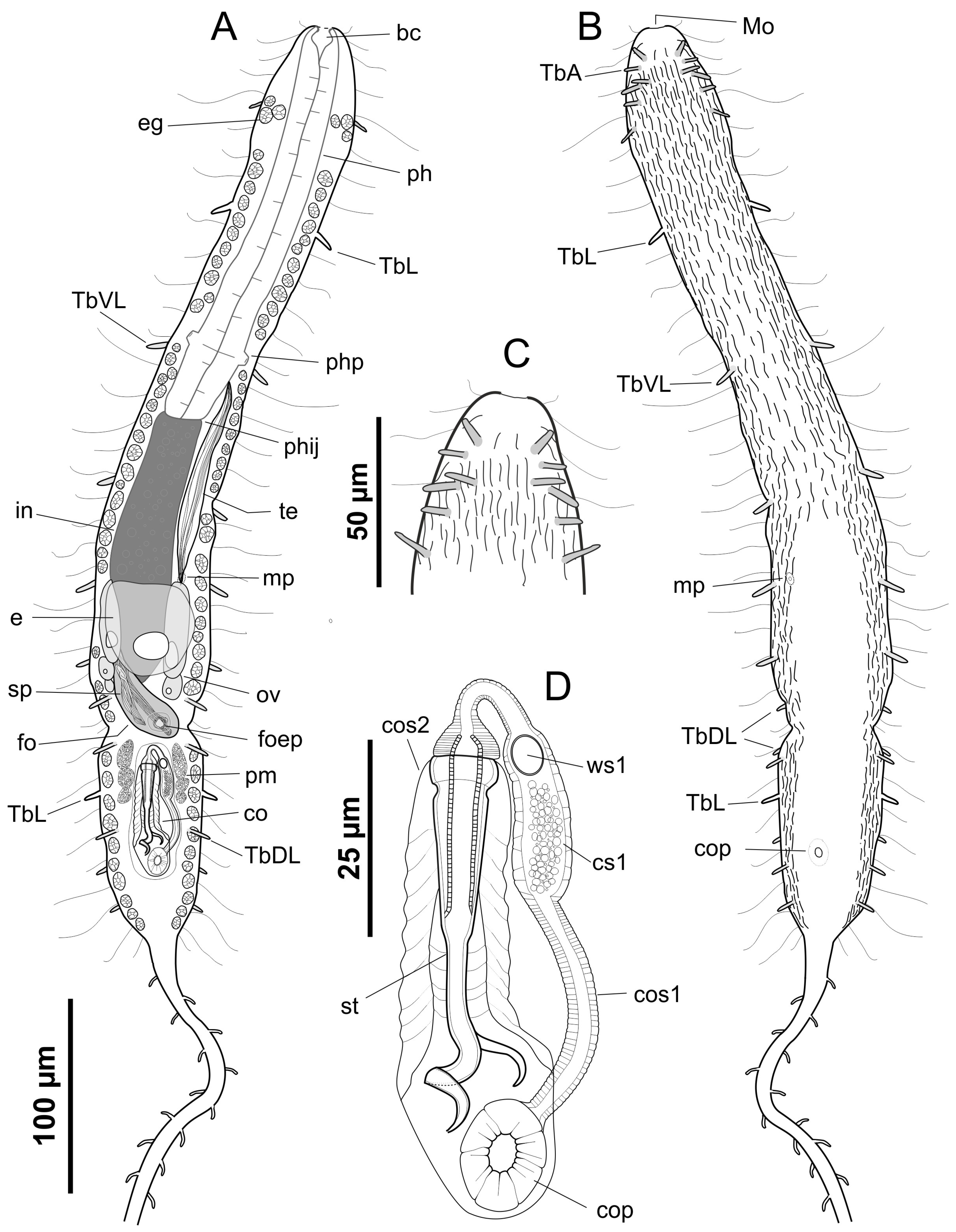
Figure 1. Line art illustration of Urodasys bifidostylis sp. nov. (A) Habitus, showing the internal anatomy seen from a dorsal view; (B) habitus, ventral view; (C) detail of the anterior end, ventral view, showing the anterior adhesive tubes; (D) detail of the caudal organs, ventral view, showing the internal organisation in two connected muscular structures. bc—buccal cavity, co—caudal organ, cop—caudal organ pore, cos1—muscular structure 1 of the caudal organ, cos2—muscular structure 2 of the caudal organ, cs1—chamber of structure 1, e—egg, eg—epidermal gland, fo—frontal organ, foep—external pore of the frontal organ, mo—mouth, mp—male pore, ov—ovary, ph—pharynx, phij—pharyngo-intestinal junction, pm—prostatic glandular material, sp—allosperm packets inside the frontal organ (=spermatophore), st—stylet, te—testis, ws1—window of structure 1, TbA—anterior adhesive tubes, TbDL—dorsolateral adhesive tubes, TbL—lateral adhesive tubes, TbVL—ventrolateral adhesive tubes.
Water 15 01106 g001
Figure 2. Photomicrographs of Urodasys bifidostylis sp. nov. holotype (Nomarski optics). (A) Habitus, showing the internal anatomy seen from a dorsal view; the arrowhead indicates the external pore of the frontal organ. (B) Details of the anterior end, ventral view, showing the anterior adhesive tubes (arrowheads). (C) Details past the mid-trunk region showing the internal organs. (D,E) Details of the posterior trunk region, showing the internal organs; the arrowhead indicates the external pores of the frontal organ, inside which two packets of allosperm are seen. (F,G) Details of the copulatory stylet seen at different focal planes, showing the forked posterior end; the arrow indicates the corkscrew-shaped injecting portion while the arrowhead indicated the secondary hooked extremity. bc—buccal cavity, co—caudal organ, cop—caudal organ pore, cos1—muscular structure 1 of the caudal organ, cos2—muscular structure of the caudal organ, cs1—chamber of structure 1, e—egg, eg—epidermal gland, eo—early oocyte, fo—frontal organ, ph—pharynx, phij—pharyngo-intestinal junction, pm—prostatic glandular material, sp—allosperm packet inside the frontal organ (=spermatophore), st—stylet, stc—stylet constriction, ta—tail, tc—trunk constriction, te—testis. Scale bars: (A) = 100; (BE) = 50 µm; (F,G) = 25 µm.
Water 15 01106 g002
Figure 3. Photomicrographs of Urodasys bifidostylis sp. nov. (Nomarski optics). (A) Habitus of an adult specimen, with detail of the internal anatomy as seen from the ventral side. Inserts (B,C) details from the dorsal side showing the external pore of the frontal organ. Insert (D) testicular spermatozoa. bc—buccal cavity, co—caudal organ, cop—caudal organ pore, fo—frontal organ, ph—pharynx, st—stylet, tc—trunk constriction, te—testis. Scale bars: (A) = 100; (B,C) = 50 µm; (D) = 20 µm.
Water 15 01106 g003
Figure 4. Photomicrographs of Urodasys bifidostylis sp. nov. (muscular system visualised with CLSM). (A) Whole adult specimen. (B) Close-up of the pharyngeal and mid trunk region. (C) Close-up of the mid- and posterior trunk region. (D) Details of the caudal organ, arrow indicates the connection between structure 1 and 2. (E) Details of the caudal organ of a different specimen; details of the caudal organ, arrow indicates the connection between structure 1 and 2. co—caudal organ, cop—caudal organ pore, cos1—caudal organ structure 1, cos2—caudal organ structure 2, cs1—chamber of structure 1, fo—frontal organ, mps—male pore sphincter, ph—pharynx, phij—pharyngo-intestinal junction, php—pharyngeal pore, ws1—window of structure 1. Scale bars: (A) = 100; (B,C) = 50 µm; (D,E) = 20 µm.
Water 15 01106 g004
Disclaimer/Publisher’s Note: The statements, opinions and data contained in all publications are solely those of the individual author(s) and contributor(s) and not of MDPI and/or the editor(s). MDPI and/or the editor(s) disclaim responsibility for any injury to people or property resulting from any ideas, methods, instructions or products referred to in the content.