Article

The Use of Photo-Biological Parameters to Assess the Establishment Success of Posidonia oceanica Cuttings after Transplantation

by
Arnaud Boulenger
1,2,*,
Stéphane Roberty
3,
Maria Margarita Lopez Velosa
1,
Michel Marengo
2 and
Sylvie Gobert
1,2
1
Laboratory of Oceanology, MARE Centre, UR FOCUS, University of Liège, 11 Allée du Six Août, 4000 Liege, Belgium
2
STAtion de REcherches Sous-Marines et Océanographiques (STARESO), 20260 Calvi, France
3
Laboratory of Animal Physiology and Ecophysiology, University of Liège, 4 Chemin de la Vallée, 4000 Liege, Belgium
*
Author to whom correspondence should be addressed.
Water 2024, 16(12), 1702; https://doi.org/10.3390/w16121702
Submission received: 22 April 2024 / Revised: 12 June 2024 / Accepted: 13 June 2024 / Published: 14 June 2024
(This article belongs to the Special Issue Coastal Restoration and Ecological Functions Enhancement)

Abstract

:
Seagrass meadows are increasingly threatened by anthropogenic activities and climate change, necessitating restoration efforts such as cutting transplantation. Understanding the complex interactions between plant morphology and physiology is crucial for designing robust restoration strategies and assessing the success of transplantation and recovery processes. A pilot transplantation experiment with the Mediterranean seagrass Posidonia oceanica (L.) Delile was conducted in Northwestern Corsica (Calvi, France) to evaluate the feasibility of meadows degraded due to boat anchoring. The effects of the cuttings’ origin and transplanting depth were investigated. The establishment success of transplanted fragments was assessed by investigating the photo-physiological parameters, carbohydrate content, and biometric parameters of both transplanted and control plants one year after transplantation at depths of 20 and 28 m. After one year, there was a high survival rate (90%) of the transplants, but their leaf surface area and biomass were significantly reduced compared to the control plants. Photosynthetic activity remained consistent between both depths, emphasizing the ability of P. oceanica cuttings to acclimate to a new light environment in a relatively short period of time (<3 months). Furthermore, light-harvesting pigments, photoprotective pigments, and carbohydrate concentration were greater at the deeper sites. This implies that transplantation at greater depths might be more effective. Furthermore, additional research is necessary to enhance our understanding of the relationship between photosynthesis and the overall health of the plant. This study emphasizes the essential integration of morphological and physiological investigations to offer an ecologically meaningful understanding of how marine ecosystems respond to various restoration methods.

1. Introduction

In recent decades, human activities and climate change have had a significant impact on seagrass meadows, with global loss rates increasing from 0.9% per year in the 1940s to 7% per year by the end of the 20th century [1]. The reduction in seagrass coverage is mainly attributed to agricultural activities causing sediment and nutrient runoff, coastal urbanization, marine heatwaves, dredging, trawling, anchoring, and disease [2]. The degradation of and decline in seagrass habitats are undermining their crucial ecosystem services, prompting a growing global push for their conservation to secure their future [3,4]. As a result of seagrass habitat loss, restoration has been implemented as a management and mitigation strategy to restore the ecological functions and services of seagrass meadows affected by anthropogenic pressures [5,6]. There has been a recent surge in research focused on restoring degraded marine environments, resulting in a proliferation of strategies and methodologies aimed at improving the effectiveness of restoration plans. However, results vary widely depending on the species, methods used, and different environmental conditions encountered. A universally accepted and standardized approach is still far from being realized [7,8,9]. Several tools can be used to determine the impact of disturbances on seagrasses, such as those caused by the transplantation of seagrass cuttings to a new environment. Classical monitoring metrics are measurements related to structure/morphology, such as shoot density or foliar indices, but little is known about physiological recovery process [10,11]. Transplantation may also have some effect on photosynthetic activity, which can be detected by observing specific characteristics of chlorophyll a fluorescence [10,12] (Figure 1). Indeed, when a chlorophyll molecule captures photon energy, that energy can either drive photosynthesis, be emitted as fluorescence, or be converted into heat (Figure 1). Variable chlorophyll a fluorescence measured by Pulse-Amplitude-Modulated (PAM) fluorometry has been widely used to assess the health status of seagrasses, allowing for the detection of plant stress before visible morphological or density-related changes occur [13,14,15]. The analysis of fluorometric and derived photosynthetic parameters provides valuable insights into the photo-physiological response of a plant [15,16,17]. In a new light environment, such as may occur after transplantation to a different depth or density of meadow, photo-acclimation can lead to changes in the content and ratio of different photosynthetic pigments [18]. In fact, light-harvesting systems consist not only of both chlorophyll a and b but also of other pigments such as xanthophyll and carotenoid. For instance, leaves that thrive under high-light conditions have fewer chlorophylls, higher photosynthetic capacity, and active photoprotective mechanisms like xanthophyll pigments. Conversely, leaves adapted to low-light conditions typically exhibit the opposite characteristics [19,20]. Photosynthetically produced reserves are stored as carbohydrates in the rhizomes (Figure 1). As an energy source, they are essential for plant growth and are remobilized to survive the winter period when photosynthesis is greatly decreased [21,22,23]. As mentioned previously, most restoration studies emphasize the need for a proper assessment of the health status of transplants and consider monitoring them a crucial stage for the success of the transplantation and recovery process. While PAM fluorometry has been extensively used to detect physiological stress in seagrasses, little research has been conducted to investigate the photo-physiological response of seagrasses when subjected to transplantation [12,24]. The objective of this study was to investigate the establishment of seagrass transplants after one year by monitoring the temporal dynamics of their morphological and photo-physiological parameters measured using the morphology, biomass, chlorophyll a fluorescence parameters, pigment content, and carbohydrate content of both the transplants and reference plants.

2. Material and Methods

2.1. Study Area and Seagrass Transplantation

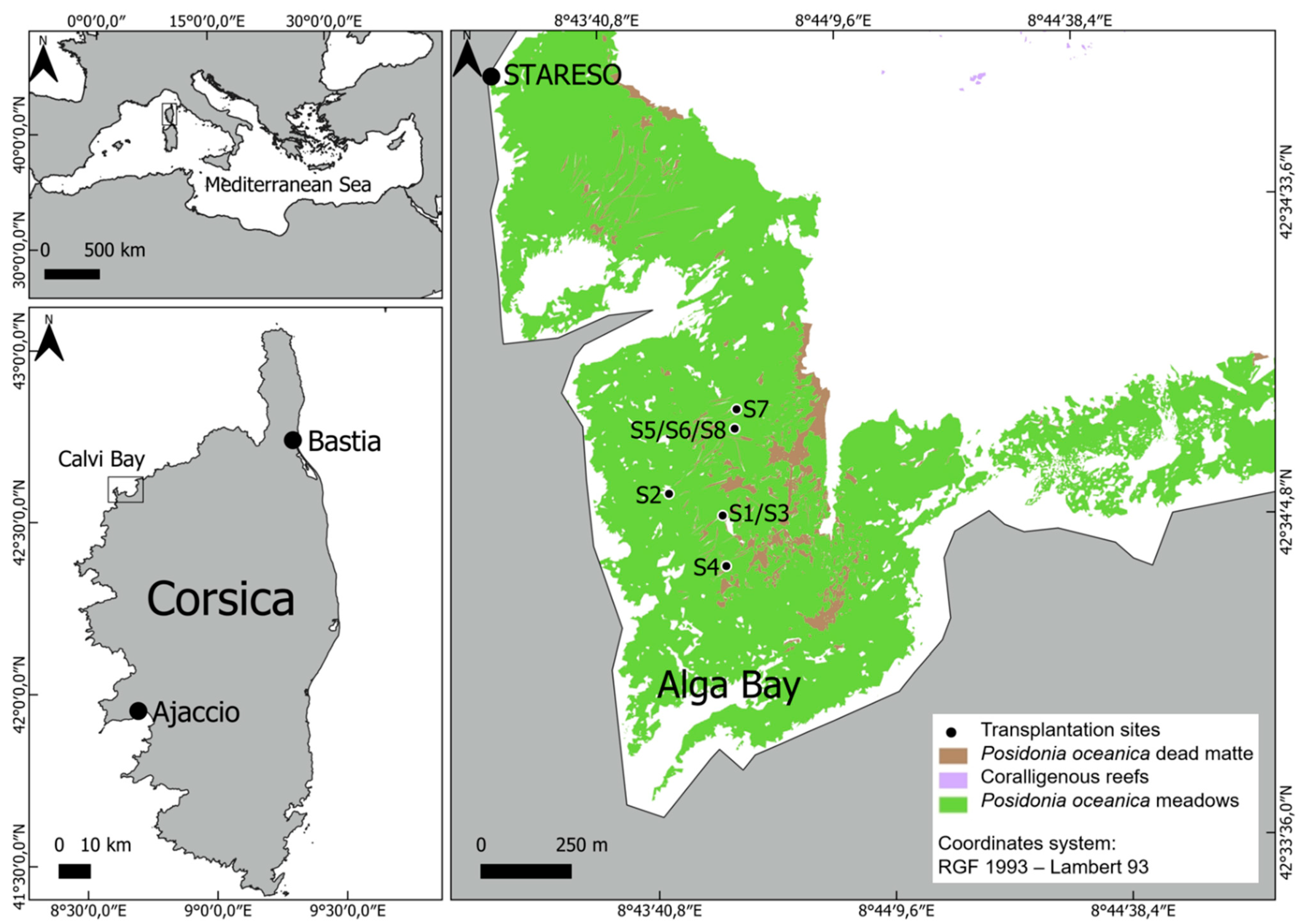
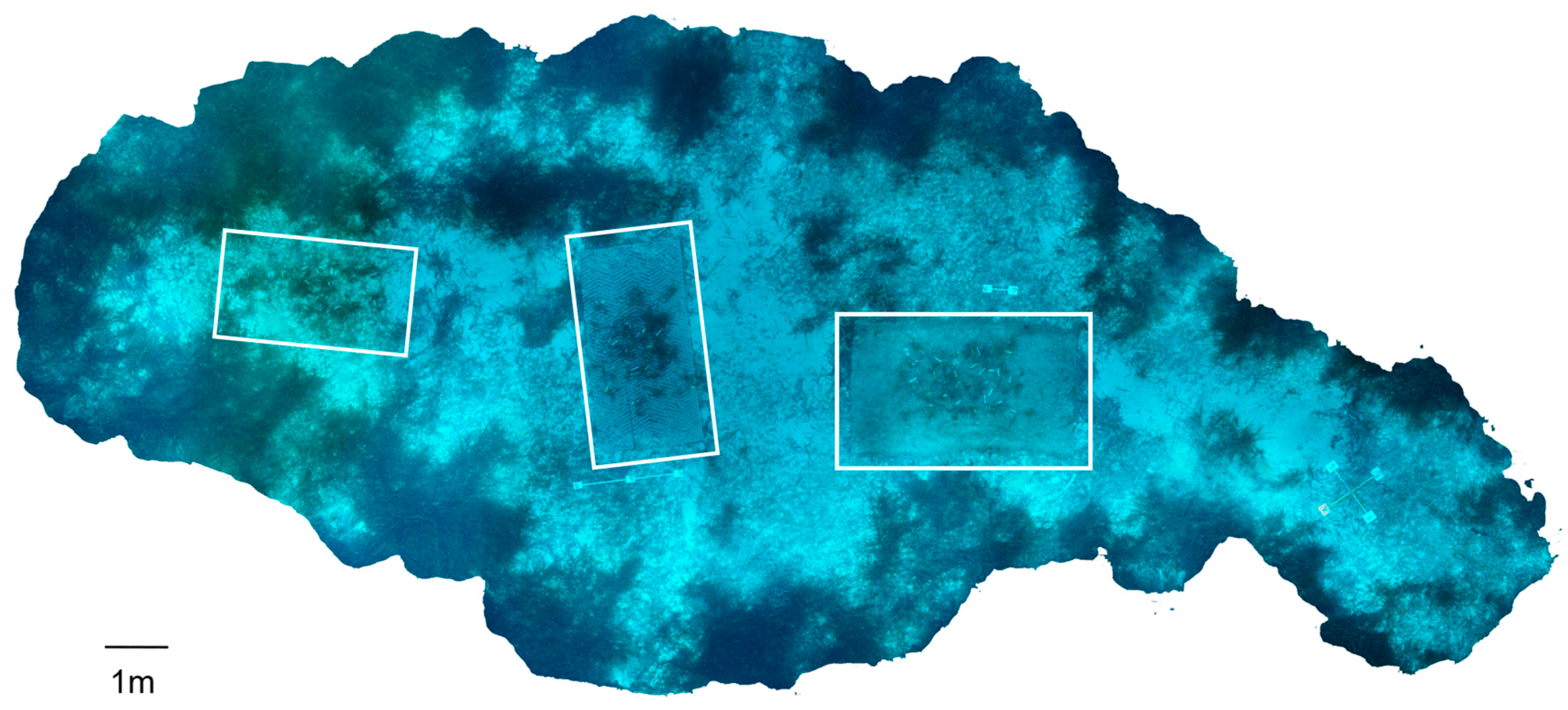
This study was carried out in a sub-bay of Calvi Bay named Alga Bay (8°43′52″ E; 42°34′20″ N), situated in front of the oceanographic station STARESO (Calvi, Western Corsica, France), between May 2022 and May 2023. This bay is occupied with extensive seagrass meadows of the species Posidonia oceanica (L.) Delile covering around 0.78 km² at depths ranging from 3 to 37 m [25] and has experienced significant anchoring activity resulting in drastic seagrass meadow regression [26]. The experimental sites were positioned along eight designated anchoring tracks at depths ranging from 20 to 28 m and were characterized by a mix of bare sediment and dead matte (see Figure 2 and Figure 3). Between May and July 2022, a total of 792 P. oceanica fragments were transplanted at these experimental sites (Figure 2 and Figure 3). To minimize the impact on the surrounding meadows, the transplantation mainly involved naturally uprooted fragments referred to as storm fragments (528 fragments). These storm fragments were collected from various locations near STARESO during SCUBA dives ranging in depth from 6 to 28 m. Additionally, a smaller part (264 fragments) of the transplants was collected from the erosion side of a natural sandy intermatte at a 15 m depth [27]. Cuttings were collected in situ by divers and then kept in outdoor flow-through seawater aquaria until initial biometric measurements (the number of shoots, maximum leaf length, and rhizome length) were taken. Only cuttings with at least 3 shoots and a plagiotropic rhizome at least 15 cm long were retained, while those with excessive leaf necrosis were discarded. After biometric measurements, the cuttings were attached to biodegradable artificial structures and transported by boat to the transplantation sites in Alga Bay where they were anchored to the seabed using rebars.

2.2. Sampling Campaigns and Biometric Measurements

During the first campaign (May 2022), 20 P. oceanica fragments were collected from surrounding control meadows at depths between 20 and 28 m. In addition, 20 cuttings, including both storm fragments and cuttings from intermattes, were preserved for biometric measurements and further laboratory analyses. For each of the two following sampling campaigns (September 2022 and May 2023), the field survival rate was assessed by counting the number of living shoots on each of the 792 transplants. At each experimental site, 12 shoots were collected for subsequent biometric and physiological measurements conducted onshore. Fewer shoots were collected in May 2023 because one of the experimental sites (S2) was partially washed away by strong hydrodynamics and low sediment stability. Similarly, 10 control shoots were collected from the surrounding meadows and brought back to the laboratory for further examination. All the shoots were collected using the non-destructive shoot sampling method (NDSM) recommended by Gobert et al. [29] except when harvesting the whole fragment was necessary for the analysis of the carbohydrate concentration in the rhizomes. The number of leaves was counted for each sampled shoot, and the length and width of each leaf were measured. Epiphytes were scraped from all sampled leaves using a ceramic scalpel blade [30]. They were then weighed for their fresh weight, subsequently freeze-dried for 48 h (BenchTop 3L, VirTis Company Inc., Gardener, NY, USA), and weighed again to determine their dry weight.

2.3. Photosynthetic Activity Measurements

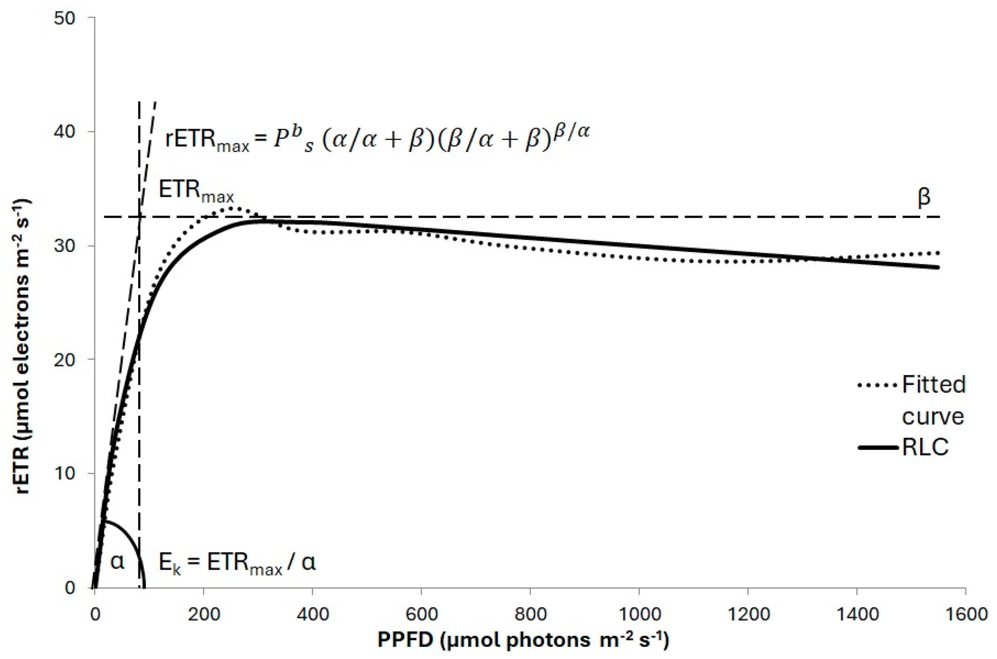
Photosynthetic activity was measured in the laboratory using a DIVING-PAM-I (Heinz Walz GmbH; hereafter referred to as a PAM device). The sampled leaves were placed in plastic trays shaded from ambient sunlight. PAM measurements were taken on the convex middle section of the second intermediate leaf, which showed the strongest correlation with the photosynthetic rate of the whole shoot [31,32]. Visible epiphytic growth on this section was removed by rubbing the leaf with a finger. To ensure standardized measurements, leaf holder clips were utilized during PAM procedures to maintain a constant distance between the tip of the fiber optic and the leaf surface [32]. Rapid light curves [20] were obtained by exposing the samples for 10 s to 9 sequential increasing light steps (0, 38, 117, 237, 377, 564, 775, 1139, and 1548 µmol photons m−2 s−1). These RLCs were obtained with the following settings: GAIN = 5, DAMP = 2, MEAS-INT = 2, SAT-INT = 8, and SAT-WIDTH = 0.8. The maximum photochemical quantum yield of the Photosystem II (Fv/Fm) was measured at the beginning of each RLC, i.e., at a photosynthetic photon flux density (PPFD) of 0 µmol photons m−2 s−1. The effective photochemical quantum yield of the Photosytem II (Y(II)) and relative electron transport rates (rETRs) were calculated at the end of each of light step as Y(II) = (Fm′–F)/Fm′ and rETRPSII = Y(II) × PPFD. Three parameters were derived from the RLCs and plotted as the rETR versus the photosynthetic photon flux density (PPFD): the maximum relative electron transport rate (rETRmax), the initial slope of the curve (α) related to photosynthetic efficiency, and the minimum saturating PPFD (Ek) (Figure 4). These parameters were derived from the equation introduced by Platt et al. [33], considering photoinhibition. Data acquisition and modeling were carried out using WinControl-3 software version 3.33 (Heinz Walz GmbH, Effeltrich, Germany).

2.4. Pigment Concentration and Analysis by HPLC

To quantify pigment concentration in the leaves, 4 cm long segments were sampled from the middle part of the second intermediate leaf of the shoot [34], corresponding to the area where photosynthetic activity was measured with the PAM device. Leaf segments were weighed for their fresh weight, freeze-dried for 48 h (BenchTop 3L, VirTis Company Inc.), and reweighed again for their dry weight. The samples were then homogenized on ice using a mortar and pestle with 2 mL of 100% methanol under dim light. The extracts were centrifuged twice for 10 min at 12,000× g to remove cellular debris. As described by Roberty et al. [35], the pigments were then separated by reverse-phase HPLC, using a Shimadzu Prominence HPLC system comprising a DGU-20A5R degassing unit, an LC-20AT liquid chromatograph, a SIL-20AC autosampler, a CTO-10ASVP column oven, and an SPD-M20A diode array detector (Shimadzu, Japan). The HPLC column (Nova Pak C18; 150 mm in length and a pore size of 4 µm) was eluted with a mobile phase gradient (1 mL min−1). Absorbance chromatograms were obtained at 430 nm and compared to the elution patterns of pigment standards (DHI Lab, Horstholm, Denmark) for the quantification of pigment quantities. Data acquisition and analysis were carried out using Shimadzu LabSolutions software version 5.92 (Shimadzu, Kioto, Japan).

2.5. Carbohydrate Content in Rhizomes

During the first campaign, 20 P. oceanica plagiotropic rhizomes were harvested from the two control sites (20 and 28 m depth) and 20 plagiotropic rhizome cuttings, including both storm fragments (10 samples) and cuttings from intermattes (10 samples). One year later, 10 plagiotropic rhizomes from the control sites and 46 plagiotropic rhizome cuttings from the different transplantation sites were harvested. They were cleaned of scale, stored at −20 °C, and sent to MicroPolluants Technology SA (Saint Julien Les Metz, France) for an analysis of their soluble carbohydrates and starch content. Each rhizome sample was placed in ethanol (v/v) and heated at 80 °C for 15 min to extract sucrose; the extract was then centrifuged to separate the solid part from the organic phase. The solvent was removed, and the extraction process was repeated twice [36]. The combined ethanol extract obtained was evaporated to dryness at room temperature, and the residue was dissolved in hot water. Starch was extracted from the sample pre-extracted from ethanol by incubation in sodium hydroxide solution for 24 h at room temperature [37] or by boiling it in sodium hydroxide for 30 min [38]. After cooling, the pH was adjusted to 5.5 with acetic acid. The content of sucrose and starch was then determined by spectrophotometry after a reaction with anthrone [39]. The result is expressed as the Total Carbohydrate Reserve (TCR) to an accuracy of 1%.

2.6. Data Analysis

Statistical analysis was performed using RStudio version 4.3.2 (RStudio Inc., Boston, MA, USA). Prior to analysis, the data set was examined to identify potential outliers. The PAM measurements were often highly variable, and data points with a significant deviation from the mean were excluded. Generalized linear models were fitted to the various morphological and physiological response variables and the explanatory variables “Origin”, “Bathymetry” and the interaction between the two (2-way interaction). “Origin” had three factors: “Control” refers to seagrass meadow control groups in close vicinity to the experimental transplantation sites; “Intermatte” cuttings are transplanted seagrass cuttings harvested on the erosion side of a natural sandy intermatte at a 15 m depth; and “Storm-fragment” samples are transplanted seagrass fragments that were naturally uprooted from seagrass meadows and collected drifting in the seafloor. Bathymetry had two factors: “Shallow” refers to the experimental and control groups at 20 m, and “Deep” refers to the experimental and control groups at 28 m. Models were generated for September 2022 and May 2023 separately to reduce the number of interactions. For the first sampling campaign (May 2022), only “Origin” was used as an explanatory variable as this was the initial campaign before the P. oceanica fragments were transplanted to the different experimental sites. A Poisson error distribution with a log link function was used for the response variables “Survival” and “Number of leaves” due to their discrete non-negative values. A gamma error distribution with an inverse link function was used for all the other response variables as they all had continuous non-negative values. Model selection (a comparison of models containing different numbers of explanatory variables) was used to identify the optimal model for each separate response variable in a stepwise process of removing non-significant explanatory variables until no more variables could be removed from the model. Overdispersion was checked by comparing the residual deviance with the degrees of freedom. When significant differences were obtained, the analysis was followed by using the lsmean() function with the Bonferroni method, which calculated the least squares means for the previously identified significant effects. Spearman’s rank correlation analysis was performed to identify the strength of the relationship between pairs of response variables, i.e., pigments, photosynthetic activity, biometry, biomass, and carbohydrates [40,41]. The interpretation of the meaning of the Spearman’s rank correlation coefficients was taken from Fowler and Cohen [42]. A principal component analysis (PCA) was used to synthesize the information in all response variables. Differences were considered statistically significant when p < 0.01. All values were reported as mean ± standard error values.

3. Results

3.1. Survival, Shoot Morphology, and Biomass

The survival rate of the transplants was calculated in relation to live and dead material left in place or lost. It was 98.74% after three months and 90.40% after one year. The survival rate was not influenced by the origin of the transplanted fragments (intermatte and storm fragment) or the bathymetry of the transplantation sites (Table 1). The number of leaves was not significantly influenced by any of the explanatory variables (Table 1, Figure 5A). However, the leaf surface area and leaf dry weight both showed higher values for the control plants compared to both types of cuttings (intermatte and storm fragment) in September 2022 and May 2023 (Table 1, Figure 5B,C).

3.2. Photosynthetic Activity

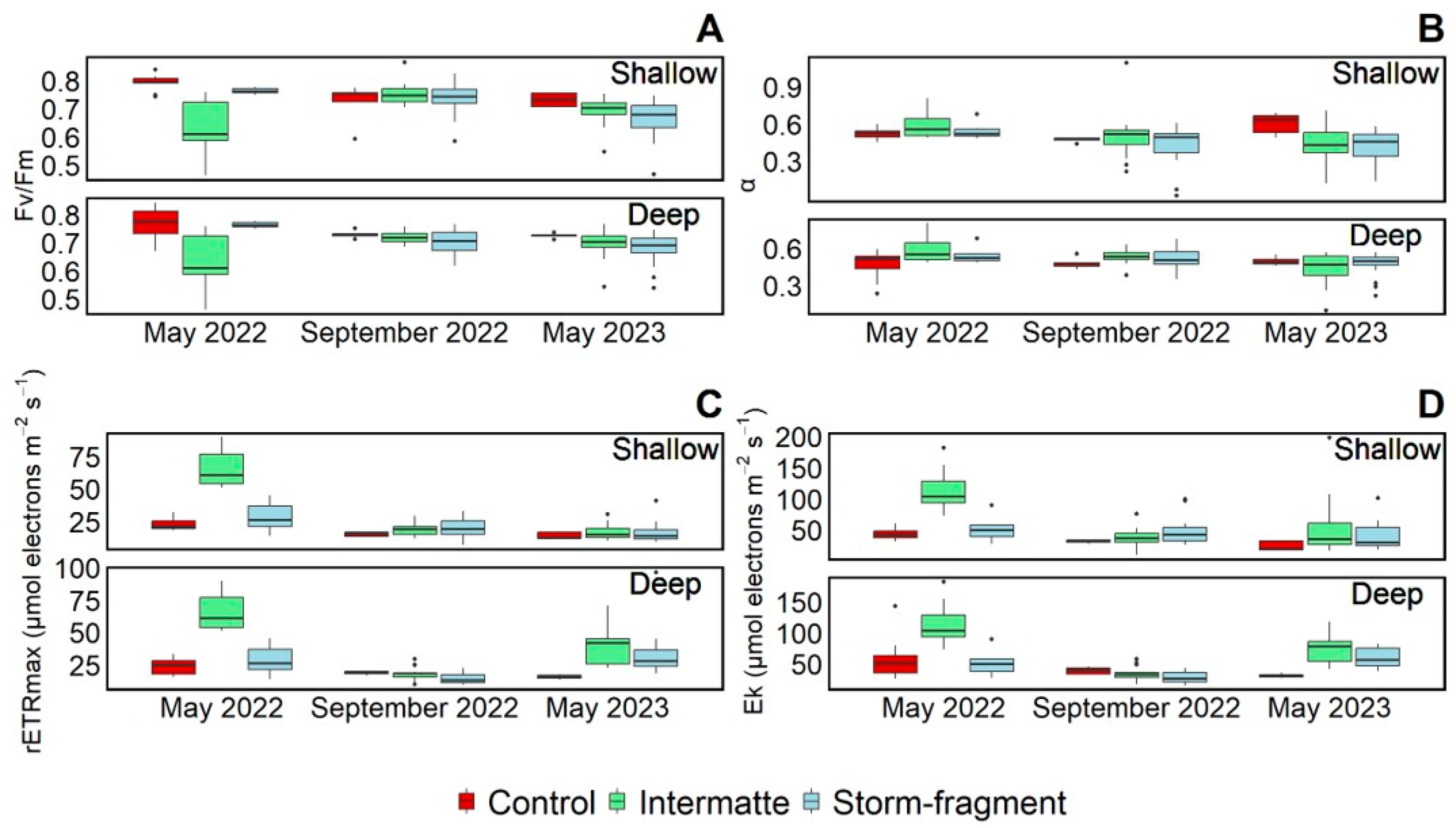
The photo-physiological response of the transplanted shoots was monitored throughout the study using the Fv/Fm and parameters calculated from rapid light curves (i.e., α, the rETRmax, and the Ek). They all behaved differently depending on the bathymetry and the origin of the fragments. While the Fv/Fm was significantly higher in May 2022 for the control plants compared to the intermatte cuttings, this difference was no longer present in September 2022 (Table 1, Figure 6A). In May 2023, control plants also showed higher Fv/Fm values, but only a significant difference was found between them and the storm fragments (Table 1, Figure 6A). The initial slope of the RLC (α) was not significantly different between control and transplants, intermatte cuttings and storm-fragments or depth (Table 1, Figure 6B). Initial differences between plant origins were found for the rETRmax but did not show the same pattern as it can be seen for the Fv/Fm (Figure 6A,C). The rETRmax was significantly higher for the intermatte cuttings than for the control plants and storm fragments (Table 1, Figure 6C). However, no significant difference in origin or bathymetry was observed for the other two sampling campaigns (Table 1, Figure 6C). As with the rETRmax, the saturating irradiance (Ek) was significantly higher in May 2022 for the intermatte cuttings compared to the control plants but not compared to the storm fragments, as was the case for the rETRmax (Table 1, Figure 6C,D). Moreover, in September 2022, the Ek was significantly higher for the plants at the shallow sites, whereas in May 2023, the opposite was true, with the plants at the deep sites showing significantly higher values compared to those at the shallow sites (Table 1, Figure 6D).

3.3. Pigment Content

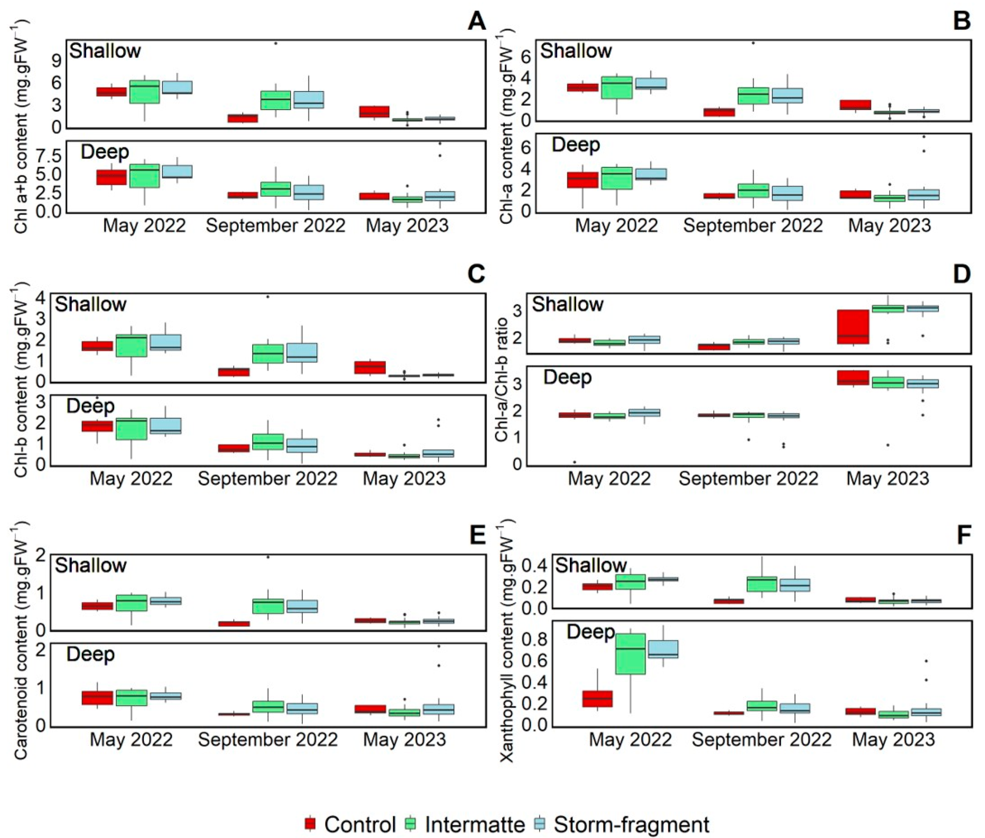
Chlorophyll a, b, and their sum were significantly higher for the shallow sites in September 2022 compared to the deeper sites, but the opposite pattern was observed in May 2023 (Table 1, Figure 7A–C). In addition, in September 2022, higher values were found for the intermatte cuttings compared to the control plants (Table 1, Figure 7A–C). In May 2023, higher values of chlorophyll b were found for the control plants compared to the intermatte cuttings (Table 1, Figure 7C). The ratio Chl-a/Chl-b was not significantly influenced by origin, bathymetry, or their interaction in any of the three sampling campaigns (Table 1, Figure 7D). The carotenoid and xanthophyll pigments were significantly higher for the shallow sites in September 2022 compared to the deeper sites (Table 1, Figure 7E,F). However, the opposite pattern was found in May 2023, with lower values for the shallow sites compared to the deep sites (Table 1, Figure 7E,F). Moreover, in September 2022, higher values were found for the transplants (intermatte cuttings and storm fragments) compared to the control plants (Table 1, Figure 7E,F).

3.4. Carbohydrate Storage in Rhizomes

The only significant difference for the total carbohydrate reserve (TCR) in the rhizomes was that the TCR was higher for the deep sites in May 2023 compared to the shallow sites at the same time, although there is a large variability in TCR values from the deep sites (Table 1, Figure 8).

3.5. Correlation within and between Morphological and Photo-Biological Parameters

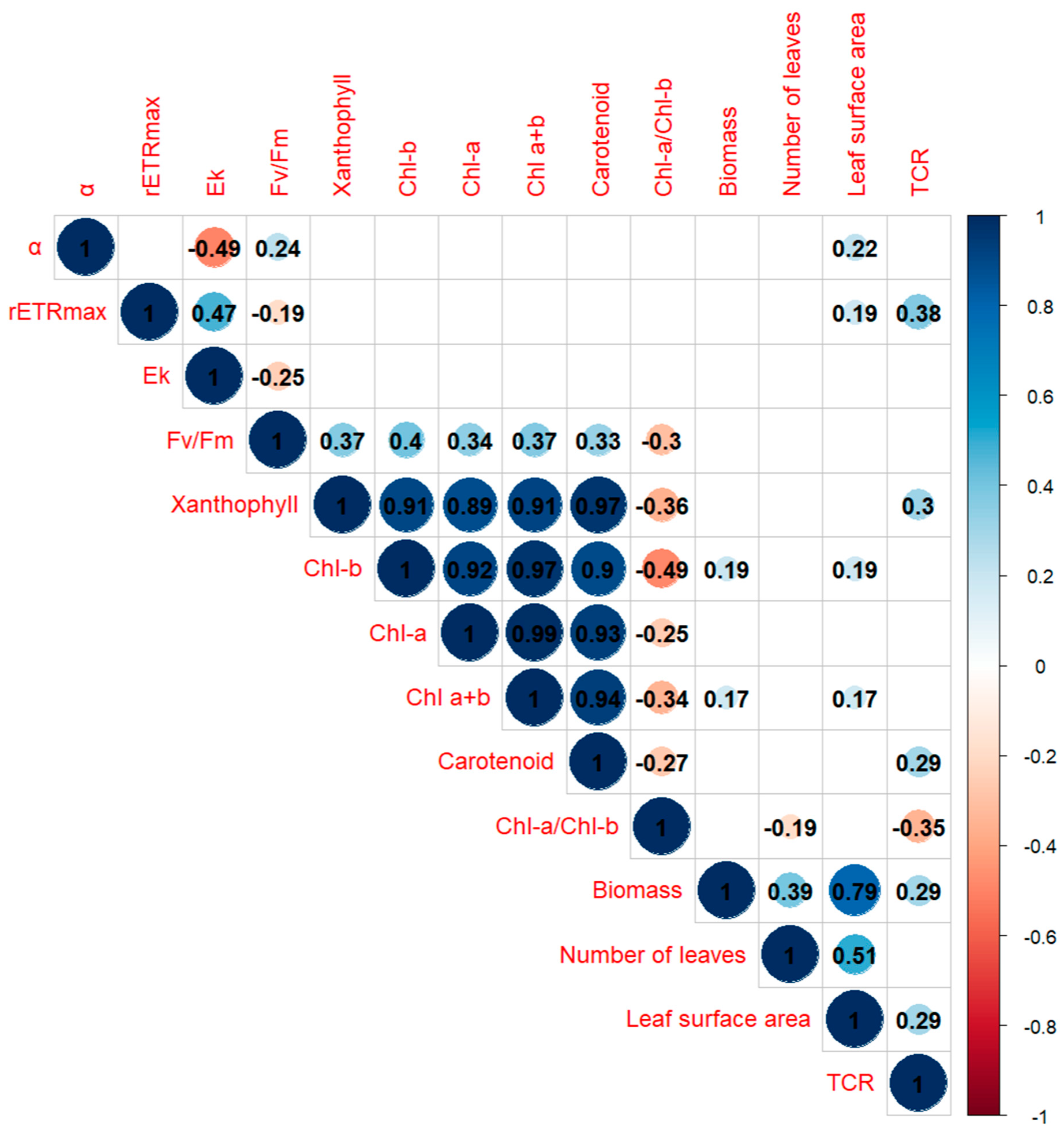
All the individual pigments and their sum are strongly positively correlated with each other, with Spearman’s rho coefficients higher or equal to 0.9 except for xanthophyll and chlorophyll-b, for which it was equal to 0.87 (Figure 9). Morphological variables also displayed strong correlations, such as a 0.79 for biomass and leaf surface area and 0.51 for the number of leaves and leaf surface area (Figure 9). None of the other variables showed strong correlations between each other (−0.5 < Spearman’s rho coefficient < 0.5) (Figure 9). This is corroborated by the PCA with all the individual pigments and their sum that are grouped together, with the Chl-a/Chl-b ratio being orthogonal to them (Figure 10). Similarly, the Fv/Fm and α are anti-correlated (Figure 9). The rETRmax, TCR, and Ek are roughly grouped together, as are biomass, leaf surface area, and the number of leaves (Figure 10). Individual pigments and their sum contributed the most to the variance, followed by Chl-a/Chl-b, morphological parameters, and, finally, the TCR and parameters related to photosynthesis efficiency (Figure 10).

4. Discussion

Despite numerous endeavors to restore P. oceanica meadows over the last 30 years, there is still a lack of well-established methodologies and guidelines for their successful restoration. Therefore, any new knowledge derived from new experimental trials is an important contribution to the overall understanding of the subject [7,43]. In this study, we investigated the photo-biological and morphological characteristics and survival of P. oceanica transplants under various conditions, specifically considering the origin of the transplanted fragments and the bathymetry of the transplantation sites. The results of this study shed light on critical aspects that influence the complex dynamic of P. oceanica meadows. Wave exposure and low sediment stability at some transplantation stations led to the loss of some P. oceanica transplants, but the survival rate of transplants after one year remained high. The relatively high survival rate observed (90.40%) underlines the resilience and adaptability of the species after transplantation on dead matte. Such a high survival rate was consistent with previous work carried out in the same area [44]. Although there were no initial differences between control plants and transplants, the leaf surface area and leaf dry weight of the transplants were significantly reduced even one year after transplantation. This could be indicative of potentially non-optimal growth conditions [44,45], or it could be due to breakage caused by increased water movement in a thinned canopy due to transplantation on a bare area compared to dense control meadows [46]. In addition, our results indicate that differences in light intensity between shallow and deep sites did not significantly affect transplant morphology, which is consistent with the idea of acclimation observed in previous studies [47,48]. Furthermore, our results are consistent with the emphasis in the literature on the acclimation of P. oceanica to different light conditions, demonstrating the ability of the plant to adapt without exceeding critical tolerance thresholds [49].
The photosynthetic parameters measured here indicate limited physiological differences among the explanatory variables Origin and Bathymetry. Many studies across different species highlight the utility of variable chlorophyll a fluorescence in understanding the photo-physiological dynamics of seagrasses thriving in different light environment, for example, due to different depths [15,18,20,47,50,51]. Previous studies on seagrasses revealed acclimation responses from the plant along a decreasing light gradient, including an increasing α for enhanced photosynthetic efficiency under low light intensities, a decreasing ETRmax, a reduction in the saturating irradiance (Ek), and an increasing Fv/Fm [10,50,52,53,54]. However, there are many studies in which these trends are inconsistent. There are many reports of no response or the response deviating from the expected light gradient; there are also many reports of the response conforming to expected patterns [55,56,57,58,59]. The reasons for these discrepancies could be related to the individual acclimation statuses of different plants, the intrinsic diversity of beds, some experimental pitfalls, and the heterogeneity of the environment [55,60]. Here, the photosynthetic efficiency remained constant across the three sampling campaigns and did not differ significantly between depths or origins. While there were initial differences in the Fv/Fm, rETRmax, and Ek between the control plants and intermatte cuttings, these differences disappeared in subsequent sampling campaigns. It is likely that these differences were due to the light conditions from the initial environment of the intermatte cuttings. Indeed, these cuttings were plagiotropic rhizomes that were harvested from the erosion side of a natural sandy intermatte, where light intensity is more important than in dense meadows. Moreover, the sandy intermatte lays at a 15 m depth, whereas the control plants were harvested at a deeper depth, near the experimental sites at 20 and 28 m. It seems that plants rapidly acclimated to their new light environment since the Fv/Fm and rETRmax values were similar between the controls and transplants 3 months after their transplantation. It is likely that the acclimation period was much faster in our case. Indeed, in the study of Horn et al. [10], a 3-month post-transplant acclimation period was required for transplants that had experienced desiccation during the transport from the donor site to the transplantation site [10], and our experimental set-up prevented this type of stress. Our results also contrast with some previous transplantation experiments involving P. oceanica cuttings, showing low survival and development rates when they were transplanted deeper than their original depth [22,61]. Although the storm fragments were collected at different depths and it was not possible to estimate their original depths, the intermatte cuttings were taken from a depth of 15 m and were transplanted to depths of 20 m and 28 m. Despite this deeper transplantation, the photosynthetic activity is like that of the control seagrass beds, and the survival results exceed 90% after one year. After the same period after transplantation, Molenaar and Meinesz [61] observed that transplants taken from a depth of 3 m and transplanted to deeper waters showed reduced survival rates of 59 and 41% at 14 and 29 m, respectively.
In new light environments after transplantation to different environmental conditions, photo-acclimation can induce changes in the concentration and distribution of photosynthetic pigments [18]. With the exception of the Chl-a/Chl-b ratio, the individual pigments and their sum show a similar behaviour as a function of bathymetry, with significantly higher values for shallow sites in September 2022 and higher values for deep sites in May 2023. The higher concentrations in September 2022 for the shallow sites (i.e., a 20 m depth) compared to the deep sites (i.e., a 28 m depth) are consistent with previous work on pigments in P. oceanica which measured chlorophyll concentrations in leaves from different depths and found higher values at 20 m compared to 30 m [22,62]. However, one year after transplantation, the opposite pattern is observed. Spring is usually a time of sustained growth and rapid increase in leaf length [22,63,64]. Differences in light intensity and temperature at different depths are known to induce differential growth rates, leading to delayed peaks in leaf surface area and biomass at deeper sites [31]. In this case, it is likely that leaf area and leaf biomass will increase faster than pigment biomass, leading to a reduction in the total amount of photopigments present at the deep sites compared to the shallow sites [22,59]. However, in this study, no significant differences were found for leaf area and biomass in May 2023. Furthermore, higher pigment levels were found in September 2022 for intermatte cuttings compared to control plants. Although this could have been expected as the intermatte cuttings were transplanted deeper than their initial depth of origin, it is surprising as no initial differences were found between the two before transplantation, indicating similar pigment contents. This also contradicts other studies which have shown that transplantation to deeper or shallower water does not systematically have a significant effect on the concentration of photosynthetic pigments [22,65].
The photosynthetic carbon fixation of P. oceanica is maximized during summer (June–September) when the light intensity is optimal, allowing the plant to accumulate sufficient reserves (i.e., carbohydrates) for overwintering and subsequent regrowth [21]. The photosynthetically produced reserves stored as carbohydrates in the rhizomes did not seem to be significantly affected by transplantation stress, as indicated by the parameter related to variable chlorophyll a fluorescence. A higher concentration of carbohydrates could have been expected for the shallow sites, as higher percentages of carbohydrates are usually found in environments with a higher light intensity [22,37], although the 8 m depth difference between the shallow and deep sites may not be sufficient to cause significant differences in light intensity and associated physiological parameters. Between depths of 5 and 20 m, Genot et al. [22] showed that plants transplanted from shallow to deeper waters photosynthesize less efficiently, store fewer carbohydrates, and have poor survival rates. The positive correlation between carbohydrates and nitrogen content pointed out by previous studies [37,66] could be a possible explanation for these significant differences, emphasizing the importance of physiological parameters not only directly related to photosynthesis to achieve a more exhaustive understanding of a plant’s health status.
The almost constant photosynthetic activity emphasizes the need for caution when attempting to use chlorophyll fluorescence alone to estimate absolute photosynthesis or primary production as it can become decoupled from the electron transport rate at PSII, along with O2 evolution and/or carbon fixation, under specific prevailing environmental conditions [67]. Further investigations are required to deepen our comprehension of the correlation between photo-biological parameters and the overall health condition of the plant. This research area holds significant importance for the estimation of seagrass primary production and blue carbon stocks, aligning with global initiatives aiming to safeguard the resilience of Mediterranean blue carbon ecosystems in mitigating climate change. Moreover, the application of these photo-biological parameters as monitoring tools to identify the establishment success of P. oceanica cuttings after transplantation to new environmental conditions is to be completed with further physiological and genetic analyses to assist in identifying the extent and nature of stress potentially experienced by the transplants [48]. In particular, further studies on this research topic should be completed with metabolomic fingerprinting, which includes monitoring growth-promoting and stress-related metabolites [68,69]. Indeed, previous studies have shown that metabolomics shows a more consistent response with photosynthetic activity and carbon fixation than biometry and biomass changes [69,70,71]. Therefore, it should be considered when selecting new indicators for assessing seagrass health after transplantation.

5. Conclusions

The combined analysis of a range of different morphological and physiological parameters offers interesting insights into the complex response of P. oceanica to transplantation stress. One year after transplantation, the survival rate of transplants remains quite high (90%), although morphological differences between control plants and transplants persist. In contrast to morphological responses, the different physiological parameters related to photosynthetic activity do not accompany changes in morphology and suggest that transplanted fragments are acclimated to their new environmental conditions. A contrast also emerges between the two depths of the transplantation sites, with higher values found in light-harvesting pigments, photoprotective pigments, and carbohydrate concentration found for the deep sites after one year, although photosynthetic activity remains unchanged. This could suggest that transplanting at a deeper depth could be more efficient, but longer-term monitoring is needed to assess whether the morphological disparities between transplants and control plants eventually diminish over time and align with photo-biological parameters. This study demonstrates the importance of a multidisciplinary approach in marine ecological research for which the integration of morphological and physiological studies is fundamental to providing an ecologically relevant interpretation of the response of marine ecosystems to different kind of restoration methods.

Author Contributions

A.B.: conceptualization, field sampling, methodology, formal analysis, investigation, writing, and visualization; S.R.: conceptualization, methodology, formal analysis, investigation, writing, visualization, and supervision; M.M.L.V.: formal analysis, investigation, and writing; M.M.: conceptualization, writing, and supervision. S.G.: methodology, conceptualization, writing, and supervision. All authors have read and agreed to the published version of the manuscript.

Funding

This work was supported by the University of Liege and the Fonds National de la Recherche Scientifique—FNRS (grants ASP 40006932 and CDR J.0076.23). This study is part of the STARECAPMED (STAtion of Reference and rEsearch on Change of local and global Anthropogenic Pressures on Mediterranean Ecosystems Drifts) project funded by the Territorial Collectivity of Corsica and by The French Water Agency (PACA-Corsica).

Data Availability Statement

The datasets generated during the current study are available upon request by contacting the first author.

Acknowledgments

The authors are grateful to STARESO for facilities and field assistance. Moreover, the authors are endebted to three anonymous reviewers for their valuable suggestions.

Conflicts of Interest

The authors declare that they have no known competing financial interests or personal relationships that could have appeared to influence the work reported in this paper.

References

  1. Waycott, M.; Duarte, C.M.; Carruthers, T.J.B.; Orth, R.J.; Dennison, W.C.; Olyarnik, S.; Calladine, A.; Fourqurean, J.W.; Heck, K.L.; Hughes, A.R.; et al. Accelerating Loss of Seagrasses across the Globe Threatens Coastal Ecosystems. Proc. Natl. Acad. Sci. USA 2009, 106, 12377–12381. [Google Scholar] [CrossRef] [PubMed]
  2. Turschwell, M.P.; Connolly, R.M.; Dunic, J.C.; Sievers, M.; Buelow, C.A.; Pearson, R.M.; Tulloch, V.J.D.; Côté, I.M.; Unsworth, R.K.F.; Collier, C.J.; et al. Anthropogenic Pressures and Life History Predict Trajectories of Seagrass Meadow Extent at a Global Scale. Proc. Natl. Acad. Sci. USA 2021, 118, e2110802118. [Google Scholar] [CrossRef] [PubMed]
  3. Cullen-Unsworth, L.C.; Unsworth, R.K.F. A Call for Seagrass Protection. Science 2018, 361, 446–448. [Google Scholar] [CrossRef] [PubMed]
  4. Unsworth, R.K.F.; McKenzie, L.J.; Nordlund, L.M.; Cullen-Unsworth, L.C. A Changing Climate for Seagrass Conservation? Curr. Biol. 2018, 28, R1229–R1232. [Google Scholar] [CrossRef] [PubMed]
  5. Rezek, R.J.; Furman, B.T.; Jung, R.P.; Hall, M.O.; Bell, S.S. Long-Term Performance of Seagrass Restoration Projects in Florida, USA. Sci. Rep. 2019, 9, 1–11. [Google Scholar] [CrossRef] [PubMed]
  6. van Katwijk, M.M.; Thorhaug, A.; Marbà, N.; Orth, R.J.; Duarte, C.M.; Kendrick, G.A.; Althuizen, I.H.J.; Balestri, E.; Bernard, G.; Cambridge, M.L.; et al. Global Analysis of Seagrass Restoration: The Importance of Large-Scale Planting. J. Appl. Ecol. 2016, 53, 567–578. [Google Scholar] [CrossRef]
  7. Boudouresque, C.F.; Blanfuné, A.; Pergent, G.; Thibaut, T. Restoration of Seagrass Meadows in the Mediterranean Sea: A Critical Review of Effectiveness and Ethical Issues. Water 2021, 13, 1034. [Google Scholar] [CrossRef]
  8. Calvo, S.; Calvo, R.; Luzzu, F.; Raimondi, V.; Assenzo, M.; Cassetti, F.P.; Tomasello, A. Performance Assessment of Posidonia oceanica (L.) Delile Restoration Experiment on Dead Matte Twelve Years after Planting—Structural and Functional Meadow Features. Water 2021, 13, 724. [Google Scholar] [CrossRef]
  9. Rende, S.F.; Bosman, A.; Menna, F.; Lagudi, A.; Bruno, F.; Severino, U.; Montefalcone, M.; Irving, A.D.; Raimondi, V.; Calvo, S.; et al. Assessing Seagrass Restoration Actions through a Micro-Bathymetry Survey Approach (Italy, Mediterranean Sea). Water 2022, 14, 1285. [Google Scholar] [CrossRef]
  10. Horn, L.E.; Paling, E.I.; van Keulen, M. Photosynthetic Recovery of Transplanted Posidonia sinuosa, Western Australia. Aquat. Bot. 2009, 90, 149–156. [Google Scholar] [CrossRef]
  11. Ralph, P.J.; Tomasko, D.; Moore, K.; Seddon, S.; Macinnis-Ng, C.M.O. Human Impacts on Seagrasses: Eutrophication, Sedimentation and Contamination. In Seagrasses: Biology, Ecology and Conservation; Springer: Dordrecht, The Netherlands, 2006; pp. 567–593. [Google Scholar]
  12. Li, W.-T.; Park, J.-I.; Park, S.-R.; Zhang, X.-M.; Lee, K.-S. Chlorophyll a Fluorescence as an Indicator of Establishment of Zostera marina Transplants on the Southern Coast of Korea. Algae 2010, 25, 89–97. [Google Scholar] [CrossRef]
  13. Belshe, E.F.; Durako, M.J.; Blum, J.E. Photosynthetic Rapid Light Curves (RLC) of Thalassia testudinum Exhibit Diurnal Variation. J. Exp. Mar. Biol. Ecol. 2007, 342, 253–268. [Google Scholar] [CrossRef]
  14. Gobert, S.; Lepoint, G.; Silva, J.; Santos, R.; Lejeune, P.; Dujardin, P.; Delvaux, B.; Cornelis, J.-T.; Richir, J. A Consensual Diving-PAM Protocol to Monitor Posidonia oceanica Photosynthesis. In Proceedings of the 4th Mediterranean Seagrass Workshop—MSW’15, Oristano, Italy, 18–22 May 2015. [Google Scholar] [CrossRef]
  15. Madonia, A.; Caporale, G.; Penna, M.; Bonamano, S.; Marcelli, M. Assessment of the Photosynthetic Response of Posidonia oceanica (Linneaus) Delile, 1813 along a Depth Gradient in the Northern Tyrrhenian Sea (Latium, Italy). Geosciences 2021, 11, 202. [Google Scholar] [CrossRef]
  16. Gera, A.; Alcoverro, T.; Mascaró, O.; Pérez, M.; Romero, J. Exploring the Utility of Posidonia oceanica Chlorophyll Fluorescence as an Indicator of Water Quality within the European Water Framework Directive. Environ. Monit. Assess. 2012, 184, 3675–3686. [Google Scholar] [CrossRef] [PubMed]
  17. Larkum, A.W.; Drew, E.A.; Ralph, P.J. Photosynthesis and Metabolism in Seagrasses at the Cellular Level. In Seagrasses: Biology, Ecology and Conservation; Springer: Dordrecht, The Netherlands, 2007; pp. 323–345. [Google Scholar] [CrossRef]
  18. Léger-Daigle, R.; Noisette, F.; Bélanger, S.; Cusson, M.; Nozais, C. Photoacclimation and Light Thresholds for Cold Temperate Seagrasses. Front. Plant Sci. 2022, 13, 805065. [Google Scholar] [CrossRef] [PubMed]
  19. Demmig-Adams, B.; Adams, W., III; Ebbert, V.; Logan, B.A. Ecophysiology of the Xanthophyll Cycle. In The Photochemistry of Carotenoids; Frank, H.A., Young, A.J., Britton, G., Cogdell, R.J., Eds.; Kluwer Academic Publishers: Alphen aan den Rijn, The Netherlands, 1999; pp. 245–269. [Google Scholar]
  20. Ralph, P.J.; Gademann, R. Rapid Light Curves: A Powerful Tool to Assess Photosynthetic Activity. Aquat. Bot. 2005, 82, 222–237. [Google Scholar] [CrossRef]
  21. Alcoverro, T.; Manzanera, M.; Romero, J. Annual Metabolic Carbon Balance of the Seagrass Posidonia oceanica: The Importance of Carbohydrate Reserves. Mar. Ecol. Prog. Ser. 2001, 211, 105–116. [Google Scholar] [CrossRef]
  22. Genot, I.; Caye, G.; Meinesz, A.; Orlandini, M. Role of Chlorophyll and Carbohydrate Contents in Survival of Posidonia oceanica Cuttings Transplanted to Different Depths. Mar. Biol. 1994, 119, 23–29. [Google Scholar] [CrossRef]
  23. Pirc, H. Seasonal Changes in Soluble Carbohydrates, Starch, and Energy Content in Mediterranean Seagrasses. Mar. Ecol. 1989, 10, 97–105. [Google Scholar] [CrossRef]
  24. Horn, L. The Measurement of Seagrass Photosynthesis Using Pulse Amplitude Modulated (PAM) Fluorometry and Its Practical Applications, Specifically in Regard to Transplantation. Ph.D. Thesis, Murdoch University, Perth, Australia, 2006. [Google Scholar]
  25. Abadie, A.; Lejeune, P.; Pergent, G.; Gobert, S. From Mechanical to Chemical Impact of Anchoring in Seagrasses: The Premises of Anthropogenic Patch Generation in Posidonia oceanica Meadows. Mar. Pollut. Bull. 2016, 109, 61–71. [Google Scholar] [CrossRef]
  26. Fullgrabe, L.; Fontaine, Q.; Marengo, M.; Donnay, A.; Sirjacobs, D.; Iborra, L.; Cnudde, S.; Mellet, N.; Seveno, J.; Boulenger, A.; et al. STARECAPMED (STAtion of Reference and Research on Change of Local and Global Anthropogenic Pressures on Mediterranean Ecosystems Drifts)—Year 2021; Research Report; STARESO: Calvi, France, 2022; 123p. [Google Scholar]
  27. Gobert, S.; Lepoint, G.; Pelaprat, C.; Remy, F.; Lejeune, P.; Richir, J.; Abadie, A. Temporal Evolution of Sand Corridors in a Posidonia oceanica Seascape: A 15-Year Study. Mediterr. Mar. Sci. 2016, 17, 777–784. [Google Scholar] [CrossRef]
  28. DONIA EXPERT. Available online: https://plateforme.medtrix.fr (accessed on 2 February 2024).
  29. Gobert, S.; Lefebvre, L.; Boissery, P.; Richir, J. A Non-Destructive Method to Assess the Status of Posidonia oceanica Meadows. Ecol. Indic. 2020, 119, 106838. [Google Scholar] [CrossRef]
  30. Dauby, P.; Poulicek, M. Methods for Removing Epiphytes from Seagrasses: SEM Observations on Treated Leaves. Aquat. Bot. 1995, 52, 217–228. [Google Scholar] [CrossRef]
  31. Buia, M.C.; Zupo, V.; Mazzella, L. Primary Production and Growth Dynamics in Posidonia oceanica. Mar. Ecol. 1992, 13, 2–16. [Google Scholar] [CrossRef]
  32. Lassauque, J. Ecophysiological Early Bio-Indicators of Anthropic-Induced Stress on Posidonia oceanica Meadows. Ph.D. Thesis, Université de Nice Sophia Antipolis, Nice, France, 2009. [Google Scholar]
  33. Platt, T.; Gallegos, C.L.; Harrison, W.G. Photoinhibition of Photosynthesis in Natural Assemblages of Marine Phytoplankton. J. Mar. Res. 1980, 38, 687–701. [Google Scholar]
  34. Marín-Guirao, L.; Sandoval-Gil, J.M.; Bernardeau-Esteller, J.; Ruíz, J.M.; Sánchez-Lizaso, J.L. Responses of the Mediterranean Seagrass Posidonia oceanica to Hypersaline Stress Duration and Recovery. Mar. Environ. Res. 2013, 84, 60–75. [Google Scholar] [CrossRef] [PubMed]
  35. Roberty, S.; Vega de Luna, F.; Pierangelini, M.; Bomhals, J.; Plumier, J.C.; Levy, O.; Cardol, P. Shallow and mesophotic colonies of the coral Stylophora pistillata share similar regulatory strategies of photosynthetic electron transport but differ in their sensitivity to light. Coral Reefs 2023, 42, 645–659. [Google Scholar] [CrossRef]
  36. Zimmerman, R.C.; Smith, R.D.; Alberte, R.S. Thermal acclimation and whole-plant carbon balance in Zostera marina L. (eelgrass). J. Exp. Mar. Biol. Ecol. 1989, 130, 93–109. [Google Scholar] [CrossRef]
  37. Gera, A.; Pagès, J.F.; Romero, J.; Alcoverro, T. Combined effects of fragmentation and herbivory on Posidonia oceanica seagrass ecosystems. J. Ecol. 2013, 101, 1053–1061. [Google Scholar] [CrossRef]
  38. Huber, S.C.; Israel, D.W. Biochemical Basis for Partitioning of Photosynthetically Fixed Carbon between Starch and Sucrose in Soybean (Glycine max Merr.) Leaves. Plant Physiol. 1982, 69, 691–696. [Google Scholar] [CrossRef]
  39. Yemn, E.W.; Willis, A.J. The estimation of carbohydrates in plant extracts by anthrone. Biochem. J. 1954, 57, 508–514. [Google Scholar] [CrossRef] [PubMed]
  40. Pearson, R.K. Exploring process data. J. Process Control 2001, 11, 179–194. [Google Scholar] [CrossRef]
  41. Schober, P.; Boer, C.; Schwarte, L.A. Correlation Coefficients: Appropriate Use and Interpretation. Anesth. Analg. 2018, 126, 1763–1768. [Google Scholar] [CrossRef] [PubMed]
  42. Fowler, J.; Cohen, L.; Jarvis, P. Practical Statistics for Field Biology; Wiley: Chichester, UK, 1998. [Google Scholar]
  43. Pansini, A.; Bosch-Belmar, M.; Berlino, M.; Sarà, G.; Ceccherelli, G. Collating Evidence on the Restoration Efforts of the Seagrass Posidonia oceanica: Current Knowledge and Gaps. Sci. Total Environ. 2022, 851, 158320. [Google Scholar] [CrossRef] [PubMed]
  44. Lepoint, G.; Vangeluwe, D.; Eisinger, M.; Paster, M.; Van Treeck, P.; Bouquegneau, J.M.; Gobert, S. Nitrogen Dynamics in Posidonia oceanica Cuttings: Implications for Transplantation Experiments. Mar. Pollut. Bull. 2004, 48, 465–470. [Google Scholar] [CrossRef] [PubMed]
  45. Gobert, S.; Lepoint, G.; Bouquegneau, J.M.; Vangeluwe, D.; Eisinger, M.; Paster, M.; Schuhmaker, H.; Van Treeck, P. Restoration of Seagrass Meadows: Means and Limitations. In Proceedings of the 7th International Conference on the Mediterranean Coastal Environment, MEDCOAST, Kusadasi, Turkey, 25–29 October 2005; Volume 1, pp. 461–472. [Google Scholar]
  46. Collier, C.J.; Lavery, P.S.; Ralph, P.J.; Masini, R.J. Shade-Induced Response and Recovery of the Seagrass Posidonia sinuosa. J. Exp. Mar. Biol. Ecol. 2009, 370, 89–103. [Google Scholar] [CrossRef]
  47. Dattolo, E.; Marín-Guirao, L.; Ruiz, J.M.; Procaccini, G. Long-term acclimation to reciprocal light conditions suggests depth-related selection in the marine foundation species Posidonia oceanica. Ecol. Evol. 2017, 7, 1148–1164. [Google Scholar] [CrossRef] [PubMed]
  48. Stipcich, P.; Resaikos, V.; Ceccherelli, G. Experimental thermocline deepening highlights the resilience of the seagrass Posidonia oceanica: An opportunity to investigate shoot adaptability. Mar. Pollut. Bull. 2023, 189, 114824. [Google Scholar] [CrossRef] [PubMed]
  49. Ruiz, J.M.; Romero, J. Effects of disturbances caused by coastal constructions on spatial structure, growth dynamics and photosynthesis of the seagrass Posidonia oceanica. Mar. Pollut. Bull. 2003, 46, 1523–1533. [Google Scholar] [CrossRef]
  50. Dattolo, E.; Ruocco, M.; Brunet, C.; Lorenti, M.; Lauritano, C.; D’Esposito, D.; de Luca, P.; Sanges, R.; Mazzuca, S.; Procaccini, G. Response of the seagrass Posidonia oceanica to different light environments: Insights from a combined molecular and photo-physiological study. Mar. Environ. Res. 2014, 101, 225–236. [Google Scholar] [CrossRef]
  51. Shafer, D.J.; Kaldy, J.E. Comparison of photosynthetic characteristics of the seagrass congeners Zostera marina L. and Zostera japonica Ascher. & Graeb. Aquat. Bot. 2014, 112, 91–97. [Google Scholar] [CrossRef]
  52. Campbell, S.; Miller, C.; Steven, A.; Stephens, A. Photosynthetic responses of two temperate seagrasses across a water quality gradient using chlorophyll fluorescence. J. Exp. Mar. Biol. Ecol. 2003, 291, 57–78. [Google Scholar] [CrossRef]
  53. Figueroa, F.L.; Jiménez, C.; Viñegla, B.; Pérez-Rodríguez, E.; Aguilera, J.; Flores-Moya, A.; Altamirano, M.; Lebert, M.; Häder, D.P. Effects of solar UV radiation on photosynthesis of the marine angiosperm Posidonia oceanica from southern Spain. Mar. Ecol. Prog. Ser. 2002, 230, 59–70. [Google Scholar] [CrossRef]
  54. Major, K.M.; Dunton, K.H. Variations in light-harvesting characteristics of the seagrass, Thalassia testudinum: Evidence for photoacclimation. J. Exp. Mar. Biol. Ecol. 2002, 275, 173–189. [Google Scholar] [CrossRef]
  55. Collier, C.J.; Lavery, P.S.; Ralph, P.J.; Masini, R.J. Physiological characteristics of the seagrass Posidonia sinuosa along a depth-related gradient of light availability. Mar. Ecol. Prog. Ser. 2008, 353, 65–79. [Google Scholar] [CrossRef]
  56. Dawes, C.J. Biomass and photosynthetic responses to irradiance by a shallow and a deep water population of Thalassia testudinum on the west coast of Florida. Bull. Mar. Sci. 1998, 62, 89–96. [Google Scholar]
  57. Durako, M.J.; Kunzelman, J.I.; Kenworthy, W.J.; Hammerstrom, K.K. Depth-related variability in the photobiology of two populations of Halophila johnsonii and Halophila decipiens. Mar. Biol. 2003, 142, 1219–1228. [Google Scholar] [CrossRef]
  58. Olesen, B.; Enríquez, S.; Duarte, C.M.; Sand-Jensen, K. Depth-acclimation of photosynthesis, morphology and demography of Posidonia oceanica and Cymodocea nodosa in the Spanish Mediterranean Sea. Mar. Ecol. Prog. Ser. 2002, 236, 89–97. [Google Scholar] [CrossRef]
  59. Pirc, H. Seasonal aspects of photosynthesis in Posidonia oceanica: Influence of depth, temperature and light intensity. Aquat. Bot. 1986, 26, 203–212. [Google Scholar] [CrossRef]
  60. Lorenti, M.; Gera, A.; Lassauque, J.; Mattera, F.; Buia, M.C. Comparative in situ estimates of the photosynthetic activity of Posidonia oceanica: RLC and maximum quantum yield measurements. Biol. Mar. Mediterr. 2006, 13, 56–59. [Google Scholar]
  61. Molenaar, H.; Meinesz, A. Vegetative Reproduction in Posidonia oceanica II. Effects of Depth Changes on Transplanted Orthotopic Shoots. Mar. Ecol. 1992, 13, 175–185. [Google Scholar] [CrossRef]
  62. Augier, H.; Maudinas, B. Variations de la croissance et de la teneur en pigments de la phanérogame marine Posidonia oceanica dans le parc national de Port-Cros en fonction de la profondeur et du degré de pollution. Etude préliminaire des paramètres physiologiques et biochimiques susceptibles de caractériser le degré d’impact de la pollution sur l’herbier de posidonies. Trav. Sci. Parc Natl. Port-Cros 1977, 3, 36–56. [Google Scholar]
  63. Caye, G. Sur la morphogenèse et le cycle végétatif de Posidonia oceanica (L.) Delile. Ph.D. Thesis, Université d’Aix-Marseille II, Marseille, France, 1980. [Google Scholar]
  64. Caye, G.; Rossignol, M. Etude des variations saisonnières de la croissance des feuilles et des racines de Posidonia oceanica. Mar. Biol. 1983, 75, 79–88. [Google Scholar] [CrossRef]
  65. Dennison, W.C.; Alberte, R.S. Photoadaptation and growth of Zostera marina L. (eelgrass) transplants along a depth gradient. J. Exp. Mar. Biol. Ecol. 1986, 98, 265–282. [Google Scholar] [CrossRef]
  66. Duarte, C.M. Seagrass nutrient content. Mar. Ecol. Prog. Ser. 1990, 67, 201–207. [Google Scholar] [CrossRef]
  67. Bhagooli, R.; Mattan-Moorgawa, S.; Kaullysing, D.; Louis, Y.D.; Gopeechund, A.; Ramah, S.; Soondur, M.; Pilly, S.S.; Beesoo, R.; Wijayanti, D.P.; et al. Chlorophyll fluorescence—A tool to assess photosynthetic performance and stress photophysiology in symbiotic marine invertebrates and seaplants. Mar. Pollut. Bull. 2021, 165, 112059. [Google Scholar] [CrossRef] [PubMed]
  68. de Kock, W.; Hasler-Sheetal, H.; Holmer, M.; Tsapakis, M.; Apostolaki, E.T. Metabolomics and Traditional Indicators Unveil Stress of a Seagrass (Cymodocea Nodosa) Meadow at Intermediate Distance from a Fish Farm. Ecol. Indic. 2020, 109, 105765. [Google Scholar] [CrossRef]
  69. Jung, E.M.U.; Abdul Majeed, N.A.B.; Booth, M.W.; Austin, R.; Sinclair, E.A.; Fraser, M.W.; Martin, B.C.; Oppermann, L.M.F.; Bollen, M.; Kendrick, G.A. Marine Heatwave and Reduced Light Scenarios Cause Species-Specific Metabolomic Changes in Seagrasses under Ocean Warming. New Phytol. 2023, 239, 1692–1706. [Google Scholar] [CrossRef]
  70. Griffiths, L.L.; Melvin, S.D.; Connolly, R.M.; Pearson, R.M.; Brown, C.J. Metabolomic Indicators for Low-Light Stress in Seagrass. Ecol. Indic. 2020, 114, 106316. [Google Scholar] [CrossRef]
  71. Costa, M.M.; Silva, J.; Barrote, I.; Santos, R. Heatwave Effects on the Photosynthesis and Antioxidant Activity of the Seagrass Cymodocea Nodosa under Contrasting Light Regimes. Oceans 2021, 2, 448–460. [Google Scholar] [CrossRef]
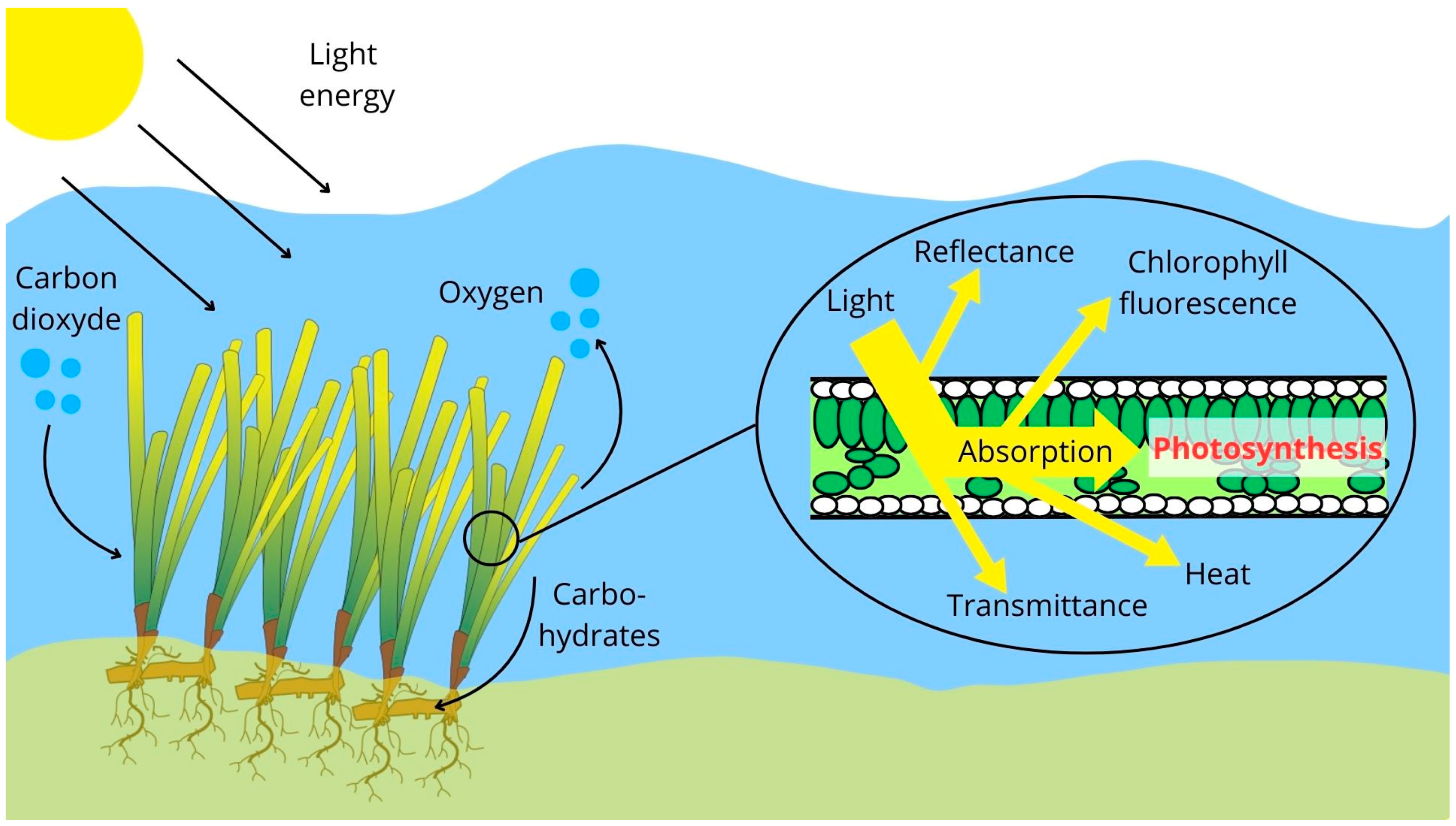
Figure 1. Schematic diagram of a simplified representation of photosynthesis in seagrasses.
Water 16 01702 g001
Figure 2. The location of the study area. The top-left figure displays the location of Corsica Island in the Mediterranean Sea. The bottom-left figure shows Corsica Island and Calvi Bay. The right figure displays the experimental sites in Alga Bay (Calvi, Corsica). The biocenosis map used in the right figured was retrieved from [28].
Water 16 01702 g002
Figure 3. An orthomosaic of an anchoring track with P. oceanica cuttings transplanted onto three different types of biodegradable artificial structures (white rectangles).
Water 16 01702 g003
Figure 4. Rapid light curve on which the relative electron transport rate is plotted against the PPFD. The fitted curve is plotted with a dotted line, and the rETRmax, Ek, and α are displayed.
Water 16 01702 g004
Figure 5. Temporal dynamics of biometric parameters and biomass in controls and cuttings (intermatte and storm fragment) as function of depth: (A) number of leaves, (B) leaf surface area, and (C) shoot dry weight. Vertical error bars are standard errors.
Water 16 01702 g005
Figure 6. Temporal dynamics of photosynthetic parameters in controls and cuttings (intermatte and storm fragment) as function of depth: (A) maximum photochemical quantum yield (Fv/Fm), (B) photosynthetic efficiency (α), (C) maximum relative electron transport rate (rETRmax), (D) and saturating irradiance (Ek). Vertical error bars are standard errors.
Water 16 01702 g006
Figure 7. The temporal dynamics of leaf chlorophyll, carotenoid, and xanthophyll pigments in controls and cuttings (intermatte and storm fragment) as a function of depth: (A) Chl a+b content, (B) Chl-a content, (C) Chl-b content, (D) Chl-a/Chl-b ratio, (E) carotenoid content, and (F) xanthophyll content. Vertical error bars are standard errors.
Water 16 01702 g007
Figure 8. The temporal dynamics of the total carbohydrate reserve (sucrose and starch) in controls and cuttings (intermatte and storm fragment) as a function of depth. Vertical error bars are standard errors.
Water 16 01702 g008
Figure 9. Correlation matrix (Spearman’s rho coefficient) of morphological and photo-biological parameters on control plants and transplants of different origins (intermatte and storm fragment) as function of depth and sampling campaign. Positive correlations are displayed in blue and negative correlations in red. Color intensity and size of circle are proportional to correlation coefficients. Non-significant correlations (p > 0.01) are left blank.
Water 16 01702 g009
Figure 10. PCA displaying differences in morphological and photo-biological response variables. Colors of biplot are relative to variance contribution (Cos2).
Water 16 01702 g010
Table 1. Results of the chi-squared tests performed on different generalized linear models at the three sampling campaigns for the survival, morphological, and photo-biological variables due to the effect of the Origin (Control (C), intermatte cuttings (I), and storm fragments (E)), bathymetry (Shallow (S) vs. Deep (D) sites), and the interaction between Origin and Bathymetry (OxB). “NA” indicates that no data were available as “TCR” was only measured in May 2022 and May 2023. “n.s.” indicates non-significant difference; “*” indicates significant difference. At the bottom of the table are the pairwise tests for the significant Origin and Origin x Bathymetry effects.
Effect TypeMay 2022September 2022May 2023
OriginOriginBathymetryOxBOriginBathymetryOxB
Morphology
Survivaln.s.n.s.n.s.n.s.n.s.n.s.n.s.
Number of leavesn.s.n.s.n.s.n.s.n.s.n.s.n.s.
Leaf surface arean.s.*n.s.n.s.*n.s.n.s.
Biomassn.s.*n.s.n.s.*n.s.n.s.
Photo-biology
Fv/Fm*n.s.n.s.n.s.*n.s.n.s.
αn.s.n.s.n.s.n.s.n.s.n.s.n.s.
rETRmax*n.s.n.s.n.s.n.s.n.s.n.s.
Ek*n.s.*n.s.n.s.*n.s.
Chl-an.s.**n.s.n.s.*n.s.
Chl-bn.s.**n.s.***
Chl a+bn.s.**n.s.n.s.*n.s.
Chl-a/Chl-bn.s.n.s.n.s.n.s.n.s.n.s.n.s.
Xanthophylln.s.**n.s.n.s.*n.s.
Carotenoidn.s.**n.s.n.s.*n.s.
TCRn.s.NANANAn.s.*n.s.
Pairwise testMay 2022September 2022May 2023
OriginOrigin OxBOrigin OxB
Leaf surface area C ≠ I
C ≠ E
C ≠ I
C ≠ E
Biomass C ≠ I
C ≠ E
C ≠ I
C ≠ E
Fv/FmC ≠ I C ≠ E
rETRmaxC ≠ I
I ≠ E
EkC ≠ I
Chl-a C ≠ I
Chl-b C ≠ I C ≠ I SI ≠ DE
SE ≠ DE
Chl a+b C ≠ I
Xanthophyll C ≠ I
C ≠ E
Carotenoid C ≠ I
C ≠ E
Disclaimer/Publisher’s Note: The statements, opinions and data contained in all publications are solely those of the individual author(s) and contributor(s) and not of MDPI and/or the editor(s). MDPI and/or the editor(s) disclaim responsibility for any injury to people or property resulting from any ideas, methods, instructions or products referred to in the content.