Review

Influencing Factors for the Growth of Cladophora and Its Cell Damage and Destruction Mechanism: Implication for Prevention and Treatment

by
Yuyao Wang
1,2,
Kuo Wang
2,
Xiaojie Bing
2,3,
Yidan Tan
2,
Qihao Zhou
2,
Juan Jiang
2,4 and
Yuanrong Zhu
2,*
1
College of Geography and Environmental Science, Northwest Normal University, Lanzhou 730070, China
2
State Key Laboratory of Environment Criteria and Risk Assessment, Chinese Research Academy of Environmental Sciences, Beijing 100012, China
3
School of Energy and Environmental Engineering, University of Science and Technology Beijing, Beijing 100083, China
4
College of Environment, Hohai University, Nanjing 210098, China
*
Author to whom correspondence should be addressed.
Water 2024, 16(13), 1890; https://doi.org/10.3390/w16131890
Submission received: 23 May 2024 / Revised: 26 June 2024 / Accepted: 30 June 2024 / Published: 1 July 2024

Abstract

:
Cladophora is commonly found in marine and freshwater around the globe and provides productivity for littoral zone microorganisms and invertebrates. The eutrophication of the water body has led to the abnormal proliferation of Cladophora in some water, often in river coast channel outbreaks. Even under the nutritional deficiency systems, such as the central trunk canal of the South-to-North Water Diversion in China, Cladophora’s blooms affect water quality and seriously jeopardize human health. Thus, the structural characteristics of Cladophora cells and spores, the factors affecting the growth of Cladophora cells, and the mechanisms of damage and destruction of Cladophora cells and spores were investigated. Cladophora cells are cylindrical with very thick cell walls. The inner layer of the spore wall is a thin membrane which contains the nucleus of the spore. The growth and spreading of Cladophora cells are affected by various environmental factors such as light, temperature, water depth, water level, nutrient salts, pH, etc. Some physical treatment measures, such as ultrasounds, would destroy the cell walls and membranes of Cladophora by its high-intensity mechanical action. Chemicals and aquatic plant measures can destroy Cladophora cells’ photosynthesis system, antioxidant enzyme systems, proteins, and ultrastructure. Based on the mechanisms for these cell damage and destruction, a combination of measures that are likely to inhibit the growth of Cladophora cells effectively was suggested. Furthermore, the damaged cells of Cladophora could provide some environmental benefits. Aggregated results could provide a scientific basis for further research on the control of blooms of Cladophora or the reuse of Cladophora cells as a natural resource.

1. Introduction

Cladophora is Chlorophyceae, a member of the family of Cladophoraceae, which is a filamentous, green, branching algae [1]. Cladophora belongs to the higher algae and contains chlorophyll identical to that of higher plants. The water environment of Cladophora is well adapted to low CO2 and can rapidly absorb CO32− as a supplementary carbon source when there is insufficient CO2 in the water [2]. Thus, Cladophora is more able to adapt to low CO2 environments than algae in general. Cladophora is ubiquitous in marine and freshwater worldwide, as it provides productivity for littoral zone microorganisms and invertebrates [3]. Cladophora can grow exceptionally well in eutrophic waters and has applications in a number of industries [4]. Wastewater can be purified with the use of Cladophora, which is simpler and less costly in terms of process compared to traditional treatment methods [5]. The biofilm area of Cladophora is large, and studies have shown that it is effective in intercepting and adsorbing microplastics [6]. The effect of Cladophora in removing N and P from eutrophic water is significantly higher than that of Diatoms and Cyanobacteria [7]. Cladophora also has good adsorption capacity of Pb, Cd, and As in the water body [8,9]. Therefore, the moderate growth of Cladophora would have a positive effect on water’s consumption of nutrient salts, purifying water quality and maintaining the balance and stability of the water ecosystem. However, excessive proliferation of Cladophora can inhibit the growth of other aquatic organisms, produce harmful substances such as H2S, and damage water quality. This leads to negative impacts on water ecosystems and water quality safety [10].
Currently, abnormal algal accretion and algal bloom outbreaks are environmental problems that are extremely difficult to solve. Due to its abnormal proliferation, it has led to significant threats to the availability of water bodies, biodiversity, and landscape [11]. Cladophora has become one of the classic algae species for algal bloom outbreaks due to its strong reproduction ability and fast growth cycle. The proliferation of Cladophora has been reported in many rivers, lakes, and other water bodies in different regions of the world, such as Lake Baikal (East Siberia), Laurentian Great Lakes (USA), Lake Budworth (UK), and Qinghai Lake (China) [12,13,14,15]. It was found that a large number of Cladophora grow in Qinghai Lake, China, and most of the areas with significant Cladophora biomass are submerged grasslands (>500 g/m2) [12]. In recent years, Cladophora has been prone to bloom in August. Their unique food chain is simpler than that of other lakes at lower elevations, so outbreaks of Cladophora can wreak havoc on their water quality [14]. Transitional proliferation of Cladophora has gradually become a widespread global problem. With the intensification of eutrophication in global water bodies, Cladophora has appeared in different scales of outbreaks in coastal zones, which may endanger the growth of fish fry and cause severe environmental problems. At the same time, the abnormal increase in the value of Cladophora also affects the recovery of submerged plants in the environmental restoration process of some lakes. Compared with eutrophic water bodies, the problem of abnormal proliferation of Cladophora is currently occurring even in some low-nutrient water bodies. For example, under low nutrient conditions (TN < 1.50 mg/L, TP < 0.026 mg/L) in the central trunk canal of the South-to-North Water Diversion in China [16], abnormal proliferation of Cladophora still existed for some years. This led to clogging of the filter media layer, affecting the regular operation of the filtration system [17]. Due to the large size of Cladophora cells, it is easy to block the filter device. In addition, Cladophora cells attach siliceous cell walls to hard shells. These create easy-to-adsorb sticky substances that are not easily destroyed by disinfectants. These make water quality treatment difficult and increase costs [18].
The Cladophora not only reproduce rapidly but are also very difficult to control. Patrick et al. [19] reported a series of biological control measures for freshwater Cladophora at an early stage but did not find any animals feeding on Cladophora. Some studies have shown that other chemical methods, such as CuSO4, can also inhibit the growth of Cladophora. However, it can have toxic effects if added in excess [20]. In general, the more widely used means of algae removal have shortcomings, such as low efficiency, maintenance difficulties, and secondary pollution. At the same time, there are few reports on the methods to effectively inhibit the propagation of Cladophora in the actual water environment. Factors affecting the growth and propagation of Cladophora cells, as well as the mechanism of damage to their cells by means of prevention and control, are the basis for scientific prevention and control of the abnormal proliferation of Cladophora in the aquatic environment and even for the recycling and utilization of Cladophora cell resources. Therefore, the present study is intended to summarize the factors affecting the cell growth of Cladophora based on a large number of reports in the literature. Based on the structural characteristics of the cells of Cladophora and the damage mechanism of the cells during the prevention and control of algal species. This study will analyze the mechanism of the destruction of the cellular structure of Cladophora through physical, chemical, and biological methods. Ultimately, theoretical methods for the integrated control and utilization of Cladophora cells will be proposed. Acquiring these results is favorable for the development of devices and engineering applications in the future.

2. Characterization of the Cell Structure and the Process of Matter-Energy Cycling in Cladophora

Cladophora larvae are embedded and float when they grow. The cells are long and cylindrical, with a thick cell wall [21], as shown in Figure 1. The cells have a large central vacuole, and the pigment bodies are granular during the spore period of the Cladophora and reticulate when the cell matures [21]. When mature, the cell can be dispersed into multiple protein nuclei and nuclei [22]. Cladophora contain chlorophyll a (Chla) and chlorophyll b (Chlb). The cell wall is layered with a cuticle that contains 70% protein. It is often epiphytic with many algae [23]. The basal cells form pseudoradical branches that are 25–35 μm wide. They are 5–8 times as long as wide [22]. Cladophora grows by apical or intermediate cell division and fixes by basal cells, pseudoroots, or fixators [22]. Cladophora cell division is caused by the cytoplasm producing ring-like constriction grooves and septa formed by the deposition of cell wall material in the grooves, separating the cell in two. The cytoplasm does not divide at the same time [24].
The Cladophora can reproduce asexually or produce spores [25]. The shape and size of Cladophora spores vary from short and thick rod-shaped, elongated, and spindle-shaped, with sizes ranging from 2 to 20 μm [21]. The surface of the spore wall has short and dense setae, which are essential structures for the spores to float, move, and reproduce in the water. The inner layer of the spore wall is a thin membrane that contains the nucleus of the spore. The outer layer of the spore wall is a hard shell with many tiny holes on the surface of the shell, which allow for the exchange of substances inside and outside the spore wall [26].
Compared with other planktonic algal cells and aquatic plant cells, the Cladophora cell has a unique morphology and structure. As mentioned earlier, the Cladophora cells are cylindrical, which differs from most planktonic algal cells and aquatic plant cells. Cladophora cells form elongated rod-like projections at the edge of the cell that are encapsulated by tiny vacuoles that contain specialized enzymes that may be used to break down organic matter [6]. In addition, the chloroplasts of the Cladophora cells also exhibit a unique structure compared to other planktonic algal cells. In chloroplasts, Cladophora cells contain a large number of vacuoles, and the membranes of these vacuoles are rich in transmembrane proteins, which may be one of the essential reasons why chloroplasts maintain efficient photosynthesis [21]. Cyanobacteria do not have organelles such as chloroplast, mitochondrion, centrosome, endoplasmic reticulum, and vacuole [27]. However, Cyanobacteria contain Chla and phycocyanin, which can photosynthesize with the help of enzymes and pigments in the cytoplasm [28].
Cladophora cells use photosynthesis and respiration for material and energy cycling (Figure 1). Cladophora cells absorb light energy and convert it into chemical energy. Cladophora cells can photosynthesize and synthesize organic matter, which is stored inside the cell [2]. At the same time, photosynthesis produces oxygen, which is taken up by the algal cells and replenishes the oxygen needed. Cladophora also converts organic substances, such as glucose, into energy through aerobic respiration, which powers the cells to maintain life activities. In addition, there are different types of enzymes that are involved in various metabolic processes within the cell, which break down organic substances into smaller molecules, releasing energy to power the cell’s life activities [21]. Inside the cells of the Cladophora, organic and inorganic substances are transferred through organelles in the cytoplasm and the cell membrane, through which CO2 is expelled from the cell. The Cladophora is very adaptable to low CO2. When there is insufficient CO2 in the water, they can utilize HCO3 as a source of carbon very quickly, unlike other algae that need a period of time to adapt. Cladophora cells can use HCO3 for the photosynthesis of carbonic anhydrase. Through metabolic activities such as photosynthesis, they consume HCO3 in the water and deposit CaCO3 [2].

3. Factors Influencing Cell Growth and Dissemination and Critical Processes in Cladophora

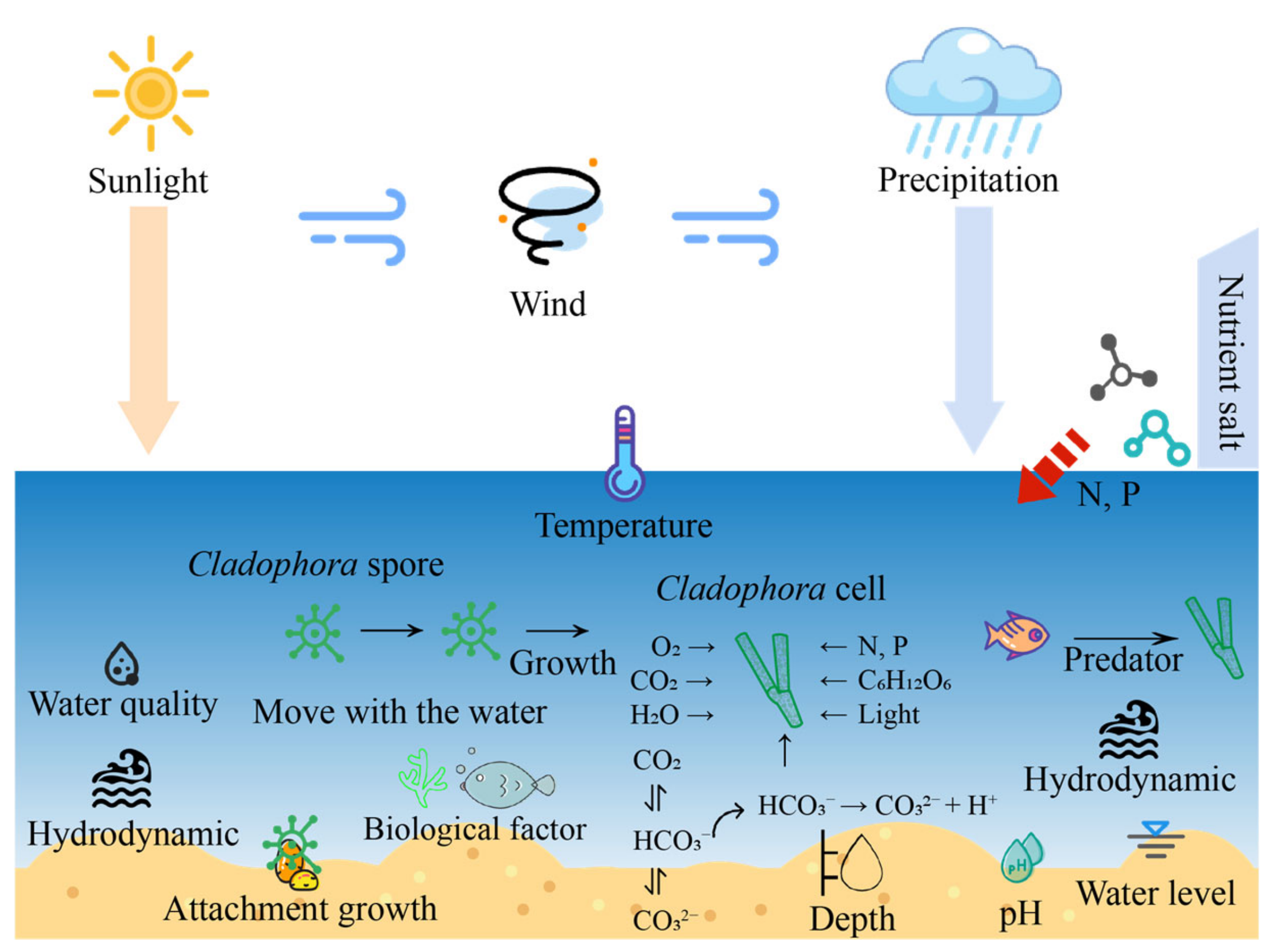
The growth and propagation of Cladophora cells can be influenced and regulated by a variety of environmental factors. The conceptual diagram of the factors and processes affecting the propagation and growth of Cladophora cells in the actual aquatic environment is shown in Figure 2. The environmental factors related to the growth of Cladophora cells are generally light, temperature, hydrodynamics, water depth, water level, nutrient salts, and pH.
Light is an important environmental factor influencing the distribution of Cladophora communities over time and space. Sometimes, there are interactions between environmental factors [29]. When the light intensity reaches saturation point, the photosynthetic rate decreases or even stops, and photoinhibition occurs [30]. According to Graham et al. [31], the light saturation point of Cladophora was in the range of 15.4–30.8 µmolphotons/m2/s under different temperature conditions. Cladophora is very sensitive to light and prefers to stay in more adequate light conditions [32]. However, there are algae that do not like bright light; for example, Rhodophyta reproduces faster under moderate light conditions [33]. Seasonal changes in the biomass or abundance of Cladophora tend to be a response to temperature. Low-temperature conditions in winter and spring inhibit the growth of Cladophora, which requires a minimum water temperature of about 5 °C for growth, and studies have shown that Cladophora gradually dies when the water temperature exceeds 23.5 °C in summer [34]. According to Stewart and Lowe [35], the optimal water temperature for growth of Cladophora ranges from 5–23 °C. The optimal temperature for growth of Cladophora differs significantly from that of planktonic algae. For example, Cyanobacteria grow at 28–35 °C, while Diatoms maximize their specific cell growth rate and significant physico-chemical content at 15–25 °C [31].
Changes in hydrodynamic conditions can lead to changes in water column material transport, affecting algal growth [36]. Water flow can enable the exchange of oxygen and carbon dioxide in the water environment with the atmosphere, which also helps to ensure the supply of nutrients. Water movement also affects the growth of Cladophora through stretching, dragging, and churning. Hydrodynamics also has an essential effect on the shedding of Cladophora cells. The speed and direction of water flow can directly affect the state and stability of Cladophora cells [37]. In the case of fast water currents, Cladophora cells face greater tensile forces and are easily dislodged by the current. Slow currents make the Cladophora cells more stable and allow for better attachment to the surface of the object. When the direction of water flow changes, the Cladophora cells need to re-adapt to the new flow environment, which may lead to cell detachment. It has been found that Cladophora collected from reservoirs have many branches, while those collected from flowing water, such as rivers, have fewer branches [22]. Whereas Cladophora usually grows in slower currents, planktonic algae can adapt to a variety of current conditions and can grow in faster currents [38]. There is also a relationship between Cladophora cell growth and water depth. Light intensity in nature also decreases with increasing water depth, mainly due to the influence of suspended solids in the water column, including plankton. It has been found that the biomass of Cladophora decreases with increasing water depth [39,40]. Water level is one of the critical factors determining the distribution, biomass, and population structure of aquatic plants, and moderate changes in water level would benefit the diversity of aquatic plant species [41]. Some studies have shown that in areas with water depths <1.0 m, sufficient sunlight can be allowed to penetrate to support Cladophora outbreaks [12].
Nutrients necessary for algae growth include N, P, Si, Fe, and other elements. Life processes such as photosynthesis, synthesis of organic compounds, and construction of tissue cells in Cladophora require the participation of various nutrient elements. Nutrient salt concentration affects physiological and biochemical conditions [42]. Phosphorus is the primary nutrient limiting the growth of Cladophora [35]. In eastern Lake Erie, mean concentrations of soluble phosphorus in the spring and summer growth periods of Cladophora ranged from 0.9 to 3.5 μg L−1 [43]. An appropriate increase in pH favors photosynthesis and promotes the growth of Cladophora, but when the pH is too high, it is detrimental to the growth of Cladophora [44]. Alkaline water can easily absorb atmospheric CO2 and promote photosynthesis of Cladophora. The main factors affecting the pH change of the reaction system are the photosynthesis and the respiration of Cladophora. Photosynthesis is stronger than respiration, CO2 in the water is utilized, the acid-base equilibrium is disrupted, and the pH increases [45]. Cladophora utilizes HCO3 for photosynthesis; when HCO3 consumes H+ to produce CO2, the amount of H+ decreases, the amount of OH increases, and the pH also increases. The predominant carbon form at pH 5 to 9 is HCO3; at low pH, it is CO2, and at high pH, it is CO32− [46]. Tsutsui et al. showed that there was an interaction between salinity and pH and a highly significant effect on the growth of Cladophora [46].
The process of the spreading of Cladophora cells is also affected by a number of other factors, including temperature, light, water quality, hydrodynamics, and biological factors. Generally, water temperature between 15–30 °C is the optimal temperature for cell spreading of Cladophora [47]. Below 15 °C, cell spreading slows down. Water temperature levels above 30 °C lead to cell death. Cell spreading in Cladophora is accelerated under adequate light conditions and slowed down under inadequate light conditions [48]. Water quality also affects the cell spread of Cladophora [49]. Poor water quality and low levels of nutrients and oxygen can lead to limited growth and reproduction of Cladophora, thus affecting cell spread. Hydrodynamics is also an essential factor influencing the cell spread of Cladophora. Cell spreading of Cladophora is accelerated under faster water currents and slowed down under slower water currents [50]. The cellular spread of Cladophora can also be affected by other organisms. For example, some aquatic animals prey on Cladophora, thereby reducing their cellular spread [51].

4. Mechanisms of Cell Damage Destruction in Cladophora

Currently, common control methods for algae, including Cladophora, can be categorized into three main groups: physical, chemical, and biological. However, few studies have been reported on the prevention and control of Cladophora alone. The mechanism of cell damage and destruction is a critical scientific issue in the prevention and control of Cladophora and the reuse of cell fragmentation. It is essential for the research and growth of prevention and control technology and reuse means, but it has also been rarely reported. Therefore, this study will focus on physical, chemical, and biological effects and will combine this research with consideration of the characteristics of the cellular structure of the Cladophora and factors influencing the growth of Cladophora. In addition, we will analyze and explore the possible damage mechanism of Cladophora cells.

4.1. Mechanism of Cell Damage and Destruction of Cladophora under Physical Action

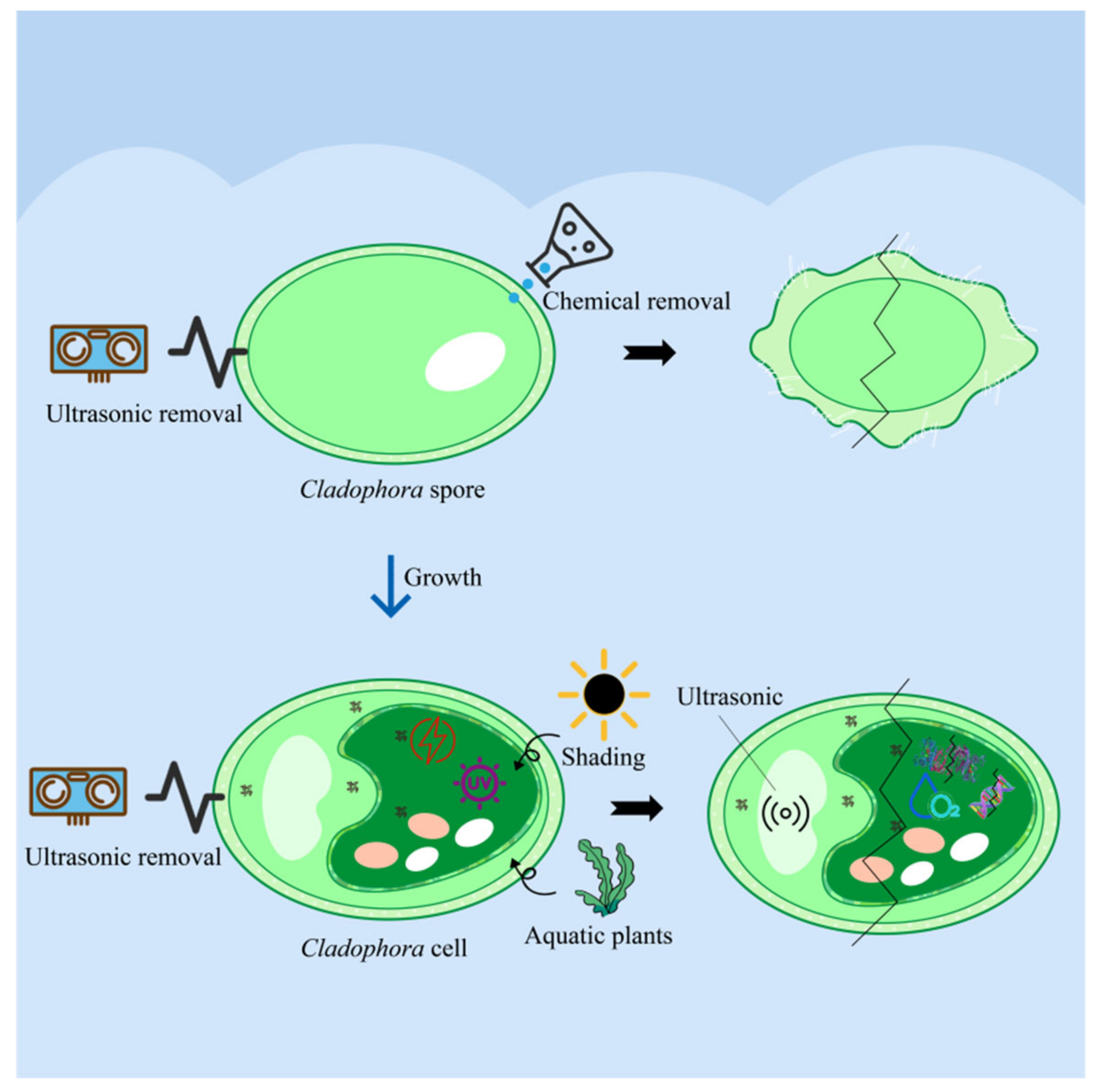
Physical methods mainly include traditional mechanical salvage, interception by interceptor poles, and hydrodynamic regulation. Some new technological methods, such as physical shading, ultrasound, micro-current, ultraviolet (UV), zeta potential, and many other methods, have been reported but are not widely used [52,53,54]. Due to the characteristics of vertical distribution and horizontal drift of Cladophora, which are directly affected by thermal stratification, light intensity, wind speed, and wind direction. When Cladophora accumulates at the littoral zone of the lake, they can be salvaged with stationary equipment [55]. However, such physical techniques may not cause damage and destruction to the cells [56], and Cladophora, which is not entirely salvaged or intercepted, will continue to grow and reproduce. Physical shading and algae control technology utilize the control of incident light illumination in the water column so that the light level obtained by the Cladophora does not satisfy their need to inhibit the growth of Cladophora [57] for photosynthesis. The main carriers of photosynthesis are photosystem I (PSI) and photosystem II (PSII) in Cladophora, which absorb light energy and transfer energy to PSI and PSII to complete the energy conversion process. Intense light reduces the electron transfer rate in Cladophora cells. Also, it causes damage to proteins in the cellular photoreaction center to outpace the rate of light repair, which greatly reduces photosynthetic activity [58]. In recent decades, it has been considered as one of the physical methods to inhibit algal blooms in specific water environments. Using physical shading and ultrasound to destroy algal cells is a common treatment method. It has been shown that physical methods such as shading and ultrasound can cause damage to cell membranes and breakage of organelles, thus disrupting the life activities of algae and affecting their growth and reproduction [59]. In addition, shading and ultrasound can cause chemical reactions within the algal cells, such as inhibition of photosynthesis and inactivation of enzymes, thus further destroying the cell structure. The advantages of the ultrasonic method are simple operation and high efficiency. In addition, this method also makes it difficult to cause secondary pollution. Ultrasonication has been found to disrupt the cellular structure of different planktonic algae, such as the air cells of Cyanobacteria [60]. According to the structure of the cells of Cladophora, ultrasonication may damage the cell structure of Cladophora to a certain extent. Therefore, after considering this, along with the current research and the characteristics of the cells of Cladophora, an ultrasound was used as an example to explore the damage mechanism of Cladophora cells under physical action (Figure 3).

4.1.1. Disruption of Cell Walls and Cell Membranes

Physical methods (such as performing ultrasounds) can cause damage to the cell walls and Cladophora cell membranes of Cladophora. The effects of ultrasound treatment on algal cells are reflected in changes in their physiological structure and function. Free radicals generated by ultrasound radiation can induce lipid peroxidation to destroy cells [61]. Ultrasonic radiation disperses aggregated algal cells [62], which increases cell number and destroys the cellular structure of Cladophora. Based on the study of the effect of ultrasound on the growth and reproduction of algae. The role of ultrasound in controlling water pollution is mainly due to its cavitation effect. The intensity of ultrasound can break the cell wall so that the material inside the cell outflow [63]. Ultrasound is a type of sound wave that produces high-frequency mechanical vibrations, the energy of which can be absorbed by the cell wall and converted into thermal energy [58]. This thermal energy can lead to cell wall rupture and damage, especially in the case of Cladophora cells, which have thick cell walls that ultrasound can effectively destroy. By generating a robust mechanical effect, it produces destructive cracks in the cell wall, thus causing the rupture of the cell membrane. Therefore, both mechanical and chemical effects caused by ultrasound may damage the cell wall and cell membrane of Cladophora, breaking the cells and releasing intracellular substances [64].

4.1.2. Disruption of the Photosynthetic System

Physical methods can cause damage to the system of photosynthesis in Cladophora. Photosynthesis is an essential pathway for Cladophora to obtain energy for growth and maintain life activities [65,66]. Ultrasound, for example, inhibits the growth rate of Cladophora by disrupting cell walls and cell membranes through cavitation, interrupting photosynthesis, and inhibiting cell division and cell cycle [67]. When an ultrasound is applied to Cladophora, the morphology and structure of the chloroplast membrane are significantly altered, and the electron transport chain of photosynthesis is inhibited, causing a failure in the light’s reaction. Ultrasounds can produce strong vibrations and a rotation in the light and system of the Cladophora, which, thus, destroys the protein structure and electron transport chain required for photosynthesis and decreases the efficiency of photosynthesis and growth rate. Ultrasounds can also cause tiny cavities and cracks in the cells of Cladophora, further aggravating the destruction of its photosynthesis. At the same time, ultrasounds can also affect the cyst-like membranes in chloroplasts so that the cell’s supply of nutrients is affected. In addition, an ultrasound can also cause the cellular vacuole to collapse, resulting in the overflow of cytosol, further affecting photosynthesis. Current studies have mainly investigated the effects of ultrasounds on the photosynthesis system in algal cells by measuring the chlorophyll and net photosynthetic oxygen release rate of algal cells [68,69]. In one study, after an ultrasound treatment, the photosynthetic system of Cladophora cells was disrupted, making it more difficult for cell activity to be restored and removed [70].

4.1.3. Destruction of Antioxidant Enzyme System

Physical methods also damage the antioxidant enzyme system of the Cladophora. Take the ultrasound as an example once again. The high temperature and pressure generated by cavitation and a large number of free radicals can destroy the active enzymes (e.g., superoxide dismutase (SOD), peroxidase (POD), catalase (CAT), and glutathione reductase (GR)) and active substances in algal cells, thus affecting the physiological and biochemical activities of the cells [71]. Ultrasounds can disrupt the normal functioning of the antioxidant enzyme system by generating high-energy sound waves, which can have a mechanical impact on the cells, leading to cell membrane rupture and damage to organelles. Ultrasounds can generate large amounts of heat, which can lead to an increase in intracellular temperature that can disrupt the structure and function of the cell. The antioxidant enzyme system is usually temperature sensitive, so ultrasounds may damage its structure and function through thermal effects. Therefore, prolonged ultrasound treatment can also affect Cladophora by disrupting their cell membranes, leading to leakage of intracellular enzymes and impairing enzyme activity [72,73].

4.2. Mechanisms of Cell Damage and Destruction of Cladophora under Chemical Action

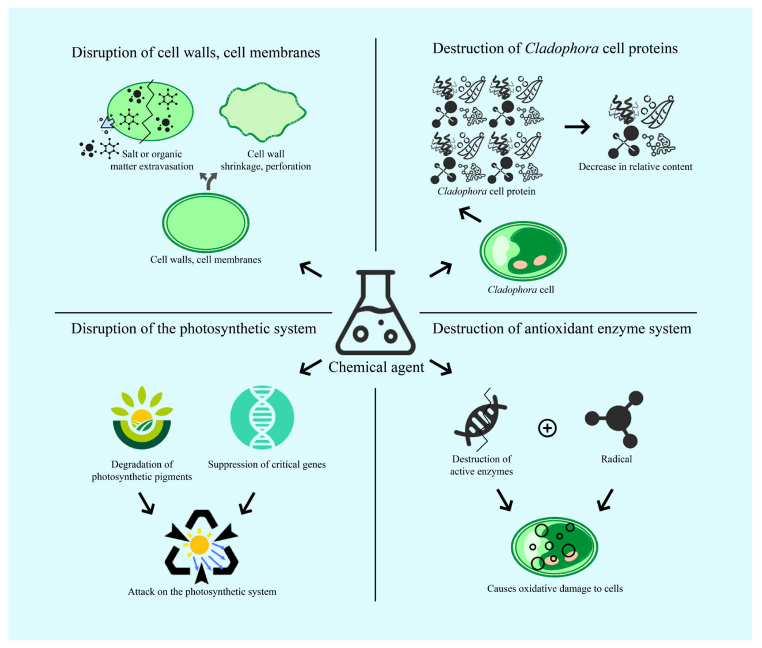
The chemical method mainly utilizes chemicals to destroy the cell structure of the Cladophora, thus leading to its death. This method entails adding chemicals through the water body, such as a variety of algaecides and flocculants, to inactivate the Cladophora. At the same time, the redox potential of water that contains algae is changed, and the Cladophora colloids are destabilized to achieve the purpose of Cladophora removal [74]. The algaecides commonly used include metallic substances (e.g., CuSO4 and copper-containing chelates), oxidizing agents (e.g., KMnO4, O3, H2O2, and ClO2), and herbicides (e.g., metsulfuron-methyl and bensulfuron methyl) [75]. The primary mechanism of chemical damage to algae cells is to directly or indirectly damage the physiological and genetic structures through different chemicals, causing changes in cell membrane permeability and protein denaturation, which leads to algae cell death or growth inhibition [76]. It has been found that algae cell walls contain large amounts of cellulose and algal-specific gums that maintain the morphology and structure of algal cells [77]. Chemistry can disrupt these structures, making algae cells more fragile. Chemicals can inhibit the photosynthesis of algae cells, reducing the growth rate and metabolic rate of the algae, thus inhibiting the growth and reproduction of the algae. Chemicals can also damage the cell membranes of algae cells, causing substances to leak out of the algae cells, which can lead to the death of the algae cells. In addition, chemicals can destroy the genetic material of algae cells so that the algae cells cannot divide and reproduce normally, thus achieving the purpose of controlling the growth and reproduction of algae [78]. Therefore, according to the structure of Cladophora cells, this section mainly focuses on the mechanism of damage and destruction of Cladophora cells by chemicals, and the mechanism is shown in Figure 4.

4.2.1. Disruption of Cell Walls and Cell Membranes

Chemical damage to Cladophora cells is reflected in the cell wall and cell membrane. Chemicals can affect the cell wall, leading to changes in cell membrane permeability, which in turn kills the algal cells [79]. Treatment of Cladophora with high dosages of chemicals can cause severe damage to their cell membrane integrity, leading to extravasation of intracellular salts or organic matter. Chemicals can disrupt the structure of the cell wall and the permeability of the cell membrane by destroying the components of the cell wall and the cell membrane. This can lead to the efflux of substances from the cell, such as cellulose and proteins, which can lead to cell death. Chemicals can release organic acids that penetrate the cell walls and membranes of Cladophora and react with the cellulose and proteins in them, leading to the rupture and breakdown of the cell walls and membranes. The surface of the cells of Cladophora is flat and smooth [80], but after treatment with chemicals, the surface of the cells appears to be wrinkled. The cell wall was shrunk and perforated, and its shape became an irregular ellipsoid. There was also a large amount of flocculent material around the cell wall.

4.2.2. Destruction of Algal Cell Proteins

Chemicals can cause damage to Cladophora cell proteins. Algae are rich in protein resources, and the protein content in algae varies depending on the species and season [81]. The cellular protein content of Cladophora varied with seasonal changes, and the relative protein content of algal cells showed a significant fluctuating decrease after chemical treatment. Chemicals increase protease activity in Cladophora cells, which disrupts the structure of proteins and causes them to lose their original properties and functions. Hydrolyzing the peptide bonds in the protein molecules, the proteins are broken down into smaller peptide segments or amino acids. The administration of chemicals can promote the level of membrane lipid peroxidation in cells, leading to the accumulation of malondialdehyde (MDA) in the cells of Cladophora [82]. Accumulation of MDA causes cross-linking polymerization of life macromolecules such as proteins and nucleic acids. It also destroys the structure of the cell membrane, leading to a decrease in the metabolic activity of the cell and affecting the average growth and division of the cell.

4.2.3. Disruption of the Photosynthetic System

Chemicals can likewise cause damage to the photosynthetic system of Cladophora. Chemicals attack the photosynthetic system by degrading photosynthetic pigments and inhibiting the expression of critical genes. This results in the inability of the Cladophora to properly synthesize photosynthetic pigments such as chlorophyll, thus affecting the process of photosynthesis [83]. By inhibiting the activity of photosynthetic enzymes, the process of photosynthesis is hindered. It can directly damage the structure of the photosynthetic membrane of Cladophora, leading to the impaired function of the photosynthetic membrane. In addition, chemicals can interfere with the photosynthetic electron transfer process, resulting in a disruption of the energy production of photosynthesis. Basic metabolic activities such as photosynthesis are impeded, thus blocking the energy capture pathway of the bristlecone algae cells and accelerating cell death. Pigment content is also closely related to photosynthesis, with Chla gradually decreasing as algal cell mortality increases [84]. It is noteworthy that Chla directly affects the capture and utilization of light energy and must be involved in the subsequent conversion of light energy to chemical energy.

4.2.4. Destruction of Antioxidant Enzyme System

The damage that chemicals can do to Cladophora is also reflected in damage to the antioxidant enzyme system. Chemicals can damage the antioxidant enzyme system of Cladophora cells and produce large amounts of free radicals that cause oxidative damage to the cells [85]. Chemicals can react with enzyme molecules in the antioxidant enzyme system, altering their structure and function, thus affecting the proper functioning of the antioxidant enzyme system. Chemicals may also affect the expression and activity of the antioxidant enzyme system by affecting intracellular signaling pathways. High concentrations of chemicals inhibit enzyme activity, and low concentrations promote enzyme activity [86]. When the concentration of chemicals exceeds a specific range, it will damage the antioxidant mechanism of Cladophora. The antioxidant enzymes in the cells cannot clean up the rapidly produced large amount of free radicals in time, which disturbs the metabolism of the cells and even causes the death of Cladophora cells. This led to the phenomenon that the activities of SOD, POD, and CAT first increased and then decreased [87].

4.3. Mechanisms of Cell Damage and Destruction of Cladophora under Biological Action

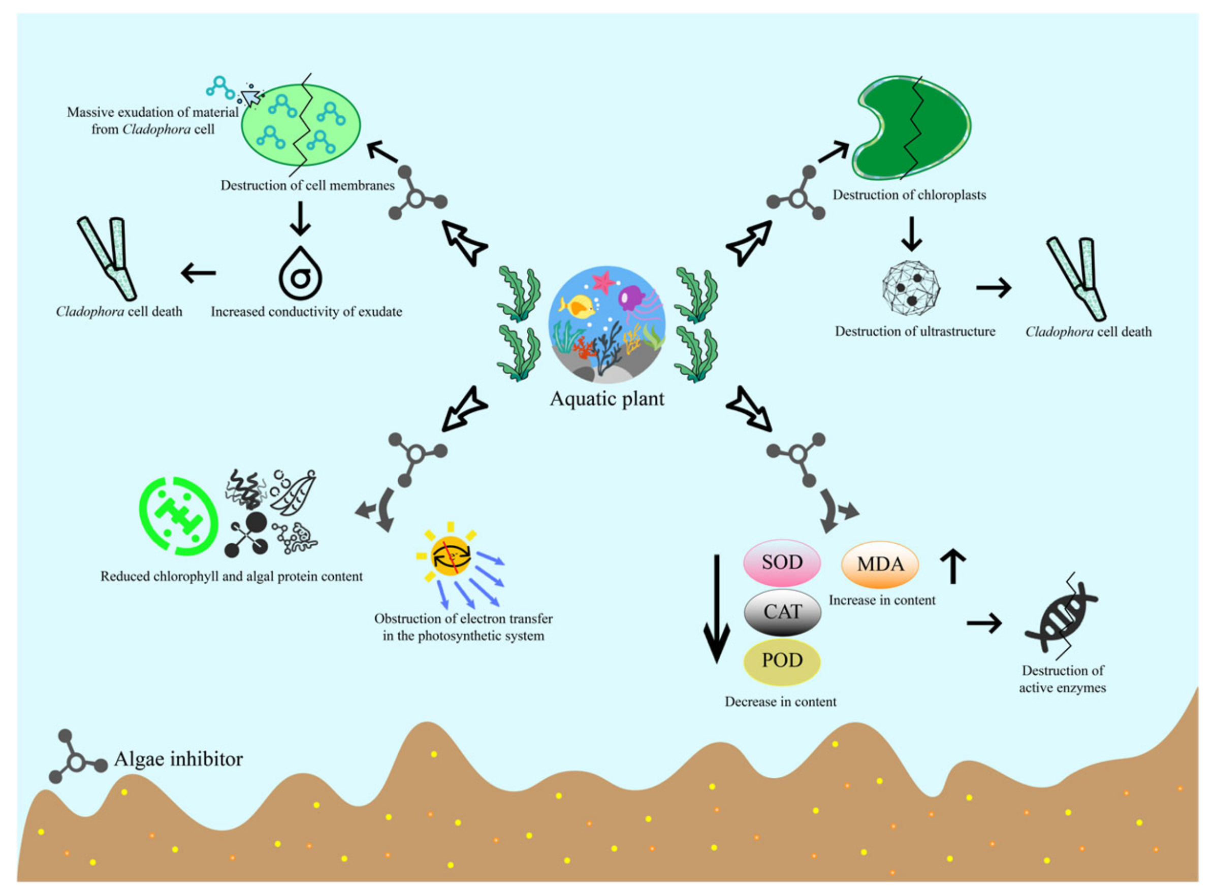
Biological methods include using aquatic plants, animals, and microorganisms to control algae. Traditional biological means of algae control are mainly aquatic animal control, which utilizes competition or predation among organisms and introduces other organisms to inhibit algal growth [88,89]. The hypophthalmichthys molitrix, which feeds exclusively on plankton and can be used for water purification, has rapidly colonized the Great Lakes and surrounding water systems in North America, causing devastating disasters in the local ecosystems [90]. Microbial algal control includes viral algal control, protozoan algal control, and bacterial algal control [91]. Aquatic plant algal control is a more researched method, which mainly utilizes specific aquatic plants in the water to inhibit algal growth by absorbing, transporting, and metabolizing nutrient-rich substances. Or utilizes the chemosensory inhibition of aquatic plants to control the presence of algae [83]. The main mechanisms of aquatic plant chemosensory substances to inhibit algae include alteration of algal cell enzyme activity, effects on photosynthesis, destruction of algal cell structure, and effects on the ultrastructure of the cell [92]. The Cladophora cells also have structures such as cell membranes and photosynthetic systems. The aquatic plant algal control method will have similar inhibitory effects and mechanisms on Cladophora cells. Therefore, this section mainly focuses on the damage destruction mechanism of aquatic plant algae control on Cladophora cells, and the mechanism is shown in Figure 5.

4.3.1. Disruption of Cell Walls and Cell Membranes

Aquatic plants can cause damage to the cell walls and membranes of Cladophora. Plants directly or indirectly affect other plants by releasing algae control substances into the environment. Algae-suppressive substances can disrupt cell membranes, reducing their integrity and leading to the exudation of large quantities of intracellular material, increasing the conductivity of the exudate [93]. Studies have shown that lysine contained in algal inhibitory substances can cause a physiological stimulation signal to algal cells, causing algal cells to rupture and cell membrane lysis. Leakage of inclusions also occurs, resulting in apoptosis and achieving the effect of algal inhibition [94]. Due to changes in the permeability of the cell membrane, the cell membrane is prone to take up nutrients from the environment, and exceeding the threshold of the cell membrane will destroy the cell membrane [95]. Para-hydroxybenzoic acid can cause a significant increase in the production of oxygen radicals in algal cells, and free radicals such as O2− can quickly react with cell membrane components, causing damage to the cell membrane and ultimately leading to cell death [96]. Phenolic acids can affect the structure and function of algal cell membranes, causing changes in membrane integrity and permeability [97]. Aquatic plants can secrete several enzymes, including cellulases, amylases, and proteases, which can break down protein, polysaccharides, and lipid molecules in the cell walls and cell membranes of Cladophora, thus destroying the structure and function of Cladophora.

4.3.2. Destruction of Ultrastructure

Notably, aquatic plants can cause damage to the ultrastructure of Cladophora cells. When the cell membrane is damaged, the organelles, such as chloroplasts, are also damaged, and the ultrastructure present in them is damaged. Various physiological activities of the cell are impeded, and the cellular operating system collapses, ultimately leading to cell death and inhibiting the growth of algae [98]. This method has a significant destructive impact on other algae. Studies have shown that the cell shape of Microcystis aeruginosa changed irregularly after treatment. The cell membrane began to deform and crumple until rupture, the central nucleoid region was inconspicuous until it disappeared, and the chlorophyll-like vesicles were evident from the previous lamellae [99]. Many ultrastructures, such as nuclei and protein nuclei, existed in the cells of Cladophora. Aquatic plants cause the cells to swell and rupture, releasing enzymes to further damage the Cladophora cells. Therefore, the method will damage the ultrastructure of the Cladophora cells.

4.3.3. Disruption of the Photosynthetic System

Aquatic plants can similarly cause damage to the photosynthetic system of Cladophora cells. Photosynthesis is the most critical physiological and biochemical process in algal cells. Inhibition pathways mainly include reducing chlorophyll and phycobiliprotein content and blocking electron transfer in the photosynthetic system [100]. Biological algal inhibition can be achieved simultaneously by blocking the photosynthetic system electron transfer to achieve the effect of algal inhibition. Electron transfer plays an important role in maintaining normal photosynthesis in algal cells. Electron transfer is driven by a redox potential difference between the donor and acceptor sides of PSII. Water is used as a donor of reduced hydrogen to synthesize ATP, which provides energy for the activity of the Cladophora cell [101]. Phenolic compounds in plants can reduce protein levels in Cladophora cells, thereby reducing their photosynthetic capacity. In addition, respiration in plants can release gases such as carbon dioxide and methane that are detrimental to the growth of Cladophora, further disrupting the photosynthetic system.

4.3.4. Destruction of Antioxidant Enzyme System

Aquatic plants also cause some damage to the antioxidant enzyme system of Cladophora cells. The antioxidant system consists of an enzymatic scavenging system and a non-enzymatic scavenging system, in which the enzymatic scavenging system is mainly an antioxidant enzyme. These include major antioxidant enzymes such as CAT, POD, GR, aseorbate peroxidase (ApX), and SOD, which enhance plant stress tolerance [102]. The inhibitory effect of most of the substances is manifested by making a decrease in the content of SOD, CAT, and POD and an increase in the content of MDA. Aquatic plants attack the antioxidant enzyme system of Cladophora cells by secreting toxins or enzymes. These enzymes can react with the antioxidant enzyme system in the Cladophora cells and inactivate it. Aquatic plants can also secrete some organic acids, phytohormones, and other substances. These substances can inhibit the growth and metabolism of Cladophora cells, thus further destroying their antioxidant enzyme system.

5. Integrated Control and Potential Utilization Pathways for Cladophora Cells

Different control measures can cause different levels of damage to Cladophora cells. Based on the damage mechanism of physical, chemical, and biological methods to the cells of Cladophora, the study proposes a potentially comprehensive measure for the control of Cladophora (Figure 6). This measure gives suitable control measures for different growth periods of Cladophora. During the sporulation period, the main structures of Cladophora are the cell wall, cell membrane, and nucleus. Ultrasound and chemicals were chosen based on their mechanisms of destroying the cell wall, cell membrane, and nucleus of Cladophora. Ultrasound can effectively destroy the cell wall and cell membrane of Cladophora spores due to its high mechanical strength. Some chemicals can cause irreversible damage to the nucleus of Cladophora spores. However, the environmental friendliness of these chemicals and their potential to cause secondary pollution must be considered. Therefore, for different water environment needs, the two methods can effectively destroy the cell structure of Cladophora spores, making it difficult to grow and achieve the effect of prevention and control. After the growth of the Cladophora, its cell structure is more complete, and it is necessary to use a variety of methods in combination to destroy the cell structure and inhibit its growth effectively. Based on the mechanism of destroying the photosynthetic system, cell membrane, cell wall, proteins, enzyme system, and ultrastructure of Cladophora, various control methods were selected. Shading affects the photosynthetic system by inhibiting photosynthesis in the cells of Cladophora, preventing it from obtaining nutrients and fundamentally inhibiting its growth. Ultrasonic waves will disperse the aggregated Cladophora cells, thus increasing the number of cells, destroying cell walls and membranes, and improving the removal rate. After UV treatment, it will lead to the blockage of DNA replication and a decrease in the protein content of the Cladophora cells, which can not meet the demand for growth of the Cladophora cells. Micro-current inhibition will lead to the oxidation of intracellular enzymes, damage to enzyme activity, and the destruction of the enzyme system, thus inactivating the cells. Aquatic plants can damage the ultrastructure existing in the cell organelles, hinder the cell’s physiological activities, cause the cell operating system to collapse, and ultimately lead to cell death. Therefore, the use of various methods is effective in destroying the cells of Cladophora and causing their apoptosis. In the future, according to the requirements of the water body, further combined with the reproduction and growth of bristle algae factors affecting the regulation. The use of different methods of combining, optimizing, and engineering practice based on the formation of effective integrated control measures for Cladophora outbreaks.
After the cells of the Cladophora are destroyed, they are still richly useful. The cells of Cladophora are stimulated by the external environment, such as chemicals and temperature changes, which leads to an increase in the permeability of the cell membrane and changes in the activity of proteins and enzymes [103]. The degree of destruction mainly depends on the growth state of the Cladophora and environmental factors. For example, the degree of destruction of Cladophora is affected by environmental factors such as water quality, temperature, and salinity. Cladophora cells are a natural organic fertilizer after being destroyed. It has adsorption capacity, provides nutrients, improves soil texture, promotes plant growth, enhances plant disease resistance, improves plant adaptability, and environmental abatement [104]. It has been found that Cladophora is able to accumulate more chemical elements from the environment, which makes them ecologically relevant for wastewater treatment, as well as a potentially high-quality fertilizer product [105]. Cladophora cells are rich in proteins, polysaccharides, and fats, which can be extracted as beneficial active pharmaceutical ingredients. In the study of Munir et al., the main activities of algae were described in detail, and Cladophora is a potential raw material for pharmaceutical applications due to its chemical composition [21]. The Cladophora cells are also efficient photosynthetic organisms with high oxygen production capacity, which can be converted into biofuel to replace traditional fossil fuels. Dorella et al. proposed the production of biogas by using Cladophora mixed with wheat straw [106]. Sharmila and Jeyanthi utilized the biomass of freshwater Cladophora for the production of biodiesel [107]. The organic matter and nutrients released from the destruction of Cladophora cells also help other organisms to grow and reproduce, thus achieving the effect of ecosystem restoration, as shown by the study of Anh et al. Cladophora were used as a protein source when feeding Penaeus monodon [108].

6. Conclusions

The Cladophora cells are cylindrical, with very thick, rough cell walls. The surface of the spore wall has short and dense setae. The inner layer of the spore wall is a thin membrane which contains the nucleus of the spore. Cladophora plays a positive role in maintaining the balance and stability of water ecosystems, but overgrowth can cause damage to water quality. Light is an important environmental factor that influences the distribution of Cladophora communities in time and space. Low temperatures in winter and spring inhibit the growth of Cladophora. Changes in hydrodynamic conditions can lead to changes in water column material transport, which can affect the growth of Cladophora. The biomass of Cladophora decreased with increasing water depth. Nutrient salt concentration affects its physiological and biochemical conditions. The Cladophora are strong bicarbonate utilizers and are more tolerant to alkaline environments, and pH can control the growth of the Cladophora. The process of cell spreading of Cladophora is affected by temperature, light, water quality, hydrodynamics, and biological factors.
In order to discuss further the preventive and control measures for the abnormal proliferation of Cladophora and even the reuse of their cells, the mechanisms of damage and destruction of Cladophora cells and spores were investigated. Physical actions, chemicals, and aquatic plants all damage Cladophora by disrupting cell walls, cell membranes, photosynthetic systems, and antioxidant enzyme systems. The chemicals destroy the cellular proteins of the Cladophora cells. Aquatic plants damage the ultrastructure of the Cladophora. In the future, combining various methods will be an effective measure to inhibit the growth of Cladophora. Although the cells of Cladophora are dead after treatment with multiple techniques, they still have many uses and realize the sustainable use of resources. In the future, combined with the water quality requirements of the water body, as well as the actual conditions of the water environment, a combination of a variety of control methods and devices will be applied to inhibit the growth of Cladophora. In addition, the cell damage and destruction mechanism of Cladophora, as well as further engineering design and validation of the effect of the basis, may provide an essential basis for the sustainable resources of the Cladophora cells.

Author Contributions

Y.W.: conceptualization, methodology, software, formal analysis, and writing —original draft. Y.Z.: conceptualization, methodology, supervision, funding acquisition, and writing—review & editing. K.W.: conceptualization, investigation, and data curation. X.B.: investigation, validation, and data curation. Y.T.: supervision and writing—review & editing. Q.Z.: investigation and validation. J.J.: investigation and validation. All authors have read and agreed to the published version of the manuscript.

Funding

This research was partially supported by the National Key Research and Development Program of China (No. 2023YFC3207804), the National Natural Science Foundation of China (No. 41877380), and the Construction and Visualization of Water Ecological Security Monitoring and Early Warning System in Key River Basins of Chenzhou City (2021SFQ26).

Data Availability Statement

No new data were created or analyzed in this study. Data sharing is not applicable to this article.

Acknowledgments

The authors thank the anonymous reviewers for their helpful and constructive comments and feedback.

Conflicts of Interest

The authors declare no conflicts of interest.

References

  1. Sun, L.; Fang, L.; Zhang, Z.; Chang, X.; Penny, D.; Zhong, B. Chloroplast Phylogenomic Inference of Green Algae Relationships. Sci. Rep. 2016, 6, 20528. [Google Scholar] [CrossRef] [PubMed]
  2. Burlacot, A.; Dao, O.; Auroy, P.; Cuiné, S.; Li-Beisson, Y.; Peltier, G. Alternative photosynthesis pathways drive the algal CO2-concentrating mechanism. Nature 2022, 605, 366–371. [Google Scholar] [CrossRef] [PubMed]
  3. Zhou, Z.; Tran, P.Q.; Kieft, K.; Anantharaman, K. Genome diversification in globally distributed novel marine Proteobacteria is linked to environmental adaptation. ISME J. 2020, 14, 2060–2077. [Google Scholar] [CrossRef] [PubMed]
  4. Lawton, R.J.; Cole, A.J.; Roberts, D.A.; Paul, N.A.; de Nys, R. The industrial ecology of freshwater macroalgae for biomass applications. Algal Res. 2017, 24, 486–491. [Google Scholar] [CrossRef]
  5. Cui, J.; Xu, H.; Cui, Y.; Song, C.; Qu, Y.; Zhang, S.; Zhang, H. Improved eutrophication model with flow velocity-influence function and application for algal bloom control in a reservoir in East China. J. Environ. Manag. 2023, 348, 119209. [Google Scholar] [CrossRef] [PubMed]
  6. Peller, J.; Nevers, M.B.; Byappanahalli, M.; Nelson, C.; Ganesh Babu, B.; Evans, M.A.; Kostelnik, E.; Keller, M.; Johnston, J.; Shidler, S. Sequestration of microfibers and other microplastics by green algae, Cladophora, in the US Great Lakes. Environ. Pollut. 2021, 276, 116695. [Google Scholar] [CrossRef] [PubMed]
  7. Wu, Z.; Yuan, X.; Xiong, X.; Ao, H.; Wu, C.; Liu, G.; Zhu, H. Cladophora as ecological engineer: A new test from the largest lake of Qinghai-Tibet plateau with filamentous algal blooms. Water Biol. Secur. 2024, 3, 100210. [Google Scholar] [CrossRef]
  8. Zhang, H.-M.; Geng, G.; Wang, J.-J.; Xin, Y.; Zhang, Q.; Cao, D.-J.; Ma, Y.-H. The remediation potential and kinetics of cadmium in the green alga Cladophora rupestris. Environ. Sci. Pollut. Res. 2019, 26, 775–783. [Google Scholar] [CrossRef]
  9. Cao, D.-J.; Shi, X.-D.; Li, H.; Xie, P.-P.; Zhang, H.-M.; Deng, J.-W.; Liang, Y.-G. Effects of lead on tolerance, bioaccumulation, and antioxidative defense system of green algae, Cladophora. Ecotoxicol. Environ. Saf. 2015, 112, 231–237. [Google Scholar] [CrossRef]
  10. Li, B.; Yin, Y.; Kang, L.; Feng, L.; Liu, Y.; Du, Z.; Tian, Y.; Zhang, L. A review: Application of allelochemicals in water ecological restoration—Algal inhibition. Chemosphere 2021, 267, 128869. [Google Scholar] [CrossRef]
  11. Vörösmarty, C.J.; McIntyre, P.B.; Gessner, M.O.; Dudgeon, D.; Prusevich, A.; Green, P.; Glidden, S.; Bunn, S.E.; Sullivan, C.A.; Liermann, C.R.; et al. Global threats to human water security and river biodiversity. Nature 2010, 467, 555–561. [Google Scholar] [CrossRef] [PubMed]
  12. Zhu, H.; Xiong, X.; Ao, H.; Wu, C.; He, Y.; Hu, Z.; Liu, G. Cladophora reblooming after half a century: Effect of climate change-induced increases in the water level of the largest lake in Tibetan Plateau. Environ. Sci. Pollut. Res. 2020, 27, 42175–42181. [Google Scholar] [CrossRef]
  13. Guo, L.L.; Li, L.-J.; Li, D.-H.; Li, G.-B. The characteristics of Cladophora community and its influencing factors in a eutrophic lake. China Environ. Sci. 2017, 37, 4667–4674. [Google Scholar]
  14. Hao, M.-Y.; Zhu, H.; Xiong, X.; He, Y.-B.; Ao, H.-Y.; Yu, G.-L.; Wu, C.-X.; Liu, G.-X.; Luo, Z.; Liu, J.-T.; et al. Analysis on the distribution and origin of Cladophora in the nearshore water of Qinghai lake. Acta Hydrobiol. Sin. 2020, 44, 1152–1158. [Google Scholar] [CrossRef]
  15. Zhang, L.; Huang, S.; Peng, X.; Liu, B.; Zhang, X.; Ge, F.; Zhou, Q.; Wu, Z. Potential ecological implication of Cladophora oligoclora decomposition: Characteristics of nutrient migration, transformation, and response of bacterial community structure. Water Res. 2021, 190, 116741. [Google Scholar] [CrossRef] [PubMed]
  16. Xu, Y.; Lin, J.; Lei, X.; Zhang, D.; Peng, Q.; Wang, J.; Zhu, B. Assessment of the spatiotemporal water quality variations in the Middle Route of China’s South-to-North Water Diversion Project by multivariate analysis. Environ. Sci. Pollut. Res. 2023, 30, 44206–44222. [Google Scholar] [CrossRef] [PubMed]
  17. Long, Y.; Yang, T.; Gao, W.; Liu, Y.; Xu, C.; Yang, Y. Prevention and control of algae residue deposition in long-distance water conveyance project. Environ. Pollut. 2024, 344, 123294. [Google Scholar] [CrossRef] [PubMed]
  18. Raeder, U.; Ruzicka, J.; Goos, C. Characterization of the light attenuation by periphyton in lakes of different trophic state. Limnologica 2010, 40, 40–46. [Google Scholar] [CrossRef]
  19. Patrick, R.; Rhyne, C.F.; Richardson, R.W.; Larson, R.A.; Bott, T.L.; Rogenmuser, K. The Potential for Biological Controls of Cladophora Glomerata; EPA 600/3-83-065: 153; United States Environmental Protection Agency: Washington, DC, USA, 1983. [Google Scholar]
  20. Liu, H.; Chen, S.; Zhang, H.; Wang, N.; Ma, B.; Liu, X.; Niu, L.; Yang, F.; Xu, Y.; Zhang, X. Effects of copper sulfate algaecide on the cell growth, physiological characteristics, the metabolic activity of Microcystis aeruginosa and raw water application. J. Hazard. Mater. 2023, 445, 130604. [Google Scholar] [CrossRef]
  21. Munir, M.; Qureshi, R.; Bibi, M.; Khan, A.M. Pharmaceutical aptitude of Cladophora: A comprehensive review. Algal Res. 2019, 39, 101476. [Google Scholar] [CrossRef]
  22. Michalak, I.; Messyasz, B. Concise review of Cladophora spp.: Macroalgae of commercial interest. J. Appl. Phycol. 2021, 33, 133–166. [Google Scholar] [CrossRef]
  23. Bar-Shai, N.; Sharabani-Yosef, O.; Zollmann, M.; Lesman, A.; Golberg, A. Seaweed cellulose scaffolds derived from green macroalgae for tissue engineering. Sci. Rep. 2021, 11, 11843. [Google Scholar] [CrossRef] [PubMed]
  24. Buschmann, H.; Zachgo, S. The Evolution of Cell Division: From Streptophyte Algae to Land Plants. Trends Plant Sci. 2016, 21, 872–883. [Google Scholar] [CrossRef] [PubMed]
  25. Whitton, B.A. Biology of Cladophora in freshwaters. Water Res. 1970, 4, 457–476. [Google Scholar] [CrossRef]
  26. Wellman, C.H. 3—Origin, function and development of the spore wall in early land plants. In The Evolution of Plant Physiology; Hemsley, A.R., Poole, I., Eds.; Academic Press: Oxford, UK, 2004; pp. 43–63. [Google Scholar]
  27. Jarvis, P.; López-Juez, E. Biogenesis and homeostasis of chloroplasts and other plastids. Nat. Rev. Mol. Cell Biol. 2013, 14, 787–802. [Google Scholar] [CrossRef] [PubMed]
  28. Rousso, B.Z.; Bertone, E.; Stewart, R.; Aguiar, A.; Chuang, A.; Hamilton, D.P.; Burford, M.A. Chlorophyll and phycocyanin in-situ fluorescence in mixed cyanobacterial species assemblages: Effects of morphology, cell size and growth phase. Water Res. 2022, 212, 118127. [Google Scholar] [CrossRef] [PubMed]
  29. Lorenz, R.C.; Monaco, M.E.; Herdendorf, C.E. Minimum Light Requirements for Substrate Colonization by Cladophora glomerata. J. Great Lakes Res. 1991, 17, 536–542. [Google Scholar] [CrossRef]
  30. Ma, Z.; Gao, K. Photosynthetically active and UV radiation act in an antagonistic way in regulating buoyancy of Arthrospira (Spirulina) platensis (cyanobacterium). Environ. Exp. Bot. 2009, 66, 265–269. [Google Scholar] [CrossRef]
  31. Graham, J.M.; Auer, M.T.; Canale, R.P.; Hoffmann, J.P. Ecological Studies and Mathematical Modeling of Cladophora in Lake Huron: 4. Photosynthesis and Respiration as Functions of Light and Temperature. J. Great Lakes Res. 1982, 8, 100–111. [Google Scholar] [CrossRef]
  32. Turner, M.A.; Schindler, D.W.; Graham, R.W. Photosynthesis-irradiance relationships of epilithic algae measured in the laboratory and in situ. In Proceedings of the Periphyton of Freshwater Ecosystems, Växjö, Sweden, 14–17 September 1982; Springer: Dordrecht, The Netherlands, 1983; pp. 73–87. [Google Scholar]
  33. Parr, L.B.; Perkins, R.G.; Mason, C.F. Reduction in photosynthetic efficiency of Cladophora glomerata, induced by overlying canopies of Lemna spp. Water Res. 2002, 36, 1735–1742. [Google Scholar] [CrossRef]
  34. Wong, S.L.; Clark, B.; Kirby, M.; Kosciuw, R.F. Water Temperature Fluctuations and Seasonal Periodicity of Cladophora and Potamogeton in Shallow Rivers. J. Fish. Res. Board Can. 1978, 35, 866–870. [Google Scholar] [CrossRef]
  35. Stewart, T.; Lowe, R. Benthic Algae of Lake Erie (1865–2006): A Review of Assemblage Composition, Ecology, and Causes and Consequences of Changing Abundance. Ohio J. Sci. 2008, 108, 82–94. [Google Scholar]
  36. Song, Y. Hydrodynamic impacts on algal blooms in reservoirs and bloom mitigation using reservoir operation strategies: A review. J. Hydrol. 2023, 620, 129375. [Google Scholar] [CrossRef]
  37. Patil, R.; Wei, Y.; Pullar, D.; Shulmeister, J. Effects of change in streamflow patterns on water quality. J. Environ. Manag. 2022, 302, 113991. [Google Scholar] [CrossRef] [PubMed]
  38. Kuhlisch, C.; Shemi, A.; Barak-Gavish, N.; Schatz, D.; Vardi, A. Algal blooms in the ocean: Hot spots for chemically mediated microbial interactions. Nat. Rev. Microbiol. 2024, 22, 138–154. [Google Scholar] [CrossRef]
  39. Higgins, S.N.; Todd Howell, E.; Hecky, R.E.; Guildford, S.J.; Smith, R.E. The Wall of Green: The Status of Cladophora glomerata on the Northern Shores of Lake Erie’s Eastern Basin, 1995–2002. J. Great Lakes Res. 2005, 31, 547–563. [Google Scholar] [CrossRef]
  40. Higgins, S.N.; Pennuto, C.M.; Howell, E.T.; Lewis, T.W.; Makarewicz, J.C. Urban influences on Cladophora blooms in Lake Ontario. J. Great Lakes Res. 2012, 38, 116–123. [Google Scholar] [CrossRef]
  41. Yang, W.; Xu, M.; Li, R.; Zhang, L.; Deng, Q. Estimating the ecological water levels of shallow lakes: A case study in Tangxun Lake, China. Sci. Rep. 2020, 10, 5637. [Google Scholar] [CrossRef] [PubMed]
  42. Ross, M.E.; Davis, K.; McColl, R.; Stanley, M.S.; Day, J.G.; Semião, A.J.C. Nitrogen uptake by the macro-algae Cladophora coelothrix and Cladophora parriaudii: Influence on growth, nitrogen preference and biochemical composition. Algal Res. 2018, 30, 1–10. [Google Scholar] [CrossRef]
  43. Scott, N.H.; Robert, E.H.; Stephanie, J.G. Environmental Controls of Cladophora Growth Dynamics in Eastern Lake Erie: Application of the Cladophora Growth Model (CGM). J. Great Lakes Res. 2006, 32, 629–644. [Google Scholar] [CrossRef]
  44. Shafiq, I.; Hussain, S.; Raza, M.A.; Iqbal, N.; Asghar, M.A.; Raza, A.; Fan, Y.-F.; Mumtaz, M.; Shoaib, M.; Ansar, M.; et al. Crop photosynthetic response to light quality and light intensity. J. Integr. Agric. 2021, 20, 4–23. [Google Scholar] [CrossRef]
  45. Chevalier, P.; Proulx, D.; Lessard, P.; Vincent, W.F.; de la Noüe, J. Nitrogen and phosphorus removal by high latitude mat-forming cyanobacteria for potential use in tertiary wastewater treatment. J. Appl. Phycol. 2000, 12, 105–112. [Google Scholar] [CrossRef]
  46. Tsutsui, I.; Miyoshi, T.; Aue-umneoy, D.; Songphatkaew, J.; Meeanan, C.; Klomkling, S.; Sukchai, H.; Pinphoo, P.; Yamaguchi, I.; Ganmanee, M.; et al. High tolerance of Chaetomorpha sp. to salinity and water temperature enables survival and growth in stagnant waters of central Thailand. Int. Aquat. Res. 2015, 7, 47–62. [Google Scholar] [CrossRef]
  47. Singh, S.P.; Singh, P. Effect of temperature and light on the growth of algae species: A review. Renew. Sustain. Energy Rev. 2015, 50, 431–444. [Google Scholar] [CrossRef]
  48. Lehmuskero, A.; Skogen Chauton, M.; Boström, T. Light and photosynthetic microalgae: A review of cellular- and molecular-scale optical processes. Prog. Oceanogr. 2018, 168, 43–56. [Google Scholar] [CrossRef]
  49. Ren, Y.; Shi, W.; Chen, J.; Li, J. Water quality drives the reconfiguration of riverine planktonic microbial food webs. Environ. Res. 2024, 249, 118379. [Google Scholar] [CrossRef] [PubMed]
  50. Xia, J.; Liu, Z.-Y.; Han, Z.-Y.; Yuan, Y.; Shao, Y.; Feng, X.-Q.; Weitz, D.A. Regulation of cell attachment, spreading, and migration by hydrogel substrates with independently tunable mesh size. Acta Biomater. 2022, 141, 178–189. [Google Scholar] [CrossRef] [PubMed]
  51. Galluzzi, L.; Yamazaki, T.; Kroemer, G. Linking cellular stress responses to systemic homeostasis. Nat. Rev. Mol. Cell Biol. 2018, 19, 731–745. [Google Scholar] [CrossRef] [PubMed]
  52. Kibuye, F.A.; Zamyadi, A.; Wert, E.C. A critical review on operation and performance of source water control strategies for cyanobacterial blooms: Part I-chemical control methods. Harmful Algae 2021, 109, 102099. [Google Scholar] [CrossRef]
  53. Zhou, Q.; Chen, W.; Shan, K.; Zheng, L.; Song, L. Influence of sunlight on the proliferation of cyanobacterial blooms and its potential applications in Lake Taihu, China. J. Environ. Sci. 2014, 26, 626–635. [Google Scholar] [CrossRef]
  54. Peng, Y.; Yang, X.; Ren, B.; Zhang, Z.; Deng, X.; Yin, W.; Zhou, S.; Yang, S. Algae removal characteristics of the ultrasonic radiation enhanced drinking water treatment process. J. Water Process Eng. 2023, 55, 104154. [Google Scholar] [CrossRef]
  55. Mascia, M.; Vacca, A.; Palmas, S. Electrochemical treatment as a pre-oxidative step for algae removal using Chlorella vulgaris as a model organism and BDD anodes. Chem. Eng. J. 2013, 219, 512–519. [Google Scholar] [CrossRef]
  56. Zhan, M.-M.; Liu, P.-R.; Liu, X.-Y.; Hong, Y.; Xie, X. Inactivation and Removal Technologies for Algal-Bloom Control: Advances and Challenges. Curr. Pollut. Rep. 2021, 7, 392–406. [Google Scholar] [CrossRef]
  57. Phull, S.S.; Newman, A.P.; Lorimer, J.P.; Pollet, B.; Mason, T.J. The development and evaluation of ultrasound in the biocidal treatment of water. Ultrason. Sonochem. 1997, 4, 157–164. [Google Scholar] [CrossRef] [PubMed]
  58. Wang, Y.; Zhu, Y.; Wang, K.; Tan, Y.; Bing, X.; Jiang, J.; Fang, W.; Chen, L.; Liao, H. Principles and research progress of physical prevention and control technologies for algae in eutrophic water. iScience 2024, 27, 109990. [Google Scholar] [CrossRef] [PubMed]
  59. Li, J.; Xia, Z.; Cai, S.; Xia, S.; Zhang, X. The non-thermal influences of ultrasound on cell membrane: A molecular dynamics study. J. Mol. Struct. 2024, 1299, 137140. [Google Scholar] [CrossRef]
  60. Khan, W.; Park, J.W.; Maeng, S.K. Fluorescence descriptors for algal organic matter and microalgae disintegration during ultrasonication. J. Water Process Eng. 2022, 45, 102517. [Google Scholar] [CrossRef]
  61. Zhang, X.; Hou, L.; Guo, Z.; Wang, G.; Xu, J.; Zheng, Z.; Sun, K.; Guo, F. Lipid peroxidation in osteoarthritis: Focusing on 4-hydroxynonenal, malondialdehyde, and ferroptosis. Cell Death Discov. 2023, 9, 320. [Google Scholar] [CrossRef] [PubMed]
  62. Yamamoto, K.; King, P.M.; Wu, X.; Mason, T.J.; Joyce, E.M. Effect of ultrasonic frequency and power on the disruption of algal cells. Ultrason. Sonochem. 2015, 24, 165–171. [Google Scholar] [CrossRef]
  63. Huang, Y.; Zhang, W.; Li, L.; Wei, X.; Li, H.; Gao, N.; Yao, J. Evaluation of ultrasound as a preventative algae-controlling strategy: Degradation behaviors and character variations of algal organic matter components during sonication at different frequency ranges. Chem. Eng. J. 2021, 426, 130891. [Google Scholar] [CrossRef]
  64. Wu, X.; Joyce, E.M.; Mason, T.J. Evaluation of the mechanisms of the effect of ultrasound on Microcystis aeruginosa at different ultrasonic frequencies. Water Res. 2012, 46, 2851–2858. [Google Scholar] [CrossRef]
  65. Huisman, J.; Codd, G.A.; Paerl, H.W.; Ibelings, B.W.; Verspagen, J.M.H.; Visser, P.M. Cyanobacterial blooms. Nat. Rev. Microbiol. 2018, 16, 471–483. [Google Scholar] [CrossRef]
  66. Watanabe, M.; Ikeuchi, M. Phycobilisome: Architecture of a light-harvesting supercomplex. Photosynth. Res. 2013, 116, 265–276. [Google Scholar] [CrossRef] [PubMed]
  67. Liu, Y.; Liu, X.; Cui, Y.; Yuan, W. Ultrasound for microalgal cell disruption and product extraction: A review. Ultrason. Sonochem. 2022, 87, 106054. [Google Scholar] [CrossRef] [PubMed]
  68. Peng, Y.; Zhang, Z.; Kong, Y.; Li, Y.; Zhou, Y.; Shi, X.; Shi, X. Effects of ultrasound on Microcystis aeruginosa cell destruction and release of intracellular organic matter. Ultrason. Sonochem. 2020, 63, 104909. [Google Scholar] [CrossRef]
  69. Tang, J.W.; Wu, Q.Y.; Hao, H.W.; Chen, Y.; Wu, M. Effect of 1.7 MHz ultrasound on a gas-vacuolate cyanobacterium and a gas-vacuole negative cyanobacterium. Colloids Surf. B Biointerfaces 2004, 36, 115–121. [Google Scholar] [CrossRef]
  70. Lewandowska, S.; Dziergowska, K.; Galek, R.; Michalak, I. Cladophora glomerata extracts produced by Ultrasound-Assisted Extraction support early growth and development of lupin (Lupinus angustifolius L.). Sci. Rep. 2023, 13, 17867. [Google Scholar] [CrossRef]
  71. Mallick, N.; Mohn, F.H. Reactive oxygen species: Response of algal cells. J. Plant Physiol. 2000, 157, 183–193. [Google Scholar] [CrossRef]
  72. Krehbiel, J.D.; Schideman, L.C.; King, D.A.; Freund, J.B. Algal cell disruption using microbubbles to localize ultrasonic energy. Bioresour. Technol. 2014, 173, 448–451. [Google Scholar] [CrossRef]
  73. Tan, X.; Shu, X.; Guo, J.; Parajuli, K.; Zhang, X.; Duan, Z. Effects of Low-Frequency Ultrasound on Microcystis aeruginosa from Cell Inactivation to Disruption. Bull. Environ. Contam. Toxicol. 2018, 101, 117–123. [Google Scholar] [CrossRef]
  74. Bhatt, P.; Bhandari, G.; Turco, R.F.; Aminikhoei, Z.; Bhatt, K.; Simsek, H. Algae in wastewater treatment, mechanism, and application of biomass for production of value-added product. Environ. Pollut. 2022, 309, 119688. [Google Scholar] [CrossRef] [PubMed]
  75. Yang, F.; Hu, Y.; Qiu, G.; Li, Q.; Wang, G. Complexation of copper algaecide and algal organic matter in algae-laden water: Insights into complex metal–organic interactions. Environ. Pollut. 2023, 333, 122032. [Google Scholar] [CrossRef] [PubMed]
  76. Wang, M.; Chen, S.; Zhou, W.; Yuan, W.; Wang, D. Algal cell lysis by bacteria: A review and comparison to conventional methods. Algal Res. 2020, 46, 101794. [Google Scholar] [CrossRef]
  77. Zanchetta, E.; Damergi, E.; Patel, B.; Borgmeyer, T.; Pick, H.; Pulgarin, A.; Ludwig, C. Algal cellulose, production and potential use in plastics: Challenges and opportunities. Algal Res. 2021, 56, 102288. [Google Scholar] [CrossRef]
  78. Xu, Z.; Qi, J.; Wang, S.; Liu, X.; Li, M.; Mann, S.; Huang, X. Algal cell bionics as a step towards photosynthesis-independent hydrogen production. Nat. Commun. 2023, 14, 1872. [Google Scholar] [CrossRef] [PubMed]
  79. Vaahtera, L.; Schulz, J.; Hamann, T. Cell wall integrity maintenance during plant development and interaction with the environment. Nat. Plants 2019, 5, 924–932. [Google Scholar] [CrossRef] [PubMed]
  80. Li, M.; Zamyadi, A.; Zhang, W.; Dumée, L.F.; Gao, L. Algae-based water treatment: A promising and sustainable approach. J. Water Process Eng. 2022, 46, 102630. [Google Scholar] [CrossRef]
  81. Geada, P.; Moreira, C.; Silva, M.; Nunes, R.; Madureira, L.; Rocha, C.M.R.; Pereira, R.N.; Vicente, A.A.; Teixeira, J.A. Algal proteins: Production strategies and nutritional and functional properties. Bioresour. Technol. 2021, 332, 125125. [Google Scholar] [CrossRef]
  82. Moldogazieva, N.T.; Zavadskiy, S.P.; Astakhov, D.V.; Terentiev, A.A. Lipid peroxidation: Reactive carbonyl species, protein/DNA adducts, and signaling switches in oxidative stress and cancer. Biochem. Biophys. Res. Commun. 2023, 687, 149167. [Google Scholar] [CrossRef]
  83. Li, D.; Kang, X.; Chu, L.; Wang, Y.; Song, X.; Zhao, X.; Cao, X. Algicidal mechanism of Raoultella ornithinolytica against Microcystis aeruginosa: Antioxidant response, photosynthetic system damage and microcystin degradation. Environ. Pollut. 2021, 287, 117644. [Google Scholar] [CrossRef]
  84. Kato, K.; Shinoda, T.; Nagao, R.; Akimoto, S.; Suzuki, T.; Dohmae, N.; Chen, M.; Allakhverdiev, S.I.; Shen, J.-R.; Akita, F.; et al. Structural basis for the adaptation and function of chlorophyll f in photosystem I. Nat. Commun. 2020, 11, 238. [Google Scholar] [CrossRef]
  85. Rezayian, M.; Niknam, V.; Ebrahimzadeh, H. Oxidative damage and antioxidative system in algae. Toxicol. Rep. 2019, 6, 1309–1313. [Google Scholar] [CrossRef] [PubMed]
  86. Ooka, H.; Chiba, Y.; Nakamura, R. Thermodynamic principle to enhance enzymatic activity using the substrate affinity. Nat. Commun. 2023, 14, 4860. [Google Scholar] [CrossRef]
  87. Jena, A.B.; Samal, R.R.; Bhol, N.K.; Duttaroy, A.K. Cellular Red-Ox system in health and disease: The latest update. Biomed. Pharmacother. 2023, 162, 114606. [Google Scholar] [CrossRef] [PubMed]
  88. Pal, M.; Yesankar, P.J.; Dwivedi, A.; Qureshi, A. Biotic control of harmful algal blooms (HABs): A brief review. J. Environ. Manag. 2020, 268, 110687. [Google Scholar] [CrossRef]
  89. Yang, K.; Chen, Q.; Zhang, D.; Zhang, H.; Lei, X.; Chen, Z.; Li, Y.; Hong, Y.; Ma, X.; Zheng, W.; et al. The algicidal mechanism of prodigiosin from Hahella sp. KA22 against Microcystis aeruginosa. Sci. Rep. 2017, 7, 7750. [Google Scholar] [CrossRef] [PubMed]
  90. Giraldo, C.; Cresson, P.; MacKenzie, K.; Fontaine, V.; Loots, C.; Delegrange, A.; Lefebvre, S. Insights into planktonic food-web dynamics through the lens of size and season. Sci. Rep. 2024, 14, 1684. [Google Scholar] [CrossRef]
  91. Vincent, F.; Gralka, M.; Schleyer, G.; Schatz, D.; Cabrera-Brufau, M.; Kuhlisch, C.; Sichert, A.; Vidal-Melgosa, S.; Mayers, K.; Barak-Gavish, N.; et al. Viral infection switches the balance between bacterial and eukaryotic recyclers of organic matter during coccolithophore blooms. Nat. Commun. 2023, 14, 510. [Google Scholar] [CrossRef]
  92. Sinang, S.C.; Daud, N.; Kamaruddin, N.; Poh, K.B. Potential growth inhibition of freshwater algae by herbaceous plant extracts. Acta Ecol. Sin. 2019, 39, 229–233. [Google Scholar] [CrossRef]
  93. Abudaqqa, W.S.K.; Madhuranthakam, C.M.R.; Chaalal, O. Algae-based membrane bioreactors: A mini review on their progress and processes for wastewater treatment. J. Water Process Eng. 2024, 59, 104937. [Google Scholar] [CrossRef]
  94. Hua, Q.; Liu, Y.-G.; Yan, Z.-L.; Zeng, G.-M.; Liu, S.-B.; Wang, W.-J.; Tan, X.-F.; Deng, J.-Q.; Tang, X.; Wang, Q.-P. Allelopathic effect of the rice straw aqueous extract on the growth of Microcystis aeruginosa. Ecotoxicol. Environ. Saf. 2018, 148, 953–959. [Google Scholar] [CrossRef]
  95. Yu, H.; Lei, P.; Ma, J.; Jin, J.; Ma, Y.; Fang, Y.; Zeng, G.; Zhang, K.; Jin, L.; Sun, D. The potential of white-rot fungi for algal control: Mechanisms, Strategies, and Challenges. Environ. Res. 2023, 236, 116738. [Google Scholar] [CrossRef] [PubMed]
  96. Liang, G.; Niu, Y. The allelopathic effect of para-hydroxybenzoic acid on the gene expression of photosynthesis and respiration in Solanum lycopersicum. Curr. Plant Biol. 2022, 32, 100261. [Google Scholar] [CrossRef]
  97. Wu, P.; Zhang, Z.; Luo, Y.; Bai, Y.; Fan, J. Bioremediation of phenolic pollutants by algae—Current status and challenges. Bioresour. Technol. 2022, 350, 126930. [Google Scholar] [CrossRef]
  98. Woodson, J.D. Control of chloroplast degradation and cell death in response to stress. Trends Biochem. Sci. 2022, 47, 851–864. [Google Scholar] [CrossRef]
  99. Zhou, S.; Yin, H.; Tang, S.; Peng, H.; Yin, D.; Yang, Y.; Liu, Z.; Dang, Z. Physiological responses of Microcystis aeruginosa against the algicidal bacterium Pseudomonas aeruginosa. Ecotoxicol. Environ. Saf. 2016, 127, 214–221. [Google Scholar] [CrossRef] [PubMed]
  100. Katayama, M. Chapter 2—Fundamental physiological processes: Photosynthesis, light-harvesting complex, and carbon-concentrating mechanisms. In Cyanobacterial Physiology; Kageyama, H., Waditee-Sirisattha, R., Eds.; Academic Press: Cambridge, MA, USA, 2022; pp. 17–28. [Google Scholar]
  101. Rochaix, J.-D. Regulation of photosynthetic electron transport. Biochim. Biophys. Acta (BBA)-Bioenerg. 2011, 1807, 375–383. [Google Scholar] [CrossRef] [PubMed]
  102. Kapoor, D.; Singh, S.; Kumar, V.; Romero, R.; Prasad, R.; Singh, J. Antioxidant enzymes regulation in plants in reference to reactive oxygen species (ROS) and reactive nitrogen species (RNS). Plant Gene 2019, 19, 100182. [Google Scholar] [CrossRef]
  103. Tang, Y.; Wang, L.; Zhao, L.; Qian, C.; Lun, F.; Wang, C.; Zheng, H.; Tang, B.; Cheng, Y.; Guo, X. Inhibitory effects and oxidative damages in Cladophora sp. (Cladophoraceae) exposed to berberine. Aquac. Rep. 2022, 27, 101357. [Google Scholar] [CrossRef]
  104. Ma, C.-Y.; Zhang, W.; Luo, D.-L.; Jiang, H.-J.; Wu, X.-H.; Sun, K.; Dai, C.-C. Fungal endophyte promotes plant growth and disease resistance of Arachis hypogaea L. by reshaping the core root microbiome under monocropping conditions. Microbiol. Res. 2023, 277, 127491. [Google Scholar] [CrossRef]
  105. Prazukin, A.V.; Anufriieva, E.V.; Shadrin, N.V. Biomass of Cladophora (Chlorophyta, Cladophorales) is a promising resource for agriculture with high benefits for economics and the environment. Aquac. Int. 2024, 32, 3637–3673. [Google Scholar] [CrossRef]
  106. Dorella, M.; Romagnoli, F.; Gruduls, A.; Collotta, M.; Tomasoni, G. Design of a biogas plant fed with Cladophora Sp. algae and wheat straw. Energy Procedia 2018, 147, 458–466. [Google Scholar] [CrossRef]
  107. Sharmila, D.; Rebecca, J. GC-MS Analysis of esters of fatty acid present in biodiesel produced from Cladophora vagabunda. J. Chem. Pharm. Res. 2012, 4, 4883–4887. [Google Scholar]
  108. Anh, N.T.N.; Hai, T.N.; Hien, T.T.T. Effects of partial replacement of fishmeal protein with green seaweed (Cladophora spp.) protein in practical diets for the black tiger shrimp (Penaeus monodon) postlarvae. J. Appl. Phycol. 2018, 30, 2649–2658. [Google Scholar] [CrossRef]
Figure 1. Diagram of spore and cell structure and the process of matter-energy cycling in Cladophora.
Water 16 01890 g001
Figure 2. Factors affecting the growth and propagation of Cladophora cells and their processes.
Water 16 01890 g002
Figure 3. Possible damage destruction mechanism of Cladophora cells by physical methods: an example of the ultrasonic technique.
Water 16 01890 g003
Figure 4. Mechanism of cell damage destruction by chemical agents on Cladophora.
Water 16 01890 g004
Figure 5. Mechanism of cell damage destruction by aquatic plant algae control on Cladophora.
Water 16 01890 g005
Figure 6. Comprehensive measures for the control of Cladophora at different growth periods.
Water 16 01890 g006
Disclaimer/Publisher’s Note: The statements, opinions and data contained in all publications are solely those of the individual author(s) and contributor(s) and not of MDPI and/or the editor(s). MDPI and/or the editor(s) disclaim responsibility for any injury to people or property resulting from any ideas, methods, instructions or products referred to in the content.