Article

Effects of pH on the Photocatalytic Activity and Degradation Mechanism of Rhodamine B over Fusiform Bi Photocatalysts under Visible Light

by
Yuli Chen
1,
Dechong Ma
1,2,*,
Guowen He
1,2 and
Sai Pan
1
1
College of Materials and Chemical Engineering, Hunan City University, Yiyang 413000, China
2
Key Laboratory of Low Carbon and Environmental Functional Materials of College of Hunan Province, Hunan City University, Yiyang 413000, China
*
Author to whom correspondence should be addressed.
Water 2024, 16(17), 2389; https://doi.org/10.3390/w16172389
Submission received: 1 August 2024 / Revised: 21 August 2024 / Accepted: 24 August 2024 / Published: 25 August 2024

Abstract

:
In this study, fusiform bismuth (Bi) was synthesized, and its photocatalytic performance, degradation mechanism, and pathways for removing rhodamine B (RhB) at different pH levels were investigated. Additionally, the morphologies, structural characteristics, surface electronic states, optical properties, active species, and potential degradation pathways of RhB over the fusiform Bi were analyzed. The comparison of the results before and after RhB degradation using the fusiform Bi revealed the formation of a Bi/BiOCl heterojunction photocatalyst. At pH 2.0, 3.0, 5.0, 7.0, and 9.0, the heterojunction exhibited excellent photocatalytic activity, with RhB removal efficiencies of ~97%, 96.7%, 72.6%, 53.5%, and 27.6%, respectively. Moreover, total organic carbon and chemical oxygen demand analyses were performed to evaluate the mineralization rates of RhB with the fusiform Bi at pH 3.0 and 7.0. Furthermore, the effects of catalyst content, initial RhB concentration, light source distance, inorganic anions, and reactant temperature on the photocatalytic performance of the fusiform Bi were investigated. Additionally, the types of active species and potential photocatalytic mechanisms for RhB degradation over the fusiform Bi at different pH levels (3.0 and 7.0) were elucidated. The appropriate degradation pathways were identified via liquid chromatography–mass spectrometry at pH 3.0 and 7.0.

1. Introduction

Green water treatment methods have emerged as effective approaches for promoting environmental sustainability and development [1,2,3,4]. Rhodamine B (RhB), a phenolic compound, is a common organic pollutant widely used in industries such as the textile, paper, cosmetics, food, pharmaceutical, water tracing, and leather industries [5,6,7]. However, RhB poses significant risks owing to its non-biodegradable nature and carcinogenic properties, making it highly toxic and harmful to the environment, microorganisms, humans, and other living organisms [8,9,10]. Therefore, to mitigate the potential harm of RhB to the environment, microorganisms, and human health, it is crucial to effectively remove RhB dyes from wastewater. Common methods for removing RhB from wastewater include biological treatment [11,12], physical adsorption [13,14], electrocatalytic degradation [15,16], flocculation [17], ion exchange [18,19], and photocatalytic degradation [20,21,22]. Among these methods, photocatalytic degradation has emerged as an effective and environmentally friendly method for RhB removal [23,24]. Therefore, investigating photocatalysts with minimal environmental impact, easy preparation, low toxicity, and high efficiency for RhB degradation is crucial.
Research on the photocatalytic degradation of RhB using metal nanomaterials is attracting increasing global attention, with significant advancements being achieved in recent years [25,26,27,28]. Bi is globally recognized as a safe and harmless “green metal”. Unlike its bulk form, Bi can undergo a semi-metal to semiconductor transition owing to the reduction of its crystal size to the nanometer level [29,30]. Consequently, Bi nanomaterials can be used as effective photocatalysts to generate hole–electron (h+–e) pairs under visible light, which enhances the photocatalytic degradation of RhB [31,32]. To date, the effectiveness of various Bi forms, such as metallic Bi nanomaterials [33,34], metallic bismuth/semiconductor heterojunctions [35,36], Bi nanocomposites [37], and Bi-doped nanomaterials [38], has been investigated in photocatalytic degradation for removing RhB pollutants.
Recently, Bi nanomaterials have been recognized as effective photocatalysts owing to their semiconductor properties. Generally, major free radicals such as • O 2 , •OH, e, and free h+ are produced under different light sources to degrade RhB pollutants [39,40]. Wang et al. [41] reported that the synthesized metallic Bi nanospheres exhibited good activity in the photodegradation of RhB pollutants under visible light irradiation. Li et al. [42] found that immobilizing bismuth nanoparticles on activated carbon significantly improved the photocatalytic degradation efficiency of RhB. Additionally, radical scavenging experiments confirmed that •OH and • O 2 are the main active species involved in the RhB photocatalytic degradation process using bismuth nanoparticles. Naing et al. [43] reported that depositing bismuth nanoparticles onto the surface of BiOCl nanosheets enhanced their photocatalytic activity. The improved removal efficiency was mainly attributed to the formation of • O 2 and h+, which facilitated RhB photodegradation under visible light. However, the types of active species, the mechanisms, and the pathways for RhB photodegradation using the fusiform Bi photocatalyst at different pH levels have been underexplored. The treatment of fusiform Bi photocatalysts with acid is considered a simple modification method for improving their photoactivity under visible light compared with their performance under UV light or sunlight. Research on improving the photocatalytic activity of Bi through pH adjustments of the RhB solution and modifications to the chemical composition of Bi with HCl is limited. Therefore, investigating the HCl-assisted photocatalytic removal of RhB using fusiform Bi is crucial.
In this study, fusiform Bi catalytic materials were fabricated via an aqueous chemical reduction method. The photocatalytic activity, structural characteristics, morphologies, optical properties, surface chemical components, and active species of the as-fabricated fusiform Bi materials were analyzed through various techniques. These methods included X-ray diffraction (XRD), Brunauer–Emmett–Teller (BET) analysis, scanning electron microscopy (SEM), electron paramagnetic resonance (EPR) spectroscopy, Fourier-transform infrared spectroscopy (FTIR), total organic carbon (TOC) analysis, UV–vis diffuse reflectance spectroscopy (UV–vis DRS), fluorescence spectroscopy, X-ray photoelectron spectroscopy (XPS), electrochemical measurements, and liquid chromatography–mass spectrometry (LC-MS). Additionally, the potential photocatalytic mechanism for RhB degradation over fusiform Bi at different pH values (3.0 and 7.0) was investigated through free radical capture experiments, XRD, photoluminescence (PL) spectroscopy, XPS, photoexcited current density measurements, EPR, electrochemical impedance spectroscopy (EIS), and FTIR. RhB intermediates were identified, and the corresponding degradation pathway of RhB during the photocatalytic reaction was determined via LC-MS.

2. Materials and Methods

2.1. Materials

The nitric acid (HNO3, 98 wt%), hydrazine hydrate (N2H4•H2O, 80 wt%), phenol, hydrochloric acid (HCl, 36–38% wt%), tetracycline (TC), and sulfuric acid (H2SO4, 75 wt%) used were industrial grade and were purchased from Aladdin. All other reagents, including sodium nitrate (NaNO3), sodium sulfate (Na2SO4), bismuth nitrate pentahydrate (Bi(NO3)3•5H2O), 5,5-dimethyl-1-pyrroline N-oxide (DMPO), polyethylene glycol (PEG, Mw 20,000), sodium chloride (NaCl), sodium acetate (CH3COONa), ethylenediaminetetraacetic acid (EDTA), potassium dichromate (K2Cr2O7), silver sulfate (Ag2SO4), isopropyl alcohol (IPA), benzoquinone (BQ), and RhB, were obtained from Sinopharm Chemical Reagent Co., Ltd. (Tianjin, China). These reagents were used without further purification.

2.2. Synthesis of Fusiform Bi

Fusiform Bi was synthesized through an aqueous chemical reduction method. First, 0.03 g of PEG 20,000 was dissolved in 5 mL of deionized water in a three-necked flask at room temperature to form a mixed solution. Subsequently, 5 mL of 1 M Bi(NO3)3 (containing 20 mmol of HNO3) was added to this solution. The mixture was transferred to the three-necked flask, heated to 90 °C, and stirred for 2 min. Afterward, 30 mL of N2H4•H2O was added to the reaction mixture. After 55 min, the reaction was complete, and a deep black precipitate was isolated through filtration. Finally, the precipitate was washed thrice with distilled water and dried in an oven at 60 °C for 4 h.

2.3. Characterizations

The morphology, physical characteristics, and optical properties of the as-synthesized fusiform Bi were characterized via XRD (Bruker AXS D8 Advance, Bruker AXS, Karlsruhe, Germany), BET analysis (3H-2000PM1, Beishide Instrument-S&T Co., Ltd., Beijing, China), UV–vis DRS (UV-2550, Shimadzu, Tokyo, Japan), and fluorescence spectroscopy (Hitachi F-7000, Japan), with an excitation wavelength of 399 nm, voltage of 800 V, and scan speed of 240 nm/min. Additional techniques included FTIR (Nicolet IS5, Thermo Fisher Scientific Escalab, Waltham, MA, USA) and SEM (Hitachi S-4800, Tokyo, Japan). The chemical composition, photocatalytic activity, and active species of the fusiform Bi microstructures were evaluated via UV–vis DRS, XPS (Hitachi AXIS SUPRA+; Hitachi High-Technologies Corp., Japan), FTIR, and TOC-LCPH TOC analyses (Shimadzu; Shimadzu, Tokyo, Japan). Electron spin resonance spectra were obtained using an EPR spectrometer (Bruker EMXplus, Karlsruhe, Germany). EIS and photoexcitation current density measurements were conducted using an electrochemical workstation (CHI760E, Beijing newbit Technology Co., Ltd., China). The degradation products of RhB over fusiform Bi were analyzed via LC-MS (Waters I-Class).

2.4. Photocatalytic Activity

First, 30 mg of fusiform Bi and 100 mL of a 10 ppm RhB solution (with the pH adjusted to 3.0 using HCl) were mixed in a 100 mL beaker and ultrasonicated for 5 min to form a uniform suspension. The suspension was then allowed to stand in the dark for 60 min to ensure complete adsorption and desorption of RhB onto the fusiform Bi. Subsequently, the beaker was positioned under a 500 W iodine tungsten lamp, with a 20 cm distance maintained between the light source and the RhB solution surface, and stirred for 70 min. At regular intervals, 2 mL of the RhB mixture was extracted, centrifugated to separate the particles, and analyzed using a UV–vis spectrophotometer (UV-2550).

3. Results and Discussion

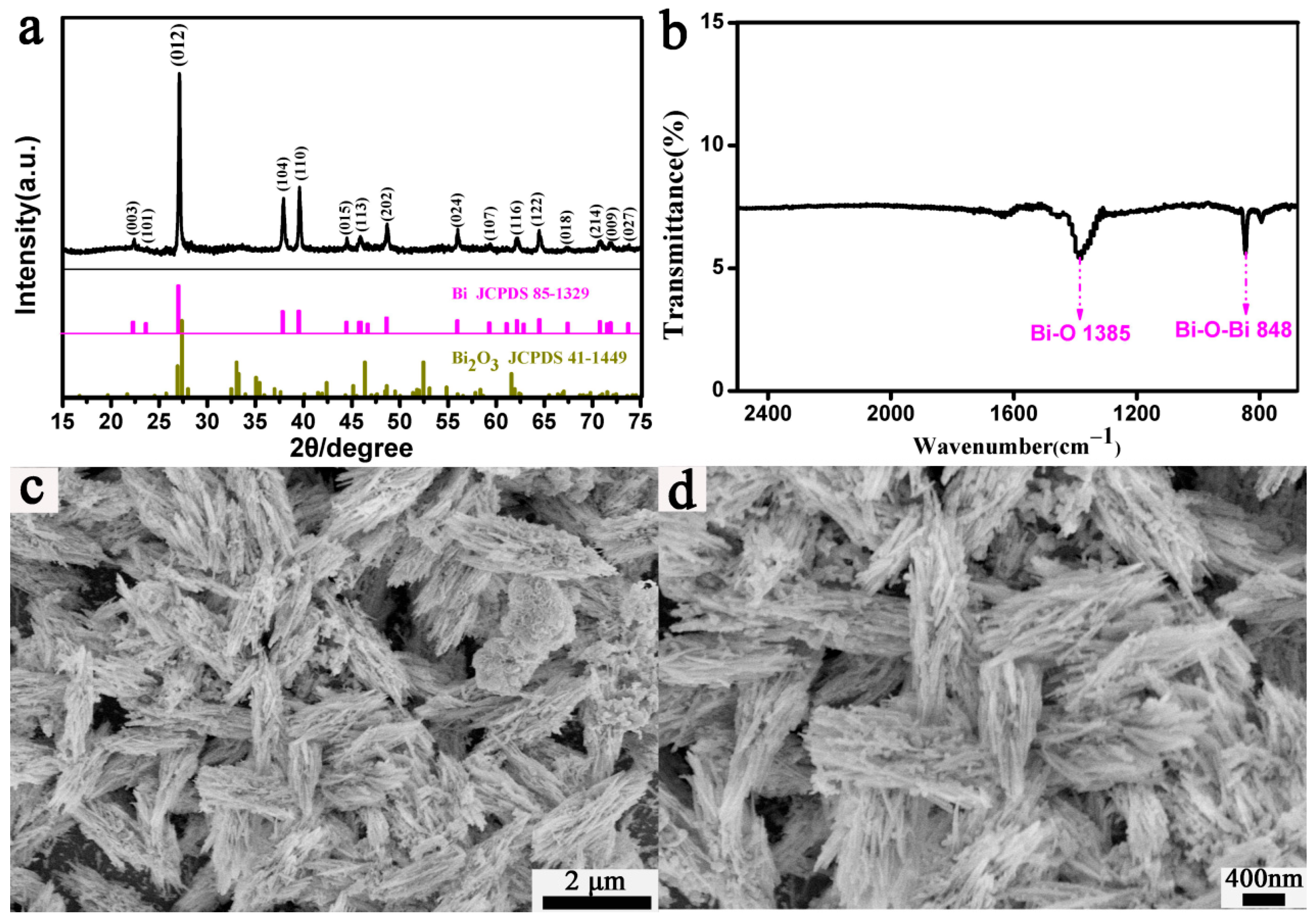
3.1. Fusiform Bi Characterization

The structural characteristics, chemical bonds, and morphologies of the fusiform Bi were characterized through XRD measurements, SEM, and FTIR spectroscopy. The samples were prepared at 90 °C for 55 min (Figure 1). Figure 1a shows the XRD pattern of the as-synthesized fusiform Bi photocatalysts. The XRD pattern of the Bi samples revealed no impurity peaks and matched the standard card peaks (JCPDS No. 85-1329), confirming the successful preparation of pure fusiform Bi. However, a further FTIR analysis confirmed the presence of Bi–O or Bi–O–Bi functional groups in the fusiform Bi samples (Figure 1b). The FTIR spectrum (Figure 1b) of the fusiform Bi exhibited peaks at 845 and 1384 cm−1, corresponding to Bi–O–Bi (symmetrical stretching) and Bi–O (stretching vibration) bonds in bismuth oxide (Bi2O3), respectively [44,45,46]. These results indicate that Bi2O3 formed on the fusiform Bi surface after drying in an oven at 60 °C for 4 h. Figure 1c,d show the SEM images of the fusiform Bi prepared with N2H4•H2O as a reducing agent. These images revealed that the resulting sample mainly comprised uniform fusiform Bi particles (with lengths of 2–4 µm and diameters of 0.5–1 µm). Additionally, some small irregular particles were either isolated or attached to the fusiform Bi structures.

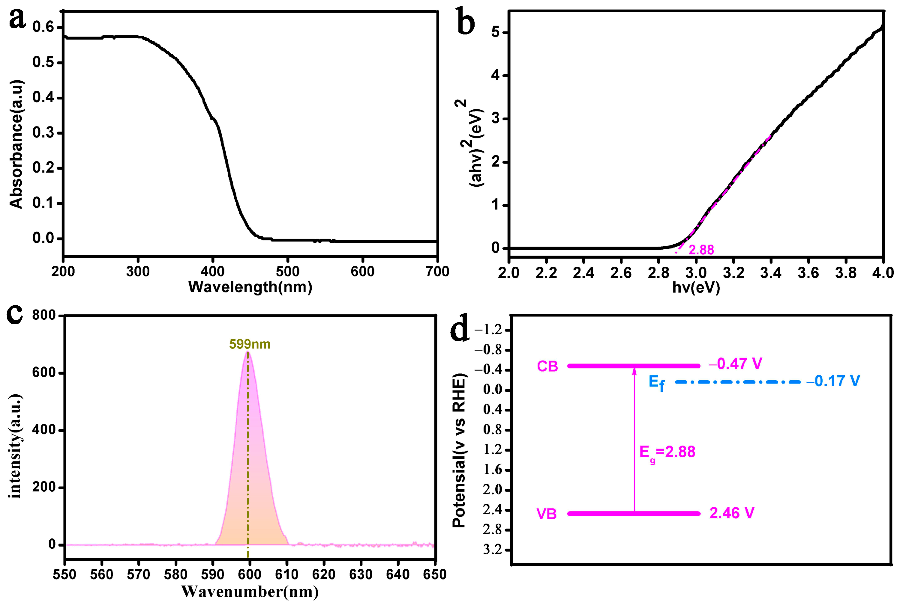
3.2. Analysis of Optical Properties

The band gap value and the separation efficiency of photogenerated e and h+ pairs for the as-synthesized fusiform Bi were assessed via UV–vis DSR, XPS, and fluorescence spectroscopy (Figure 2).
The UV–vis DRS spectra revealed that the fusiform Bi had a band gap energy value of 2.88 eV, as determined using the Kubelka–Munk model [47] (Figure 2a,b). This indicates that the as-prepared fusiform Bi photocatalyst exhibited strong visible light absorption, thereby improving its effectiveness in RhB degradation. The PL spectrum (Figure 2c) of fusiform Bi exhibited a weak emission peak at 599 nm owing to the charge carrier recombination, indicating a low separation efficiency of photogenerated e and h+. Additionally, the VB of the fusiform Bi was measured to be 2.46 eV through VB-XPS analysis (Figure S1). The CB of the fusiform Bi was calculated to be −0.47 V (vs. RHE) using the formula EVB = Eg + ECB [48,49]. The band structure and Eg value of the fusiform Bi samples are illustrated in Figure 2d. The results revealed that the fusiform Bi exhibited a strong reduction ability with a CB potential of −0.47 V (vs. RHE).

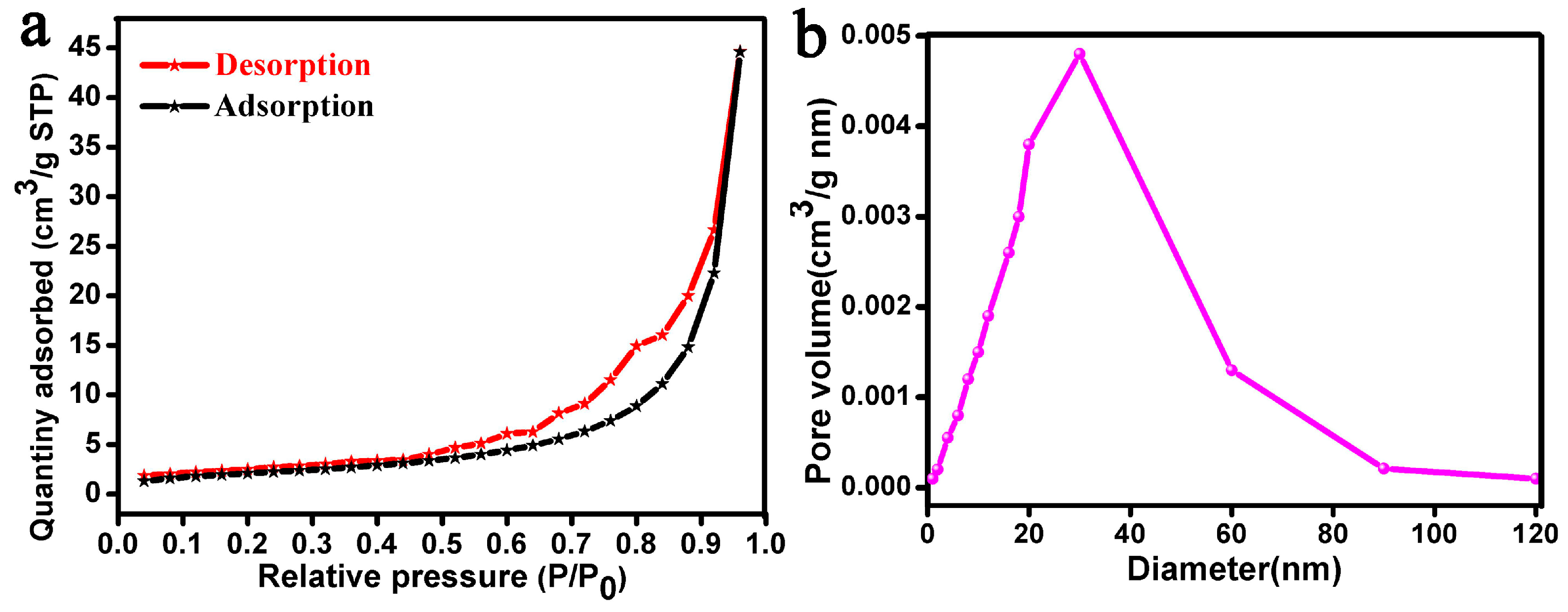
3.3. BET Analysis and Adsorption

BET analysis was performed to evaluate the SSA and average pore size of the fusiform Bi photocatalysts (Figure 3). The N2 adsorption–desorption isotherms (Figure 3a) of the fusiform Bi exhibited a type IV curve with an H3 hysteresis loop, indicating that the fusiform Bi is a mesoporous material [50]. The fusiform Bi had an average pore size of 25.6 nm (Figure 3b). The BET analysis of the fusiform Bi revealed a pore volume of 0.04 cm3/g and a BET surface area of 6.7405 m2/g. These results confirmed that the fusiform Bi exhibited more attachment sites and a higher adsorption capacity, thereby enhancing its photocatalytic and adsorption performance for RhB. A blank control experiment without the use of fusiform Bi achieved a RhB removal efficiency of 54.3% after 70 min (Figure S2). This result indicates that the blank experiment had significantly lower RhB removal than the HCl-assisted fusiform Bi.

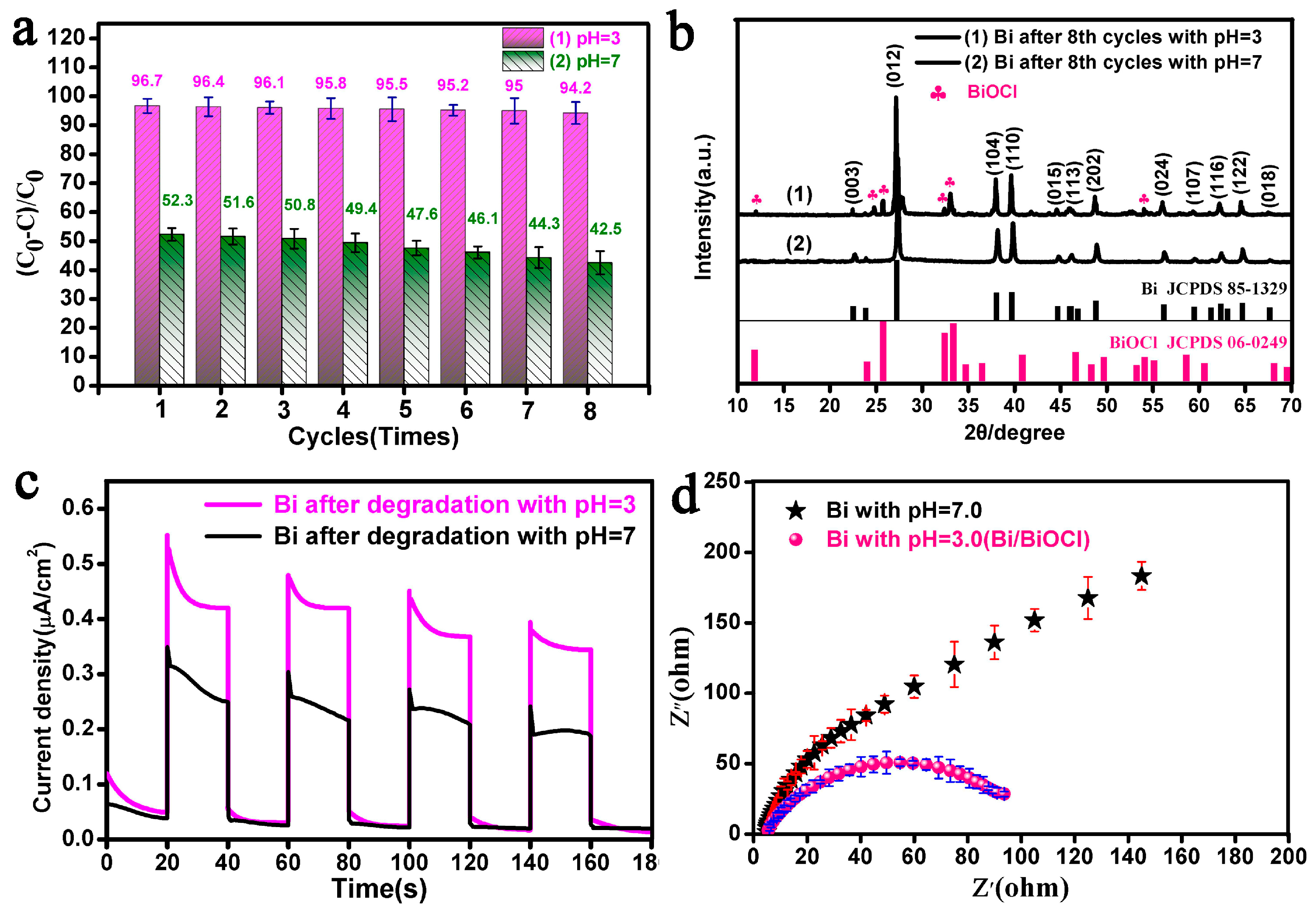
3.4. Photodegradation of RhB over Fusiform Bi

The removal efficiency of RhB was analyzed at pH 3.0 under visible light to assess the photocatalytic properties of the fusiform Bi (Figure 4). Figure 4a presents the time-dependent plots of RhB degradation and λmax shifts over the fusiform Bi photocatalysts. After 70 min of irradiation, the fusiform Bi photocatalysts achieved 96.7% degradation of RhB (Figure 4a(1)). Moreover, with prolonged exposure to visible light, the maximum absorption band (λmax shifts) of RhB blue-shifted from 554 to 498 nm (Figure 4a(2)). This blue shift in λmax resulted from the breakdown of the RhB structure and gradual deethylation, respectively, as confirmed by the FTIR spectra of RhB (Figure S3). Figure 4b shows the COD and TOC removal efficiencies in the RhB solution under different irradiation times using the fusiform Bi catalyst. After 70 min of irradiation, the fusiform Bi achieved maximum COD and TOC removal rates of 69.5% and 58.6%, respectively, indicating that RhB molecules were eventually mineralized to CO2 and H2O (Figure 4b(1,2)). Additionally, the COD and TOC rate constants (min−1) were 0.01452, and 0.01029, respectively (Figure S4). The TOC analysis (Figure 4b(2)) indicated that over 58.6% of the C was mineralized into CO2 [51,52]. To further investigate the photocatalytic activity of the as-synthesized fusiform Bi, the degradation of TC and colorless phenol was evaluated at pH 3.0. After 70 min of irradiation, the fusiform Bi achieved removal efficiencies of 87.2% and 37.3% for TC and phenol, respectively (Figure S5). This result indicates that the fusiform Bi exhibited a higher removal rate for RhB compared with that for both TC and phenol.
The effects of pH and inorganic salts on RhB degradation were investigated under different irradiation times, and the results are presented in Figure 5. At lower pH levels, the fusiform Bi exhibited a higher adsorption capacity for RhB in the dark over a 60 min period (Figure 5a). This was mainly due to the presence of RhB molecules in their cationic form (RhB+) at lower pH levels, which enhanced their interaction with the active centers on the fusiform Bi. Lower pH values corresponded to a stronger adsorption effect of the fusiform Bi [53,54]. Additionally, at lower pH values, the as-prepared fusiform Bi exhibited excellent photocatalytic degradation efficiency for RhB removal within 70 min of irradiation. At pH values of 2.0, 3.0, 5.0, 7.0, and 9.0, the fusiform Bi achieved RhB removal efficiencies of 97%, 96.7%, 72.6%, 53.5%, and 27.6%, respectively. The apparent rate constants (min−1) for these pH values were 0.0457, 0.0452, 0.01649, 0.0097, and 0.00397 (inset of Figure 5b). These results indicate that the fusiform Bi catalyst exhibited poor RhB degradation rates at both very low and high pH values. At very low pH (pH 2.0), the increased Cl concentration can trap h+ and •OH radicals, leading to a reduced degradation efficiency for RhB [55,56]. At pH 9.0, the fusiform bismuth exhibited a low RhB removal rate owing to the interaction between OH ions and h+ [57]. At lower pH values, the Bi catalyst effectively removed RhB owing to the protonation of the COO group in RhB to COOH by H+ under acidic conditions. Moreover, RhB interacted with the photocatalyst surface through hydrogen bonding, which facilitated its adsorption onto the catalyst and enhanced electron transfer [58]. However, these results indicate that under the same photocatalytic conditions, the RhB solution was slightly degraded by the fusiform Bi at pH 3.0 (regulated with H2SO4) (Figure S6). The H2SO4-treated fusiform Bi acted as an •OH scavenger, which effectively inhibited RhB photodegradation [59,60]. The H2SO4 molecules on the fusiform Bi surface can interact with the positively charged n-ethyl group of RhB [61], leading to lower electron transfer efficiency (Figure S7). This indicates that the H2SO4-treated fusiform bismuth exhibited a lower RhB removal rate than HCl-treated samples.
These results suggest that inorganic anions in the RhB solution were key factors influencing the photocatalytic removal of RhB using the as-prepared fusiform Bi. To evaluate the effects of inorganic ions on RhB photodegradation, particularly Cl ions introduced by HCl for pH adjustments, the impacts of Cl, N O 3 , S O 4 2 , and CH3COO ions on RhB photodegradation were assessed. The results are presented in Figure 5b. In the presence of N O 3 , Cl, S O 4 2 , and CH3COO, the fusiform Bi featured RhB degradation rates of 94.8%, 92.3%, 80.2%, and 31.4%, respectively. Notably, the presence of these anions significantly inhibited the degradation process. The pH-dependent and inorganic salt experiments revealed that the ion concentration in the RhB solution, rather than the presence of Cl, was the main factor affecting the photocatalytic removal of RhB. Similar trends in RhB photodegradation were observed in a neutral environment (pH 7.0) (Figure S8). In the presence of Cl, blank (Cl), and CH3COO ions, the corresponding apparent kinetic rate constants (min−1) were 0.0377, 0.03348, 0.0452, 0.02138, and 0.0049, respectively (Figure 5d). Additionally, the effects of the catalyst dosage, RhB initial concentration, light source distance, and reactant temperature on the RhB dye solution were examined (Figure S9). In particular, an increased light source distance led to a lower degradation rate of the RhB dye solution owing to the lower light intensity reaching the catalyst surface and reduced irradiation efficiency. At a light distance of 20 cm, the temperature of the RhB solution increased to 50 °C, leading to 96.7% removal of RhB after 70 min. This enhancement can be attributed to improved free radical transfer on both the RhB and fusiform Bi surfaces at higher temperatures [62,63].
Figure 6 presents the excellent reproducibility and XRD patterns of the fusiform Bi photocatalysts after eight degradation cycles of RhB at different solution pH levels. At pH 3.0 and 7.0, the removal rate of RhB significantly decreased by 2.5% and 9.8%, respectively. After eight cycles of degradation, the final RhB removal rates were 94.2% and 42.5% (Figure 6a). Even after 20 cycles at pH 3.0, the RhB removal rate remained at 79.4% (Figure S10). The XRD pattern of the fusiform Bi after eight cycles of RhB photodegradation at different pH values (3.0 and 7.0) was examined to further assess the stability of the catalyst. After eight degradation cycles at pH 3.0, the XRD patterns (Figure 6b(1)) of the fusiform Bi revealed a relatively a low purity with multiple new BiOCl peaks (marked with solid red clubs). This decrease in purity was mainly due to the dissolution of Bi2O3 (Figure 1b) on the surface of the fusiform Bi by HCl during the photodegradation of RhB, forming BiOCl and p-n-type Bi/BiOCl heterojunctions on the fusiform Bi surface. Under enriched Bi conditions, the BiOCl formed on the Bi surface exhibits inherent n-type conductivity owing to Cl vacancy defects [64]. For the metal bismuth, it is a typical p-type transition metal. Furthermore, the oxygen-rich vacancy of Bi/BiOCl can enhance the p-type conductivity of Bi [65,66]. Additionally, the synergistic effect of Bi surface plasmon resonance (SPR) further enhanced the photocatalytic activity of the Bi/BiOCl heterojunction photocatalyst for RhB removal [67]. Moreover, after eight cycles at pH 7.0, the XRD pattern (Figure 6b(2)) of the Bi samples revealed no impurity peaks and matched the standard card peaks (JCPDS No. 85-1329), indicating that the degraded fusiform Bi retained relatively high purity.
To further confirm that fusiform Bi samples generated more photocharge carriers at lower pH values under visible light irradiation, a photocurrent density experiment was conducted. The results are shown in Figure 6c. At pH 3.0, the Bi/BiOCl heterojunction exhibited a higher photocurrent density than the fusiform Bi at pH 7.0 (Figure 6b). This indicates that the formation of the Bi/BiOCl heterojunction photocatalyst can enhance carrier transfer and separation efficiency. Similarly, Figure 6d shows the EIS spectra of the as-fabricated fusiform Bi. Compared with Bi/Bi2O3 samples, the Bi/BiOCl heterojunction photocatalyst exhibited a smaller semicircle (Figure 6d), indicating lower charge-transfer resistance [68,69,70]. The PL analysis revealed that the formation of the Bi/BiOCl heterojunction photocatalyst reduced the recombination of h+–e pairs at pH 3.0 compared with pH 7.0 (Figure S7).

3.5. XPS and FTIR Analyses

To effectively elucidate the surface electronic states and chemical states of the fusiform Bi before and after photodegradation, XPS and FTIR analyses were performed at different pH values (3.0 and 7.0) (Figure 7). The full survey XPS spectra (Figure 7a) indicated the presence of Bi, C, and O in the fusiform Bi before and after degradation. Among these elements, the presence of C in the XPS spectra suggested that C may have been introduced into the fusiform Bi from the environment [71,72]. After the photodegradation of the fusiform Bi at pH 3.0, a new Cl signal was detected (Figure 7a(3)).
Figure 7b shows the high-resolution (HR) XPS spectra of Bi4f for the fusiform Bi before and after degradation. The XPS spectrum of the fusiform Bi exhibited two peaks at 157.1 and 162.4 eV, corresponding to the Bi4f7/2 and Bi4f5/2 signals of zerovalent bismuth (Bi0, metallic Bi) [73,74], respectively. Moreover, the peaks at 159.2 and 164.5 eV can be attributed to the Bi4f7/2 and Bi4f5/2 signals of trivalent bismuth (Bi3+) [75,76], respectively, indicating that Bi0 and Bi3+ were present in the fusiform Bi after photocatalytic degradation. The HR-XPS spectra (Figure 7c) of the degraded fusiform Bi displayed two peaks at 199.8 (Cl 2p1/2) and 198.0 eV (Cl 2p3/2), confirming the formation of BiOCl crystals on the fusiform Bi at pH 3.0 [77].
The structural characteristics of the fusiform Bi after degradation at pH 7.0 and 3.0 were further analyzed via FTIR (Figure 7d). At pH 7.0, the FTIR spectra (Figure 7d(1)) of the degraded fusiform Bi exhibited peaks at 845 and 1384 cm−1, corresponding to the Bi–O–Bi (symmetrical stretching) and Bi–O (stretching vibration) bonds, respectively [78,79,80]. At pH 3.0, the FTIR spectra of the degraded fusiform Bi exhibited new adsorption peaks at 1463 and 1076 cm−1, corresponding to the O–Cl and Bi–Cl stretching vibrations, respectively [81,82]. Therefore, these results indicate that BiOCl crystals were easily produced to form a Bi/BiOCl heterojunction on the fusiform Bi crystal surface at lower pH values (3.0).

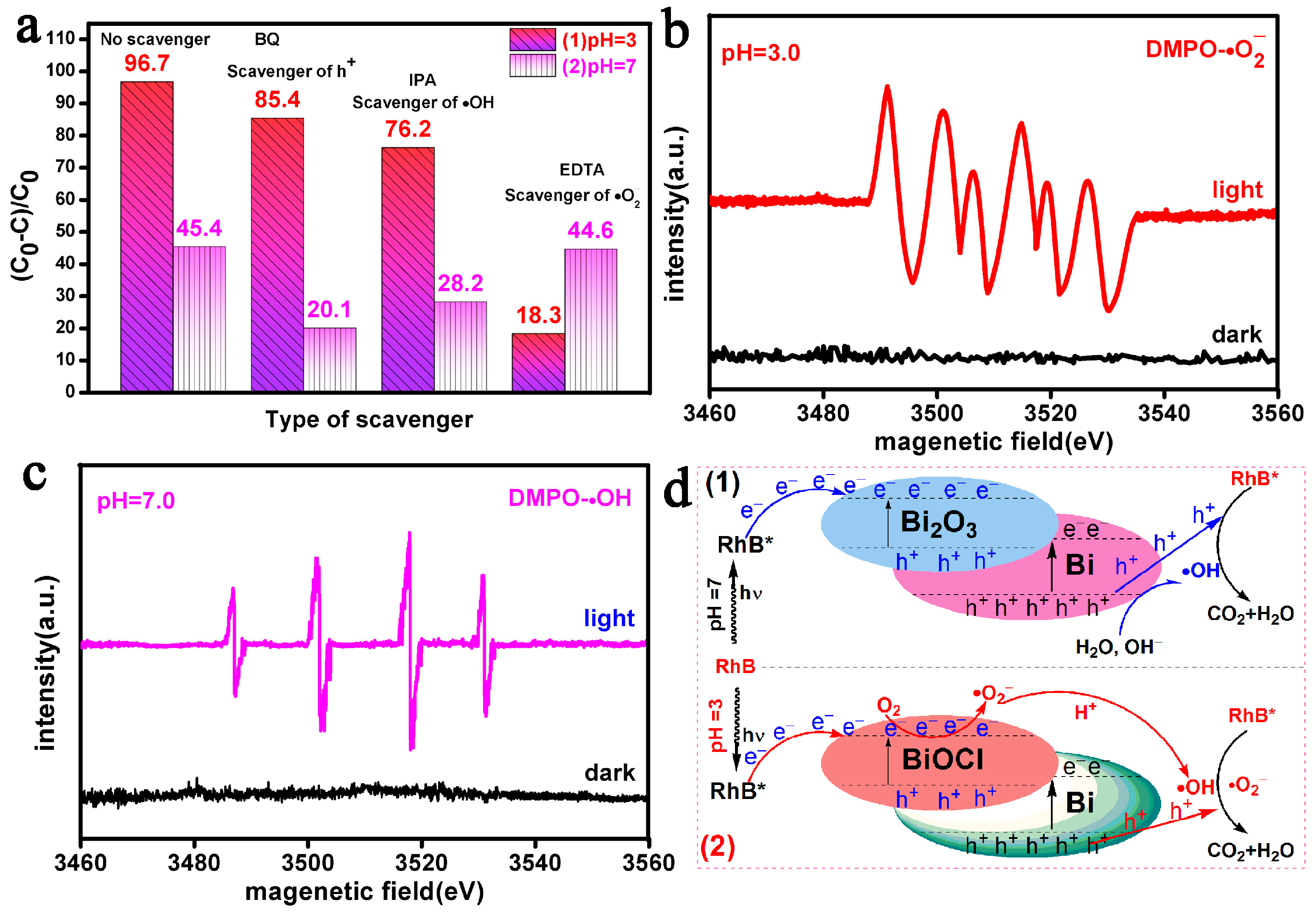
3.6. Potential Photocatalytic Mechanisms

To investigate the effects of different free radicals, a potential mechanism for RhB degradation using the fusiform Bi at pH 7.0 and 3.0 was examined through free radical quenching experiments (Figure 8). During the photocatalytic degradation of RhB, BQ, EDTA, and IPA were used as scavengers for h+, •OH, and • O 2 radicals [83,84]. At pH 3.0, the addition of EDTA and IPA resulted in RhB removal efficiencies of 18.3% and 76.2%, respectively, over a 70 min period (Figure 8a(1)). However, the photocatalytic removal rate of RhB slightly decreased to 85.4% with the addition of BQ compared with the 96.7% rate without any scavengers. This suggests that h+ was not the primary active species, while •OH and • O 2 were the main contributors to RhB removal using the fusiform Bi at pH 3.0. In contrast, at pH 7.0, the addition of BQ and IPA to the catalytic system significantly reduced the RhB removal rate by the fusiform Bi, with only ~20.1% and 28.2% of the RhB degraded, respectively (Figure 8a(2)). This indicates that • O 2 was not effective under these conditions, while h+ and •OH were the key active species in the RhB degradation process by the fusiform Bi in a neutral environment (pH 7.0).
EPR characterization is an effective method for detecting the type and quantity of active species in the Bi/RhB system at pH 3.0 and 7.0. The production of •OH and • O 2 was further confirmed through EPR characterization using DMPO as a capture agent (Figure 8b,c). Under dark conditions, no EPR signals for •OH and • O 2 free radicals were detected. The EPR spectra (Figure 8b) revealed four characteristic peaks, corresponding to DMPO-• O 2 adducts, indicating the presence of the • O 2 radical in the reaction system. Similarly, under light irradiation, the •OH spectrum (Figure 8c) exhibited four distinct characteristic peaks, confirming the presence of •OH radicals. These results confirmed that •OH and • O 2 free radicals were the main active species responsible for RhB removal, consistent with the findings from the free radical capture experiments.
According to these observations, the proposed potential photocatalytic mechanism for RhB degradation over the fusiform Bi at pH 7.0 and 3.0 is illustrated in Figure 8d. The mechanism for RhB degradation over the fusiform Bi at pH 7.0 is shown in Figure 8d(1). Under visible light, the photoinduced h+–e pairs on the fusiform Bi surface were effectively separated. Some of these photogenerated h+ could react with H2O or OH to generate •OH radicals. These radicals and h+ attacked RhB* molecules, causing the cleavage of the RhB structure and the formation of multiple small intermediates, which were eventually mineralized into CO2 and H2O. Therefore, •OH and h+ were the main radicals responsible for RhB degradation at a neutral pH (7.0). Additionally, a rational potential mechanism for RhB photodegradation by the fusiform Bi under acidic conditions (pH 3.0) is illustrated in Figure 8d(2). In the initial stage of photodegradation, Bi2O3 on the fusiform bismuth surface layer was dissolved by HCl to produce BiOCl, which then formed a Bi/BiOCl heterojunction with the fusiform Bi. The formation of the Bi/BiOCl heterojunction photocatalyst facilitated the transfer of photocharge carriers [67]. Under visible light, electrons (e) in the VB of Bi/BiOCl were excited and transferred to the CB of the fusiform Bi, forming h+ in the VB. These excited e reacted with adsorbed O2 on the Bi/BiOCl heterojunction surface to produce • O 2 radicals. Some of these • O 2 radicals could be further converted into •OH in the presence of HCl. The generated • O 2 and •OH active species were released into the Bi/RhB solution system, which attacked RhB molecules. This process degraded RhB into small inorganic molecules, thereby achieving complete degradation.

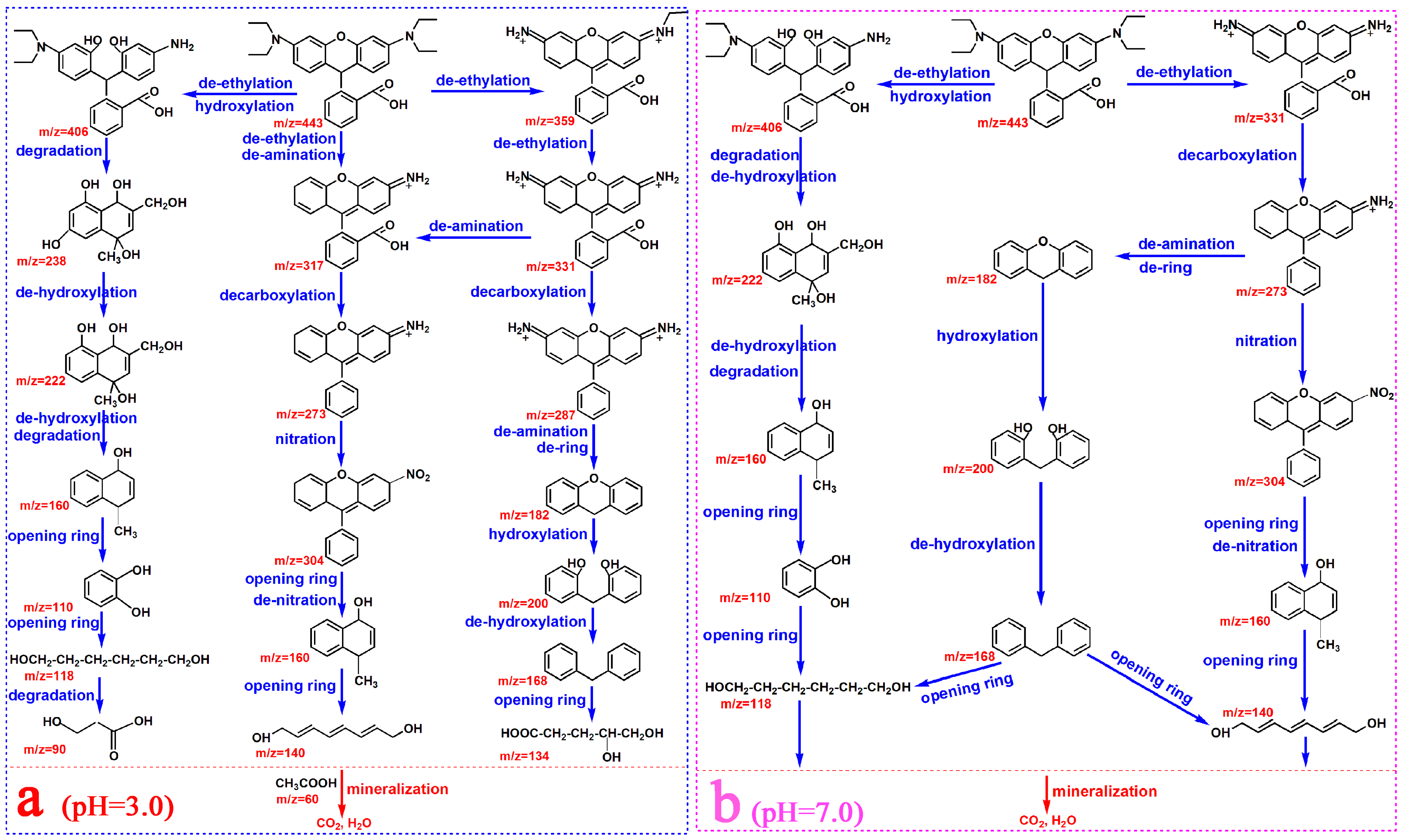
3.7. Degradation Pathways of RhB

To further investigate the potential degradation pathways of RhB using the fusiform Bi photocatalyst, the RhB and intermediates in the photocatalytic process were analyzed at different pH values via LC-MS (Figure S11). The LC–MS data (Figure S11) revealed several degradation products at pH 3.0, with strong signals at (mass charge ratio) m/z values of 443, 406, 359, 331, 317, 304, 287, 273, 238, 222, 200, 182, 168, 160, 140, 134, 118, 110, 90, and 60. At pH 7.0, the degradation products were observed at m/z values of 443, 406, 331, 304, 273, 222, 200, 182, 168, 160, 140, 118, and 110.
According to the LC–MS analysis of RhB and its intermediates during the photocatalytic process at different pH values, a potential RhB degradation pathway at pH 3.0 and 7.0 was proposed (Figure 9). At pH 3.0 and 7.0, the RhB degradation process mainly involved nine steps: hydroxylation, deethylation, decarboxylation, nitration, deamination, dehydroxylation, ring opening, denitration, and mineralization (Figure 9). First, the amino, carboxyl groups, and ethyl groups of the RhB molecule were degraded to form several intermediates. Additionally, the hydroxyl and nitro groups were introduced into the RhB molecules. The interaction of RhB molecules with reactive oxygen species (• O 2 , •OH, or h+) formed multiple intermediates, such as C24H26N2O4 (m/z 406), C22H19N2O3 (m/z 359), C20H15N2O3 (m/z 331), C20H15NO3 (m/z 317), C19H14NO3 (m/z 304), C19H15N2O (m/z 287), C19H15NO (m/z 273), C12H14O5 (m/z 238), C12H14O4 (m/z 222), C13H12O2 (m/z 200), C13H10O (m/z 182), C13H12 (m/z 168), and C11H12O (m/z 160). Subsequently, a key ring-opening reaction occurred, breaking down aromatic rings into low-molecular-weight organics. Finally, these small molecules were further oxidized and mineralized to obtain CO2 and H2O.
However, at pH 7.0, the number of RhB degradation intermediates was relatively small compared with pH 3.0, particularly for low-molecular-weight compounds. This was mainly due to the limited interaction between •OH and RhB in the solution at pH 7.0 (Figure 8a) and the oxidation of various C atoms. In particular, •OH has a short lifespan and low oxidation activity [85]. Additionally, the use of photogenerated h+ as an active species for RhB degradation led to its complete confinement to the surface, which limited its ability to effectively contact and attack C atoms in RhB [85]. At higher pHs, further degradation of RhB was challenging owing to the reduced effectiveness of both •OH and h+ on low-molecular-weight organic compounds, such as C5H10O4(m/z 134), C3H6O3(m/z 90), and C2H4O2(m/z 60). Overall, this indicates that the pH of the RhB solution influenced the types, quantities, and patterns of degradation products.

4. Conclusions

A fusiform Bi catalytic material with non-uniform spherical structures was synthesized via an aqueous chemical reduction method. UV–vis DRS and PL analyses revealed that the fusiform Bi photocatalyst exhibited strong visible light absorption, which improved its performance in degrading RhB. The photodegradation results confirmed that at pH 2.0, 3.0, 5.0, 7.0, and 9.0, the fusiform Bi catalyst exhibited RhB removal efficiencies of ~97%, 96.7%, 72.6%, 53.5%, and 27.6%, respectively, within 70 min. These results indicate that the prepared Bi catalyst achieved the maximum RhB removal efficiency and rate constant at pH 3.0. The TOC analysis revealed that after a 70 min irradiation, over 58.6% of the carbon in the RhB solution was converted into CO2 products. Additionally, the effects of the catalyst content, initial RhB concentration, light source distance, inorganic anions, and reactant temperature on the photocatalytic performance of the fusiform Bi were investigated. The addition of acid to the RhB/fusiform Bi system accelerated RhB degradation, generated different active species, and altered the degradation pathway over the fusiform Bi. The quenching experiments and EPR analysis revealed that at pH 3.0, • O 2 , •OH, and h+ were the main active species responsible for RhB removal using the fusiform Bi. In contrast, at pH 7.0, •OH was the main active species involved in the removal process of RhB. These results indicate that at pH 3.0, a p-n type Bi/BiOCl heterojunction photocatalyst formed on the Bi surface. The as-formed Bi/BiOCl heterojunction photocatalyst exhibited higher photocatalytic activity than the fusiform Bi. Moreover, the potential photocatalytic mechanisms for RhB degradation over the fusiform Bi at different pH values were investigated through XRD, XPS, radical capture experiments, EPR spectroscopy, and FTIR analysis. This study provides new methods and insights for designing bismuth-based photocatalysts and improving their effectiveness in dye removal through pH adjustments.

Supplementary Materials

The following supporting information can be downloaded at: https://www.mdpi.com/article/10.3390/w16172389/s1, Figure S1: VB-XPS analysis of fusiform Bi; Figure S2: Blank contrast experiment results for the removal efficiency of RhB; Figure S3: FTIR spectra of RhB before and after photodegradation; Figure S4: The corresponding rate constants (min−1) of COD and TOC; Figure S5: Removal rate of TC and phenol over fusiform Bi; Figure S6: RhB removal efficiency at a pH value of 3.0 (regulated with H2SO4); Figure S7: The PL spectra of fusiform Bi at different pH values (7.0 and 3.0) and different acid treatments (HCl and H2SO4); Figure S8: Degradation rate of RhB using fusiform Bi as photocatalyst on Cl ions at a pH of 7.0; Figure S9: The effect of (a) catalyst dosage, (b) RhB initial concentration, (c) light source distance, and (d) reactant temperature on degradation of RhB; Figure S10: Removal rate of RhB after 21-cycle experiment with pH of 3.0; Figure S11; LC-MS spectra of RhB solution (3) pre- and post- photodegradation with fusiform Bi as photocatalyst at pH values of (1) 3.0 and (2) 7.0.

Author Contributions

Methodology, Y.C. and D.M.; formal analysis, G.H. and S.P.; investigation, Y.C. and D.M.; data curation, Y.C., G.H., and S.P.; writing—original draft preparation, Y.C.; writing—review and editing, D.M.; supervision, G.H.; funding acquisition, D.M. All authors have read and agreed to the published version of the manuscript.

Funding

This work was supported by the Natural Science Foundation of Hunan Province (Grant No. 2022JJ50269) and the Key Project Foundation of Hunan Provincial Education Department (Grant No. 20A094).

Data Availability Statement

The data presented in this study are available upon request from the corresponding author. The data are not publicly available due to data copyright issues.

Conflicts of Interest

The authors declare no conflicts of interest.

Nomenclature

RhBRhodamine B
CODChemical oxygen demand
TOCTotal organic carbon
pHPotential of hydrogen
O 2 Superoxide anion radical
•OHHydroxyl radical
eElectron
h+Hole
VBValence band
CBConduction band
RHEReversible hydrogen electrode
SSASpecific surface area
EISElectrochemical impedance spectroscopy
EPRElectron paramagnetic resonance
m/zMass charge ratio
EgBand gap energy value
SPRSurface plasmon resonance

References

  1. Parsa, S.M.; Norozpour, F.; Elsheikhc, A.H.; Kabeel, A.E. Solar desalination/purification (solar stills, humidification-dehumidification, solar disinfection) in high altitude during COVID19: Insights of gastrointestinal manifestations and systems’mechanism. J. Hazard. Mater. Adv. 2023, 10, 100259. [Google Scholar] [CrossRef]
  2. Parsa, S.M. Mega-scale desalination efficacy (Reverse Osmosis, Electrodialysis, Membrane Distillation, MED, MSF) during COVID-19: Evidence from salinity, pretreatment methods, temperature of operation. J. Hazard. Mater. Adv. 2023, 9, 100217. [Google Scholar] [CrossRef] [PubMed]
  3. Parsa, S.M.; Norouzpour, F.; Shoeibi, S.; Shahsavar, A.; Aberoumand, S.; Said, Z.; Guo, W.S.; Ngo, H.H.; Ni, B.-J.; Afrand, M.; et al. A comprehensive study to find the optimal fraction of nanoparticle coated at the interface of solar desalination absorbers: 5E and GHGs analysis in different seasons. Sol. Energ. Mat. Sol. C 2023, 256, 112308. [Google Scholar] [CrossRef]
  4. Parsa, S.M.; Rahbar, A.; Koleini, M.H.; Aberoumand, S.; Afrand, M.; Amidpour, M. A renewable energy-driven thermoelectric-utilized solar still with external condenser loaded by silver/nanofluid for simultaneously water disinfection and desalination. Desalination 2020, 480, 114354. [Google Scholar] [CrossRef]
  5. Al-Buriahi, A.K.; Al-Gheethi, A.A.; Kumar, P.S.; Mohamed, R.M.S.R.; Yusof, H.; Alshalif, A.F.; Khalifa, N.A. Elimination of rhodamine B from textile wastewater using nanoparticle photocatalysts: A review for sustainable approaches. Chemosphere 2022, 287, 132162. [Google Scholar] [CrossRef]
  6. Venkatraman, S.K.; Vijayakumar, N.; Bal, D.K.; Mishra, A.; Gupta, B.; Mishra, V.; Wysokowski, M.; Koppala, S.; Swamiappan, S. Degradation of environmentally harmful textile dye rhodamine B using silicate ceramic photocatalysts. Inorg. Chem. Commun. 2022, 142, 109674. [Google Scholar] [CrossRef]
  7. Danish, M.; Khanday, W.A.; Hashim, R.; Sulaiman, N.S.B.; Akhtar, M.N.; Nizami, M. Application of optimized large surface area date stone (Phoenix dactylifera) activated carbon for rhodamin B removal from aqueous solution: Box-Behnken design approach. Ecotoxicol. Environ. Saf. 2017, 139, 280–290. [Google Scholar] [CrossRef]
  8. Lim, S.; Shi, J.L.; Gunten, U.; McCurry, D.L. Ozonation of organic compounds in water and wastewater: A critical review. Water Res. 2022, 213, 118053. [Google Scholar] [CrossRef]
  9. Lal, M.; Sharma, P.; Singh, L.; Ram, C. Photocatalytic degradation of hazardous Rhodamine B dye using sol-gel mediated ultrasonic hydrothermal synthesized of ZnO nanoparticles. Results Eng. 2023, 17, 100890. [Google Scholar] [CrossRef]
  10. Sharma, J.; Sharma, S.; Bhatt, U.; Soni, V. Toxic effects of Rhodamine B on antioxidant system and photosynthesis of Hydrilla verticillata. J. Hazard. Mater. Lett. 2022, 3, 100069. [Google Scholar] [CrossRef]
  11. Çelik, M.S.; Çetinus, Ş.A.; Yenidünya, A.F.; Çetinkaya, S.; Tüzün, B. Biosorption of Rhodamine B dye from aqueous solution by Rhus coriaria L. plant: Equilibrium, kinetic, thermodynamic and DFT calculations. J. Mol. Struct. 2023, 1272, 134158. [Google Scholar] [CrossRef]
  12. Souza, F.H.M.; Leme, V.F.C.; Costa, G.O.B.; Castro, K.C.; Giraldi, T.R.; Andrade, G.S.S. Biosorption of Rhodamine B using a low-cost biosorbent prepared from inactivated aspergillus oryzae cells: Kinetic, equilibrium and thermodynamic studies. Water Air Soil. Pollut. 2020, 242, 231. [Google Scholar] [CrossRef]
  13. Al-Gheethi, A.A.; Azhar, Q.M.; Kumar, P.S.; Yusuf, A.A.; Al-Buriahi, A.K.; Mohamed, R.M.S.R.; Al-shaibani, M.M. Sustainable approaches for removing Rhodamine B dye using agriculturalwaste adsorbents: A review. Chemosphere 2022, 287, 132080. [Google Scholar] [CrossRef] [PubMed]
  14. Saigl, Z.M. Various adsorbents for removal of Rhodamine B dye: A review. Indones. J. Chem. 2021, 21, 1039–1056. [Google Scholar] [CrossRef]
  15. Deng, D.L.; Li, Y.; Wu, M.Z.; Song, Y.; Huang, Q.J.; Duan, Y.Q.; Chang, Y.; Zhao, Y.Y.; He, C.L. Electrocatalytic degradation of Rhodamine B on the Sb-doped SnO2/Ti electrode in alkaline medium. ACS Omega 2023, 8, 48480–48490. [Google Scholar] [CrossRef] [PubMed]
  16. Hu, J.F.; Zhu, H.; Lin, M.; Wu, D.E.; Yao, J.G.; Sun, T.Y.; Ma, X.J.; Xia, Y.J. Highly-efficient removal of Rhodamine B using a flow-through electrocatalytic filtration system: Characteristics, efficiency and mechanism. J. Electroanal. Chem. 2023, 946, 117742. [Google Scholar] [CrossRef]
  17. Ma, X.L.; Duan, D.M.; Chen, X.; Feng, X.M.; Ma, Y.H. A polysaccharide-based bioflocculant BP50-2 from banana peel waste: Purification, structure and flocculation performance. Int. J. Biol. Macromol. 2022, 205, 604–614. [Google Scholar] [CrossRef]
  18. Vadivel, M.; Jayakumar, S.; Philip, J. Rapid removal of rhodamine dye from aqueous solution using casein-surfactant complexes: Role of casein-surfactant interaction. J. Disper. Sci. Technol. 2022, 43, 928–943. [Google Scholar] [CrossRef]
  19. Saruchi; Kumar, V.; Kaith, B.S.; Jindal, R. Synthesis of hybrid ion exchanger for Rhodamine B dye removal: Equilibrium, kinetic and thermodynamic studies. Ind. Eng. Chem. Res. 2016, 55, 10492–10499. [Google Scholar] [CrossRef]
  20. Thakur, V.; Singh, S.; Kumar, P.; Rawat, S.; Srivastava, V.C.; Lo, S.-L.; Štangar, U.L. Photocatalytic behaviors of bismuth-based mixed oxides: Types, fabrication techniques and mineralization mechanism of antibioticsm. Chem. Eng. J. 2023, 475, 146100. [Google Scholar] [CrossRef]
  21. Jain, S.; Mittal, A.; Kumari, V.; Sharma, A.; Jindal, J.; Makgwane, P.R.; Kumar, V.; Kumar, N.; Kumari, K. A facile synthesized Z-scheme Bi2O3/SnS/Ag ternary nanocomposite: An expedited visible photocatalysis by plasmonic silver for efficient organic decontamination. Opt. Mater. 2023, 145, 114434. [Google Scholar] [CrossRef]
  22. Astuti, Y.; Elesta, P.P.; Widodo, D.S.; Widiyandari, H.; Balgis, R. Hydrazine and urea fueled-solution combustion method for Bi2O3 synthesis: Characterization of physicochemical properties and photocatalytic activity. Bull. Chem. React. Eng. Catal. 2020, 15, 104–111. [Google Scholar] [CrossRef]
  23. Kansaard, T.; Ishihara, K.N.; Pecharapa, W. Structural, optical and photo-induced catalytic properties of derived-Leucoxene/BiVO4 composite prepared by sonochemical process. Optik 2022, 267, 169665. [Google Scholar] [CrossRef]
  24. Monisha, K.; Kavipriya, S.; Silambarasan, A.; Arulmozhi, R.; Abirami, N.; Ramesh, R. Hydrothermal synthesis of hierarchically structured cobalt doped bismuth tungstate with improved photocatalytic activity. Optik 2020, 206, 164366. [Google Scholar] [CrossRef]
  25. Khalid, N.R.; Ishtiaq, H.; Ali, F.; Tahir, M.B.; Naeem, S.; Ul-Hamid, A.; Ikram, M.; Iqbal, T.; Kamal, M.R.; Alrobei, H.; et al. Synergistic effects of Bi and N doped on ZnO nanorods for efficient photocatalysis. Mater. Chem. Phys. 2022, 289, 126423. [Google Scholar] [CrossRef]
  26. Chen, L.Q.; Tian, L.J.; Zhao, X.; Hu, Z.; Fan, J.J.; Lv, K.L. SPR effect of Au nanoparticles on the visible photocatalytic RhB degradation and NO oxidation over TiO2 hollow nanoboxes. Arab. J. Chem. 2022, 13, 4404–4416. [Google Scholar] [CrossRef]
  27. Chen, X.G.; Zhu, L.; Ma, Z.P.; Wang, M.L.; Zhao, R.; Zou, Y.Y.; Fan, Y.J. Ag nanoparticles decorated ZnO nanorods as multifunctional SERS substrates for ultrasensitive detection and catalytic degradation of Rhodamine B. Nanomaterials 2022, 12, 2394. [Google Scholar] [CrossRef]
  28. Pinchujit, S.; Phuruangrat, A.; Wannapop, S.; Sakhon, T.; Kuntalue, B.; Thongtem, T.; Thongtem, S. Bi2WO6 thin nanoplates decorated with Pt nanoparticles possessing enhanced visible-light-driven photocatalytic performance for Rhodamine B degradation. Russ. J. Inorg. Chem. 2022, 67, S199–S209. [Google Scholar] [CrossRef]
  29. Nevinskas, I.; Stanionyte, S.; Devenson, J.; Krotkus, A. Direct bandgap dependence of bismuth films on their thickness. J. Appl. Phys. 2022, 132, 055301. [Google Scholar] [CrossRef]
  30. Huang, H.; Liu, G.S.; Wang, X.H. Bismuth microsphere for photo-assisted nitrate removal: Experimental and theoretical investigations. Chem. Eng. J. 2022, 431, 133239. [Google Scholar] [CrossRef]
  31. Zhang, J.J.; Yang, Z.W.; Wang, L.S.; Chao, L. Carbon covered Ag and Bi nanoparticles uniformly dispersed in porous carbon matrix: Synergistic effect for removal of RhB. Mater. Lett. 2020, 275, 128099. [Google Scholar] [CrossRef]
  32. Zhang, F.N.; Zhao, Y.; Li, Y.W.; Wu, G.J.; Zhao, J.Z. CTAB induced hierarchical bismuth microspheres for visible-light photocatalytic study. J. Colloid Interf. Sci. 2017, 505, 519–527. [Google Scholar] [CrossRef]
  33. Ozer, M.S.; Eroglu, Z.; Yalin, A.S.; Kılıç, M.; Rothlisberger, U.; Metin, O. Bismuthene as a versatile photocatalyst operating under variable conditions for the photoredox C-H bond functionalization. Appl. Catal. B 2022, 304, 120957. [Google Scholar] [CrossRef]
  34. Zhang, Q.; Zhou, Y.; Wang, F.; Dong, F.; Li, W.; Li, H.; Patzke, G.R. From semiconductors to semimetals: Bismuth as a photocatalyst for NO oxidation in air. J. Mater. Chem. 2014, 2, 11065–11072. [Google Scholar] [CrossRef]
  35. Zhang, M.L.; Ke, J.; Xu, D.S.; Zhang, X.Y.; Liu, H.Y.; Wang, Y.R.; Yu, J.X. Construction of plasmonic Bi/Bismuth oxycarbonate/Zinc bismuth oxide ternary heterojunction for enhanced charge carrier separation and photocatalytic performances. J. Colloid Interf. Sci. 2022, 615, 663–673. [Google Scholar] [CrossRef]
  36. Zhang, L.P.; Ghimire, P.; Phuriragpitikhon, J.; Jiang, B.J.; Gonçalves, A.A.S.; Jaroniec, M. Facile formation of metallic bismuth/bismuth oxide heterojunction on porous carbon with enhanced photocatalytic activity. J. Colloid Interf. Sci. 2018, 513, 82–91. [Google Scholar] [CrossRef]
  37. Khavar, A.H.C.; Mahjoub, A.R.; Najafi, S. Enhanced photocatalytic degradation of tetracycline by improving electron flux at the Schottky junction between Bi nanoparticles and MoS2-anchored RGO. J. Photoch. Photobio. A Chem. 2024, 447, 115270. [Google Scholar] [CrossRef]
  38. Liu, D.; Li, C.L.; Zhao, C.Y.; Nie, E.; Wang, J.Q.; Zhou, J.; Zhao, Q. Efficient dye contaminant elimination and simultaneously electricity production via a Bi-doped TiO2 photocatalytic fuel cell. Nanomaterials 2022, 12, 210. [Google Scholar] [CrossRef]
  39. Yue, C.L.; Zhu, L.L.; Qiu, Y.X.; Du, Z.L.; Qiu, J.L.; Liu, F.Q.; Wang, F.H. Recent advances of plasmonic elemental Bi based photocatalysts in environmental remediation and energy conversion. J. Clean. Prod. 2023, 392, 136017. [Google Scholar] [CrossRef]
  40. Song, Y.K.; Bao, Z.Q.; Gu, Y.Y. Photocatalytic enhancement strategy with the introduction of metallic Bi: A review on Bi/semiconductor photocatalysts. Chem. Rec. 2023, 24, e202300307. [Google Scholar] [CrossRef]
  41. Wang, Z.; Jiang, C.L.; Huang, R.; Peng, H.; Tang, X.D. Investigation of optical and photocatalytic properties of bismuth nanospheres prepared by a facile thermolysis method. J. Phys. Chem. C 2014, 118, 1155–1160. [Google Scholar] [CrossRef]
  42. Li, Y.L.; Zhu, J.Z. Synthesis of Bi nanoparticles supported on activated carbon for efficient photocatalytic degradation of high concentration Rhodamine B. Micro Nano Lett. 2020, 15, 627–631. [Google Scholar] [CrossRef]
  43. Naing, H.H.; Li, Y.; Ghasemi, J.B.; Wang, J.T.; Zhang, G.K. Enhanced visible-light-driven photocatalysis of in-situ reduced of bismuth on BiOCl nanosheets and montmorillonite loading: Synergistic effect and mechanism insight. Chemosphere 2022, 304, 135354. [Google Scholar] [CrossRef]
  44. Astuti, Y.; Andianingrum, R.; Haris, A.; Darmawan, A. The role of H2C2O4 and Na2CO3 as precipitating agents on the physichochemical properties and photocatalytic activity of bismuth oxide. Open Chem. 2020, 18, 129–137. [Google Scholar] [CrossRef]
  45. Astuti, Y.; Amri, D.; Widodo, D.S.; Widiyandari, H.; Balgis, R.; Ogi, T. Effect of fuels on the physicochemical properties and photocatalytic activity of bismuth oxide, synthesized using solution combustion method. Int. J. Technol. 2020, 11, 26–36. [Google Scholar] [CrossRef]
  46. Astuti, Y.; Listyani, B.M.; Suyati, L.; Darmawan, A. Bismuth oxide prepared by sol-gel method: Variation of physicochemical characteristics and photocatalytic activity due to difference in calcination temperature. Indones. J. Chem. 2021, 21, 108–117. [Google Scholar] [CrossRef]
  47. Landi, S.; Segundo, I.R.; Freitas, E.; Vasilevskiy, M.; Carneiro, J.; Tavares, C.J. Use and misuse of the Kubelka-Munk function to obtain the band gap energy from diffuse reflectance measurements. Solid. State Commun. 2022, 341, 114573. [Google Scholar] [CrossRef]
  48. Ye, F.; Qian, J.; Xia, J.J.; Li, L.F.; Wang, S.J.; Zeng, Z.X.; Mao, J.; Ahamad, M.; Xiao, Z.R.; Zhang, Q.R. Efficient photoelectrocatalytic degradation of pollutants over hydrophobic carbon felt loaded with Fe-doped porous carbon nitride via direct activation of molecular oxygen. Environ. Res. 2024, 249, 118497. [Google Scholar] [CrossRef]
  49. Wang, P.L.; Fan, S.Y.; Li, X.Y.; Duan, J.; Zhang, D.K. Modulating the molecular structure of graphitic carbon nitride for identifying the impact of the piezoelectric effect on photocatalytic H2O2 production. ACS Catalysis 2023, 13, 9515–9523. [Google Scholar] [CrossRef]
  50. Rong, J.; Zhang, T.; Qiu, F.; Chen, M. Structural evolution of hierarchical porous NiO/Al2O3 composites and their application for removal of dyes by adsorption. Korean J. Chem. Eng. 2017, 34, 41–53. [Google Scholar] [CrossRef]
  51. Qi, Y.; Zhao, J.J.; Wang, H.T.; Zhang, A.M.; Li, J.P.; Yan, M.F.; Guo, T.Y. Shaddock peel-derived N-doped carbon quantum dots coupled with ultrathin BiOBr square nanosheets with boosted visible light response for high-efficiency photodegradation of RhB. Environ. Pollut. 2023, 325, 121424. [Google Scholar] [CrossRef] [PubMed]
  52. Rathi, A.; Basu, S.; Barman, S. Efficient eradication of antibiotic and dye by C-dots@zeolite nanocomposites: Performance evaluation, and degraded products analysis. Chemosphere 2022, 298, 134260. [Google Scholar] [CrossRef]
  53. Tun, P.P.; Wang, J.T.; Khaing, T.T.; Wu, X.Y.; Zhang, G.K. Fabrication of functionalized plasmonic Ag loaded Bi2O3/montmorillonite nanocomposites for efficient photocatalytic removal of antibiotics and organic dyes. J. Alloys Compd. 2020, 818, 152836. [Google Scholar] [CrossRef]
  54. Sharma, A.; Mitta, A.; Sharma, S.; Tahir, M.; Parmar, D.; Singh, P.; Kumar, N. Integrating Ni, Pt, and Pd on biphasic Cu-doped Bi2O3 for physicochemical characteristics and superior light driven elimination of pollutants. Catal. Surv. Asia 2024, 28, 101–116. [Google Scholar] [CrossRef]
  55. Shi, Y.; Luo, L.; Zhang, Y.; Chen, Y.; Wang, S.; Li, L.; Long, Y.; Jiang, F. Synthesis and characterization of α/β-Bi2O3 with enhanced photocatalytic activity for 17α- Ethynylestradiol. Ceram. Int. 2017, 43, 7627–7635. [Google Scholar] [CrossRef]
  56. Bera, K.K.; Chakraborty, M.; Mondal, M.; Banik, S.; Bhattacharya, S.K. Synthesis of α-β Bi2O3 heterojunction photocatalyst and evaluation of reaction mechanism for degradation of RhB dye under natural sunlight. Ceram. Int. 2020, 46, 7667–7680. [Google Scholar]
  57. Bera, K.K.; Majumdar, R.; Chakraborty, M.; Bhattacharya, S.K. Phase control synthesis of α, β and α/β Bi2O3 hetero-junction with enhanced and synergistic photocatalytic activity on degradation of a toxic dye, Rhodamine-B under natural sunlight. J. Hazard. Mater. 2018, 352, 182–191. [Google Scholar]
  58. Mittal, A.; Sharma, S.; Kumari, V.; Yadav, S.; Chauhan, N.S.; Kumar, N. Highly efficient, visible active TiO2/CdS/ZnS photocatalyst, study of activity in an ultra low energy consumption LED based photo reactor. J. Mater. Sci. Mater. Electron. 2019, 30, 17933–17946. [Google Scholar]
  59. Sakkas, V.A.; Arabatzis, I.M.; Konstantinou, I.K.; Dimou, A.D.; Albanis, T.A.; Falaras, P. Metolachlor photocatalytic degradation using TiO2 photocatalysts. Appl. Catal. B 2004, 49, 195–205. [Google Scholar] [CrossRef]
  60. Park, S.K.; Shin, H. Effect of HCl and H2SO4 treatment of TiO2 powder on the photosensitized degradation of aqueous rhodamine B under visible light. J. Nanosci. Nanotechnol. 2014, 14, 8122–8128. [Google Scholar] [CrossRef] [PubMed]
  61. Jayaweera, P.M.; Sanjeewa, T.; Tennakone, K. Formation of ion-associated complexes between SPADNS and rhodamine B: Antenna effect and enhancement of photovoltaic properties. Sol. Energ. Mat. Sol. C 2007, 91, 944–950. [Google Scholar] [CrossRef]
  62. Zhu, J.; Zhu, Z.; Zhang, H.; Lu, H.; Qiu, Y. Efficient degradation of organic pollutants by peroxymonosulfate activated with MgCuFe-layered double hydroxide. RSC Adv. 2019, 9, 2284–2291. [Google Scholar] [CrossRef]
  63. Li, W.; Wu, P.-X.; Zhu, Y.; Huang, Z.-J.; Lu, Y.-H.; Li, Y.-W.; Dang, Z.; Zhu, N.-W. Catalytic degradation of bisphenol A by CoMnAl mixed metal oxides catalyzed peroxymonosulfate: Performance and mechanism. Chem. Eng. J. 2015, 279, 93–102. [Google Scholar] [CrossRef]
  64. Kong, B.; Zeng, T.X.; Wang, W.T. The n-type and p-type conductivity mechanisms of the bulk BiOCl photocatalyst from hybrid density functional theory calculations. Phys. Chem. Chem. Phys. 2021, 23, 19841. [Google Scholar] [CrossRef]
  65. Raza, A.; Qumar, U.; Haider, A.; Naz, S.; Haider, J.; Ul-Hamid, A.; Ikram, M.; Ali, S.; Goumri-Said, S.; Kanoun, M.B. Liquid-phase exfoliated MoS2 nanosheets doped with p-type transition metals: A comparative analysis of photocatalytic and antimicrobial potential combined with density functional theory. Dalton Trans. 2021, 50, 6598–6619. [Google Scholar] [CrossRef]
  66. Wu, S.Q.; Wang, J.B.; Li, Q.C.; Huang, Z.A.; Rao, Z.Q.; Zhou, Y. Bi/BiOCl Nanosheets Enriched with Oxygen Vacancies to Enhance Photocatalytic CO2 Reduction. Trans. Tianjin Univ. 2021, 27, 155–164. [Google Scholar] [CrossRef]
  67. Yu, Y.; Yang, Z.X.; Shang, Z.C.; Wang, X.H. One-step solution combustion synthesis of Bi/BiOCl nanosheets: Reaction mechanism and photocatalytic RhB degradation. J. Phys. Chem. Solids 2023, 174, 111172. [Google Scholar] [CrossRef]
  68. Shi, Y.; Li, L.; Xu, Z.; Sun, H.; Guo, F.; Shi, W. One-step simple green method to prepare carbon-doped graphitic carbon nitride nanosheets for boosting visible-light photocatalytic degradation of tetracycline. J. Chem. Technol. Biotechnol. 2021, 96, 3122–3133. [Google Scholar] [CrossRef]
  69. Shi, W.; Wang, J.; Yang, S.; Lin, X.; Guo, F.; Shi, J. Fabrication of a ternary carbon dots/CoO/g-C3N4 nanocomposite photocatalyst with enhanced visible-light-driven photocatalytic hydrogen production. J. Chem. Technol. Biotechnol. 2020, 95, 2129–2138. [Google Scholar] [CrossRef]
  70. Zhao, D.X.; Lu, G.P.; Cai, C. Efficient visible-light-driven Suzuki coupling reaction over Co-doped BiOCl/Ce-doped Bi2O2CO3 composites. Green. Chem. 2021, 23, 1823–1833. [Google Scholar] [CrossRef]
  71. Nie, G.D.; Lu, X.F.; Lei, J.Y.; Yang, L.; Wang, C. Facile and controlled synthesis of bismuth sulfide nanorods-reduced graphene oxide composites with enhanced supercapacitor performance. Electrochim. Acta 2015, 154, 24–30. [Google Scholar] [CrossRef]
  72. Zatsepin, A.F.; Kuznetsova, Y.A.; Zatsepin, D.A.; Wong, C.-H.; Law, W.-C.; Tang, C.-Y.; Gavrilov, N.V.; Boukhwalov, D.V. Bismuth-doped gadolinium oxide films for UV-Vis multicolor conversion: Combined XPS, DFT and photoluminescence study. J. Alloys Compd. 2023, 949, 169815. [Google Scholar] [CrossRef]
  73. Wang, S.; Yin, H.S.; Li, P.H.; Ding, J.; Wang, L.S.; Zhou, Y.L.; Wang, J. Controlled preparation of Bi/BiOCl with enhanced catalytic activity for organic pollutant under visible light using one-pot hydrothermal technology. Chemosphere 2022, 307, 136188. [Google Scholar] [CrossRef] [PubMed]
  74. Wang, L.S.; Yin, H.S.; Wang, S.; Wang, J.; Ai, S.Y. Ni2+-assisted catalytic one-step synthesis of Bi/BiOCl/Bi2O2CO3 heterojunction with enhanced photocatalytic activity under visible light. Appl. Catal. B-Environ. 2022, 305, 121039. [Google Scholar] [CrossRef]
  75. Song, D.X.; Li, M.X.; Yang, F.; Yu, M.H.; Li, Z.Z.; Chen, J.; Zhang, X.S.; Zhou, W. Plasmon Bi in-situ anchored on BiOCl nanosheets assembled microspheres towards optimized photothermal-photocatalytic performance. Chinese Chem. Lett. 2024, 35, 108591. [Google Scholar] [CrossRef]
  76. Sun, X.; Zhai, H.J.; Sun, Z.; Liu, Z.; Yin, Z.M.; Zhang, S.; Shi, L.; Qu, X.F. From one to three: In-situ transformation of Bi3O4Cl to Bi/BiOCl/Bi3O4Cl core-shell nanocomposites with highly photocatalytic activities. Surf. Interfaces 2023, 40, 103017. [Google Scholar] [CrossRef]
  77. Zheng, J.; Liu, J.C.; Feng, X.Y.; Liu, J.R.; Zong, S.; Liu, L.L.; Fang, Y.X. Outstanding photo-thermo synergy in aerobic oxidation of cyclohexane by bismuth tungstate-bismuth oxychloride high-low heterojunction. J. Colloid Interf. Sci. 2023, 651, 304–318. [Google Scholar] [CrossRef]
  78. Saeed, M.; Alwadai, N.; Farhat, L.B.; Baig, A.; Nabgan, W.; Iqbal, M. Co3O4-Bi2O3 heterojunction: An effective photocatalyst for photodegradation of rhodamine B dye. Arab. J. Chem. 2022, 15, 103732. [Google Scholar] [CrossRef]
  79. Gupta, G.; Kaur, M.; Kansal, S.K.; Umar, A.; Ibrahim, A.A. α-Bi2O3 nanosheets: An efficient material for sunlight-driven photocatalytic degradation of Rhodamine B. Ceram. Int. 2022, 48, 29580–29588. [Google Scholar] [CrossRef]
  80. Bouziani, A.; Park, J.; Ozturk, A. Effects of fluorination and thermal shock on the photocatalytic activity of Bi2O3 nanopowders. Colloids Surf. A Physicochem. Eng. Asp. 2021, 626, 127049. [Google Scholar] [CrossRef]
  81. Pizarro-Castillo, L.; Mera, A.C.; Cabello-Guzmán, G.; Bernal, C.; Bizarro, M.; Carrasco, C.; Blesa, M.-J.; Rodríguez, C.A. The effect of sintering temperature on the properties of the BiOCl films for potential application in DSSC. Ceram. Int. 2023, 49, 16305–16313. [Google Scholar] [CrossRef]
  82. Younis, S.; Taj, A.; Mujahid, A.; Yazdi, A.A.; Xu, J.; Bhatti, H.N.; Khan, W.S.; Bajwa, S.Z. Ultrathin 2D bismuth oxychloride nanosheets as novel catalytic interfaces for detection of biomolecules. Mater. Sci. Eng. B 2022, 278, 115651. [Google Scholar] [CrossRef]
  83. Kanagaraj, T.; Kumar, P.S.M.; Thomas, R.; Kulandaivelu, R.; Subramani, R.; Mohamed, R.N.; Lee, S.; Chang, S.W.; Chung, W.J.; Nguyen, D.D. Novel pure α-, β-, and mixed-phase α/β-Bi2O3 photocatalysts for enhanced organic dye degradation under both visible light and solar irradiation. Environ. Res. 2022, 205, 112439. [Google Scholar] [CrossRef]
  84. Sharma, A.; Sharma, S.; Makgwane, P.R.; Kumari, V.; Kumari, K.; Kataria, J.; Kumar, N. Ag@AgCl/Cu2+-Bi2O3 nanocomposite for decontamination of Rhodamine B: Adsorption, kinetics, thermodynamics, and photocatalytic aspects. Eur. Phys. J. Plus 2022, 137, 825. [Google Scholar] [CrossRef]
  85. Zhang, Y.T.; Shen, Z.R.; Xin, Z.K.; Hu, Z.F.; Ji, H.M. Interfacial charge dominating major active species and degradation pathways: An example of carbon based photocatalyst. J. Colloid Interf. Sci. 2019, 554, 743–751. [Google Scholar] [CrossRef]
Figure 1. Structural characteristics of as-synthesized fusiform Bi at 90 °C for 55 min: (a) XRD patterns; (b) FTIR spectra; (c,d) SEM images.
Water 16 02389 g001
Figure 2. (a) UV–vis DSR, (b) band gap, (c) PL spectra, and (d) band structure of as-synthesized fusiform Bi.
Water 16 02389 g002
Figure 3. (a) N2 adsorption–desorption isotherms and (b) pore size distribution curve of fusiform Bi.
Water 16 02389 g003
Figure 4. (a) (1) Removal rates and (2) λmax shifts of RhB; (b) COD and TOC removal rates in the RhB solution using fusiform Bi photocatalysts.
Water 16 02389 g004
Figure 5. Effects of (a) degradation rates and (b) linear fitting of kinetic data (histogram of rate constant) for RhB over the fusiform Bi photocatalyst at different pH values of 2.0, 3.0, 5.0, 7.0, and 9.0. (c) Degradation rates and (d) linear fitting of kinetic data (rate constants) for RhB in the presence of Cl, N O 3 , S O 4 2 , and CH3COO ions.
Water 16 02389 g005
Figure 6. (a) Reproducibility of RhB degradation experiments; (b) XRD patterns of fusiform Bi after eight cycles of RhB degradation; (c) transient photocurrent curves; (d) EIS spectra of fusiform Bi at pH 3.0 and 7.0.
Water 16 02389 g006
Figure 7. (ac) XPS and (d) FTIR spectra of fusiform Bi after degradation.
Water 16 02389 g007
Figure 8. (a) Degradation profiles of RhB over the fusiform Bi catalyst with and without scavengers, including BQ, IPA, and EDTA for h+, •OH, and • O 2 radicals. (b) EPR spectra of DMPO-• O 2 and (c) DMPO-•OH adducts with Bi. (d) Proposed degradation mechanisms for RhB by fusiform Bi at pH (1) 7.0 and (2) 3.0.
Water 16 02389 g008
Figure 9. Potential degradation pathways for RhB over fusiform Bi at pH (a) 3.0 and (b) 7.0.
Water 16 02389 g009
Disclaimer/Publisher’s Note: The statements, opinions and data contained in all publications are solely those of the individual author(s) and contributor(s) and not of MDPI and/or the editor(s). MDPI and/or the editor(s) disclaim responsibility for any injury to people or property resulting from any ideas, methods, instructions or products referred to in the content.

