Review

Interaction between Microplastics and Pathogens in Subsurface System: What We Know So Far

1
Key Laboratory of Groundwater Resources and Environment (Jilin University), Ministry of Education, Changchun 130021, China
2
College of New Energy and Environment, Jilin University, Changchun 130021, China
3
Technical Centre for Soil, Agriculture and Rural Ecology and Environment, Ministry of Ecology and Environment, Beijing 100012, China
*
Authors to whom correspondence should be addressed.
Water 2024, 16(3), 499; https://doi.org/10.3390/w16030499
Submission received: 4 January 2024 / Revised: 26 January 2024 / Accepted: 29 January 2024 / Published: 4 February 2024
(This article belongs to the Topic Microplastics Pollution)

Abstract

:
Microplastics (MPs) are abundant in soil and the subsurface environment. They can co-transport with pathogens or act as vectors for pathogens, potentially causing severe ecological harm. The interaction of MPs with pathogens is an important topic. To describe the origins and features of MPs in the subsurface environment, we evaluated relevant studies conducted in the laboratory and field groundwater habitats. We explore the interactions between pathogens and microplastics from three perspectives including the respective physicochemical properties of microplastics and pathogens, external environmental factors, and the binding between microplastics and pathogens. The effects of some interaction mechanisms and environmental factors on their co-transport are discussed. The key factors affecting their interaction are the particle size, specific surface area, shape and functional groups of MPs, the zeta potential and auxiliary metabolic genes of pathogens, and the hydrophobicity of both. Environmental factors indirectly affect MPs and the interaction and co-transport process of pathogens by changing their surface properties. These findings advance our knowledge of the ecological behavior of MPs–pathogens and the associated potential health hazards.

1. Introduction

Thompson developed the term “microplastics” (MPs) in 2004 to refer to the tiny plastics found in the ocean [1]. The concept was then used to describe tiny plastic trash fragments found in the environment as a result of consumer and industrial waste disposal and decomposition. Recent studies and government reports put the particle size of MPs between 5 mm and 1 μm, with nanoplastics having particle sizes less than 1 μm [2,3,4,5]. Currently, there are two classification methods for MPs: primary and secondary [6,7]. Primary MPs are plastic fragments or fibers that are less than or equal to 5 mm in size before entering the environment [8]. These include microfibers from clothes and plastic fragments from cosmetics and industrial manufacturing [9]. Secondary MPs comprise a range of plastic fragments with an initial size greater than 5 mm. These MPs can be reduced in size over time by a variety of biological, physical, and chemical weathering processes [4,10]. Nanoplastics have been less studied and are generally considered to be generated during the degradation, manufacturing, and use of plastics [11,12,13]. There is a public misconception that MPs in the environment are a single contaminant rather than a mixture of multiple plastic particles [14,15]. In actuality, the environment contains more than seven different kinds of MPs (Table 1) [16].
Even though there have been recent restrictions on the use of plastic in some countries, its use in everyday life inevitably poses potential risks to the ecosystem [17,18]. The government of Brazil has promoted economic growth in the Amazon Basin, leading to a dramatic increase in the region’s population. Increasing human activity has accelerated the industrialization of the Amazon Basin. Although the Amazon Basin covers 4.7 percent of the world’s land area and has only 0.4 percent of the global population, it is thought to generate 10 percent of the plastic waste in the world’s oceans [19]. The concentration of MPs in water bodies in southern India’s coastal regions can reach 19.9 items/L, with the most common types being polyamide (PA), polypropylene (PP), polyethylene (PE), and polyvinyl chloride (PVC) [20]. The average abundance of MPs in the Pearl River’s urban portion and estuary is 19.86 items/L and 8.902 items/L, respectively, with the primary types being PA and cellophane [21]. MPs have also been found in the sedimentary aquifer of the Bacchus Swamp in Australia, with an average concentration of 38 ± 8 items/L [22]. PE, PP, polystyrene (PS), and PVC were the most common. More than 90% of the fish sampled in the Nandu River carried MPs, with an average concentration of 0.6–5.8 [23]. MPs have also been discovered in mussels, raptors, and even drinking water [24,25,26]. According to Cox et al. [27], the number of MPs consumed per adult every year ranges between 74,000 and 121,000, which is the equivalent of 52 bank cards. The majority of MPs in an organism collect in the gut. MP accumulation and toxicity in the fish gut have been studied, and it appears that MP accumulation in the fish gut causes a range of toxic consequences such as intestinal mucosal damage and inflammation, and may also contribute to the dysbiosis of gut microbes [28]. Yong et al. [29] evaluated the relevant findings in a mouse model, where long-term MPs ingestion may result in intestinal liver lesions and other metabolic issues such as decreased energy metabolism.
Multiple outbreaks of viral and bacterial infections such as H1N1, COVID-19, pulmonary tuberculosis, and cholera in the past few decades have aroused serious concerns regarding the movement of dangerous germs. There is evidence that MPs can affect how harmful germs are transported while acting as carriers [30]. Nevertheless, it is unclear whether pathogens adsorbing on MPs influence the health of organisms when they consume such MPs. According to research on an e-waste disposal site in Guangdong, MPs form a new biological niche in the soil environment, and bacteria are well-suited to dwell on the surface of MPs [31]. More than 20 species were found colonizing the surface of MPs in a well-urbanized river in Chicago, USA, with average cell densities ranging from 0.037 to 0.063 cells μm−2 [32]. It has been shown that floating microplastic contaminants can facilitate the spread of pathogens over long distances to pristine locations far from land-based sources of contamination, potentially mediating the spread of pathogens in the marine environment, with important implications for wildlife and human health. Studies have shown that floating microplastic contaminants can facilitate the long-range dispersal of pathogens to pristine locations far from land-based sources of contamination, potentially mediating the spread of pathogens in the marine environment, with significant impacts on wildlife and human health [33]. Harmful microorganisms and gut-associated pathogens in wastewater colonize the surface of microplastics upon entering the aquatic environment [34]. Therefore, the ingestion of microplastics may pose a threat to aquatic organisms not only because of their inherent toxicity, but also because of their potential to act as vectors for disease transmission. Fabra et al. [35] studied the uptake, accumulation, and physiological responses of oysters to virgin and E. coli-coated MPs and discovered that oysters exhibited a greater uptake of E. coli-coated MPs. Although oysters retain less than 0.5% of total MPs and bioaccumulate minimally over short periods, germs on their surfaces may be transferred along the food chain to higher trophic levels by eating MPs. This could endanger both the ecosystem and human health. Microplastics can act as collectors of pathogens and transporters in the trophic chain.
Due to the diversity of MPs and pathogens, it is of great significance to study the interaction mechanisms between MPs and pathogens in the environment. Several research studies have attempted to investigate the potential interaction mechanisms between pathogenic microorganisms and MPs in the marine environment. For example, Khalid et al. [30] analyzed the possibility of marine MPs as vectors of pathogenic microorganisms and Jiang et al. [36] used high-throughput sequencing to examine the dominating bacterial populations on the surface of MPs in the intertidal zone of the Yangtze River estuary in China. Between 4.8 million and 12.7 million tons of plastic waste are discharged from land into the oceans each year. In total, 98 percent of primary MPs come from land-based activities and only 2 percent from marine activities. Soil has become the greatest repository of MPs, with an abundance of 4–23 times that of the ocean [37]. However, due to the concealment and complexity of hydrogeological conditions, there are significant differences in the interaction mechanism between MPs and pathogens in aquifer media compared to other environmental systems. There has been no extensive research on the processes of interaction between pathogens and MPs in the subsurface environment.
A total of 212 keywords (filtered by keyword frequency ≥ 5) were screened in the study of microplastic–pathogen interactions, with the most common keywords being “microplastics”, “pathogens”, “wastewater”, “biofilm”, “biofilm formation”, “marine-environment”, and “temperature”, with a total of five clusters, as shown in Figure 1. The larger clusters are blue, green and red, which represent the various elements of the microplastics research field. The blue color indicates microplastics research in wastewater; green indicates microplastic species and pollution studies. Red represents research on pathogen colonization on microplastic surfaces, etc.
The goals of this review were to (1) Summarize the sources and features of MPs in groundwater; (2) Discuss the possible interaction mechanisms between MPs and pathogens in aquifer media; (3) Explore the impact of environmental conditions on interaction mechanisms.

2. Sources and Features of Groundwater Microplastics

The sources of microplastics in the subsurface environment are complex (Figure 2). Because groundwater receives recharge from atmospheric precipitation, MPs on the soil surface may enter the soil pore space through leaching and eventually enter the groundwater system [38]. Soil not only stores the most MPs, but is also a potential pathway for MPs to enter the groundwater system [39,40]. Understanding the properties of MP distribution in soil is crucial to conducting an accurate assessment of MPs in groundwater. Trash, sewage sludge, and plastic film are the main three sources of plastics in global topsoil [16]. In the United States, 24.3 million tons of plastic were dumped in landfills in 2017 [41]. MPs from decomposition enter the soil with landfill leachate and may be harmful to the groundwater environment. The analytical results of landfill leachate from various places in China show that PE and PP are the most dominant MPs [7,42,43]. Plastic packaging, sludge, and medical supplies are its primary sources [44,45]. The predominant size of these fragmented or fibrous MPs is 20–80 μm, with larger sizes significantly decreasing with soil depth [42,46]. Additionally, agricultural films made of PE and PVC as well as sludge wastewater discharged from sewage disposal plants are the primary sources of MPs in agricultural soils [47,48]. The analysis of 384 soil samples by Huang et al. [47] revealed a substantial linear association between the number of MPs in soil and the use of plastic films. Sewage sludge released from wastewater disposal plants is frequently high in organic matter and is commonly utilized for agricultural irrigation [49]. However, due to the limits of wastewater filtering technology, around 1.59% of MPs remain in already treated wastewater [50]. These MPs include microfibers from clothing, personal skin care products, and fertilizers. The abundance of MPs in sewage varies greatly depending on the local population density and lifestyle [51]. In eastern Spain, the abundance of MPs in agricultural land, which is irrigated by effluent from sewage treatment plants in various regions, can vary by more than threefold. Atmospheric deposition is the main source of MPs in natural terrestrial environments such as forests [52,53].
Another significant source for recharging groundwater is river water. Isotopic evidence suggests that riverbank groundwater is recharged by rivers [54,55]. Past studies have confirmed that rivers are not only sinks that trap microplastics, but also media for the transport of microplastics from terrestrial to aquatic environments [56]. Microplastics in rivers can enter the groundwater environment through the lateral recharge of rivers to groundwater. Therefore, it is critical to comprehend the sources and distribution of MPs in rivers. Studies of MPs in rivers have shown that the type and concentration of MPs are closely related to population density and land use type [21,56,57,58,59]. Generally speaking, the river reaches in agriculturally developed areas have more MPs in the form of films, with the highest percentage of PE [59]. This is due to the widespread usage of plastic mulch films made from PE in agricultural farming. The highest abundance of MPs was found in urban rivers. This can be explained by the greater MP discharge rates per capita and population density [58]. More than 80% of these MPs are less than 0.5 mm in diameter, with the most common form being fibers with fragments [21,57,59]. Nylon fibers (polyamide, PA) from clothing and plastic particles (polyethylene, PP) from cosmetics are commonly found in waterways via sewage drainage systems [8,9]. Furthermore, plastic dust (styrene block copolymers, SBCs) from tire wear can be carried to water bodies by rainfall and runoff [60]. The abundance of MPs is expected to be higher in the downstream section of the river due to the transportation of MPs by the river [61]. However, studies by Yan et al. [21] and He et al. [56] yielded different results. It is not possible to move all MPs from upstream to downstream. Changes in hydrodynamic circumstances, where some MPs are deposited on the river bottom, may reduce MP accumulation in estuaries [62]. Furthermore, industrial MPs such as PET are common in the downstream area, which is related to industrial wastewater discharges in the area [39].

3. Interactions between Pathogens and Microplastics

Micro- and nanoplastics can be used as carriers to carry pathogens for long-distance migration [53,63], and Fabra et al. [35] defined this adsorption–desorption behavior in terms of a “Trojan horse”. Thus, the ability of MPs to carry pathogens for migration in porous surfaces is primarily determined by the strength of their pathogen adsorption capability. Furthermore, MPs may cause the long distance transmission of pathogenic microorganisms in aquifers by impeding their contact with the porous media or by competing for deposition sites of pathogenic microorganisms [5].
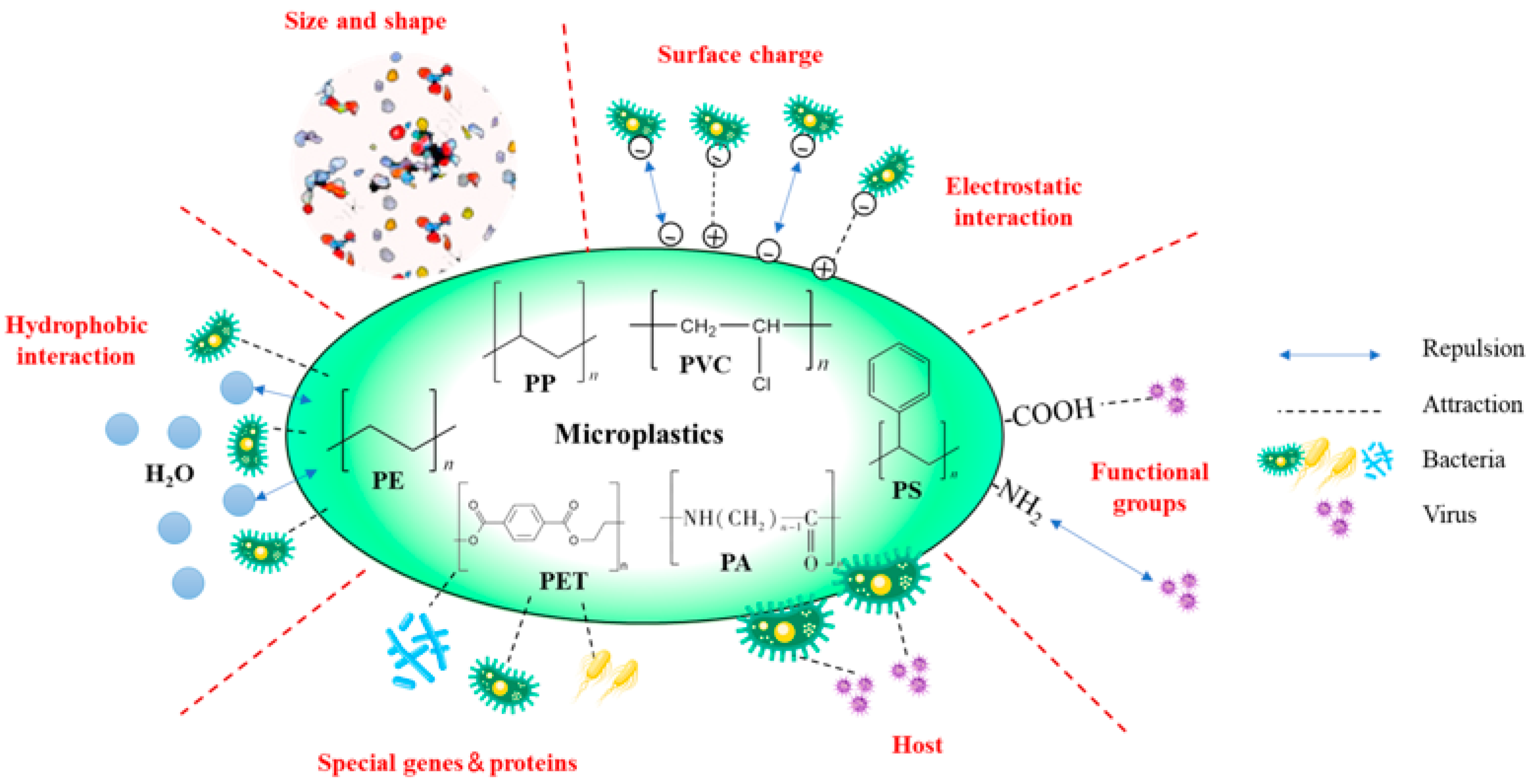
Most of the time, electrostatic repulsion occurs between pathogens and MPs because they both have net negatively charged surfaces. However, the repulsion is overcome due to the complexity of pathogen flagella, proteins, and surface charges, and the hydrophobicity of the cell surface [64]. A large number of studies have shown that microplastics are a vector that can be colonized by various algae and microorganisms. Laboratory investigations have shown that as the soil depth increases, the variety of bacteria on the surface of MPs diminishes [65]. The pathogen characteristics also influence their co-transport mechanisms with MPs. In this section, we will address the mechanism of interaction between microplastics and pathogens in aquifer media from their perspectives, respectively.

3.1. Effects from the Physical Properties of Microplastics

A large specific surface area and small particle size are key factors in the adsorption and transport of MPs in aquifer media. Li et al. [59] found that the adsorption capacity of Legionella at 50 μm MPs was 1–2 orders of magnitude higher than that at 3000 μm MPs. The increase in bacterial adsorption on MPs with decreasing particle size can be explained by a greater specific surface area. Therefore, for MPs to be able to travel vertically in the soil, a particle size smaller than the soil pore space is necessary [66]. Liu et al. [65] discovered that 0.5 mm MPs recovered in soil were less than 1 mm. However, adsorption is no longer the primary interaction mechanism when the particle size of MPs is comparable to that of pathogens. When the particle size of MPs is sufficiently tiny, MPs will compete with E. coli for adsorption sites in the soil, reducing E. coli adsorption in the soil and facilitating the transfer of E. coli in the aquifer medium through electrostatic repulsion [5].
Similarly, microplastic species is one of the key factors influencing the relationship between microplastic–pathogen interactions. Fibers were shown to be the most common type of MPs in different places in China, accounting for 86.37%, 59.4%, and 49% of marine, river, and soil MPs, respectively [67,68,69]. Microfibers have a higher potential for interaction with pathogens than other types of MPs. When compared to microspheres, microfibers had a better ability to adsorb pathogens on their surface [33]. The primary explanation was that increased surface roughness and heterogeneity promoted the microfibers’ ability to attach to pathogens. Some earlier studies [70,71] suggested that nanoscale roughness on colloidal surfaces tends to minimize the repulsive interaction energy barriers between colloids and enhances the aggregation of MPs with bacteria, even in adverse conditions. However, the scanning electron microscopy (SEM) and atomic force microscopy (AFM) of various polymers [72] revealed that PVC had stronger cell adhesion than PP, even though the surface roughness of PVC (13.78 ± 0.65 nm) was somewhat lower than that of PP (14.07 ± 1 nm). Therefore, surface roughness is not a significant element affecting pathogen adsorption. One study reported the same conclusion that surface roughness did little to affect the adsorption of S. sanguinis on titanium surfaces [73]. Cohen and Radian [74] first described the alterations that microfibers undergo when migrating across aquifer media. As a result of friction with the coarse soil particles, microfibers break apart and release smaller, more mobile pieces during flow, which may enable pathogens adsorbed on surfaces to move farther, increasing the environmental and ecological health risks.
Different shapes of microplastics have also been detected in organisms [75]. However, the comparison of bioaccumulation and the toxicity of microplastics with different shapes is still largely unknown. It has been shown that shape-dependent effects should not be ignored when conducting health risk assessments of microplastics. In comparison, non-spherical microplastics had more severe effects on the gut microbiota. Some specific species of gut bacteria are very sensitive to microplastic exposure. For example, microplastics induced a significant increase in the abundance of Proteobacteria. Microbial diversity studies continue to demonstrate the important role of Proteobacteria in gut inflammation. The increased Proteobacteria may produce more bacterial products such as lipopolysaccharide (LPS), which trigger inflammation, disrupt the intestinal mucosal barrier, and increase intestinal permeability. Microplastic fibers also reduce the relative abundance of Pseudomonas and Aeromonas, which can secrete signals to promote the proliferation and renewal of intestinal epithelial cells [76], compared to microplastic beads and microplastic debris [77]. Decreases in Pseudomonas and Aeromonas may further inhibit the regeneration of intestinal epithelial cells and reduce the coverage of cup cells [78]. In addition, fibrous microplastics lead to a decrease in the abundance of actinobacteria, which may weaken the function of the intestinal barrier and increase its susceptibility to immune stimulation, as actinobacteria play a key role in the synthesis of secondary metabolites that can be used as invasive antibiotics for invasive pathogens. In addition, Gordonia abundance was significantly increased in the gut of fish treated with microplastic fibers. Gordonia has a strong ability to degrade plastic-related compounds such as plasticizers [79], polypropylene [80], and phthalates [81]. These findings suggest that a high accumulation of microplastic fibers in the gut leads to specific changes in the gut microbiota associated with plastic exposure, and that gut flora dysbiosis would be a potential new mechanism by which microplastics cause or exacerbate gut toxicity in fish [28].

3.2. Effects from Chemical Characteristics of Microplastics

In addition to their physical characteristics, the chemical properties of MPs such as hydrophobicity and surface functional groups influence their adsorption behavior toward pathogens. Microplastics, as exogenous particles with a hydrophobic surface, are highly likely to provide new substrates for heterotrophic microbial activities, making their surface microbial communities significantly different from those of the surrounding environment and other organic residues [82]. As a result, a richer bacterial community exists on the surface of hydrophobic MPs compared to hydrophilic MPs [83,84]. Thanks to the hydrophobicity of MPs, the interaction affinity of SARS-CoV-2 with MPs in water is more than 10 times higher than that in air [85]. The impact of MP surface functional groups on the adsorption of pathogens is mostly observed in viruses. Liu et al. [86] found that compared to MP-NH2 (average zeta potential before adsorption −7.85 mV, virus adsorption rate 51.4 ± 12.5%) and MP with no groups (average zeta potential before adsorption −16.06 mV, virus adsorption rate 83.6 ± 0.8%), MP-COOH (average zeta potential before adsorption −23.72 mV, virus adsorption rate 94.3 ± 0.8%) could adsorb more viruses. The absolute zeta potential values of MPs were favorably linked with the viral adsorption rate under various functional group alteration settings. As a result, while studying the interaction process between pathogenic microbes and MPs, hydrophobicity and surface functional groups must be considered.

3.3. Effects from Characteristics of Pathogens

3.3.1. Hydrophobicity

Hydrophobic interactions occur due to the mutual repulsion of hydrophobic nonpolar groups with water, and this action draws the hydrophobic groups closer together. Therefore, hydrophobicity is crucial in the early adsorption of germs to hydrophobic surfaces. The non-specific adsorption of germs on abiotic surfaces is favored by strong hydrophobicity [87], while hydrophilic viruses and bacteria exhibit greater migratory potential in aquifer media [88,89]. In a study of the colonization of microplastic surfaces by different bacteria in the presence of Tween-80, it was found that highly hydrophobic strains colonized the microplastic surfaces with significantly higher biomass [90]. When there are enough carbon sources in the environment, bacteria with high cell surface hydrophobicity are better able to colonize. Interfacial tension may also be used to describe how hydrophobicity affects the growth of biofilms on MP surfaces. The decrease in interfacial tension is directly related to the hydrophobicity of bacterial cells [64]. During growth, some bacteria create surfactants, which might result in the replacement of certain proteins, lowering the surface tension and changing viscous moduli [91,92,93]. Therefore, bacterial adsorption is made easier by the development of protein networks with lower surface tension, particularly on the surfaces of MPs with low interfacial tension.

3.3.2. Surface Charge

In the DLVO theory, the positive and negative valves of the interaction energy (Eint) respectively indicate intermolecular interactions of mutual repulsion or attraction [94]. The Eint values of MPs containing SARS-CoV-2 RNA fragments were negative across the whole temperature range of water, which means that the two may assemble into a stable complex [85]. Further research revealed that the Eint values derived from the electrostatic energy between the two are often more similar to the Eint values derived from the total energy/potential energy than those derived from the van der Waals energy. This suggests that the primary mode of interaction between MPs and viruses is electrostatic contact. Electrostatic interaction is the primary adsorption method used by PS-MPs to capture 98.6% of the viral dose [86]. Experimental research by Dika et al. [95] demonstrated the role of electrostatic interactions in viral adsorption. The fact that viruses are normally negatively charged means that the electrostatic interaction force between viruses and positively charged MPs must grow in proportion to the overall negative charge carried by phages. When MPs have a negative charge, the higher the phage bulk charge density, the stronger the electrostatic repulsion and the lower the phage adsorption capability. Unlike the definition of relevant parameters about viruses, bacteria typically use the zeta potential or isoelectric point to assess the electrostatic interactions with non-biological surfaces. Positively charged MPs and negatively charged bacteria typically associate to create heterogeneous aggregates (with negative total zeta potential), which facilitates the movement of MPs across aquifer media [71]. However, when both bacteria and MPs are negatively charged, the zeta potentials of the different types of plastics are comparable, and electrostatic interaction is of little importance in the adhesion of bacteria to plastic surfaces [72].

3.3.3. Specific Properties

Pathogen adsorption levels on the surface of MPs varied substantially due to changes in the pathogen properties. Proteobacteria and Actinobacteria made up the majority of the bacterial communities that predominated on the surface of MPs in soil, together comprising more than 65% of the community [65]. On the one hand, both exhibited the highest relative abundance in the agricultural soil bacterial community, which favors their adsorption on plastics [96]. On the other hand, certain bacteria from these two phyla have unique genes for auxiliary metabolism that enable them to use additives and polymer resins as carbon sources and energy to promote their development [97,98]. I. sakaiensis may release two enzymes that break down PET and PET degradation pathways, respectively [99]. P. aeruginosa and Achromobacter sp. can be involved in the degradation of PVC in the presence of epoxidized Mesua ferrea L. seed oil [100]. Enterobacter sp., Bacillus sp., and Pseudomonas sp. are considered to be involved in the degradation of PS [101,102]. In addition, surface-associated proteins of Streptomycetaceae can reduce the surface tension to very small levels within minutes, which facilitates their adhesion to various surfaces [103].
Phage presence in biofilms is determined by the makeup of the biofilm matrix [104]. Consequently, phage diversity is strongly influenced by the variety of bacterial populations. Biofilms are known to be selective for phage enrichment, with Caudovirales being the most prevalent [98]. In addition, phages such as Podoviridae and Autographiviridae were more likely to be abundant in MP biofilms compared to stone. Podoviridae and Autographiviridae have a limited host range including Enterobacteriaceae, Pseudomonas, Bacillus, and Streptococcus, which include the majority of MP-degrading bacteria. Furthermore, the pathogenicity surface charge distribution may be heterogeneous. For example, the phage tail structure is positively charged, but the entire phage is negatively charged [105]. This heterogeneous surface charge can have a substantial impact on how it interacts with MPs.
We plotted all the factors that could affect the interaction between microplastics and pathogens in Figure 3. In addition to the nature of the pathogens and microplastics themselves, external environmental factors are also important in influencing the interaction between them.

4. Effects of Environmental Factors on the Interactions between Microplastics and Pathogens

4.1. Soil Physicochemical Properties

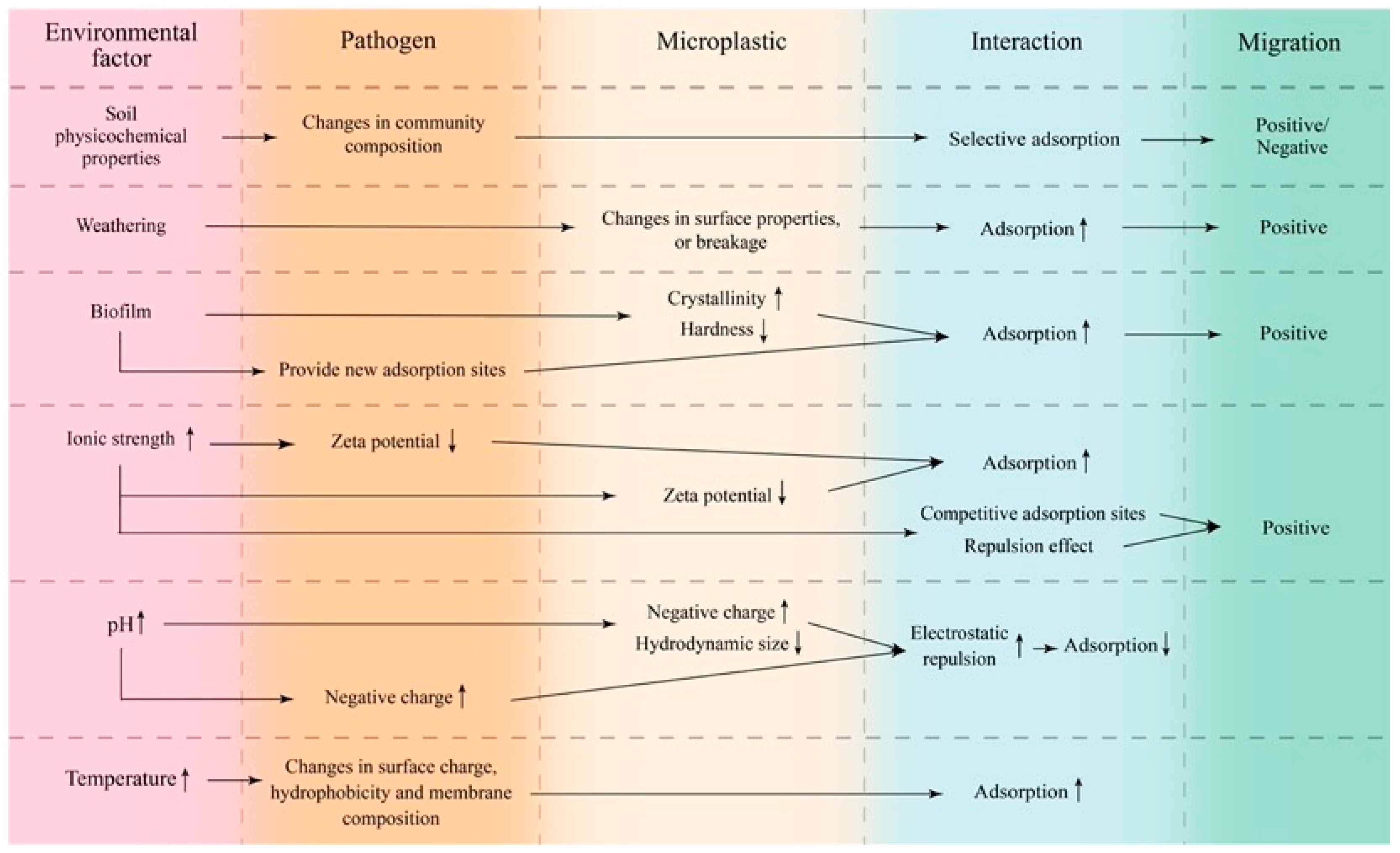
Soil physicochemical parameters have a significant impact on the structure and variety of the bacterial community, which in turn impacts the adsorption of pathogens by MPs [106,107,108]. Chai et al. [31] discovered a high percentage of common species on multiple MPs from the same plot of soil, showing that different MPs had highly similar core microbiota. Furthermore, when the physicochemical characteristics of soils from various plots were varied, the organization of bacterial communities populating the surface of the same MPs differed significantly. For example, heavy metal ions such as Cu, Pb, and Zn are found in high quantities in the soil of e-waste disposal sites, as are polymers such as PP and PC, which are commonly used in electronic devices [109]. Through surface complexation, electrostatic interactions, and hydrogen bonds, MPs adsorb heavy metals and serve as carriers for transfer in aquifer media [110,111,112]. Likewise, several heavy metals are utilized as additives in the manufacture of plastics [113]. Correspondingly, Anaerobicbacteria, represented by Desulfovibrio, made up the majority of the bacterial community composition on the surface of MPs in the target soil. This is because Desulfovibrio may thrive in severe oligotrophic settings and precipitate certain heavy metals via hydrogen sulfide synthesis [114]. Long-term MP persistence in the soil will alter the soil physicochemical features including a drop in soil organic matter and total nitrogen [115]. This would reduce the number of actinomycetes adsorbed on soil particles and the surface of MPs while increasing the prevalence of Proteus, Bacillus, and Sphingomonas [31,65]. Sphingomonas are more suited to the surface environment of MPs and have a higher adsorption capability than actinomycetes [116]. Therefore, the physicochemical characteristics of the soil can either increase or decrease the MP adsorption capability for certain diseases.

4.2. Weathering

On the one hand, weathering has a direct impact on MP adsorption by changing their surface characteristics through physical fragmentation and photo-oxidation. Yuan et al. [117] demonstrated that the adsorption capability of aged MPs for E. coli, plasmid, and phage harboring antibiotic resistance genes was 6.6, 5.2, and 8.3 times greater than that of virgin MPs. Plastic aging is often characterized by polymer chain breaking and the formation of surface cracks and pores, which eventually fragment into micro- and nanoparticles. This means that weathered MPs have a larger specific surface area, microporous area, surface roughness, and attraction for antibiotic resistance gene (ARG) vectors. Long-term UV irradiation initiates the photolytic destruction of plastics, resulting in many microscopic pieces that serve as colonization sites for certain functional bacteria [90]. However, Lu et al. [86] showed that prolonged UV irradiation significantly decreased the absolute zeta potential of carboxyl group-modified MPs, which reduced their virus adsorption capacity, but increased the absolute zeta potential of amino group-modified MPs, which facilitated their virus adsorption capacity. On the other hand, weathering can have an indirect effect on the adsorption ability of MPs for pathogens. Aged PP-MPs can absorb more antibiotics because of a more developed pore structure [118,119]. This will encourage pathogens with ARG to bind to the surface of MPs. Weathering therefore directly influences the surface characteristics of MPs, which subsequently directly or indirectly affects the pathogen adsorption behavior.

4.3. Biofilm

Biofilms, a community of microorganisms accumulated in the matrix of self-developed extracellular polymeric substances (EPS), are present ubiquitously in both natural aquatic environments and engineering systems. Some studies have shown that the biofilms clinging to sand surfaces enhance the surface roughness of aquifer media and reduce flow channels, which can impede pathogen–MP co-transport [120]. Low crystallinity and high hardness matrix surfaces typically exhibit higher pathogenic microbial diversity [121]. However, McGivney et al. [122] discovered that biofilm-mediated weathering increases the crystallinity of PE and decreases the hardness of PP. This may lead to the selective adsorption of microorganisms by MPs and impact the diversity of adsorbed pathogens. However, it has also been demonstrated that the surface hydrophobicity and crystallinity of MPs are reduced by biofilm adherence [65]. Compared to the uncertainty of bacterial adsorption by biofilm-coated MPs, biofilms play a positive role in virus adsorption by MPs. Some viruses can interact with and bind to lipopolysaccharides (LPS) and peptidoglycans (PG) in biofilms [123]. This may provide novel locations for virus adsorption while also improving virus stability and transmission [124]. Biofilms can be crucial in the adsorption of MPs since they have colonized the majority of the surfaces of MPs in the water bodies.
In addition to microorganisms that can colonize the surface of microplastics, nanomaterials (NMs) can also be adsorbed onto the surface of microplastics. In 2013, Fries et al. classified MPs from sediment samples collected from Norderney and found TiO2 nanoparticles on their surfaces, confirming that MPs can act as carriers of NMs [125,126]. Existing studies have reported that the oral administration of Ag nanoparticles decreases the Firmicutes/Bacteroidetes values and microbial community density in the gut of mice [127]. In addition, Sprague-Dawley rats orally exposed to Ag nanoparticles showed an increase in the proportion of Gram-negative bacteria and a decrease in the abundance of Firmicutes and Lactobacillus [128]. Zhao et al. found that TiO2 nanoparticles further interfered with the diversity, composition, and KEGG pathways of the gut microbiota and led to inflammatory damage in the colon of mice with metabolic syndrome.
Antibiotics are widely used in the pharmaceutical aquaculture industry, and the overuse of antibiotics has led to antibiotics entering the water column. Despite the low concentration of antibiotics in this fraction, they can promote the development of antibiotic resistance in natural bacteria [129]. Natural bacteria may then transfer resistance to other bacteria including human pathogens [130]. At the same time, antibiotics selectively adsorb onto aged microplastics, favoring opportunistic pathogen colonization.

4.4. Ionic Strength

Ionic strength changes the surface charge of MPs and pathogens, influencing their interaction mechanism and movement pathway. He et al. [5] evaluated the migration of E. coli with MPs in aquifer media under various circumstances and discovered that MPs impact pathogen migration via diverse pathways at different ionic strengths. Because of the great mobility of E. coli and the poor deposition rates of MPs at low ionic strengths, the presence of MPs (<2 μm) had no influence on E. coli movement in aquifer media. Ionic strength changes impact the charge balance of colloids, influencing their stability. This may cause MPs to aggregate and depose, affecting their migration behavior in groundwater. The zeta potential of MPs and bacterial surfaces drops dramatically when the solution ionic strength increases, which is due to a large number of ions in the solution compressing the electrical double layer of the colloidal particles [131]. As a result, MPs and pathogens are more likely to overcome energy barriers and adsorb onto the media [132,133]. However, He et al. [5] demonstrated that MPs continue to facilitate E. coli transport at high ionic strengths. In a similar manner to E. coli, MPs with a size smaller than 2 μm exhibit increased deposition rates with higher ionic strength. This leads to competition between MPs and bacteria for deposition sites, ultimately resulting in a decrease in bacterial deposition in high ionic strength solutions. In contrast, nanoplastics maintain high mobility under high ionic strength. The suspended nanoplastics reduce the potential for E. coli attachment through repulsive effects, eventually increasing bacterial transportation in aquifer media.

4.5. pH

pH determines the fate and transport of pathogens and MPs in the subsurface environment by altering surface charge and adsorption–desorption processes. The transport of MPs in aquifer media is significantly affected by pH [132]. Slightly alkaline solutions facilitate the transport of MPs in aquifer media. At higher pH, MPs tend to carry more negative charges along with the aquifer media particles. As a result, MP colloids find it challenging to hit and adhere to the media surface because of the higher electrostatic repulsion and potential energy barrier between the two. In addition, the hydrodynamic size of the MPs shrinks with increasing pH, which aggravates Brown motion. Pathogens exhibit similar migration patterns. Decreasing pH increases the pathogen attachment to aquifer media and colloids, resulting in increased pathogen retention in the subsurface environment [82]. It is worth noting that pathogens can group due to weak electrostatic interaction and deposit in aquifer media when the pH of the solution is near the isoelectric point of the pathogens [134]. Therefore, when the solution pH is slightly acidic, the electrostatic repulsion between MPs and pathogens is minimized, and the adsorption capacity of MPs on pathogens is enhanced. A larger pH range may result in different surface charges for pathogens and MPs, thereby affecting their form of interaction. In contrast, if the pH range in the subsurface environment is modest, the changes in surface charges for both may be meaningless, and pH may not be the primary factor influencing their interaction.

4.6. Temperature

Temperature affects the interaction of MPs with pathogens mainly by altering the physicochemical characteristics, physiological properties, and adsorption thermodynamics. Many researchers have found that temperature affects the pathogen adhesion to solid surfaces and migration behavior in the subsurface environment [135,136]. Based on the results of rank correlation analysis [137], temperature is a major factor determining the colonization of MPs by dominant pathogens. There are studies that illustrate the temporal and successional dynamics of biofilms and clearly show that an increase in temperature plays a role in the formation of plastic-specific microbial communities [138]. Cappello and Guglielmino [139] investigated P. aeruginosa adherence to polystyrene at 15 °C, 30 °C, and 47 °C and discovered that bacterial adhesion increased with temperature rise. The surrounding temperature affects the bacterial surface charge, hydrophobicity, and outer membrane components (e.g., lipopolysaccharides and flagella), which explains the variance in pathogen adhesion to MP surfaces [140,141]. Higher temperatures often boost pathogen attachment and limit their ability to move in groundwater [82]. Nevertheless, MP migratory behavior increases the uncertainty of the mentioned processes. According to the releasing behavior of various plastics in aqueous settings, higher temperatures stimulate plastic breaking, which in turn promotes MP migration [142,143]. Therefore, the co-transport behavior of MPs and pathogens in groundwater has to be researched further.
The influence of environmental factors on the interaction and synergistic transport between MPs and pathogens is extremely complex. Weathering processes such as physical fragmentation, photo-oxidation, and UV irradiation affect interactions due to changes in the morphological structure and surface characteristics of MPs. Ionic strength, pH, and temperature alter the surface charge of MPs and pathogens, thereby affecting their adsorption and transport capacities. Soil physicochemical characteristics and biofilms alter the composition of pathogen communities (Figure 4).

5. Effects of Combined Exposure to MPs and Pathogens on Organisms

Microbial colonization boosts MP absorption by marine species. Filter feeders consume colonized MPs 10 times more than virgin MPs, and sea urchins exhibit a similar tendency [35,144]. These marine invertebrates seek for and absorb particulate matter through chemical sensing, and they prefer aged MPs over virgin particles [145]. Biofilm colonization may have changed the characteristics of MPs, making them more appealing. Environmental MPs may increase pathogen virulence. Disposable plastic tubes have been shown to promote significant expression of the central virulence-associated protein (VapA) of Rhodococcus equi at lower temperatures than standard glass tubes [146]. The surface nature of the plastic can help it activate transcriptional control of VirR and VirS proteins, thereby boosting the mRNA levels of VapA by 70-fold. The combined activity of MPs and their adsorbed pathogens may affect the organism’s immune system. Sea urchins exposed to colonized MPs had a substantial decrease in coelomocytes but an increase in vibratile cells and red/white amoebocyte ratio [144]. In addition, the exposed individuals’ digestive systems were shown to have increased levels of total antioxidant activity and catalase. It has been reported that colonized MPs have a greater toxic effect on mussels than single MPs. V. parahaemolyticus-attached MPs affect hemocyte function in mussels, causing apoptosis and inhibiting antioxidant enzyme activity in the gills [147]. The combined impacts of pathogens and MPs may harm not just animals but even humans. In cancer patients with indwelling central venous catheters, Rhodococcus equi produces biofilms on the surface of the catheters made of polyurethane, which can cause bacteremia [148]. Although marine species have an exceptional ability to consume MPs, MPs may not be transferred up the food chain to higher levels after ingestion and excretion. Furthermore, it should be further researched to determine the risks that MPs or colonized MPs have to human health.

6. Conclusions

The damage that MPs cause to the environment has started to draw attention since the idea of them was first proposed. People have also started to pay attention to the transmission of microbes, particularly pathogens, in the natural environment out of concern for their health. Both are transported through the environment similarly to colloids, and their interactions are quite complicated. Groundwater, a critical supply of drinking water, is under threat from industrial and agricultural activities as well as the vertical migration of tiny particle pollutants from landfills, which may increase the degree of pollution and ecological risk from MPs and pathogens. This study focused on the major processes of MP–pathogen interactions as well as the sources and characteristics of MPs in the subsurface environment. The primary elements influencing their interactions include particle size, specific surface area, shape, hydrophobicity, and surface functional groups of MPs as well as pathogen hydrophobicity, zeta potential, and physiological features. The surface characteristics, crystallinity, and surface charge of MPs as well as changes in pathogens and the environment (e.g., soil physicochemical parameters and ionic strength) all have an impact on interactions. Furthermore, we investigated the co-transport of pathogens and MPs in aquifer media. The cohabitation of the two in subsurface environments may mutually enhance or prevent their migration, depending on the interactions. Future research should concentrate on (i) the synergistic transport behavior of pathogens and MPs in aquifer media under various environmental circumstances, and (ii) the combined toxicity of MPs and pathogens to organisms.

Author Contributions

W.Z. and H.H. conceived and supervised the current review. H.Z. conducted the literature review, collated and analyzed the data, and wrote the manuscript. X.H. conducted the literature review, collated, and analyzed the data. J.C., B.W., K.Z. and C.H. collated and analyzed the data. All authors have read and agreed to the published version of the manuscript.

Funding

This study was supported by the National Natural Science Foundation of China [grant number 42177047].

Conflicts of Interest

The authors declare no competing interests.

References

  1. Thompson, R.C.; Olsen, Y.; Mitchell, R.P.; Davis, A.; Rowland, S.J.; John, A.W.G.; McGonigle, D.; Russell, A.E. Lost at Sea: Where Is All the Plastic? Science 2004, 304, 838. [Google Scholar] [CrossRef] [PubMed]
  2. Arthur, C.; Baker, J.E.; Bamford, H.A. International Research Workshop on the Occurrence, Effects, and Fate of Microplastic Marine Debris. In Proceedings of the Conference Proceedings, Tacoma, WA, USA, 9–11 September 2008; University of Washington Tacoma: Tacoma, WA, USA, 2009. [Google Scholar]
  3. Collignon, A.; Hecq, J.-H.; Galgani, F.; Collard, F.; Goffart, A. Annual variation in neustonic micro- and meso-plastic particles and zooplankton in the Bay of Calvi (Mediterranean–Corsica). Mar. Pollut. Bull. 2014, 79, 293–298. [Google Scholar] [CrossRef] [PubMed]
  4. Gigault, J.; ter Halle, A.; Baudrimont, M.; Pascal, P.-Y.; Gauffre, F.; Phi, T.-L.; El Hadri, H.; Grassl, B.; Reynaud, S. Current opinion: What is a nanoplastic? Environ. Pollut. 2018, 235, 1030–1034. [Google Scholar] [CrossRef] [PubMed]
  5. .He, L.; Wu, D.; Rong, H.; Li, M.; Tong, M.; Kim, H. Influence of Nano- and Microplastic Particles on the Transport and Deposition Behaviors of Bacteria in Quartz Sand. Environ. Sci. Technol. 2018, 52, 11555–11563. [Google Scholar] [CrossRef]
  6. Auta, H.; Emenike, C.; Fauziah, S. Distribution and importance of microplastics in the marine environment: A review of the sources, fate, effects, and potential solutions. Environ. Int. 2017, 102, 165–176. [Google Scholar] [CrossRef] [PubMed]
  7. Su, Y.; Zhang, Z.; Wu, D.; Zhan, L.; Shi, H.; Xie, B. Occurrence of microplastics in landfill systems and their fate with landfill age. Water Res. 2019, 164, 114968. [Google Scholar] [CrossRef] [PubMed]
  8. Boucher, J.; Friot, D. Primary Microplastics in the Oceans: A Global Evaluation of Sources; IUCN: Gland, Switzerland, 2017. [Google Scholar]
  9. Yurtsever, M. Glitters as a Source of Primary Microplastics: An Approach to Environmental Responsibility and Ethics. J. Agric. Environ. Ethics 2019, 32, 459–478. [Google Scholar] [CrossRef]
  10. Wang, L.; Wu, W.-M.; Bolan, N.S.; Tsang, D.C.; Li, Y.; Qin, M.; Hou, D. Environmental fate, toxicity and risk management strategies of nanoplastics in the environment: Current status and future perspectives. J. Hazard. Mater. 2021, 401, 123415. [Google Scholar] [CrossRef]
  11. Bouwmeester, H.; Hollman, P.C.H.; Peters, R.J.B. Potential Health Impact of Environmentally Released Micro- and Nanoplastics in the Human Food Production Chain: Experiences from Nanotoxicology. Environ. Sci. Technol. 2015, 49, 8932–8947. [Google Scholar] [CrossRef]
  12. da Costa, J.P.; Santos, P.S.; Duarte, A.C.; Rocha-Santos, T. (Nano)plastics in the environment–Sources, fates and effects. Sci. Total Environ. 2016, 566–567, 15–26. [Google Scholar] [CrossRef]
  13. Birch, Q.T.; Potter, P.M.; Pinto, P.X.; Dionysiou, D.D.; Al-Abed, S.R. Sources, transport, measurement and impact of nano and microplastics in urban watersheds. Rev. Environ. Sci. Bio/Technol. 2020, 19, 275–336. [Google Scholar] [CrossRef] [PubMed]
  14. Rochman, C.M.; Brookson, C.; Bikker, J.; Djuric, N.; Earn, A.; Bucci, K.; Athey, S.; Huntington, A.; McIlwraith, H.; Munno, K.; et al. Rethinking microplastics as a diverse contaminant suite. Environ. Toxicol. Chem. 2019, 38, 703–711. [Google Scholar] [CrossRef] [PubMed]
  15. Deng, L.; Cai, L.; Sun, F.; Li, G.; Che, Y. Public attitudes towards microplastics: Perceptions, behaviors and policy implications. Resour. Conserv. Recycl. 2020, 163, 105096. [Google Scholar] [CrossRef]
  16. Chia, R.W.; Lee, J.-Y.; Kim, H.; Jang, J. Microplastic pollution in soil and groundwater: A review. Environ. Chem. Lett. 2021, 19, 4211–4224. [Google Scholar] [CrossRef]
  17. Chatziparaskeva, G.; Papamichael, I.; Zorpas, A.A. Microplastics in the coastal environment of Mediterranean and the impact on sustainability level. Sustain. Chem. Pharm. 2022, 29, 100768. [Google Scholar] [CrossRef]
  18. Kyriakopoulos, G.L.; Zamparas, M.G.; Kapsalis, V.C. Investigating the Human Impacts and the Environmental Consequences of Microplastics Disposal into Water Resources. Sustainability 2022, 14, 828. [Google Scholar] [CrossRef]
  19. Gerolin, C.R.; Pupim, F.N.; Sawakuchi, A.O.; Grohmann, C.H.; Labuto, G.; Semensatto, D. Microplastics in sediments from Amazon rivers, Brazil. Sci. Total Environ. 2020, 749, 141604. [Google Scholar] [CrossRef] [PubMed]
  20. Selvam, S.; Jesuraja, K.; Venkatramanan, S.; Roy, P.D.; Kumari, V.J. Hazardous microplastic characteristics and its role as a vector of heavy metal in groundwater and surface water of coastal south India. J. Hazard. Mater. 2021, 402, 123786. [Google Scholar] [CrossRef]
  21. Yan, M.; Nie, H.; Xu, K.; He, Y.; Hu, Y.; Huang, Y.; Wang, J. Microplastic abundance, distribution and composition in the Pearl River along Guangzhou city and Pearl River estuary, China. Chemosphere 2019, 217, 879–886. [Google Scholar] [CrossRef]
  22. Samandra, S.; Johnston, J.M.; Jaeger, J.E.; Symons, B.; Xie, S.; Currell, M.; Ellis, A.V.; Clarke, B.O. Microplastic contamination of an unconfined groundwater aquifer in Victoria, Australia. Sci. Total Environ. 2022, 802, 149727. [Google Scholar] [CrossRef]
  23. Chen, Y.; Shen, Z.; Li, G.; Wang, K.; Cai, X.; Xiong, X.; Wu, C. Factors affecting microplastic accumulation by wild fish: A case study in the Nandu River, South China. Sci. Total Environ. 2022, 847, 157486. [Google Scholar] [CrossRef]
  24. Bošković, N.; Joksimović, D.; Bajt, O. Microplastics in mussels from the Boka Kotorska Bay (Adriatic Sea) and impact on human health. Food Chem. Toxicol. 2023, 173, 113641. [Google Scholar] [CrossRef] [PubMed]
  25. Carlin, J.; Craig, C.; Little, S.; Donnelly, M.; Fox, D.; Zhai, L.; Walters, L. Microplastic accumulation in the gastrointestinal tracts in birds of prey in central Florida, USA. Environ. Pollut. 2020, 264, 114633. [Google Scholar] [CrossRef]
  26. Muhib, I.; Uddin, K.; Rahman, M.; Malafaia, G. Occurrence of microplastics in tap and bottled water, and food packaging: A narrative review on current knowledge. Sci. Total Environ. 2023, 865, 161274. [Google Scholar] [CrossRef] [PubMed]
  27. Cox, K.D.; Covernton, G.A.; Davies, H.L.; Dower, J.F.; Juanes, F.; Dudas, S.E. Human Consumption of Microplastics. Environ. Sci. Technol. 2019, 53, 7068–7074. [Google Scholar] [CrossRef] [PubMed]
  28. Qiao, R.; Deng, Y.; Zhang, S.; Wolosker, M.B.; Zhu, Q.; Ren, H.; Zhang, Y. Accumulation of different shapes of microplastics initiates intestinal injury and gut microbiota dysbiosis in the gut of zebrafish. Chemosphere 2019, 236, 124334. [Google Scholar] [CrossRef] [PubMed]
  29. Yong, C.Q.Y.; Valiyaveettil, S.; Tang, B.L. Toxicity of Microplastics and Nanoplastics in Mammalian Systems. Int. J. Environ. Res. Public Health 2020, 17, 1509. [Google Scholar] [CrossRef]
  30. Khalid, N.; Aqeel, M.; Noman, A.; Hashem, M.; Mostafa, Y.S.; Alhaithloul, H.A.S.; Alghanem, S.M. Linking effects of microplastics to ecological impacts in marine environments. Chemosphere 2021, 264, 128541. [Google Scholar] [CrossRef]
  31. Chai, B.; Li, X.; Liu, H.; Lu, G.; Dang, Z.; Yin, H. Bacterial communities on soil microplastic at Guiyu, an E-Waste dismantling zone of China. Ecotoxicol. Environ. Saf. 2020, 195, 110521. [Google Scholar] [CrossRef]
  32. McCormick, A.; Hoellein, T.J.; Mason, S.A.; Schluep, J.; Kelly, J.J. Microplastic is an Abundant and Distinct Microbial Habitat in an Urban River. Environ. Sci. Technol. 2014, 48, 11863–11871. [Google Scholar] [CrossRef]
  33. Zhang, E.; Kim, M.; Rueda, L.; Rochman, C.; VanWormer, E.; Moore, J.; Shapiro, K. Association of zoonotic protozoan parasites with microplastics in seawater and implications for human and wildlife health. Sci. Rep. 2022, 12, 6532. [Google Scholar] [CrossRef]
  34. Rodrigues, A.; Oliver, D.M.; McCarron, A.; Quilliam, R.S. Colonisation of plastic pellets (nurdles) by E. coli at public bathing beaches. Mar. Pollut. Bull. 2019, 139, 376–380. [Google Scholar] [CrossRef]
  35. Fabra, M.; Williams, L.; Watts, J.E.; Hale, M.S.; Couceiro, F.; Preston, J. The plastic Trojan horse: Biofilms increase microplastic uptake in marine filter feeders impacting microbial transfer and organism health. Sci. Total Environ. 2021, 797, 149217. [Google Scholar] [CrossRef]
  36. Jiang, P.; Zhao, S.; Zhu, L.; Li, D. Microplastic-associated bacterial assemblages in the intertidal zone of the Yangtze Estuary. Sci. Total Environ. 2018, 624, 48–54. [Google Scholar] [CrossRef] [PubMed]
  37. Nizzetto, L.; Futter, M.; Langaas, S. Are Agricultural Soils Dumps for Microplastics of Urban Origin? Environ. Sci. Technol. 2016, 50, 10777–10779. [Google Scholar] [CrossRef] [PubMed]
  38. Othman, A.R.; Abu Hasan, H.; Muhamad, M.H.; Ismail, N.; Abdullah, S.R.S. Microbial degradation of microplastics by enzymatic processes: A review. Environ. Chem. Lett. 2021, 19, 3057–3073. [Google Scholar] [CrossRef]
  39. Nizzetto, L.; Bussi, G.; Futter, M.N.; Butterfield, D.; Whitehead, P.G. A theoretical assessment of microplastic transport in river catchments and their retention by soils and river sediments. Environ. Sci. Process. Impacts 2016, 18, 1050–1059. [Google Scholar] [CrossRef] [PubMed]
  40. O’Connor, D.; Pan, S.; Shen, Z.; Song, Y.; Jin, Y.; Wu, W.M.; Hou, D. Microplastics undergo accelerated vertical migration in sand soil due to small size and wet-dry cycles. Environ. Pollut. 2019, 249, 527–534. [Google Scholar] [CrossRef] [PubMed]
  41. Hou, L.; Kumar, D.; Yoo, C.G.; Gitsov, I.; Majumder, E.L.-W. Conversion and removal strategies for microplastics in wastewater treatment plants and landfills. Chem. Eng. J. 2021, 406, 126715. [Google Scholar] [CrossRef]
  42. Li, N.; Han, Z.; Guo, N.; Zhou, Z.; Liu, Y.; Tang, Q. Microplastics spatiotemporal distribution and plastic-degrading bacteria identification in the sanitary and non-sanitary municipal solid waste landfills. J. Hazard. Mater. 2022, 438, 129452. [Google Scholar] [CrossRef]
  43. Yu, F.; Wu, Z.; Wang, J.; Li, Y.; Chu, R.; Pei, Y.; Ma, J. Effect of landfill age on the physical and chemical characteristics of waste plastics/microplastics in a waste landfill sites. Environ. Pollut. 2022, 306, 119366. [Google Scholar] [CrossRef]
  44. Liang, H.; Ji, Y.; Ge, W.; Wu, J.; Song, N.; Yin, Z.; Chai, C. Release kinetics of microplastics from disposable face masks into the aqueous environment. Sci. Total Environ. 2022, 816, 151650. [Google Scholar] [CrossRef]
  45. Ziajahromi, S.; Neale, P.A.; Silveira, I.T.; Chua, A.; Leusch, F.D. An audit of microplastic abundance throughout three Australian wastewater treatment plants. Chemosphere 2021, 263, 128294. [Google Scholar] [CrossRef] [PubMed]
  46. Sun, J.; Zhu, Z.-R.; Li, W.-H.; Yan, X.; Wang, L.-K.; Zhang, L.; Jin, J.; Dai, X.; Ni, B.-J. Revisiting Microplastics in Landfill Leachate: Unnoticed Tiny Microplastics and Their Fate in Treatment Works. Water Res. 2021, 190, 116784. [Google Scholar] [CrossRef] [PubMed]
  47. Huang, Y.; Liu, Q.; Jia, W.; Yan, C.; Wang, J. Agricultural plastic mulching as a source of microplastics in the terrestrial environment. Environ. Pollut. 2020, 260, 114096. [Google Scholar] [CrossRef] [PubMed]
  48. Jin, T.; Tang, J.; Lyu, H.; Wang, L.; Gillmore, A.B.; Schaeffer, S.M. Activities of Microplastics (MPs) in Agricultural Soil: A Review of MPs Pollution from the Perspective of Agricultural Ecosystems. J. Agric. Food Chem. 2022, 70, 4182–4201. [Google Scholar] [CrossRef] [PubMed]
  49. Zhang, L.; Xie, Y.; Liu, J.; Zhong, S.; Qian, Y.; Gao, P. An Overlooked Entry Pathway of Microplastics into Agricultural Soils from Application of Sludge-Based Fertilizers. Environ. Sci. Technol. 2020, 54, 4248–4255. [Google Scholar] [CrossRef]
  50. Carr, S.A.; Liu, J.; Tesoro, A.G. Transport and fate of microplastic particles in wastewater treatment plants. Water Res. 2016, 91, 174–182. [Google Scholar] [CrossRef] [PubMed]
  51. van Den Berg, P.; Huerta-Lwanga, E.; Corradini, F.; Geissen, V. Sewage sludge application as a vehicle for microplastics in eastern Spanish agricultural soils. Environ. Pollut. 2020, 261, 114198. [Google Scholar] [CrossRef]
  52. Wright, S.; Ulke, J.; Font, A.; Chan, K.; Kelly, F. Atmospheric microplastic deposition in an urban environment and an evaluation of transport. Environ. Int. 2020, 136, 105411. [Google Scholar] [CrossRef]
  53. Zhou, D.; Cai, Y.; Yang, Z. Key factors controlling transport of micro- and nanoplastic in porous media and its effect on coexisting pollutants. Environ. Pollut. 2022, 293, 118503. [Google Scholar] [CrossRef]
  54. Pang, Z.; Huang, T.; Chen, Y. Diminished groundwater recharge and circulation relative to degrading riparian vegetation in the middle Tarim River, Xinjiang Uygur, Western China. Hydrol. Process. 2010, 24, 147–159. [Google Scholar] [CrossRef]
  55. Yeh, H.-F.; Lin, H.-I.; Lee, C.-H.; Hsu, K.-C.; Wu, C.-S. Identifying Seasonal Groundwater Recharge Using Environmental Stable Isotopes. Water 2014, 6, 2849–2861. [Google Scholar] [CrossRef]
  56. He, B.; Wijesiri, B.; Ayoko, G.A.; Egodawatta, P.; Rintoul, L.; Goonetilleke, A. Influential factors on microplastics occurrence in river sediments. Sci. Total Environ. 2020, 738, 139901. [Google Scholar] [CrossRef] [PubMed]
  57. Bian, P.; Liu, Y.; Zhao, K.; Hu, Y.; Zhang, J.; Kang, L.; Shen, W. Spatial variability of microplastic pollution on surface of rivers in a mountain-plain transitional area: A case study in the Chin Ling-Wei River Plain, China. Ecotoxicol. Environ. Saf. 2022, 232, 113298. [Google Scholar] [CrossRef] [PubMed]
  58. Hu, E.; Sun, C.; Yang, F.; Wang, Y.; Hu, L.; Wang, L.; Li, M.; Gao, L. Microplastics in 48 wastewater treatment plants reveal regional differences in physical characteristics and shape-dependent removal in the transition zone between North and South China. Sci. Total Environ. 2022, 834, 155320. [Google Scholar] [CrossRef] [PubMed]
  59. Li, Q.; Hu, Y.; Kou, D.; Yang, W.; Tang, W.; Chen, Q.; Que, S.; Zhao, X. Factors Impacting Microplastic Biofilm Community and Biological Risks Posed by Microplastics in Drinking Water Sources. Water Air Soil Pollut. 2022, 233, 179. [Google Scholar] [CrossRef]
  60. Wang, L.; Luo, Z.; Zhen, Z.; Yan, Y.; Yan, C.; Ma, X.; Sun, L.; Wang, M.; Zhou, X.; Hu, A. Bacterial community colonization on tire microplastics in typical urban water environments and associated impacting factors. Environ. Pollut. 2020, 265, 114922. [Google Scholar] [CrossRef] [PubMed]
  61. Ballent, A.; Corcoran, P.L.; Madden, O.; Helm, P.A.; Longstaffe, F.J. Sources and sinks of microplastics in Canadian Lake Ontario nearshore, tributary and beach sediments. Mar. Pollut. Bull. 2016, 110, 383–395. [Google Scholar] [CrossRef] [PubMed]
  62. Besseling, E.; Quik, J.T.; Sun, M.; Koelmans, A.A. Fate of nano- and microplastic in freshwater systems: A modeling study. Environ. Pollut. 2017, 220, 540–548. [Google Scholar] [CrossRef]
  63. Zuo, L.-Z.; Li, H.-X.; Lin, L.; Sun, Y.-X.; Diao, Z.-H.; Liu, S.; Zhang, Z.-Y.; Xu, X.-R. Sorption and desorption of phenanthrene on biodegradable poly(butylene adipate co-terephtalate) microplastics. Chemosphere 2019, 215, 25–32. [Google Scholar] [CrossRef]
  64. Rühs, P.; Böcker, L.; Inglis, R.; Fischer, P. Studying bacterial hydrophobicity and biofilm formation at liquid–liquid interfaces through interfacial rheology and pendant drop tensiometry. Colloids Surf. B Biointerfaces 2014, 117, 174–184. [Google Scholar] [CrossRef]
  65. Liu, H.; Yue, L.; Zhao, Y.; Li, J.; Fu, Y.; Deng, H.; Feng, D.; Li, Q.; Yu, H.; Zhang, Y.; et al. Changes in bacterial community structures in soil caused by migration and aging of microplastics. Sci. Total Environ. 2022, 848, 157790. [Google Scholar] [CrossRef]
  66. Helmberger, M.S.; Tiemann, L.K.; Grieshop, M.J. Towards an ecology of soil microplastics. Funct. Ecol. 2020, 34, 550–560. [Google Scholar] [CrossRef]
  67. Ding, L.; Zhang, S.; Wang, X.; Yang, X.; Zhang, C.; Qi, Y.; Guo, X. The occurrence and distribution characteristics of microplastics in the agricultural soils of Shaanxi Province, in north-western China. Sci. Total Environ. 2020, 720, 137525. [Google Scholar] [CrossRef] [PubMed]
  68. Li, J.; Ouyang, Z.; Liu, P.; Zhao, X.; Wu, R.; Zhang, C.; Lin, C.; Li, Y.; Guo, X. Distribution and characteristics of microplastics in the basin of Chishui River in Renhuai, China. Sci. Total Environ. 2021, 773, 145591. [Google Scholar] [CrossRef] [PubMed]
  69. Zhang, B.; Wu, D.; Yang, X.; Teng, J.; Liu, Y.; Zhang, C.; Zhao, J.; Yin, X.; You, L.; Liu, Y.; et al. Microplastic pollution in the surface sediments collected from Sishili Bay, North Yellow Sea, China. Mar. Pollut. Bull. 2019, 141, 9–15. [Google Scholar] [CrossRef] [PubMed]
  70. Bradford, S.A.; Kim, H.; Shen, C.; Sasidharan, S.; Shang, J. Contributions of Nanoscale Roughness to Anomalous Colloid Retention and Stability Behavior. Langmuir 2017, 33, 10094–10105. [Google Scholar] [CrossRef] [PubMed]
  71. He, L.; Rong, H.; Li, M.; Zhang, M.; Liu, S.; Yang, M.; Tong, M. Bacteria have different effects on the transport behaviors of positively and negatively charged microplastics in porous media. J. Hazard. Mater. 2021, 415, 125550. [Google Scholar] [CrossRef] [PubMed]
  72. Cai, L.; Wu, D.; Xia, J.; Shi, H.; Kim, H. Influence of physicochemical surface properties on the adhesion of bacteria onto four types of plastics. Sci. Total Environ. 2019, 671, 1101–1107. [Google Scholar] [CrossRef]
  73. Rodríguez-Hernández, A.G.; Muñoz-Tabares, J.A.; Godoy-Gallardo, M.; Juárez, A.; Gil, F.-J. S. sanguinis adhesion on rough titanium surfaces: Effect of culture media. Mater. Sci. Eng. C 2013, 33, 714–720. [Google Scholar] [CrossRef]
  74. Cohen, N.; Radian, A. Microplastic Textile Fibers Accumulate in Sand and Are Potential Sources of Micro(nano)plastic Pollution. Environ. Sci. Technol. 2022, 56, 17635–17642. [Google Scholar] [CrossRef] [PubMed]
  75. Neves, D.; Sobral, P.; Ferreira, J.L.; Pereira, T. Ingestion of microplastics by commercial fish off the Portuguese coast. Mar. Pollut. Bull. 2015, 101, 119–126. [Google Scholar] [CrossRef] [PubMed]
  76. Li, J.-D.; Feng, W.; Gallup, M.; Kim, J.-H.; Gum, J.; Kim, Y.; Basbaum, C. Activation of NF-κB via a Src-dependent Ras-MAPK-pp90rsk pathway is required for Pseudomonas aeruginosa -induced mucin overproduction in epithelial cells. Proc. Natl. Acad. Sci. USA 1998, 95, 5718–5723. [Google Scholar] [CrossRef] [PubMed]
  77. Cheesman, S.E.; Neal, J.T.; Mittge, E.; Seredick, B.M.; Guillemin, K. Epithelial cell proliferation in the developing zebrafish intestine is regulated by the Wnt pathway and microbial signaling via Myd88. Proc. Natl. Acad. Sci. USA 2010, 108, 4570–4577. [Google Scholar] [CrossRef] [PubMed]
  78. Bates, J.M.; Mittge, E.; Kuhlman, J.; Baden, K.N.; Cheesman, S.E.; Guillemin, K. Distinct signals from the microbiota promote different aspects of zebrafish gut differentiation. Dev. Biol. 2006, 297, 374–386. [Google Scholar] [CrossRef] [PubMed]
  79. Drzyzga, O. The strengths and weaknesses of Gordonia: A review of an emerging genus with increasing biotechnological potential. Crit. Rev. Microbiol. 2012, 38, 300–316. [Google Scholar] [CrossRef] [PubMed]
  80. Arkatkar, A.; Juwarkar, A.A.; Bhaduri, S.; Uppara, P.V.; Doble, M. Growth of Pseudomonas and Bacillus biofilms on pretreated polypropylene surface. Int. Biodeterior. Biodegrad. 2010, 64, 530–536. [Google Scholar] [CrossRef]
  81. Ren, L.; Lin, Z.; Liu, H.; Hu, H. Bacteria-mediated phthalic acid esters degradation and related molecular mechanisms. Appl. Microbiol. Biotechnol. 2018, 102, 1085–1096. [Google Scholar] [CrossRef]
  82. Zhang, W.; Chai, J.; Li, S.; Wang, X.; Wu, S.; Liang, Z.; Baloch, M.Y.J.; Silva, L.F.; Zhang, D. Physiological characteristics, geochemical properties and hydrological variables influencing pathogen migration in subsurface system: What we know or not? Geosci. Front. 2022, 13, 101346. [Google Scholar] [CrossRef]
  83. Bao, R.; Pu, J.; Xie, C.; Mehmood, T.; Chen, W.; Gao, L.; Lin, W.; Su, Y.; Lin, X.; Peng, L. Aging of biodegradable blended plastic generates microplastics and attached bacterial communities in air and aqueous environments. J. Hazard. Mater. 2022, 434, 128891. [Google Scholar] [CrossRef]
  84. Das, M.P. Effect of cell surface hydrophobicity in microbial biofilm formation. Eur. J. Exp. Biol. 2014, 4, 254–256. [Google Scholar]
  85. Zhang, F.; Wang, Z.; Vijver, M.G.; Peijnenburg, W.J. Theoretical investigation on the interactions of microplastics with a SARS-CoV-2 RNA fragment and their potential impacts on viral transport and exposure. Sci. Total Environ. 2022, 842, 156812. [Google Scholar] [CrossRef] [PubMed]
  86. Lu, J.; Yu, Z.; Ngiam, L.; Guo, J. Microplastics as potential carriers of viruses could prolong virus survival and infectivity. Water Res. 2022, 225, 119115. [Google Scholar] [CrossRef] [PubMed]
  87. Liu, G.; Fang, Z.; Zhong, H.; Shi, L.; Yang, X.; Liu, Z. Transport of Pseudomonas aeruginosa in Porous Media Mediated by Low-Concentration Surfactants: The Critical Role of Surfactant to Change Cell Surface Hydrophobicity. Water Resour. Res. 2020, 56, 26103. [Google Scholar] [CrossRef]
  88. Armanious, A.; Aeppli, M.; Jacak, R.; Refardt, D.; Sigstam, T.; Kohn, T.; Sander, M. Viruses at Solid–Water Interfaces: A Systematic Assessment of Interactions Driving Adsorption. Environ. Sci. Technol. 2016, 50, 732–743. [Google Scholar] [CrossRef] [PubMed]
  89. Zhong, H.; Jiang, Y.; Zeng, G.; Liu, Z.; Liu, L.; Liu, Y.; Yang, X.; Lai, M.; He, Y. Effect of low-concentration rhamnolipid on adsorption of Pseudomonas aeruginosa ATCC 9027 on hydrophilic and hydrophobic surfaces. J. Hazard. Mater. 2015, 285, 383–388. [Google Scholar] [CrossRef] [PubMed]
  90. Cheng, Y.; Chen, J.; Bao, M.; Zhao, L.; Li, Y. The proliferation and colonization of functional bacteria on amorphous polyethylene terephthalate: Key role of ultraviolet irradiation and nonionic surfactant polysorbate 80 addition. Chemosphere 2022, 291, 132940. [Google Scholar] [CrossRef] [PubMed]
  91. Angelini, T.E.; Roper, M.; Kolter, R.; Weitz, D.A.; Brenner, M.P. Bacillus subtilis spreads by surfing on waves of surfactant. Proc. Natl. Acad. Sci. USA 2009, 106, 18109–18113. [Google Scholar] [CrossRef]
  92. Gunning, P.; Mackie, A.; Gunning, A.; Wilde, P.; Woodward, N.; Morris, V. The effect of surfactant type on protein displacement from the air–water interface. Food Hydrocoll. 2004, 18, 509–515. [Google Scholar] [CrossRef]
  93. Klein, B.; Bouriat, P.; Goulas, P.; Grimaud, R. Behavior of Marinobacter hydrocarbonoclasticus SP17 cells during initiation of biofilm formation at the alkane–water interface. Biotechnol. Bioeng. 2010, 105, 461–468. [Google Scholar] [CrossRef]
  94. Wu, H.; Fang, H.; Xu, C.; Ye, J.; Cai, Q.; Shi, J. Transport and retention of copper oxide nanoparticles under unfavorable deposition conditions caused by repulsive van der Waals force in saturated porous media. Environ. Pollut. 2020, 256, 113400. [Google Scholar] [CrossRef]
  95. Dika, C.; Duval, J.F.; Francius, G.; Perrin, A.; Gantzer, C. Isoelectric point is an inadequate descriptor of MS2, Phi X 174 and PRD1 phages adhesion on abiotic surfaces. J. Colloid Interface Sci. 2015, 446, 327–334. [Google Scholar] [CrossRef] [PubMed]
  96. Zhang, M.; Zhao, Y.; Qin, X.; Jia, W.; Chai, L.; Huang, M.; Huang, Y. Microplastics from mulching film is a distinct habitat for bacteria in farmland soil. Sci. Total Environ. 2019, 688, 470–478. [Google Scholar] [CrossRef] [PubMed]
  97. Campana, R.; Sabatini, L.; Giorgi, L.; Pettinari, G.; Valentini, L.; Gobbi, P. A Multidisciplinary Approach in Examining the Susceptibility to Microbial Attack of Polyacrylic and Polyurethane Resins Used in Art Restoration. Int. J. Mol. Sci. 2022, 23, 11725. [Google Scholar] [CrossRef] [PubMed]
  98. Niu, L.; Zhao, S.; Chen, Y.; Li, Y.; Zou, G.; Tao, Y.; Zhang, W.; Wang, L.; Zhang, H. Diversity and potential functional characteristics of phage communities colonizing microplastic biofilms. Environ. Res. 2023, 219, 115103. [Google Scholar] [CrossRef] [PubMed]
  99. Yoshida, S.; Hiraga, K.; Takehana, T.; Taniguchi, I.; Yamaji, H.; Maeda, Y.; Toyohara, K.; Miyamoto, K.; Kimura, Y.; Oda, K. A bacterium that degrades and assimilates poly(ethylene terephthalate). Science 2016, 351, 1196–1199. [Google Scholar] [CrossRef] [PubMed]
  100. Das, G.; Bordoloi, N.K.; Rai, S.K.; Mukherjee, A.K.; Karak, N. Biodegradable and biocompatible epoxidized vegetable oil modified thermostable poly(vinyl chloride): Thermal and performance characteristics post biodegradation with Pseudomonas aeruginosa and Achromobacter sp. J. Hazard. Mater. 2012, 209–210, 434–442. [Google Scholar] [CrossRef] [PubMed]
  101. Mohan, A.J.; Sekhar, V.C.; Bhaskar, T.; Nampoothiri, K.M. Microbial assisted High Impact Polystyrene (HIPS) degradation. Bioresour. Technol. 2016, 213, 204–207. [Google Scholar] [CrossRef]
  102. Sekhar, V.C.; Nampoothiri, K.M.; Mohan, A.J.; Nair, N.R.; Bhaskar, T.; Pandey, A. Microbial degradation of high impact polystyrene (HIPS), an e-plastic with decabromodiphenyl oxide and antimony trioxide. J. Hazard. Mater. 2016, 318, 347–354. [Google Scholar] [CrossRef]
  103. El Othmany, R.; Zahir, H.; Ellouali, M.; Latrache, H. Current Understanding on Adhesion and Biofilm Development in Actinobacteria. Int. J. Microbiol. 2021, 2021, 6637438. [Google Scholar] [CrossRef] [PubMed]
  104. Simmons, E.L.; Drescher, K.; Nadell, C.D.; Bucci, V. Phage mobility is a core determinant of phage–bacteria coexistence in biofilms. ISME J. 2018, 12, 532–543. [Google Scholar] [CrossRef] [PubMed]
  105. Penrod, S.L.; Olson, T.M.; Grant, S.B. Deposition Kinetics of Two Viruses in Packed Beds of Quartz Granular Media. Langmuir 1996, 12, 5576–5587. [Google Scholar] [CrossRef]
  106. Liu, J.; He, X.-X.; Lin, X.-R.; Chen, W.-C.; Zhou, Q.-X.; Shu, W.-S.; Huang, L.-N. Ecological Effects of Combined Pollution Associated with E-Waste Recycling on the Composition and Diversity of Soil Microbial Communities. Environ. Sci. Technol. 2015, 49, 6438–6447. [Google Scholar] [CrossRef] [PubMed]
  107. Wu, W.; Dong, C.; Wu, J.; Liu, X.; Wu, Y.; Chen, X.; Yu, S. Ecological effects of soil properties and metal concentrations on the composition and diversity of microbial communities associated with land use patterns in an electronic waste recycling region. Sci. Total Environ. 2017, 601–602, 57–65. [Google Scholar] [CrossRef] [PubMed]
  108. Zamparas, M.; Kyriakopoulos, G.L.; Kapsalis, V.C.; Drosos, M.; Kalavrouziotis, I.K. Application of novel composite materials as sediment capping agents: Column experiments and modelling. Desalination Water Treat. 2019, 170, 111–118. [Google Scholar] [CrossRef]
  109. Jiang, B.; Adebayo, A.; Jia, J.; Xing, Y.; Deng, S.; Guo, L.; Liang, Y.; Zhang, D. Impacts of heavy metals and soil properties at a Nigerian e-waste site on soil microbial community. J. Hazard. Mater. 2019, 362, 187–195. [Google Scholar] [CrossRef]
  110. Duan, J.; Bolan, N.; Li, Y.; Ding, S.; Atugoda, T.; Vithanage, M.; Sarkar, B.; Tsang, D.C.; Kirkham, M. Weathering of microplastics and interaction with other coexisting constituents in terrestrial and aquatic environments. Water Res. 2021, 196, 117011. [Google Scholar] [CrossRef]
  111. Purwiyanto, A.I.S.; Suteja, Y.; Trisno; Ningrum, P.S.; Putri, W.A.E.; Rozirwan; Agustriani, F.; Fauziyah; Cordova, M.R.; Koropitan, A.F. Concentration and adsorption of Pb and Cu in microplastics: Case study in aquatic environment. Mar. Pollut. Bull. 2020, 158, 111380. [Google Scholar] [CrossRef]
  112. Wang, X.; Zhang, R.; Li, Z.; Yan, B. Adsorption properties and influencing factors of Cu(II) on polystyrene and polyethylene terephthalate microplastics in seawater. Sci. Total Environ. 2022, 812, 152573. [Google Scholar] [CrossRef]
  113. Park, Y.-W.; Lee, G.-S.; Kwak, J.-S. A study on compound contents for plastic injection molding products of metallic resin pigment. J. Mech. Sci. Technol. 2016, 30, 5673–5677. [Google Scholar] [CrossRef]
  114. Cabrera, G.; Pérez, R.; Gómez, J.; Ábalos, A.; Cantero, D. Toxic effects of dissolved heavy metals on Desulfovibrio vulgaris and Desulfovibrio sp. strains. J. Hazard. Mater. 2006, 135, 40–46. [Google Scholar] [CrossRef] [PubMed]
  115. Qian, H.; Zhang, M.; Liu, G.; Lu, T.; Qu, Q.; Du, B.; Pan, X. Effects of Soil Residual Plastic Film on Soil Microbial Community Structure and Fertility. Water Air Soil Pollut. 2018, 229, 261. [Google Scholar] [CrossRef]
  116. Lengeler, J.W.; Drews, G.; Schlegel, H.G. Biology of the Prokaryotes; John Wiley & Sons: Hoboken, NJ, USA, 2009. [Google Scholar]
  117. Yuan, Q.; Sun, R.; Yu, P.; Cheng, Y.; Wu, W.; Bao, J.; Alvarez, P.J. UV-aging of microplastics increases proximal ARG donor-recipient adsorption and leaching of chemicals that synergistically enhance antibiotic resistance propagation. J. Hazard. Mater. 2022, 427, 127895. [Google Scholar] [CrossRef] [PubMed]
  118. Yu, X.; Du, H.; Huang, Y.; Yin, X.; Liu, Y.; Li, Y.; Liu, H.; Wang, X. Selective adsorption of antibiotics on aged microplastics originating from mariculture benefits the colonization of opportunistic pathogenic bacteria. Environ. Pollut. 2022, 313, 120157. [Google Scholar] [CrossRef] [PubMed]
  119. Papamichael, I.; Voukkali, I.; Jeguirim, M.; Argirusis, N.; Jellali, S.; Sourkouni, G.; Argirusis, C.; Zorpas, A.A. End-of-Life Management and Recycling on PV Solar Energy Production. Energies 2022, 15, 6430. [Google Scholar] [CrossRef]
  120. He, L.; Rong, H.; Wu, D.; Li, M.; Wang, C.; Tong, M. Influence of biofilm on the transport and deposition behaviors of nano- and micro-plastic particles in quartz sand. Water Res. 2020, 178, 115808. [Google Scholar] [CrossRef]
  121. Lichter, J.A.; Thompson, M.T.; Delgadillo, M.; Nishikawa, T.; Rubner, M.F.; Van Vliet, K.J. Substrata Mechanical Stiffness Can Regulate Adhesion of Viable Bacteria. Biomacromolecules 2008, 9, 1571–1578. [Google Scholar] [CrossRef]
  122. McGivney, E.; Cederholm, L.; Barth, A.; Hakkarainen, M.; Hamacher-Barth, E.; Ogonowski, M.; Gorokhova, E. Rapid Physicochemical Changes in Microplastic Induced by Biofilm Formation. Front. Bioeng. Biotechnol. 2020, 8, 205. [Google Scholar] [CrossRef]
  123. Moresco, V.; Oliver, D.M.; Weidmann, M.; Matallana-Surget, S.; Quilliam, R.S. Survival of human enteric and respiratory viruses on plastics in soil, freshwater, and marine environments. Environ. Res. 2021, 199, 111367. [Google Scholar] [CrossRef]
  124. Moresco, V.; Charatzidou, A.; Oliver, D.M.; Weidmann, M.; Matallana-Surget, S.; Quilliam, R.S. Binding, recovery, and infectiousness of enveloped and non-enveloped viruses associated with plastic pollution in surface water. Environ. Pollut. 2022, 308, 119594. [Google Scholar] [CrossRef]
  125. Fries, E.; Dekiff, J.H.; Willmeyer, J.; Nuelle, M.-T.; Ebert, M.; Remy, D. Identification of polymer types and additives in marine microplastic particles using pyrolysis-GC/MS and scanning electron microscopy. Environ. Sci. Process. Impacts 2013, 15, 1949–1956. [Google Scholar] [CrossRef]
  126. Chatziparaskeva, G.; Papamichael, I.; Voukkali, I.; Loizia, P.; Sourkouni, G.; Argirusis, C.; Zorpas, A.A. End-of-Life of Composite Materials in the Framework of the Circular Economy. Microplastics 2022, 1, 377–392. [Google Scholar] [CrossRef]
  127. Agans, R.T.; Gordon, A.; Hussain, S.; Paliy, O. Titanium Dioxide Nanoparticles Elicit Lower Direct Inhibitory Effect on Human Gut Microbiota Than Silver Nanoparticles. Toxicol. Sci. 2019, 172, 411–416. [Google Scholar] [CrossRef] [PubMed]
  128. Williams, K.; Milner, J.; Boudreau, M.D.; Gokulan, K.; Cerniglia, C.E.; Khare, S. Effects of subchronic exposure of silver nanoparticles on intestinal microbiota and gut-associated immune responses in the ileum of Sprague-Dawley rats. Nanotoxicology 2015, 9, 279–289. [Google Scholar] [CrossRef] [PubMed]
  129. Niu, X.-Z.; Busetti, F.; Langsa, M.; Croué, J.-P. Roles of singlet oxygen and dissolved organic matter in self-sensitized photo-oxidation of antibiotic norfloxacin under sunlight irradiation. Water Res. 2016, 106, 214–222. [Google Scholar] [CrossRef] [PubMed]
  130. Liu, C.; Nanaboina, V.; Korshin, G.V.; Jiang, W. Spectroscopic study of degradation products of ciprofloxacin, norfloxacin and lomefloxacin formed in ozonated wastewater. Water Res. 2012, 46, 5235–5246. [Google Scholar] [CrossRef] [PubMed]
  131. Tang, C.Y.; Fu, Q.S.; Gao, D.; Criddle, C.S.; Leckie, J.O. Effect of solution chemistry on the adsorption of perfluorooctane sulfonate onto mineral surfaces. Water Res. 2010, 44, 2654–2662. [Google Scholar] [CrossRef] [PubMed]
  132. Wang, Y.; Xu, L.; Chen, H.; Zhang, M. Retention and transport behavior of microplastic particles in water-saturated porous media. Sci. Total Environ. 2022, 808, 152154. [Google Scholar] [CrossRef]
  133. Yu, J.; Zhang, D.; Ren, W.; Liu, B. Transport of Enterococcus faecalis in granular activated carbon column: Potential energy, migration, and release. Colloids Surf. B Biointerfaces 2019, 183, 110415. [Google Scholar] [CrossRef]
  134. Schinner, T.; Letzner, A.; Liedtke, S.; Castro, F.D.; Eydelnant, I.A.; Tufenkji, N. Transport of selected bacterial pathogens in agricultural soil and quartz sand. Water Res. 2010, 44, 1182–1192. [Google Scholar] [CrossRef] [PubMed]
  135. Chrysikopoulos, C.V.; Aravantinou, A.F. Virus attachment onto quartz sand: Role of grain size and temperature. J. Environ. Chem. Eng. 2014, 2, 796–801. [Google Scholar] [CrossRef]
  136. Sasidharan, S.; Torkzaban, S.; Bradford, S.A.; Cook, P.G.; Gupta, V.V. Temperature dependency of virus and nanoparticle transport and retention in saturated porous media. J. Contam. Hydrol. 2017, 196, 10–20. [Google Scholar] [CrossRef] [PubMed]
  137. Hou, D.; Hong, M.; Wang, Y.; Dong, P.; Cheng, H.; Yan, H.; Yao, Z.; Li, D.; Wang, K.; Zhang, D. Assessing the Risks of Potential Bacterial Pathogens Attaching to Different Microplastics during the Summer–Autumn Period in a Mariculture Cage. Microorganisms 2021, 9, 1909. [Google Scholar] [CrossRef] [PubMed]
  138. Pinnell, L.J.; Turner, J.W. Temporal changes in water temperature and salinity drive the formation of a reversible plastic-specific microbial community. FEMS Microbiol. Ecol. 2020, 96, fiaa230. [Google Scholar] [CrossRef] [PubMed]
  139. Cappello, S.; Guglielmino, S.P. Effects of growth temperature on polystyrene adhesion of Pseudomonas aeruginosa ATCC 27853. Braz. J. Microbiol. 2006, 37, 205–207. [Google Scholar] [CrossRef]
  140. Bales, R.C.; Hinkle, S.R.; Kroeger, T.W.; Stocking, K.; Gerba, C.P. Bacteriophage adsorption during transport through porous media: Chemical perturbations and reversibility. Environ. Sci. Technol. 1991, 25, 2088–2095. [Google Scholar] [CrossRef]
  141. Briandet, R.; Meylheuc, T.; Maher, C.; Bellon-Fontaine, M.N. Listeria monocytogenes Scott A: Cell Surface Charge, Hydrophobicity, and Electron Donor and Acceptor Characteristics under Different Environmental Growth Conditions. Appl. Environ. Microbiol. 1999, 65, 5328–5333. [Google Scholar] [CrossRef]
  142. Ajaj, A.; J’bari, S.; Ononogbo, A.; Buonocore, F.; Bear, J.C.; Mayes, A.G.; Morgan, H. An Insight into the Growing Concerns of Styrene Monomer and Poly(Styrene) Fragment Migration into Food and Drink Simulants from Poly(Styrene) Packaging. Foods 2021, 10, 1136. [Google Scholar] [CrossRef]
  143. Ye, X.; Wang, P.; Wu, Y.; Zhou, Y.; Sheng, Y.; Lao, K. Microplastic acts as a vector for contaminants: The release behavior of dibutyl phthalate from polyvinyl chloride pipe fragments in water phase. Environ. Sci. Pollut. Res. 2020, 27, 42082–42091. [Google Scholar] [CrossRef]
  144. Murano, C.; Donnarumma, V.; Corsi, I.; Casotti, R.; Palumbo, A. Impact of Microbial Colonization of Polystyrene Microbeads on the Toxicological Responses in the Sea Urchin Paracentrotus lividus. Environ. Sci. Technol. 2021, 55, 7990–8000. [Google Scholar] [CrossRef] [PubMed]
  145. Vroom, R.J.; Koelmans, A.A.; Besseling, E.; Halsband, C. Aging of microplastics promotes their ingestion by marine zooplankton. Environ. Pollut. 2017, 231, 987–996. [Google Scholar] [CrossRef]
  146. Hansen, P.; von Bargen, K.; Jünger-Leif, A.; Haas, A. Laboratory Plasticware Induces Expression of a Bacterial Virulence Factor. mSphere 2022, 7, e0031122. [Google Scholar] [CrossRef]
  147. Sun, C.; Teng, J.; Wang, D.; Zhao, J.; Shan, E.; Wang, Q. The adverse impact of microplastics and their attached pathogen on hemocyte function and antioxidative response in the mussel Mytilus galloprovincialis. Chemosphere 2023, 325, 138381. [Google Scholar] [CrossRef]
  148. Al Akhrass, F.; Al Wohoush, I.; Chaftari, A.-M.; Reitzel, R.; Jiang, Y.; Ghannoum, M.; Tarrand, J.; Hachem, R.; Raad, I. Rhodococcus Bacteremia in Cancer Patients Is Mostly Catheter Related and Associated with Biofilm Formation. PLoS ONE 2012, 7, e32945. [Google Scholar] [CrossRef] [PubMed]
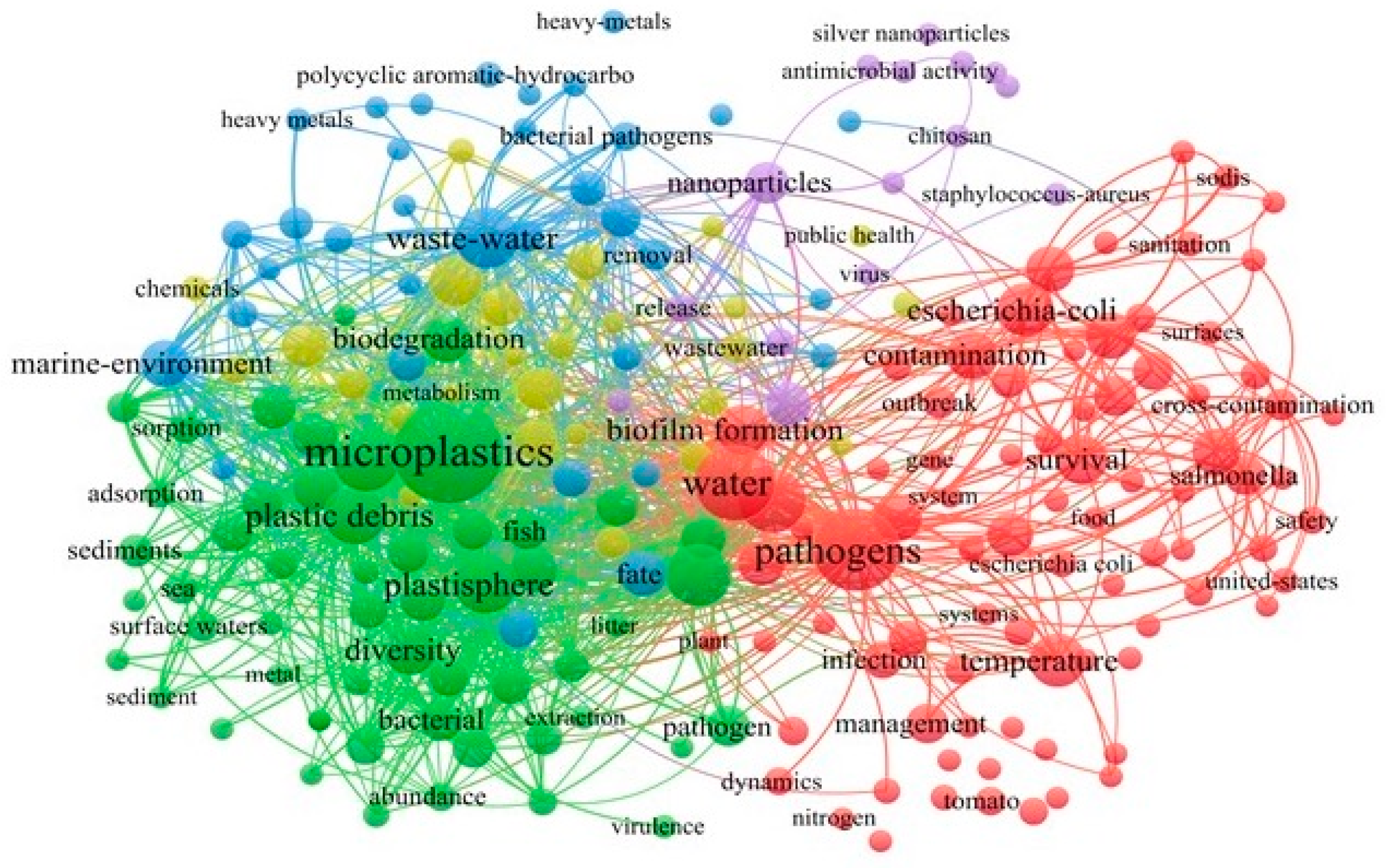
Figure 1. Keyword co-occurrence network visualization was generated by VOSviewer. Each color represents a topic cluster, where the font size and density (background color) of the keyword indicate the total link strength (TLS). A greater font indicates greater TLS, and the closer the distance between keywords, the higher the relevance of these studied topics.
Water 16 00499 g001
Figure 2. Sources and entry pathways of microplastics in groundwater.
Water 16 00499 g002
Figure 3. Mechanisms of interaction between MPs and pathogens in the subsurface environment.
Water 16 00499 g003
Figure 4. The effects of environmental factors on the interaction and co-migration between MPs and organic compounds.
Water 16 00499 g004
Table 1. Types of plastic polymers commonly found in the environment.
TypeMolecular StructureClassificationBrief
Polyethylene terephthalate
(PET)
Water 16 00499 i001
((C10H8O4)n)
Water 16 00499 i002Polyethylene terephthalate, often abbreviated as PET, is the fourth most produced polymer in the world and is commonly used in producing synthetic fibers, food, and liquid containers.
High-density polyethyleneWater 16 00499 i003
((C2H4)n)
Water 16 00499 i004HDPE, or high-density polyethylene, is recognized for its high strength-to-density ratio and is often used to produce plastic bottles and corrosion-resistant products.
Polyvinyl chloride
(PVC)
Water 16 00499 i005
((C2H3Cl)n)
Water 16 00499 i006Polyvinyl chloride, often abbreviated as PVC, is the third largest synthetic polymer produced in the world. Rigid PVC is commonly used in profile applications such as doors and windows, while flexible PVC is used for insulating cables, rainwear, and inflatable products.
Low-density polyethyleneWater 16 00499 i007
((C2H4)n)
Water 16 00499 i008Low-density polyethylene is one of the world’s most widely produced plastics with low tensile strength and high elasticity. Its most common use is in plastic bags and films.
Polypropylene(PP)Water 16 00499 i009
((C3H6)n)
Water 16 00499 i010Polypropylene is a chemically resistant material and is the second most produced polymer after polyethylene. It is used in a wide range of applications including medical, packaging, and industrial.
Polystyrene
(PS)
Water 16 00499 i011
((C8H8)n)
Water 16 00499 i012Polystyrene is one of the most frequently used polymers, with an annual production capacity of millions of tons. Its primary applications include protective packaging, disposable dinnerware, and model construction kits.
NylonWater 16 00499 i013Water 16 00499 i014Nylon, also known as polyamide, was the first synthetic fiber in the world to have extraordinarily high abrasion resistance. It works in several applications including fabrics and wear-resistant components.
Styrene block copolymersWater 16 00499 i015Water 16 00499 i016SBCs are a thermoplastic elastomer family. It has qualities comparable to natural rubber and offers high elongation, processability, and environmental stability, making it an important raw material for toys, furniture, medical, and automotive parts.
Disclaimer/Publisher’s Note: The statements, opinions and data contained in all publications are solely those of the individual author(s) and contributor(s) and not of MDPI and/or the editor(s). MDPI and/or the editor(s) disclaim responsibility for any injury to people or property resulting from any ideas, methods, instructions or products referred to in the content.