Review

Recent Advancements in Developments of Novel Fluorescent Probes: In Cellulo Recognitions of Alkaline Phosphatases

by
Madhusudan Dasnur Nanjappa
1,
Anup Pandith
2,*,
Svetlana Sankaran
1,
Dorothy Priyanka Dorairaj
3,
Anusha Anjaneya Reddy
4 and
Hari Prasad Badubanahalli Ramesh
5
1
Department of Chemistry, University of Mysore, Manasagangotri, Mysuru 570005, India
2
College of Biomedical Engineering, Taipei Medical University, Taipei City 11031, Taiwan
3
Department of Chemistry, National Institute of Technology, Tiruchirapalli 620015, India
4
Department of Chemical Engineering, National Institute of Technology, Surathkal 575025, India
5
Department of Chemistry, JC Technical Campus, JSSS & T University, Mysuru 570006, India
*
Author to whom correspondence should be addressed.
Symmetry 2022, 14(8), 1634; https://doi.org/10.3390/sym14081634
Submission received: 12 July 2022 / Revised: 1 August 2022 / Accepted: 2 August 2022 / Published: 9 August 2022
(This article belongs to the Special Issue Medicinal Chemistry: Topics and Advances)

Abstract

:
Alkaline phosphatase (ALP) is one of the vital phospho-ester bond cleaving biocatalysts that has inevitable significance in cellular systems, viz., early-stage osteoblast differentiation, cell integrity in tissues, bone mineralization, cancer biomarker, liver dysfunction, cellular osmotic pressure, protein folding and many more. Variation from optimal levels of ALP in intra and extracellular fluids can cause severe diseases, including death. Due to these reasons, ALP is considered as a vital biomarker for various preclinical and medical diagnosis. Fluorescence image-based diagnosis is the most widely used method, owing to its simplicity, robustness, non-invasive properties and excellent spatio-temporal resolution (up to the nM/pM level), as compared to conventional analytical techniques, such as the electroanalytical method, nuclear magnetic resonance (NMR) and high-performance liquid chromatography (HPLC). Most of the reviews reported for ALP’s recognition in the literature scarcely explain the structurally related, photophysical and biophysical parameters; and the sub-cellular localizations. Considering these facts, in order to enhance the opto-analytical parameters of fluorescence-based diagnostic materials at the cellular level, herein we have systematically documented recent developments in the opto-analytical capabilities of quencher-free probes for ALP, used in in vitro (biological buffers) to in cellulo conditions, along with in vivo models.

Graphical Abstract

1. Introduction

Alkaline phosphatase (ALP) is the vital Zn containing glycoprotein, generally expressed on the cell membrane and utilized for the hydrolysis of phosphorylated organic/inorganic molecules preferably at neutral to basic pH levels [1]. ALP molecular weight generally falls between 70,000 and 200,000 Da with an isoelectric point range from pH 5.4 to 6.0, and shows a high hydrolysis rate at pH 8–9 (weak alkaline conditions) [2]. Most of the biologically originated phosphorylated molecules, such as nucleotides, alkaloids and proteins, can be dephosphorylated in the presence of ALP [3]. ALP belongs to one of the ectophosphatase (kinase/phosphatase) enzymes, which generally control the extra and intracellular phosphates’ homeostasis in cells and tissues, and have significant roles in embryogenesis, bone mineralization, vascular calcification, terminiotoxin detoxification, inflammatory responses and neuronal functions [4,5]. Deviations from the optimal level of ALP in serum can lead several diseases, such as hepatitis [6]; prostate, ovarian and breast cancers [7]; bone cancer [8]; diabetes [9,10]; Paget’s disease [11]; bile duct obstruction [12]; blood malignancies [13]; leukemia; and Wilson’s disease [14]. Due to these reasons; ALP is regarded as an important indicator in clinical and medical diagnostics [15,16,17]. Recently, it has been found that children and pregnant women’s sera have as much as 500 U/L of ALP [18]. In contrast, normal healthy adults can show 35–160 U/L of ALP in their sera [19], depending on age and other biological factors.
ALP can be found in liver, kidney, bone, intestinal epithelium, lung, leukocytes and placental cells [20]. Depending on the origin, ALPs are classified as intestinal ALP (intestinal absorption), placental ALP, bone (mineralization) ALP, germ cell ALP, blood ALP (usually originating from chromosome 1) and tissue-non-specific ALPs (generally originating from chromosome 2). Irrespective of their origin, almost all ALPs typically exhibit common physiological properties in different locations.
In general, mammalian ALPs are zinc-containing metalloenzymes, functioning as dimeric biomacromolecules bearing two Zn2+ and one Mg2+ in their active sites [21,22]. Apparently, the two Zn ions are separated by approximately 4.0 A° units, bound by His331, His412, Asp327, Asp369 and His370 amino-acid residues [23]. Asp51’s carboxylate group can act as a bridge connecting Zn and Mg ions. Usually, pyrophosphates (PPi) are encapsulated in the pocket of the two Zn ions and additionally stabilized through the hydrogen bonding with guanidinium units of Arg166 residues [24,25,26]. Thermodynamically favorable nucleophilic attack of serine 102 (pKa 5.50) on the phosphorous center of the PPi-bound complex, in the active center, is the driving force for the phosphate-ester hydrolysis (Scheme 1). It is worth mentioning that Zn and Mg ions are vital metal ions responsible for proper biocatalytic activity of ALP, generally operating through the synergistic regulations between the two active subunits. ALPs can be found in both prokaryotes and eukaryotic cell lines; however, due to the higher significance of mammalian ALPs in biological systems, such as cellular signal transductions, cell division, cell differentiation and bone calcification, in the current review we highlight only mammalian ALPs and their in cellulo recognition, along with in vivo models [27,28] based on fluorescence (bioimaging) methods.
Based on ALP’s wide distributions in biological systems, such as in saliva, urine, synovial and cerebrospinal fluids, ALP is considered a vital biomarker in medical diagnostics [29,30]. Therefore, recognition of ALPs either in intra or extracellular fluids and in tissues with a rapid and accurate method is currently in high demand in biomedical science. Hence, various bioanalytical tools have been used for the recognition of ALPs in in cellulo and in vitro models, such as electro-analytical methods [31,32,33,34], surface enhanced Raman scattering [35,36], Northern blotting [37,38], enzyme linked immunosorbent assay (ELISA) [39], radioimmuno assays [40,41], quartz crystal microbalance [42,43], field effects transistors (FET) [44], colorimetry [45,46,47], magnetic resonance imaging [48] and fluorescence methods [49]. Amongst them, fluorescence methods are considered as far superior owing to their simplicity, cost-effectiveness and high-spatio temporal resolution at the molecular level [50,51,52]. In the last decade, various research groups throughout the globe have actively been involved in the development of novel fluorogenic probes for ALP recognition in both in vitro and in vivo models [53,54]. However, based on our knowledge, there are no reports which document ALP recognition alone in cellular or in vivo models. Considering the broad literature of molecular diagnostics, especially regarding fluorescence methods and in vivo models, in the current review we exclusively document the use confocal fluorescence imaging techniques for ALP recognition at the cellular level, along with the imaging of in vivo models.

2. Conceptual Strategies for the Design of ALP Fluorescent Probes in Cellulo Recognition

Most ALP recognition strategies rely on turn-on methods of optical responses in biological fluids. It is one of the essential criteria for ideal biomolecular probes, which trigger significant switch on responses upon interactions with specific analytes in physiological conditions. Selective recognition capabilities of rationally designed probes, either in chemo-dosimetric (non-reversible, chemical changes) or reversible (non-covalently held) types, substantially induce perturbation in the electronic/rotational/translational energy levels of fluorescent units. There are several photophysical properties that have been documented in the literature, such as photoinduced electron transfer (PET), excited state intramolecular proton transfer (ESIPT), intramolecular charge transfer (ICT), C=N isomerization inhibitions, twisted intramolecular charge transfer (TICT) and aggregated induced emission (AIE) mechanisms, which were exploited in the design of various types of ALP probes [55,56,57,58].
It is evident that most of the reported ALP probes specially designed for cell imaging studies are constructed based on conjugations of fluorescent reporters (fluorophore part), linkers and receptor units. Linkers usually help to hold the fluorophore moiety and receptor unit together without causing steric hindrance in the biocatalytic reaction site. Additionally, receptors (for phosphatase its phosphoester link) help to recognize the biocatalytic reaction sites within the biomolecules in question. Pertaining to the conjoining of these concepts, various ALP probes have been designed through the conjugation of fluorescent cores with phosphoester moieties either directly (without linker) or indirectly (with linker). Given the size of a fluorophore, in order to avoid steric hindrance at the dephosphorylation reaction site, most fluorescent reporters have been constructed with a structurally flexible linker. To achieve the best site-specific diagnostics, sometimes ALP probes are associated with targeting components/functional groups. Depending the requirements, subcellular localization (mitochondria, lysosome, endoplasmic reticulum, etc.) of probes will be regulated by incorporating conventional or non-classical targeting functional groups. The photophysical properties of the designed fluorescent materials depend on core unit, substituents and molecular geometry. Conventionally, symmetric and asymmetric molecular structures substantially regulate the photophysical properties of the fluorophoric units through the cumulative dipole moments of each bond, along with their transitional frequencies (energies) within the molecules. With careful consideration of these facts, various fluorescent probes with peculiar absorption and emission behavior have been designed to recognize biomolecules. Differently, based on a complexation and decomplexation strategy, metal complexes have been used to detect ALP activity [59,60,61,62]. In this strategy, ALP is indirectly monitored based on the PPi concentration in in vitro digestion assay protocols. Initially, PPi selective fluorescent ligand-metal complexes are prepared; then, metal ions are extruded from the fluorescent ligands by the sequential addition of PPi ions. Eventually, the optical properties of the fluorescent ligand are regulated based on complexation and decomplexation processes in a sequential manner. Upon performing the PPi digestion in the presence of ALP, the residual amount of PPi in the reaction mixture is monitored. Residual PPi in the reaction mixture for ALP activity has been monitored through opto-analytical methods (UV–Vis and fluorescence methods). It is worth mentioning that most efficient ALP probes are associated with the systematic conjugation of the phosphorylated terminus through the self-immobilized moiety (linkers) to the fluorescent reporter, in association with subcellular organelles targeting functional groups, such as mitochondria, lysosomes and the Golgi apparatus, in a sterically free environment (Figure 1). In contrast, indirect methods, such as the use of PPi-regulated organometallic complexes and nano-material-based techniques, could be more effective in in vitro diagnostics rather than in vivo ALP model-based recognition. If these materials are designed rationally, considering site-specific qualitative and quantitative tracking of phosphatase substrates (PPi, nucleotides, etc.), they may provide novel platforms for a future generation of fluorescent materials for ALP activity monitoring based on the indirect approach. Furthermore, this strategy using both the phosphatase and its substrates could be helpful to elucidate the disease states in a biological system at the molecular level.

3. Small-Molecule-Based Fluorescent Probes

Small molecular probes are always advantageous compared to labelled methods, where biomolecules such as peptides and nucleic acids are generally covalently attached to the fluorophores [63,64,65,66,67]. In this labelling approach, where biomacromolecules are site specifically modified with fluorescent labels, usually suffers from various drawbacks—viz., susceptibility to cellular ions, proteases and nucleases; and always demanding transfecting agents to enter the cells. In contrast, small molecules can enter the cells through passive modes without getting degraded by proteases or nucleases [68,69,70]. Furthermore, small molecules are usually not accompanied by quenching units (such as BHQ) and can have a ability to trigger dual emission (AIE-based materials monomer ↔ excimer) depending on the molecular confinements. Considering these facts, various small molecule based blue/green/red/NIR emissive fluorescent materials were designed for the recognition of ALP.
Zhang et al. reported phosphorylated tetraphenylethylene-based, AIE-based fluorescent probes ALP-1, ALP-2 and ALP-3 [71] for monitoring of ALPs during osteogenic differentiation of stem cells (Figure 2). In these probes, hydrophilicities, were tuned by incorporating mono-, di-, and tetra-phosphate units at the periphery, resulting in a very low quantum yield in aqueous conditions. Probes showed characteristics λmax at ~338 (±4) nm and λem at 460 (±10) nm in UV–Vis and fluorescence spectra, respectively. In contrast, upon incubation with ALP, the probes showed a turn response within 5 min, at the bluish green region centered at λem = ~470 nm, which was blue shifted to λem = ~450 nm after 60 min.
This substantial blue shift, attributed to complete hydrolysis of phosphorylated probes into insoluble hydroxylated tetraphenylethylene units, resulted in highly ordered aggregates. An ALP-regulated switch on response was found to be highest in the case of ALP-2, followed by ALP-4 and ALP-1, under physiological conditions. In contrast, mono-phosphorylated probe ALP-1 was found to be very sensitive compared to di- and tetra-phosphorylated probes in similar experimental conditions. The authors speculate that the presence of multiple phosphorylated units in ALP-3 induced a complex mechanism of dephosphorylation with ALP, resulting in low sensitivity. Due to these reasons, even though di-phosphorylated probe showed a higher enhancement ratio upon interaction with ALP, the lowest detection limit was found for the mono-phosphorylated probe. Stoichiometrically, at the specified concentration, the mono-phosphorylated probe has quantitatively half of the di-phosphorylated one’s units. Only mono- and di-phosphorylated probes were analyzed in biological samples because of their higher sensitivity and stability.
Cytotoxicity studies analyzed through propidium iodide revealed that both ALP-1 and ALP-2 can be used effectively at 2 to 20 μM. Upon incubation of 20 μM of probe with ALP-1/2 during osteogenic differentiation in a bone marrow mesenchymal stem cell (BMSC) culture (0 to 7 days), fluorescence intensities were increased in the green channel, and can be visualized through fluorescence confocal laser scanning microscopic images (Figure 3). Overexpression of ALP during osteogenic differentiation of stem cells was further supported through the flow cytometric Western blotting and rt-PCR analysis. It was found that, in cellular conditions, the di-phosphorylated probe ALP-2 showed significant changes compared to that of the mono-phosphorylated counterpart.
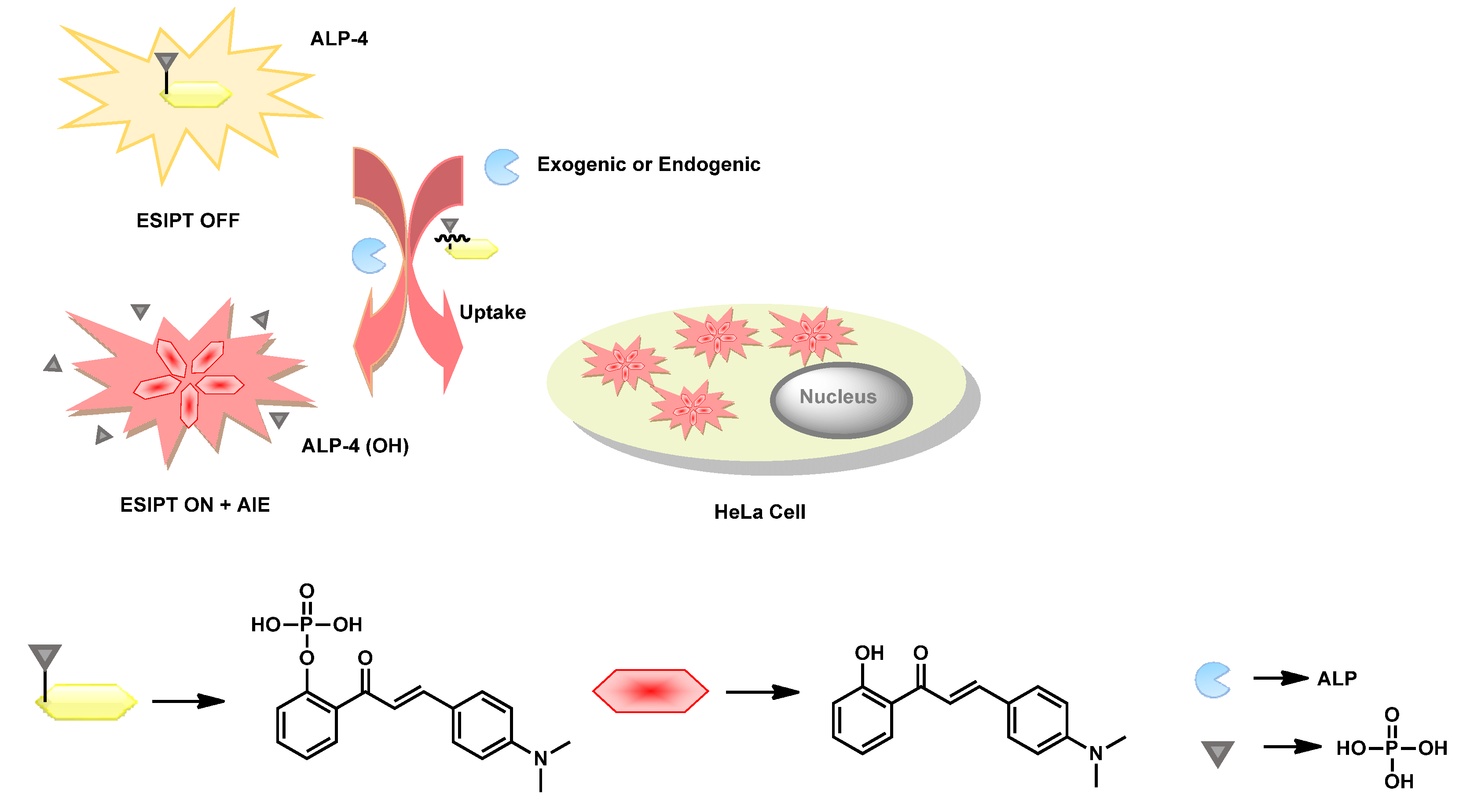
Upon exploiting the AIE properties in combination with ESIPT behavior, Tang et al. reported a 2′-hydroxychalcone-based phosphorylated probe ALP-4 (Figure 4) for ALP recognition in in vitro and in cellulo [72]. The rationally designed probe triggered far-red emission centered at 640 nm upon ALP-catalyzed dephosphorylation under physiological conditions. ALP-4 showed absorption maxima at λmax 430 and 416 nm in UV–Vis spectra and weak emission (greenish-yellow λem 538 nm). This weakly green emissive behavior in the phosphorylated state was attributed to the presence of N,N,dimethylaniline and vinyl ketone units, thereby creating an ICT process in the chalcone unit in the twisted conformation. However, upon dephosphorylation, the presence of the ketone unit at the ortho position induced strong intramolecular hydrogen bonding upon photochemical excitation, leading to the highly stable 6-membered ring, resulting in keto-enol tautomerism. Based on pH-dependent study, the authors speculated that fluorescence properties in the probe should be attributed to the combination of synergistic ESIPT and ICT processes. Single crystal analysis of ALP-4 (OH) revealed head-to-head and edge–face stacking resulted in strong intermolecular-coupling-aided AIE behavior. Exploiting such dual photophysical properties in phosphorylated and dephosphorylated 2-hydroxychalcone resulted in ratiometric fluorescence switching properties in green and red channels. The probe detected ALP as scarce as 0.15 mU/mL in a ratiometric manner. The authors did not specify the kinetic parameters in their work.
ALP-4 showed excellent biocompatibility towards HeLa cell lines. Upon incubation of ALP-4 in endogenously overexpressed HeLa cells, orange fluorescence was seen in the cytoplasm (Figure 5). The authors speculated on the existence of unusual blue-shifted emission in the orange region in cellular conditions, as compared to that of in vitro conditions (red emissive), could have been due to the twisted molecular confinements of ALP-4 (OH) upon interactions with bio-macromolecules in biological fluids. To validate the intracellular phosphate hydrolysis of ALP-4, fluorescence confocal imaging studies (FCIS) were performed in dual channels in the absence and presence of the ALP inhibitor levamisole. According to the results, the authors postulated that intercellular ALPs readily cleaved the phosphorylated probe in extracellular fluid, and thereafter the fluorescent ALP-4 (OH) was transfected passively, resulting in bright fluorescence in the cytosol (Figure 6).
Sun et al. and co-workers reported the naphthalene-based green emissive two-photon florescent probe ALP-5 (Figure 7) for in vitro and in cellulo recognition of ALPs [73]. A PO43− group was incorporated into the self-cleavable benzyl-ester unit at the ortho position in such a way that, upon dephosphorylation, it could trigger the release of an amino-naphthalene moiety. Under physiological conditions, the probe showed absorption maxima at λmax 300 nm in UV–Vis spectrum and emission maxima centered at λem 450 nm in the fluorescence spectrum. Upon dephosphorylation in the presence of ALP, the absorption and emission maxima shifted to 365 and 500 nm respectively. Such selective bathochromicity in the absorption spectrum, along with a switch on response in the green channel, made the authors interested in performing ALP recognitions in biological systems.
The phospho-ester hydrolysis kinetics of ALP-5 was analyzed through the well-established Lineweaver–Burk equations, revealing appreciably good Km (8.56 µM) and Vmax (0.073 µM) values. Under the specified experimental conditions, the probe showed excellent sensitivity of up to 0.3 U/L towards ALP, with a good linearity range from 0.1 to 10 U/L. The authors speculated that the ortho-substituted phospho-phenolic ester hydrolysis by phosphatase thermodynamically favors cascade cleavage of the para-substituted amidic linkage in 2-acetyl-naphthalene. This process restoring the strong ICT process between the amino and acetyl substituents across the naphthalene’s core resulted in the bright green fluorescence. Two-photon cross-section studies of ALP-assisted dephosphorylated product showed 90 GM at 740 nm. In contrast ALP-5’s two-photon cross-section in similar experimental conditions was found to be too small. Due to these reasons, one-photon and two-photon excitations showed similar emission spectra under physiological conditions.
Upon detailed in vitro analysis, two-photon ALP-5 probe cytotoxicity was evaluated in HeLa cell models, based on MTT assays. Upon incubation of the probe in HeLa cells, it displayed strong intracellular green fluorescence, due to two-photon excitations (λex 720 nm). However, Na3VO4-pre-treated HeLa cells also revealed fluorescence in the green channel upon two-photon excitations (Figure 8a–d). Interestingly ALP-5 did not show any fluorescence in HEK cell lines, even at higher concentrations, revealing that the two-photon probe was able to recognize ALP in a cell-dependent manner (Figure 8e,f).
Two-photon imaging studies of ALP-5 were performed in various live tissues, such as the hippocampi, livers and kidneys of rats at 30 to 230 μm depth levels. ALP-5-incubated tissues showed bright green fluorescence within 120 min in all tissues, which was not observed in the presence of the inhibitor Na3VO4 (Figure 9). This validated successful tracking of endogenous ALP in tissues and organs. Due to the green emission range, the in vivo recognition capability of the probe was not demonstrated.
Nie et al. developed a new near-infrared, emissive, hemi-cyanin-based fluorescent probe, ALP-6 (Figure 10), for in vitro and in vivo recognition of endogenous ALP activity [74]. ALP-6 involves phosphorylated chromene units conjugated with benz[e]indolium salts. In buffered conditions, the probe showed an absorption maximum at λmax at 604 nm and was very weakly emissive (λem 738 nm). Upon incubation with ALP at roughly physiological pH, 8.0, there was a 10-fold enhancement (at 738 nm) in the emission intensity, along with a substantial redshift from 604 to 738 nm in the UV–Vis spectra within 20 min. The designed hemicyanine-based probe showed a highly selective and robust switch on response in the NIR region, towards phosphatase, without allowing any interference by biologically relevant ions or macromolecules. The ALP-induced dephosphorylation of ALP-6 retained a strong ICT process, resulting in high fluorescence with redshifts in the UV–Vis spectrum, which were significantly inhibited by the phosphorylated probe due to the electron pulling effects from the electronegative phosphorous atom in the PO43− unit.
Probe ALP-6’s dephosphorylation kinetic parameters, Vmax and Km, were 0.693 μM/min and 9.32 μM, respectively, under similar experimental conditions. The probe’s LOD towards ALP was found to be 0.003 U/mL (linearity range from 0.01 to 2.0 U/mL). In detailed in vitro studies, ALP-6 was tested in HeLa cells. The MTT assay revealed the probe was negligibly toxic until 30 μM. Upon incubation of APL-6 in ALP-overexpressing HeLa cells, bright fluorescence in red channel was exhibited, but it was greatly diminished in the presence of the inhibitor Na3VO4, as can be seen in the fluorescence confocal images (FCIMs) (Figure 11a–c). Fluorescence signals in rat liver tissues collected through Z-scan mode revealed that ALP-6 has excellent tissue penetration capabilities (Figure 11d). Due to its excellent tissue penetration capabilities, the probe’s in vivo ALP recognition capabilities were demonstrated in Kunming mice. Upon intraperitoneal injection of ALP-6, mice produced deep red NIR emissions over time. Such emissive behavior was not observed in the Na3VO4-pretreated mice, which revealing that the probe can be effectively used to track endogenous ALP in in vitro and in vivo models.
Liang’s group reported fluorescent peptide ALP-7 (Figure 12) for the in vitro recognition of ALP. The designed FITC-conjugated, phosphorylated peptide undergoes dephosphorylation in the presence of ALP, leading to the three-dimensional, self-assembled, fibrous-type material—a “gel” [75] at pH 8.0 (TRIS-HCl). Enzyme-assisted sol to gel transformation resulted in quenching of fluorescence in the green channel attributed to conventional aggregation caused quenching (ACQ). The authors did not specify kinetic parameters such as Vmax and Km. ALP-7 showed strong green emission upon illumination at 365 nm (UV light), whereas upon incubation with ALP, in physiological pH buffers, a substantial decrement in emission intensity in the green channel was observed. Concomitantly, a significant color change from light green to transparent yellow was clearly seen with the naked eye. Probe was able to recognize ALP as lowest as 0.06 U/mL in similar experimental conditions.
MTT assay against human colorectal cancer (LoVo) cells did not show any cytotoxic behavior (99% cell survival was observed) until 160 μM, after a 12 h incubation. ALP overexpressed LoVo cells showed very weak fluorescence upon incubation with ALP-7, whereas in the presence of the inhibitor L-phenylalanine (L-Phe), they showed bright greenish fluorescence (Figure 13a). From these studies, the authors speculate that FITC-conjugated peptides, while dispersed in biological fluids, can cause bright greenish fluorescence. However, upon dephosphorylations with the aid of ALP, they resulted in the formation of a supramolecular assembly, causing significant ACQ. Such drastic variations in emission intensity in the green channel helped with the qualitative and quantitative recognition of ALP in in vitro and in cellulo models.
Tan et al. reported a hemicyanine-based NIR based fluorescent probe, ALP-8 (Figure 14), for in vitro and in vivo recognition of ALP [76]. In buffered conditions, the probe showed absorption maxima at λmax 600 and 650 nm in the UV–Vis spectrum, and did not induce any fluorescence band in the NIR region. Upon dephosphorylation, absorption maxima λmax were red-shifted to 650 and 680 nm, elicited with bright NIR fluorescence centered at 700 nm. They observed blue shifts in the absorption and emission wavelengths compared to ALP-6 (benz[e]-indolium core), revealing that indolyl core’s conjugation extension can significantly affect the ICT process in hemicyanine-based fluorophore units.
ALP-8 showed a detection limit of 0.07 U/mL for phosphatase, without inducing interference from structurally similar biomolecules and cell metabolites (cations, anions and other molecules). The observed dephosphorylation kinetic parameters, such as Km and Kcat, were found to be 21.2 μM and 2.14 S−1 respectively. The catalytic efficiency (Kcat/Km) value of 1.01 × 10⁵ M−1 S−1 is much higher than that of commercially available 4-methylumbelliferyl phosphate and enzyme labelled fluorescence substance 97 (ELF-97).
The intracellular ALP-recognizing capabilities of ALP-8 were evaluated in ALP-positive (HeLa cells) and negative (HEK 293) cell models. The ALP-positive HeLa cells showed a strong fluorescence pattern, whereas the ALP-negative HEK 293 cells exhibited negligible fluorescence (Figure 15A) upon ALP-8 incubation. When ALP-8 was incubated in Na3VO4 (a well-known ALP inhibitor)-pretreated cells, low emission was exhibited in both cell lines in the red channel, supporting the probe’s capability of successfully tracking endogenously produced ALP. Intraperitoneal injection of ALP-8 in Kunming mice in the absence and presence of Na3VO4 inhibitor revealed bright and low emissive behavior, respectively (Figure 15B,C). From these results, the authors speculated that bright NIR emission from cell and mouse models can be attributed to endogenously produced ALP-regulated dephosphorylation of probe.
Wei et al. reported a highly efficient, NIR-regulated far-red probe, ALP-9 (Figure 16) based on the dicyanomethylene-4H-chromene unit conjugated with dichrolorophenyl phosphate ester [77]. To achieve best possible tumor microenvironment (TME) localization and controlled time-resolved intracellular ALP imaging, ALP-9 was loaded into am NIR-responsive nano-container.
The rationale behind this design is that incorporation of dichlorination at the ortho position via phenolic OH enhances the biocompatibility, through the high degree of dissociation (pKa 6.97), which is much lower in the case of its mono-chlorinated (pKa ~ 8.43)/non-substituted (pKa ~ 9.85) counterpart. Additionally, nano-encapsulated-ALP-9 showed a peculiar absorption maximum at λab ca. ~380 nm, and was weakly emissive (λem ca. 683 nm); thus, the debilitated ICT process (dicyanomethylene as the acceptor and the phosphophenolate unit as the donor) operated between π-bridged acceptor–donor couples. ALP-regulated biocatalytic dephosphorylation of ALP-9ALP-9 (OH) exhibited a new peak at ca. ~510 nm and strong emission at ca. 683 nm (~15 fold) in UV–Vis and fluorescence spectra, respectively. Such dramatic changes in the optical behavior of the dephosphorylated probe were attributed to the presence of a strong ICT process operating between the dechlorinated phenolate unit and dicyanomethylene across the styryl unit. Dephosphorylation kinetic parameters Km and Vmax were found to be 13.58 µM and 0.083 µM/s, respectively. The lowest fluorogenic response towards ALP was 0.072 U/L under physiological conditions. In order to demonstrate the time-resolved ALP recognition, ALP-9 was loaded into NIR-responsive nano-containers consisting of a lanthanide core (NaYF4:Yb/Tm@NaYF4) and a mesoporous silica layer decorated with an azo compound. In this strategy, ALP-9 molecules loaded in the nano-container were selectively released, upon irradiating by NIR light, by up-converting it to UV–Visible light. During the up-conversion, the generated UV–Visible light induced trans to cis transition in the azo compound, thereby regulating controlled release of the phosphorylated probe from the nano-container.
HeLa cells and L02 cell lines were chosen for this study due to their up- and down- regulation of ALPs levels. When the cells were initially incubated with the nano-ALP-9, no obvious fluorescence was seen, but upon irradiation with NIR, the pre-treated HeLa cells exhibited very strong fluorescence, indicating that ALP-9 was efficiently released, followed by subsequent recognition of endogenously produced ALP. However, in the presence of the ALP inhibitor Na3VO4, fluorescence in the red channel enormously decreased, even upon NIR irradiation (Figure 17). Similarly, ALP downregulated L02 cells also exhibited extremely weak emission in the red channel, even in the presence of NIR irradiation. Based on the observed results, the authors speculated that generation of red mission in the cell lines solely resulted from phosphatase-catalyzed dephosphorylation of the probe.
Since the in vitro studies showed remarkable results, the efficiency of the nano-encapsulated probe was further studied in Kunming mice, as an in vivo model. Results revealed that the nano-ALP-9 impregnated mice showed red fluorescence only upon irradiation by NIR light. In contrast, mice treated with the ALP inhibitor (Na3VO4) with or without NIR irradiation did not exhibit any fluorescence (Figure 18). Further, HepG2/xenograft tumor-bearing nude mice were chosen to recognize the ALP activity in acidic tumor environments in in vivo models. Interestingly, intratumoral injection of the nano form of the ALP-9 probe showed excellent ALP recognition, especially in the tumor microenvironment upon irradiation by NIR radiation.
Ding et al. reported a novel D–π–A-tuned mitochondria-targeting fluorescence probe (ALP-10) whose emission properties are regulated through the ESIPT phenomenon [78]. Molecular engineering was performed by the combination of 2-(2′-hydroxyphenyl)-benzothiazole (ESIPT core) with a 4-styryl-pyridinium (robust mitochondrial targeting unit) core (Figure 19). ALP-10 showed a characteristic broad absorption peak with maximal absorptivity at ca. ~358 nm and an emission maximum at 514 nm (ϕ = 0.21) in UV–Vis and fluorescence spectra, respectively. Upon ALP induced dephosphorylation, the free phenolate unit induced a strong ESIPT process in ALP-10 (OH), resulting in the generation of a new absorption band (λmax ca. ~510 nm) and emission band (λem ca. ~650 nm). The dephosphorylated product with free phenolic OH emitted a strong red fluorescent signal in a ratiometric manner. The ALP recognition capability of the probe was found to be appreciable in the physiological pH range (5.0 to 8.0). However, at an alkaline pH (pH > 8.0) the ratiometric ALP recognition capability was significantly diminished, which was attributed to inhibition of the ESIPT process pertaining to phenolate ion formation.
The dephosphorylation kinetic parameters Km and Vmax were 1.48 µM and 0.63 µM/s, respectively, under in vitro diagnostic conditions. In physiological conditions, ALP-10 did not show any fluorescence instability due to biologically relevant ions or molecules, and showed a minimum detection limit of 0.072 mU/mL towards phosphatase.
The endogenous ALP activity of the probe was monitored in ALP positive cells, such as HeLa, A549 and ALP-negative HUVEC cell lines. ALP-10-treated HeLa and A549 cell lines exhibited bright red fluorescence with complete disappearance of emission intensity in the green channel after 30 to 60 min (Figure 20). In the HUVEC cell lines, only weak green fluorescence was observed, due to the downregulation of endogenous ALP. Colocalization experiments with mito-tracker green showed a Pearson’s correlation coefficient of >0.96, further supporting the idea that ALP-10 can recognize mitochondrial ALP.
ALP inhibition was monitored in HeLa cells with Na3VO4 and NaH2PO4. Disregarding the control experiments (without inhibitor), the remaining HeLa cell lines (with inhibitor) showed dose-dependent red and green fluorescence upon incubation with ALP-10 (Figure 21). Additionally, at a low concentration of inhibitor, the probe incubated cells’ green emission did not completely disappear. From these experiments, the authors speculated that the red emission in cells should be attributed to phosphatase-regulated dephosphorylation of ALP-10.
The in vivo ALP recognition capabilities of the probe were tested in tumor-bearing BALB/C nude mice. Upon intratumoral injection of ALP-10 into the mouse model, a time dependent enhancement in the red signal, along with a substantial intensity decrement in the green channel, was observed. In contrast, when Na3VO4-pretreated tissue was injected with ALP-10, mice exhibited their lowest red emission intensity in the tumor (Figure 22) and these results are consistent with in vitro (cell imaging) experiments.
Han et al. reported the 2′-(2′-hydroxyphenyl)-benzothiazole (HBT) fluorescent conjugates (Figure 23) were linked to the 5′ position of the ribonucleoside (adenosine/guanosine) residues for recognition of ALP in cellular conditions [79]. These 5′-ribonucleotide-fluorophore analogues showed weak emission, due to the phosphorylated phenolic OH in HBT unit. P–O bond cleavage (dephosphorylation) generates free HBT and phenolic OH, which exists in a keto–enol tautomerization equilibration. Upon photochemical excitation, the ESIPT core equilibrium shifted toward the enol tautomer (more predominant isomer), resulting in red shifts in the emission spectrum. ALP-11/12 are non-fluorescent in aqueous buffer, but otherwise have three emission maxima, 370, 375 and 380 nm. Similarly, these probes exhibited a bright fluorescent signal centered at 512 nm in the presence of ALP.
Due to the presence of two phosphoester linkages in ALP-11/12 probes, they showed two hyperbolic peaks. The apparent Km values were 1.89 and 238.6 μM for adenosine conjugates (ALP-11) and 0.1 and 65.26 μM for guanosine conjugates (ALP-12). Vmax values for ALP-11 were 9.8 × 10⁵ and 1.64 × 10⁷ pmol min−1 nmol−1. Similarly, for ALP-12, Vmax values were 3.48 × 10⁵ and 5.33 × 10⁶ pmol min−1 nmol−1, revealing that guanosine conjugates have higher affinities towards ALP in similar experimental conditions.
The cytotoxicity of ALP-11/ALP-12 probes assessed by sulforhodamine B (SRB) assays showed a small cytotoxic effect in both HeLa cells and HT29 cells. It was also observed that ALP-12 has less toxicity than ALP-11. Due to the upregulated placental ALP in the HeLa cells upon ALP-11 incubation, green fluorescence intensity in the intracellular region was gradually increased until 30 min had gone by. (Figure 24). By contrast, no fluorescence was seen in ALP-downregulated HT-29 cell lines. In cellular conditions, the adenosine analogue (ALP-11) showed better fluorescence in response to endogenous ALP than the guanosine conjugate (ALP-12).
Kim et al. reported a new iminocoumarin-benzothiazole-based fluorophore, ALP-13 (Figure 25) to recognize the phosphatase at the single-cell level [80]. In an aqueous buffer solution, probe showed an absorption maximum of 472 nm and emission maximum (λem) of 542 nm, in the UV–Vis and fluorescence spectra, respectively.
ALP-13 was very weakly emissive (ϕ = 0.002) in aqueous conditions, which was attributed to free rotation across the vinyl unit. Upon phosphor-ester hydrolysis, the ortho-substituted OH group undergoes rapid cyclisation by the nucleophilic attack on the C atom of the nitrile unit, which thereby inhibits free rotation of the vinyl units. Such planar arrangements induce hypsochromic changes (ϕ = 0.10) in the emission maxima and bathochromic shifts in the absorption maxima. The ALP-13 enzymatic dephosphorylation biocatalytic kinetic parameters were found to be KM = 19.2 µM and kcat = 0.27 s−1. The enzymatic efficiency of ALP-13 was measured as kcat/KM = 1.4 × 10⁴ M−1 s−1, which is moderate compared to that of commercially available phosphatase probes under similar experimental conditions.
Intracellular the ALP recognition capability of ALP-13 was demonstrated in HeLa cells (human cervical carcinoma) as a positive control and HT29 cell lines as a negative control due their up and downregulation of ALP levels, respectively. ALP-13-incubated HeLa cells showed bright green emission within 2 min; however, such intensified green emission was not observed in the case of inhibitor levamisole-pretreated HeLa cells. Similarly, when ALP-13 was incubated with HT29 cells, no such substantial increment in the green channel was observed (Figure 26). Based on these results, the authors speculate that the imino-coumarine–benzothiazole conjugate exhibits bright green emission only upon selective dephosphorylation by the endogenously produced ALP.
Li et al. reported the self-immolative resorufin-based probe ALP-14 (Figure 27) for an ALP assay in cellular conditions [81]. Molecular engineering was performed based on the conjugation of an ALP-recognizing substrate (phosphoester unit) to a red emissive resorufin unit through the reactive p-hydroxybenzyl unit, which acts as self-immolative linker. This design is such that ALP can easily access the phosphor-ester terminus of the probe without steric hindrance, for biocatalytic dephosphorylation. When ALP-14 is dephosphorylated, it intermediately undergoes 1,6-eliminations to produce a p-quinone methide ether derivative and a free Resorufin unit. This transformation is associated with changing of the solution’s color from orange to purple and generating intense fluorescence emission centered at the 585 nm region.
In physiological conditions, ALP-14 showed a UV–Vis absorption maximum at 484 nm with a shoulder peak at 400 nm, and was very weakly emissive (λem 585 nm and ϕ = 0.0023). This weakly emissive behavior was attributed to photoinduced electron transfer (PET) from 7-hydroxy substitution (ohydroxy-benzyl) in the resorufin unit. This phenomenon was not observed upon dephosphorylation, due to the formation of the free resorufin unit.
The dephosphorylation kinetic parameters of probe, KM and kcat, were found to be 15.38 µM and 0.26 s−1, respectively. The enzymatic efficiency kcat/KM = 1.7 × 10⁴ M−1 s−1 was greatly reduced in the absence of a self-immolative linker between resorufin (signaling unit) and the phosphate (receptor) group. ALP-14 has high affinity, as compared to commercially available probes such as 4-MUP, and was very sensitive, being able detect as little as 1.09 U/L of ALP in physiological conditions. The inhibition assay revealed IC50 values for Na3VO4 and levamisole of 7.58 and 79.4 µM, respectively.
The ALP-14 probe’s intracellular phosphatase recognition capabilities were demonstrated in HeLa cells and HEK 293T cells as ALP positive and negative controls, respectively. ALP-14-incubated HeLa cells showed intense red fluorescence within 5 min, whereas under the same conditions, no visible fluorescence signals were observed inside HEK 293T cells (Figure 28). The validated emission signals in the red channel were attributed to phosphatase-catalyzed dephosphorylation of ALP-14.
Sun et al. reported a novel hydrazone-based fluorescent probe, ALP-15 (Figure 29), for the in cellulo recognition of ALP. The opto-analytical behavior of the probe is regulated through the dual photophysical phenomena, such as aggregation-induced emission (AIE) and excited state intramolecular proton transfer (ESIPT) [82]. The design strategy targeted a simple and small molecular architecture, which showed excellent selectivity and sensitivity, by showing a large Stokes shift upon interacting with phosphatase in physiological conditions.
In ALP-15, ESIPT and AIE are regulated through the ortho-hydroxy unit. In the phosphorylated state, the probe exhibited good water solubility and prevents the ESIPT and AIE phenomena. Selective blocking of the ortho-OH group in the probe did not elicit significant fluorescence in the green channel. In buffer, ALP-15 exhibited very weak emission (λem 536 nm) in the green channel. In contrast, the dephosphorylated probe with free hydroxyl groups showed bright green emissive behavior with a large Stokes shift (Δλ = 180 nm), which was attributed to synergistic ESIPT and AIE properties. Intensity-based emission profiling revealed that the dephosphorylation rate was high at pH 9. Dephosphorylation kinetic parameters Km and Vmax, calculated through Lineweaver–Burk analysis, were 7.66 µM and 0.408 µM min−1, respectively. The probe showed a limit of detection of 0.012 UL−1 towards ALP without inducing interference from biologically relevant ions or molecules.
Intracellular ALP activity was measured in various cell lines, such as MG-63, WI-38, B6F10, RAW264.7 and HEK293, using confocal fluorescence imaging. Amongst them, MG3 showed bright green emission, owing to overexpression of ALP during differentiation of osteosarcoma cells (Figure 30). Other cells, such as WI-38, B16F10 and RAW 264.7 exhibited a mild fluorescence signal due to moderate expression of ALP. However, HEK 293 cells did not show any significant fluorescence signal, due to the complete downregulation of ALP. Hence, the authors believed that depending on the amount of ALP in cells, the intensity profiles in the green channel were different in the various cell lines.
Wu et al. developed a 1,8-naphthalimide derivative, ALP-16, for real time ratiometric recognition of (Figure 31) the organ damage biomarker ALP in cells and in vivo models [83]. A phosphor-ester group was incorporated at the 4th position of the 1,8-naphthalimide core to induce dual emission (blue–green region) and incorporation of a peripheral amine-N-oxide group facilitated water solubility and biocompatibility. Upon phosphor-ester hydrolysis, the 4th position of the naphthalimide unit is transformed into free OH. This chemical transformation enormously changes the photophysical properties of the probe, causing a dramatic color change and a ratiometric signal in the emission spectra. In the biological buffer, ALP-16 exhibited an absorption maximum λmax at ca. ~375 nm, and an emission maximum at λem 468 nm in the UV–Vis and fluorescence spectra, respectively. Upon ALP-regulated biocatalytic dephosphorylation, electron deficient substituents (phosphoester) were changed to electron donating substituents (phenolic OH) at the 4th position of the 1,8-naphthalimide unit of ALP-15 (OH). Due to these reasons, significant bathochromic behavior (λmax 375 → 450 nm) was observed in UV–Vis spectra. Similarly, significant ratiometric changes were observed through the concomitant decrement at 468 nm and a significant enhancement at 554 nm (redshift) in the emission signal.
Dephosphorylation kinetics parameters calculated through the Michaelis–Menten equation revealed appreciably good values: Vmax = 1.506 µM min−1 and Km = 22.514 µM in physiological conditions. This naphthalimide-based probe showed a detection limit of 0.38 UL−1 towards ALP without inducing interference from biologically relevant ions and molecules.
ALP-16’s cytotoxicity assay was evaluated in L929 and HeLa cell lines via MTT assay. Even at high concentrations of probe, no substantial reduction in cell viability was observed in cells. HeLa cells incubated with ALP-16 exhibited the green fluorescence within 30 min (Figure 32(I)). On the other hand, due to the downregulation of ALP in L929 cells, only blue fluorescence was observed. In support to the above studies, fluorescence images collected from the levamisole-preincubated cell lines exhibited only blue emission (Figure 32(II)). Based on these results, the authors speculated that the bright green signal originated in the cells due to the biocatalytic dephosphorylation of ALP-16 by endogenously produced phosphatase.
N-acetyl-p-aminophenol (NAPA) is a drug generally used to damage zebrafish larvae organs by the upregulation of ALP. Accordingly, NAPA dose-dependent studies in zebrafish larvae exhibited bright green emission in damaged liver, stomach and intestine due to the presence of elevated ALP (Figure 32(III)). Based on these studies, the authors speculated that ALP-16 can be used to detect organ damage in in vivo models, such as zebrafish larvae.
Zhou et al. reported a naphthalene-based two-photon fluorescent probe ALP-17 [84] for the recognition of ALP (Figure 33) in a ratiometric manner. The designed probe is associated with a typical donor–π–acceptor (D–π–A) structure and is able to emit very low fluorescence in its phosphorylated state, which is attributed to inhibition of the ICT process from the naphthalene core to the benzo-thiazole unit due to the electron withdrawing properties of the phosphoester unit. However, upon dephosphorylation, strong ICT is restored, due to the resonance effects of phenolic OH (strong electron donor) in the naphthalene core. As a result, the emission band undergoes a bathochromic shift, relative to the phosphorylated structure, along with a peculiar ratiometric response.
The probe’s fluorescence intensity ratio showed a linear relationship with ALP concentration in the range of 20 to 180 U/L, and had a detection limit of 2.3 UL−1 in physiological conditions. The authors did not specify the dephosphorylation kinetic parameters in their work. In a detailed in vitro investigation, ALP-17 was used to recognize the intracellular ALP activity using one-photon (405 nm) or two-photon (720 nm) excitation techniques. The probe’s quantum yield was found to be 0.41. When excited at 720 nm, the probe has a 65 GM two-photon action absorption cross-section at 428 nm. The probe’s cytotoxicity was evaluated based on MTT assay in HeLa cells, revealing that ALP-17 did not have a toxic effect until 20 µM. During incubation with ALP-17, HeLa cells exhibited bright green fluorescence after 30 min. In contrast, probe incubated in levamisole-hydrochloride-pre-treated HeLa cells resulted in only blue emission (Figure 34), which supports that ALP-17 is able to detect intracellular ALP. The probe-incubated HeLa cells resulted in bright fluorescence images upon excitation at λex 720 nm, supporting the idea that ALP-17 is suitable for two-photon imaging as well (Figure 35).
Podder et al. developed the rhodol-based green fluorescent probe ALP-18 (Figure 36), which was used to distinguish cancer cells from normal cells based on lysosomal phosphatase expression [85]. In vitro studies revealed that the probe causes a ~9-fold increment in UV-absorption at λabs of 490 nm and a ~33-fold emission enhancement at λem 532 nm in the presence of 1.72 U mL−1 of ALP. In this probe, fluorescence emission behavior was tuned through the closed and open structure of the conventional spiro-lactam ring in the rhodol skeleton. In the phosphorylated state, the probe exists in spirocyclic form and exhibited weak fluorescence. However, upon dephosphorylation, the spiro-lactam ring will be opened, resulting in revival of rhodol fluorescence in the green channel. Dephosphorylation kinetic parameters KM, kcat and kcat/KM were found to be 7.0 µM, 9.52 s−1, and 13.6 × 10⁵ M−1 s−1, respectively. The probe did not suffer from interference in the presence of biologically relevant ions and molecules in physiological conditions.
MTT-based toxicity assays in NIH-3T3 (normal) and HeLa (cancer) cells revealed that at µM concentrations, ALP-18 did not show antiproliferative activity towards either cell line. The probe’s adhesion to HeLa (cancer) cells was relatively stronger than to normal cells. Based on a dose-dependent assay, the relative expression of ALP was examined through fluorescence imaging in both cancer and normal cells (Figure 37). When compared to HeLa cells, the fluorescence intensity in regular NIH-3T3 cells was much lower (~2 folds).
In order to evaluate the ALP-18’s sub cellular localization and inhibitory activity in HeLa cells, colocalization experiments were carried out in the presence and absence of levamisole (inhibitor) (Figure 38). Lyso tracker co-incubated cells showed bright green and non-emissive behavior in the absence and presence of inhibitor, respectively, and a good co-localization coefficient (Pearson’s coefficient 0.93) with the red channel supported that ALP-18 is primely localized in lysosomes.
Lu et al. reported ICT tuned dicyano-based fluorogenic substrate (ALP-19) for the ratiometric recognition of ALP (Figure 39) [86]. The probe showed a significant bathochromic shift (550 → 650 nm) in the emission spectrum, upon biocatalytic dephosphorylation: a substantial color change from yellow-green to light orange. In its phosphorylated state, the probe was weakly emissive due to lowering of electron densities on the phenolic oxygen, which resulted in inhibition of the ICT phenomenon in D–π–A skeleton. However, upon phosphate hydrolysis, phenolic OH enhanced the ICT phenomenon in the probe, resulting in noticeable spectral shifts in the UV–Vis and emission signals. The absorption and emission bands on the probe ALP-19 exhibited peaks at 440 nm (ε = 2.2 × 10⁴ M−1 cm−1) and 550 nm (ϕ = 10.5%) respectively. Upon dephosphorylation, a new band appeared at 650 nm (ϕ = 7.5%). It is worth noting that due to the weak emission at 650 nm, the dephosphorylated probe exhibited a low quantum yield compared to the phosphorylated form.
The probe detected ALP very selectively with a LOD of 3.8 UL−1. The authors did not specify the kinetic parameters of the probe towards ALP.
MTT-based cell viability assays showed the probe was non-toxic to HeLa cells under the μM range. Thereafter, endogenously produced ALP activity was monitored in the presence and absence of Na3VO4 (inhibitor) in HeLa cells. In the absence of Na3VO4, ALP-19-incubated HeLa cells showed bright fluorescence in the red channel. However, such bright emissive behavior was not observed in the presence of inhibitor (Figure 40). From these studies, the authors speculate that emission intensity in the red channel solely came from ALP-catalyzed dephosphorylation of the probe.
Hu et al. reported a new flavone-based fluorescent probe, ALP-20 (Figure 41) (3-hydroxy-2-(p-tolyl)-4H-chromen-4-one), for the in vitro and in cellulo recognition of ALP [87]. Tuning of photophysical behavior in the probe is attributed to inhibition and facilitation of the ESIPT process in phosphorylated and dephosphorylated states, respectively. Due to the presence of a phosphorylated phenolic unit, it is unable to undergo a tautomerization process; as a result, emission intensity is significantly quenched. However, ALP-regulated dephosphorylation induced the keto-enol tautomers through the phenolic OH group, allowing for intensified ESIPT-based emission.
The probe showed absorption and emission maxima at 335 and 480 nm in UV–Vis and fluorescence spectra, respectively. However, upon the addition of ALP, a decrement and an increment at 335 and 410 nm were observed in UV–Vis spectra, respectively. Similarly, a dramatic peak in the emission spectrum centered at λem 480 nm was observed. The dephosphorylation kinetic parameters Km and Vmax were found to be 1.919 µM and 1.21 × 10⁻³ µmol/min, respectively. Due to high affinity of the probe towards phosphatase (affinity constant of Ka 1.84 × 10⁷ L/mol), its detection limit was good, 0.032 U/L; and it showed no interference from other proteins and biologically relevant ions. The authors also demonstrated the probe’s phosphatase recognition capabilities in serum samples.
MTT-based cytotoxicity assays performed in ALP-downregulated (L929) and upregulated (HeLa) cell lines revealed that ALP-20 is biocompatible at µM concentrations. The probe-incubated HeLa cells exhibited bright green emission; however, no such bright emission was observed in L929 cells. Furthermore, upon incubation of ALP-20 in levamisole-preincubated HeLa cells did not show any emission signal in green channel, supporting successful tracking of endogenously produced ALP (Figure 42).
Liu et al. reported a hemicyanine-based NIR fluorescent probe, ALP-21 (Figure 43), for turn-on trapping of ALP activity in cancer cells and tumor-xenografted nude mouse models [88]. The probe rationale was based on regulation of the intramolecular charge transfer (ICT) principle. The phosphate group was tethered directly to the fluorophore’s phenolic OH group, thereby preventing fluorescence in the hemicyanine core by repressing the ICT process. Upon biocatalytic dephosphorylation, free phenolic OH was regenerated, thereby reviving a strong ICT process which triggered a bright emission signal in the NIR region.
ALP-21 showed absorption maxima at λmax at 590 and 635 nm in the UV–Vis spectrum in buffer. However, upon addition of ALP, a substantial red-shift was observed (635 → 680 nm): a distinct color change from blue to blue–green (bluish green). Paradoxically, dramatic enhancement in the emission spectra (at λem 706 nm) was observed. It is worth noting that the phosphorylated probe exhibited very weak fluorescence at λem 706 nm, which was attributed to inhibition of the ICT process from the phosphoester unit to the indolium unit. The dephosphorylation kinetic parameters Km and Vmax (reaction rate) were found to be 52.45 μM and 4.41 μM min−1, respectively. The probe did not show any interference from biologically relevant proteins, ions or biomolecules, and showed a detection limit of 0.28 U/L towards ALP under physiological conditions.
Initially ALP-21’s cytotoxicity was evaluated in HeLa, HepG2 and HEK293 cells with MTT assays. At µM concentrations, the probe did not show any significant cytotoxicity in the cell lines. Upon incubation of ALP-21 in ALP-upregulated cells, such as HeLa and HepG2, it exhibited bright emission in the NIR/far-red channel. However, in the presence of Na3VO4, no such bright emission was observed. Additionally, ALP-downregulated HEK293 cells did not show significant emission in the NIR/far-red channel, which was attributed to dephosphorylation of the probe in the presence of endogenously produced ALP (Figure 44).
In vivo ALP recognition capabilities were demonstrated in BALB/c nude mouse models. Intratumoral injection of ALP-21 into tumor-bearing living mice showed concomitant increments in fluorescence intensities over time (Figure 45a). This type of NIR emission intensity increment was not observed in Na3VO4-preincubated mice, even after 1 h under (Figure 45b). However, after 2 h, a weak fluorescence signal was generated, which was not comparable with fluorescence obtained in the absence of Na3VO3.
Li et al. reported ALP-22, a water-soluble hemicyanine-based NIR fluorescent probe (Figure 46) tethered with a quinolinium ethyl iodide unit to induce water solubility [89]. ALP-22 showed absorption maxima at λmax 568 and 720 nm in UV–Vis spectrum, before and after addition of phosphatase, respectively. Substantial changes in absorption maxima involved a dramatic color transition from purple to blue. The phosphorylated probe was weakly emissive due to the inhibition of ICT by the phosphoester unit. Upon dephosphorylation, a strong ICT process was resurrected between the quinolinium unit and the chromene-type phenolic unit, resulting in bright emission centered at 770 nm (~7 fold). ALP-22 showed excellent selectivity and sensitivity towards ALP in vitro, allowing it to be used to visualize ALP activity in various cells.
The dephosphorylation kinetic parameters Vmax and Michaelis constant (Km) were found to be 0.491 μM min−1 and 8.90 μM, respectively. The probe had peak activity at pH 8.0 and exhibited a LOD of 0.017 U mL−1 towards ALP.
The cytotoxicity of probe ALP-22 was investigated in HeLa, HepG2, HCT116 and GT1 cell lines based on the MTT assay and was non-toxic at µM concentrations. ALP-22-incubated HeLa cells showed bright NIR emission in the absence of inhibitor (Figure 47). However, such emissive behavior was not observed when the probe was incubated in inhibitor (Na3VO4)-pretreated cells, validating the selective red fluorescence being attributed to successful dephosphorylation of ALP-22 by the intracellular phosphatase.
In detailed in vitro studies, ALP-22’s in vivo recognition capabilities were tested in mouse models (normal, diabetic and treatment). The fluorescence intensity in normal mice increased slowly over time, indicating that the probe detected ALP activity in normal mice. The fluorescence signal of diabetic (induced by intraperitoneal injection of streptozotocin, a toxin for pancreatic β-cells) mice was much greater than that of the normal mice, indicating upregulation of the ALP level in diabetic mice. Interestingly, when diabetic mice were treated with the hypoglycemic drug metformin, fluorescence intensity was reduced significantly (Figure 48a). From these studies, the authors believed that the generation of bright NIR fluorescence in the mouse models was attributable to ALP-catalyzed dephosphorylation of the probe. These results were found concordant with fluorescence images of normal, diabetic and treatment-given mice, in organs such as liver, heart and spleen (Figure 48b) and blood samples (Figure 48c).
Zhang et al. reported a far-red fluorescent probe based on malononitrile tethered chromene analogue ALP-23 (Figure 49) for in vitro and in vivo detection of ALP [90]. The design rationale of ALP-23 was based on the phosphorylation and dephosphorylation of the phenolic OH unit, accomplished through the inhibiting and restraining of the ICT process in D–π–A confinements. The ALP-regulated dephosphorylation reaction restores the ICT effect by unveiling the “turn-on” fluorescence in the ALP-23 (OH). The probe showed absorption spectra ranging from 420 to 580 nm with a sharp absorption maximum at 510 nm and annihilated red emission upon excitation at 600 nm. However, upon addition of ALP, the absorption band at 510 nm was decreased and saw a substantial shift to 605 nm. Additionally, a prominent emission signal was observed with maximal intensity at 640 nm. Such substantial changes in optical behavior in ALP-23 were attributed to biocatalytic dephosphorylation, via the strong ICT process in ALP-23 (OH).
The dephosphorylation kinetic parameters Michaelis constant (Km) and Vmax were found to be 2.094 μM and 0.244 μM min−1, respectively, and it exhibited a LOD of 0.28 U/L towards ALP under physiological conditions.
The endogenous ALP recognition capabilities of the probe were evaluated in LO2 and HepG2 cells lines. In the MTT-based cytotoxicity assay, ALP-23 was not toxic to either of the cell lines at below-µM concentrations. Due to the upregulated ALP expression in HepG2 cells, upon incubation with the probe, cells showed bright emission in the red channel; however, such significant emissive behavior was not observed in the case of LO2 cells due to the downregulation of phosphatase (Figure 50). Additionally, when the probe was incubated with inhibitor Na3VO4-pre-treated cells, it did not show significant emission in the NIR channel. From these results, the authors speculated that bright emission in the far-red region in cells should be attributed to dephosphorylation of the probe by endogenously produced phosphatase. ALP-23’s phosphatase recognition capabilities were successfully demonstrated in zebrafish larvae through the fluorescence confocal imaging studies (Figure 51).
Khatun et al. reported coumarin-tethered triphenylphosphonium salt ALP-24 for the recognition of (Figure 52) mitochondrial ALP in cellular conditions [91]. The rationale was based on a receptor-fluorophore-subcellular targeting group where coumarin acts as a signaling unit (blue fluorophore), in which the ALP receptor (diethyl phosphate) is attached to the 8th position, and the mitochondrial targeting moiety triphenylphosphine is connected to a coumarin unit through the ethyl spacer. In this work, the diethyl-phosphate moiety was retained in the molecule to enhance the cellular uptake, instead of the conventional negatively charged free phosphate terminus.
Biocatalytic cleaving of the diethyl-phospho-ester unit in the presence of phosphatase resulted in a significant bathochromic shift (350 → 410 nm) in the UV–Vis absorption spectrum, along with a substantial increment in the emission spectrum centered at ca. ~450 nm (~18 fold). After exposing the probe solution to a 150 W Xenon lamp for several hours, the emission intensity at 450 nm did not alter. From these results, the authors believe that the designed probe has good photostability in buffered conditions. The dephosphorylation kinetic parameters Michaelis constant (Km = 16.0 μM), catalytic efficiency constant (Kcat/Km ~ 3.12 × 10⁻¹⁶ M−1 s−1) and turnover number (Kcat 4.23 s−1) were found to be moderately good and favorable, even in cellular conditions.
Its ALP recognition capabilities were demonstrated in cancer (HeLa cells) and normal (NIH3T3 cells) cell lines. In neither the cell lines did ALP-24 show negative effects on cell proliferation at µM concentrations. It was found that, due to downregulation of ALP in normal cells, emission signal intensity in the blue channel was too low. However, probe-incubated HeLa cells exhibited bright blue emission; such emissive behavior was significantly diminished in levamisole-preincubated cell lines. Furthermore, colocalization studies with mito-tracker red showed a good correlation coefficient (~0.9) in HeLa cells (Figure 53). From these results, the authors speculated that the bright blue emissive behavior in mitochondrial regions can be attributed to dephosphorylation of the probe in the presence of endogenously produced phosphatase in subcellular organelles (mitochondria).
Park et al. reported the two dihydroxanthene-based NIR fluorescent probes ALP-25 and “ALP-26” (Figure 54) for the detection of ALP in cells and tissues [92]. To induce better water solubility, phenolic and sulfonate groups were incorporated at each terminus. The rationally designed probes were non-fluorescent in their phosphorylated states; however, upon biocatalytic dephosphorylation, the probes exhibited bright NIR fluorescence, in a dose-dependent manner. ALP-regulated off–on fluorescence switching in the probes was attributed to inhibition and restoration of the ICT process in the hemicyanine core. ALP-25 and ALP-26, due to the minor difference in the hemicyanine core substitution, both showed a characteristic electronic absorption band around 600–700 nm in their UV–Vis absorption spectra. Both probes were weakly emissive (ΦF = ~0.006) due to the effective inhibition of the ICT process. However, in the presence of ALP, a new band was generated at ~710 nm with a substantial increment in NIR emission intensity (~200 fold, ΦF = ~0.009), upon excitation at 685 nm. The probes showed a rapid switch on response towards ALP (0.1 U min−1) within 1.5 min. However, the authors did not report any kinetic parameters, such as Km and Vmax, in their work.
Upon detailed in vitro analysis, the probes’ (ALP-25 and ALP-26) binding affinities towards hydroxyapatite (HA) were studied in the presence of various types of calcium salts, such as phosphates, carbonates, chlorides and nitrates. Intriguingly, they showed high binding affinities towards the HA surface. The authors believed that the phosphate unit in the probes being able to interact with the Ca2+ ions on the HA surface is the main reason for their superior adsorption. It was found that the probe-holding calcium phosphates scaffolds have moderately good ALP recognition capabilities in physiological conditions.
The cell viability of the respective probes was analyzed through CCK-8 assay studies. It was found that the probes did not induce any serious cytotoxicity below 50 µM towards HeLa, HepG2 or MC3T3-E1 cell lines. After incubation of (ALP-25/ALP-26) probes in the ALP-upregulated cells HeLa and HepG2, bright NIR emission was seen. In contrast, ALP-downregulated cells, MC3T3-E3, exhibited extremely weak emission, supporting their successful tracking of endogenous ALP in cellular conditions (Figure 55). Their in vivo ALP recognition capabilities were demonstrated by using the probe-holding calcium phosphate scaffolds under the skin of nude mice (Figure 56). Bright NIR emission was detected on day 0, and concomitant increments were found in the emission intensities as the days passed on. From these studies, the authors concluded that the designed probes detect ALP activity during bone formation, even in in vivo models.
Yaqian et al. reported hydrazone-based AIE-tuned fluorescent probe €-2-(((9H-fluoren-9-ylidene) hydrazono)methyl)phenyl dihydrogen phosphate, ALP-27, for the recognition of cellular ALP [93]. The probe’s rationale is based on the dual photophysical phenomena ESIPT and AIE. ALP-27 exhibited a characteristic absorption maximum at 400 nm and was weakly emissive upon excitation at λex 380 nm in physiological conditions. However, upon biocatalytic dephosphorylation, the probe showed bright emission centered at 586 nm by showing minor changes (blue shift 380 →350 nm) in the UV–Vis absorption spectra (Figure 57). ALP-regulated dramatic green emissive behavior exhibited a large Stokes shift (>200 nm), which was attributed to the synergistic AIE and ESIPT mechanism.
The presence of an o-phosphorylated phenolic hydrazone in the probe makes it unable to induce an ESIPT effect with the nitrogen atoms through the hydrogen bonding mechanism. However, upon dephosphorylation, ALP-27’s (OH) free ortho-hydroxyl group (phenolic OH) interacts with nitrogen atoms (forming stable 6 membered ring) through the hydrogen bonding process, during photo-chemical excitation, which is the main reason for the ESIPT effect. The observed in vitro dephosphorylation kinetics included a Km value of 8.49 μM, revealing that the probe has a high affinity towards phosphatase in biological buffers. The probe did not suffer from optical interference from biologically relevant enzymes, ions, H2O2, bio-mercaptan or amino acids, and had a detection limit of 0.6 U/L (linear range 1 to 100 U/L) towards ALP in physiological conditions. ALP-27’s cytotoxicity assay used HeLa cells, finding that it and its dephosphorylated product have negligible cytotoxicity up to the concentration of 10 μM.
Due to the excellent biocompatible properties of ALP-27 at under μM concentrations, the probe was employed for imaging of endogenous ALP in living cells (Figure 58). Upon incubation of ALP-27 in HeLa cells, bright yellow emission was observed. In contrast, L-cysteine (ALP inhibitor)-pretreated HeLa cells incubated with probe did not show any significant emission in the yellow channel. Based on the imaging results, the authors speculate that the bright yellow emission from HeLa cells can be attributed to dephosphorylation of the probe by the endogenously produced ALP.
Yin et al. reported a novel red emissive fluorescent probe, ALP-28 (Figure 59), based on isophorone for intracellular ALP detection [94]. In physiological conditions, the probe showed an absorption maximum at 404 nm and weak emission centered at 570 nm in UV–Vis and fluorescence spectra, respectively. Upon phosphatase catalyzed hydrolysis, the probe exhibited substantial bathochromicity (404 → 421 nm) in the UV–Vis spectrum and an intense orange–yellow fluorescence signal centered at ca. 570 nm in the emission spectrum. The probe showed excellent sensitivity towards ALP with a detection limit of 0.088 U/L in in vitro assay conditions. Biocatalytic dephosphorylation of ALP-28 was authentically validated through the 31P NMR spectroscopic methods. In the studies before and after dephosphorylation, the phosphorous resonance peak was at δ −4.56 ppm or δ 0.81 ppm, respectively, unambiguously supporting the ALP-regulated phosphoester hydrolysis.
A Cell Counting Kit-8 (CCK-8)-based cytotoxicity assay in HeLa and HepG2 cell lines revealed that ALP-28 was biocompatible and did not show a toxic effect until 10 µM. Incubation of the probe in ALP-upregulated HeLa and HepG2 cell lines caused different emission intensities in the yellow channels (Figure 60a). When the probe was incubated with levamisole and NaH2PO4-pre-treated cells, there was no significant difference in the yellow channel (Figure 60b). From these results, the authors speculated that the bright emission in the yellow channel is attributable to ALP-catalyzed dephosphorylation of the probe.
Yangyang et al. reported a benzothiazole-based ratiometric probe, (4-benzamio-2-(benzo[d] thiazol-2-yl)phenyl dihydrogen phosphate (ALP-29), for the intracellular recognition of ALP [95]. Its molecular engineering associated it with the phosphatase-regulated ESIPT phenomenon (Figure 61). In order to reduce the pH-related instability in ESIPT-based probes, the phenolic‘s unit para position was functionalized with a benzoamide moiety. Under physiological conditions, the probe showed characteristic excitation and fluorescence emission peaks at ca. 340 and ca. 425 nm respectively. Upon biocatalytic dephosphorylation, bright red-shifted (120 nm) fluorescence emission was observed (425 nm → 545 nm). Such dramatic changes in fluorescence intensity were attributed to revival of the ESIPT mechanism, resulting from the phenolic OH unit with a benzothiazole nitrogen forming the partial quinone structure upon photochemical excitation.
ALP-29 showed a ratiometric emission enhancement ratio (I545/I425) of ~122-fold towards phosphatase and a low detection limit of 0.004 mU/mL in physiological conditions. Based on pH-dependent spectral measurements, pH 8.0 was found to be the most suitable for the ALP activity measurements. ALP-29 showed excellent selectivity towards ALP without showing any interference from the other enzymes, such as acetylcholinesterase, trypsin or lysozyme. The MTT-based cytotoxicity assay in HeLa cell lines revealed that the probe did not show any toxicity and showed cell viability at 5 µmol/L to 20 µmol/L concentrations.
Upon detailed in vitro analysis and cytotoxicity evaluations, ALP-29 was employed for the imaging of endogenous ALP in HeLa cells (Figure 62). Accordingly, probe-incubated HeLa cells exhibited bright yellow emission; such bright emissive behavior was not observed in the presence of levamisole-pretreated cells. From these results, the authors speculated that the dramatic fluorescence enhancement in cells should be attributed to endogenous phosphatase-catalyzed dephosphorylation of ALP-29.
Gao et al. reported a new blue–green emissive fluorescent probe, ALP-30 (Figure 63), based on a naphthalimide core, for in cellulo ratiometric imaging of ALP [96]. In physiological conditions, ALP-30 exhibited an absorption maximum at λabs ca. 355 nm and itself has an emission maximum at ca. 472 nm, due to the weak electron withdrawing effect (weak ICT) of its phosphoester group. Upon phosphatase assisted cleavage of the phosphoester unit, there was a new absorption maximum at λabs ca. 448 nm and a new emission peak at 556 nm, owing to the strong electron donating ability of -OH (strong ICT). Strong green emissive behavior in the dephosphorylated probe (ALP-30 (OH)) can be attributed to the formation of 4-hydroxyl-naphthalimide, which resulted in the generation of the ICT process with the electron donating (resonance stabilized) phenolic OH and the electron accepting imide core.
The probe showed a detection limit of 0.25 U/L towards ALP (linearity range from 0 to 200 U/L) under physiological conditions. The probe exhibited good selectivity towards ALP over other enzymes, such as lipase, peroxidase and carboxylesterase. The authors did not specify the dephosphorylation kinetic parameters, such as Km and Vm, in their work.
A cytotoxicity assay was performed with HepG2 cells based on the MTT assay. Even at high concentrations, ALP-30 exhibited low cytotoxicity towards HepG2 cells. Upon incubation with ALP-30, HepG2 cells showed significant green fluorescence. In contrast, when the cells were incubated with an ALP inhibitor (Na3VO4) beforehand, the cells showed a substantial decrement in the green emission intensity (Figure 64). From said studies, the authors speculated that the bright green emission should be attributed to the biocatalytic dephosphorylation of ALP-30 by endogenously produced ALP.
Wang et al. reported a novel hemicyanine-based water-soluble NIR fluorophore ALP-31 (OH) (Figure 65) (3-ethyl-2-(2-(6-hydroxy-2,3-dihydro-1H-xanthen-4-yl)vinyl)benzo[d]thiazol-3-ium) for exogenous and endogenous ALP imaging in cells [97]. The probe’s rationale revolved around a simple push–pull tuned ICT process between the phosphorylated/dephosphorylated phenolic OH and the benzothiazolium unit. The phosphorylated state’s ICT process is inhibited due to the electron withdrawing capability of the phosphoester unit; as a result, the probe was weakly emissive (ϕ = 0.15). However, dephosphorylated ALP-31 (OH) exhibited a strong fluorescence signal with an excitation maximum at ca. 680 nm and an emission maximum at ca. 723 nm. The strong ICT process between the phenolic OH and thiazolium units culminated in a Stokes’ shift of 43 nm.
Based on pH dependent fluorescence studies, it was found that the probe exhibited maximal dephosphorylation kinetics at pH 8. The probe showed excellent sensitivity and exhibited a detection limit of 0.042 U/L (linear range 0–8 U/L) towards phosphatase. ALP-31 did not show optical interference from other enzymes or anions under similar experimental conditions.
Upon detailed in vitro studies, the probe’s intracellular ALP recognition capabilities were monitored in ALP-downregulated HEK 293 cell lines (Figure 66). After incubation of ALP-31 in the HEK 293 cells, they exhibited low emission signals in the red channel. In contrast, a strong red fluorescence signal was observed after the cells were transfected by exogenous ALP. From these observations, the authors concluded that ALP-31 can be used even in ALP-downregulated cell lines to detect exogenously transfected ALP activity.
Thereafter, the probe’s cytotoxicity was evaluated in ALP-upregulated BEL 7402 cells through the MTT assay. Until 20 µM, the probe did not show any cytotoxic effects. Encouraged by these results, the authors tested the endogenous ALP activity in a BEL 7402 cell lines (Figure 67). As expected, untreated BEL 7402 cells did not show any fluorescence in the red channels; on the other hand, the cells treated with ALP-31 showed strong red fluorescence. When the probe was incubated in Na3VO4-pre-treated cells, there was extremely low fluorescence intensity. When the probe was incubated in exogenously transfected ALP, the cells had significant emission in the red channel, supporting that ALP-31 can be used to recognize both endogenous and exogenous ALP activity in cells.
Wu et al. reported an AIE-responsive tetraphenylenthene fluorescent probe, ALP-32 (Figure 68), for the recognition of ALP in living cells [98]. The probe’s rationale is associated with the incorporation of a hydrophilic polar phosphate terminus—a specific receptor for ALP with a hydrophobic TPE core. In biological buffers, the probe showed a characteristic absorption maximum at λmax ca. 375 nm and extremely weak emissive behavior. Upon dephosphorylation, due to the elimination and formation of polar phosphate and phenol groups, respectively, ALP-32 (OH) resulted in bright green emission. These biochemically catalytic transformations led to characteristic absorption at λmax ca. 400 nm and a ~12-fold enhancement in emission intensity at λem ca. 545 nm. Elevated hydrophobicity in ALP-32 (OH) resulted in poor water solubility and strong green fluorescence upon aggregation through the AIE mechanism.
The probe’s fluorescence intensity ratio showed a linear relationship with ALP from 0 to 200 U/L and exhibited a detection limit of 14.2 U/L under physiological conditions. ALP-32 showed good selectivity towards the ALP compared to the other biologically relevant proteins, such as BSA and trypsin, under physiological conditions.
Upon detailed in vitro analysis, ALP-32’s MTT-based cytotoxicity was evaluated in HeLa cells, L929 cells and HepG2 cells. Until 50 µM, ALP-32 did not show any significant cytotoxicity in the cell lines. After incubation of ALP-32 in the ALP-upregulated cell lines, such as HeLa and HepG2, there was strong emission in the green channel (Figure 69a,b). In contrast, when the probe was incubated with levamisole-hydrochloride-pretreated cells, there was no significant emission in the green channel (negative control). Likewise, when the probe was incubated in ALP-downregulated L929 cells, there were no remarkable changes in fluorescence under similar experimental conditions (Figure 69c). From these results, the authors speculated that the bright green emission in cells should be attributed to selective dephosphorylation of ALP-32 by the cellular phosphatase (Table 1).
The rationale was the conjugation of a phosphatase recognition unit (phosphoester unit) and subcellular targeting moieties (lysosome, mitochondria, cancer cell receptors, etc.) to the fluorophore in a sterically free environment. It is evident from the literature, most cancer cells substantially upregulate ALP; hence, it is highly desirable to develop novel fluorescent probes that selectively target the cancer cells rather than normal cells in tissues or in vivo models. Conventionally, it is possible to design novel small fluorescent probes that can exclusively target cancer cells in tissues by incorporating cancer-cell-targeting units such as biotin, nitroimidazole, anthocyanine and many more, in a sterically free environment. Several NIR emissive small-molecule-based fluorescent probes have been reported for the selective recognition of ALP in mouse (in vivo) models, in the tumor microenvironment (TME) and cancer tissues, but very few of them addressed the ALP association with cancer pathophysiology. As cancer is very broad area, there are several biochemical issues that need to be addressed, especially at the molecular level. Therefore, in the current scenario, it is highly essential to develop label-free fluorescent probes for ALP-like cancer biomarkers in the simplest way. In that sense, simple and robust non-classical small molecular probes that can exclusively address ALP-associated diseases such as cancers could provide a better platform for bioanalytical chemists to develop a future generation of novel cancer diagnosing tools.

4. Organo-Metallic-Based Fluorescent Probes

Seo et al. reported the fluorescein/hydrazine-based probe ALP-33 (Figure 70) to investigate ALP activity in cellular systems by measuring the consumption of trace amounts of PPi in the intracellular matrix [99]. With this indirect strategy, the green emissive probe ALP-33 was designed to detect Cu2+/Ppi under physiological conditions in a sequential manner. In this sequential analysis, on–off–on switching properties were observed upon sequential addition of Cu2+ and PPi ions. In addition, dramatic color transitions occurred from colorless → yellow → colorless. ALP-33 showed a characteristic absorption maximum at 370 nm in the UV–Vis absorption spectrum. However, upon addition of Cu2+ ions, a new band at 446 nm was generated, making the solution an intense yellow color. In contrast, upon sequential addition of PPi, the 446 nm band disappeared by regenerating the probe’s absorption maximum. On the other hand, emission signals centered at 570 nm underwent switching, on–off–on, upon sequential addition of Cu2+ and Ppi ions, respectively. Such significant optical changes in ALP-33 were attributed to the selective complexation (Cu2+) and decomplexation (PPi) process. Upon analyzing the ALP-digested PPi reaction mixture, the ALP-33–Cu2+ complex’s fluorescence was not recovered. Negative control samples (PPi without ALP) exhibited bright green emission, supporting the successful tracking of ALP activity in in vitro conditions. As this is an indirect method of analysis, the author’s did not report any kinetic parameters. For this indirect strategy, they demonstrated a LOD of 0.012 U/mL for ALP in mixed biological buffer conditions.
In a detailed investigation, endogenous ALP activity was monitored in normal salivary gland cells and cancer cell lines (Figure 71). By tracking the intracellular PPi, by employing a Cu2+ entity, the alkaline phosphatase activity was monitored in the presence of a fixed dose of an inhibitor (Na3VO4) in ALP-overexpressing cancer cells and normal salivary gland cell lines. In the time and dose-dependent study, the authors used specified amounts of ALP-33–Cu2+ complex for specified numbers of cells. Upon detailed analysis of PPi amounts produced in cancer cells at specified conditions, endogenous ALP activity was monitored at different time intervals. Intriguingly, complex-incubated cells (cancer and normal cells) showed bright green emission only in the presence of Na3VO4 (inhibitor). Relative signal intensities in cancer cells were higher than in normal cells due to rapid mitotic divisions, which resulted in the generation of large amounts of PPi in cells.
Even though the organometallic-based fluorescent materials recognize ALP activity based on an indirect strategy, they always demand several control experiments. However, compared to phosphorylated-fluorescent conjugates, the PPi regulated metal displacement strategy was much simpler in terms of synthesis, characterization and analysis. Furthermore, most of the transition-metal-based organometallic complexes which are colored in nature would be assets to identifying ALP activity in in vitro models through the colorimetric and fluorometric methods. It is also evident that due to the substantial upregulation of ALP activities in cancer cells, as compared to normal cells, unambiguous phosphatase recognition can be achieved through the chromo-fluorogenic methods. In cancer cells, most of the time, phosphatase and its substrates, such as pyrophosphates and nucleotides, are at elevated levels. These biochemical transformations in cells attributed to rapid mitotic divisions are usually associated with huge amounts of energy dissipation. Due to the uncontrolled energy supply and metabolism of cancer cells, they end up with several types of metabolites in the intra and extracellular matrixes. Therefore, while designing the organometallic fluorescent probes, it is very important to keep in mind that opto-analytical properties should not suffer from interference, especially from cell metabolites, such as thiols, lipids and amino acids, in in vitro and in vivo conditions, and have negligible metal-induced adverse effects.

5. Nanomaterial-Based Fluorescent Probes

Lim et al. developed a new type of nanocomposite by formulating the doping of FDA-approved indocyanine green (IcyG), a fluorescent material, onto hydroxyapatite, followed by its surface modification through gold nanoparticles. This composite, ALP-34 [100], was made for the recognition of ALP activity during the osteoblast differentiation process (Figure 72). In this work, to reduce the nonspecific background fluorescence signals during analysis, Au nanoparticles (acceptor) were adsorbed on the surface, to induce significant FRET within the confined space. The rationally designed nanocomposite ALP-34 showed a strong absorption band in the NIR region, due to the surface plasmon resonance properties of Au nanoparticles. The absorption spectra of ALP-34 were found to partially overlap with the emission spectra of IcyG, which is the vital factor for a significant FRET process from IcyG to Au nanoparticles. The authors did not specify the photophysical properties and kinetic parameters.
Initially, ALP-34’s ability to detect ALP activity in in vitro conditions was investigated in the presence of inhibitor (levamisole). Fluorescent hydroxyapatite was tested for its ability to detect ALP activity in the presence of ALP inhibitors. Upon increasing the concentration, emission signals were concomitantly decreased. However, this trend was not observed in the absence of inhibitor. In order to validate the concentration-dependent effects, cell lysates were collected during various stages of the osteoblast differentiation process. It was found that weak emission was obtained during the early stages of cell lysis due to the low amount of ALP. In contrast, during the last stage, the intensity signals were too bright due to the huge amount of ALP production during the differentiation process.
To evaluate the phosphatase activity in live cells without cell lysis, the nanoprobe ALP-34 was incubated in MC3T3-E1 cells undergoing osteogenic differentiation. As shown in Figure 73, at 17 and 21 days, nanoprobe-incubated cells exhibited bright NIR fluorescence, suggesting successful tracking of endogenously produced ALP during osteoblast differentiation.
Compared to organometallic complexes, nanomaterials are more promising candidates for the in vitro and in vivo recognition of ALP activity. This is because nano-sized functional materials usually show excellent cell transfusion capabilities, along with excellent photo-stability. Depending on surface functionalization, nanomaterials cellular localizations can be regulated. Even though not many fluorescent nano-materials have been reported for the in vitro, in cellulo recognition of phosphatase, and for their applications in cancer related systems, in near future, next generation nanomaterials could emerge as promising candidates for phosphatase-associated diagnostic tools for cancer and diagnostic tools for other diseases.

6. Miscellaneous

Ou-Yang et al. reported an NIR-based infinite coordination polymer nanoparticle (ALP-35) (Figure 74) for endogenous ALP activity detection in in cellulo and in vivo models [101]. The rationally designed nanoprobe was constructed based on doping of fluorescent dye into a metal-ion-driven co-ordinational supramolecular nano-assembly. Accordingly, NIR emissive hemicyanine dyes were doped into Tb3+-GMP, an infinite nano-co-ordinational polymer. Due to the proximity induced quenching in confined spaces in the metal nano-coordination polymer, NIR emissive behavior was diminished. However, upon dephosphorylation, self-assembled metal nanostructures gradually dissolved by releasing NIR dye. The nanoprobe showed a sharp absorption band at ca. 695 nm and weak emission centered at 738 nm upon excitation at 685 nm. Paradoxically, upon ALP-catalyzed dephosphorylation of GMP, there was bright NIR emission centered at 738 nm (~8-fold) and substantial hyperchromicity in the absorption spectrum.
ALP-35 showed a good catalytic constant (kcat ~ 0.03 s−1) and the detection limit of 0.0033 U/mL towards phosphatase, and showed no interference from biologically relevant molecules, such as GOX, EXO and trypsin. Inhibitor assay studies with sodium orthovanadate (Na3VO4) showed an IC50 of 148.6 μM for ALP in similar experimental conditions.
In detailed in vitro analysis, the probe’s phosphatase recognition was tested in HeLa and HUVEC cells, as ALP positive and negative controls (Figure 75). Only ALP-35-incubated HeLa cells showed bright NIR emission in FCIMs. On the other hand, phosphatase-downregulated cell lines did not show any significant emission in the NIR channel. In addition, upon incubation of probe in Na3VO4-pretreated cells, there was no NIR emission in cells. From these studies, the authors speculated that ALP-35 successfully tracked endogenously produced phosphatase in cellular conditions.
The in-vivo phosphatase recognition capabilities of ALP-35 were demonstrated in Kunming mouse models. To evaluate the signals from phosphatase-catalyzed dephosphorylation, mouse models were arranged in three batches. The first mouse batch was untreated (control), and in the second and third, the probe was intraperitoneally injected in the absence or presence of Na3VO4, respectively. The observed results indicate that only probe injected mice showed bright NIR emission, validating bright emission behavior in mice being attributed to phosphatase-catalyzed hemicyanine release from the nano-assembled coordination polymer (Figure 76).

7. Conclusions and Perspectives

In the review we have systematically documented fluorescent ALP probes specially designed for the cellular and in vivo model imaging, along with their photophysical properties (in a structurally oriented way). Additionally, biochemical reaction kinetics, and the cellular and subcellular localizations of rationally designed probes, were well documented based on label-free molecular engineering concepts. Most of the probes reported were made for the recognition of cellular ALP activities, mainly based on phosphorylated fluorescent conjugates with intrinsic or extrinsic targeting groups. The probe designing rationales were associated with regulation of photophysical properties, such as ICT, ESIPT and AIE, in phosphorylated and dephosphorylated states. In some of the cases, to achieve better subcellular/cytosolic localizations and excellent photostability in cell organelles, targeting functionalities (triphenylphosphonium, thiazolium, quinolinium and indolium salts, etc.) were incorporated through either an intrinsic or an extrinsic approach with D-A or ESIPT fluorescent cores. Furthermore, rationally designed fluorescent materials’ emission properties were smartly tuned beyond the green, red and NIR regions. On the other hand, very few reported materials, especially organometallics and nano-materials, have been exploited for the indirect recognition of ALP activity, especially based on pyrophosphate sensing in cellular conditions. It is also evident that probes that have been extrinsically used for the recognition based on organic, organometallic and nanomaterials have selective switch on emission behavior in cellular conditions, and few of them showed excellent phosphatase recognition capabilities even in in vivo models. It is very important to keep in mind that while designing the ALP probes, the receptor units and fluorophore unit must be conjugated in such way that they should not create any steric hindrance in the active site of the phosphatase. If not, then the biocatalytic dephosphorylation rate may decrease, or the phosphatase may not recognize the probe, based on lock and key theory. Furthermore, it is also essential to design new and novel fluorophores in small molecular architecture that can emit fluorescent signals beyond the red channel, and have long wavelength excitation properties (two-photon and upconverting fluorescent materials), thereby avoiding autofluorescence from the biological matrix and phototoxicity during imaging studies.
We envision that this review will provide novel insights for next-generation ALP recognition probes, especially for the conceptual design strategies, such as tuning of photophysical properties via the phosphorylated and dephosphorylated structure; and careful consideration of cellular/subcellular localization, deep tissue penetration capabilities, high photostability, cytotoxicity and many more factors. We strongly believe that the current review will be a valuable asset for chemical biologists in the designing of next-generation fluorescence-based ALP probes, especially for cancer diagnostics.

Author Contributions

Conceptualization, A.P.; investigation, A.P.; resources, A.P.; writing—original draft preparation, M.D.N., A.P., S.S., D.P.D., A.A.R. and H.P.B.R.; writing—reviewing and editing, M.D.N., A.P., S.S., D.P.D., A.A.R. and H.P.B.R.; visualization, A.P.; supervision, A.P. All authors have read and agreed to the published version of the manuscript.

Funding

This research received no external funding.

Institutional Review Board Statement

Not applicable.

Informed Consent Statement

Not applicable.

Data Availability Statement

Not applicable.

Acknowledgments

A.P. thanks Taipei Medical University, for providing financial assistance through the newly recruited faculty fund. M.D.N. thanks University of Mysore for providing the library facility. We drew scientific illustrations using Chemdraw 2014, MS Office (academic license) and Biorender. Enzyme structures were adapted from respective PDBIDs.

Conflicts of Interest

The authors declare no conflict of interest.

References

  1. Liang, J.; Kwok, R.T.K.; Shi, H.; Tang, B.Z.; Liu, B. Fluorescent Light-up Probe with Aggregation-Induced Emission Characteristics for Alkaline Phosphatase Sensing and Activity Study. ACS Appl. Mater. Interfaces 2013, 5, 8784–8789. [Google Scholar] [CrossRef] [PubMed]
  2. McComb, R.B.; Bowers, G.N., Jr.; Posen, S. Alkaline Phosphatase, 1st ed.; Springer: New York, NY, USA; Plenum Press: New York, NY, USA, 1979. [Google Scholar]
  3. Yu, L.; Feng, L.; Xiong, L.; Li, S.; Xu, Q.; Pan, X.; Xiao, Y. Rational design of dual-emission lanthanide metal-organic framework for visual alkaline phosphatase activity assay. ACS Appl. Mater. Interfaces 2021, 13, 11646–11656. [Google Scholar] [CrossRef]
  4. Ma, F.; Liu, M.; Zhang, C.-Y. Ligase amplification reaction-catalyzed assembly of a single quantum dot-based nanosensor for sensitive detection of alkaline phosphatase. Chem. Commun. 2019, 55, 8963–8966. [Google Scholar] [CrossRef] [PubMed]
  5. Haarhaus, M.; Brandenburg, V.; Kalantar-Zadeh, K.; Stenvinkel, P.; Magnusson, P. Alkaline phosphatase: A novel treatment target for cardiovascular disease in CKD. Nat. Rev. Nephrol. 2017, 13, 429–442. [Google Scholar] [CrossRef] [PubMed] [Green Version]
  6. Zhang, W.; Gao, Y.; Li, Y.; Zhang, Q.; Hu, Z.; Zhang, Y.; Hussain, E.; Yang, X.; Yu, D.; Yu, C. Polyphosphoric acid-induced perylene probe self-assembly and label-free fluorescence turn-on detection of alkaline phosphatase. Anal. Bioanal. Chem. 2017, 409, 1031–1036. [Google Scholar] [CrossRef]
  7. Yang, D.; Guo, Z.; Tang, Y.; Miao, P. Poly(thymine)-templated selective formation of copper nanoparticles for alkaline phosphatase analysis aided by alkyne-azide cycloaddition “Click” reaction. ACS Appl. Nano Mater. 2018, 1, 168–174. [Google Scholar] [CrossRef]
  8. Liu, X.-G.; Xing, X.-J.; Li, B.; Guo, Y.-M.; Zhang, Y.-Z.; Yang, Y.; Zhang, L.-F. Fluorescent assay for alkaline phosphatase activity based on graphene oxide integrating with λ exonuclease. Biosens. Bioelectron. 2016, 81, 460–464. [Google Scholar] [CrossRef]
  9. Hu, X.-L.; Wu, X.-M.; Fang, X.; Li, Z.-J.; Wang, G.-L. Switchable fluorescence of gold nanoclusters for probing the activity of alkaline phosphatase and its application immunoassay. Biosens. Bioelectron. 2016, 77, 666–672. [Google Scholar] [CrossRef]
  10. Liu, S.; Pang, S.; Na, W.; Su, X. Near-infrared fluorescence probe for the determination of alkaline phosphatase. Biosens. Bioelectron. 2014, 55, 249–254. [Google Scholar] [CrossRef]
  11. Halawa, M.I.; Gao, W.; Saqib, M.; Kitte, S.A.; Wu, F.; Xu, G. Sensitive detection of alkaline phosphatase by switching on gold nanoclusters fluorescence quenched by pyridoxal phosphate. Biosens. Bioelectron. 2017, 95, 8–14. [Google Scholar] [CrossRef]
  12. Rao, G.M.M.; Morghom, L.O. Correlation between serum alkaline phosphatase activity and blood glucose levels. Enzyme 1986, 35, 57–59. [Google Scholar] [CrossRef] [PubMed]
  13. Sharma, U.; Pal, D.; Prasad, R. Alkaline Phosphatase: An Overview. Ind. J. Clin. Biochem. 2014, 29, 269–278. [Google Scholar] [CrossRef] [PubMed] [Green Version]
  14. Wang, X.; Zhang, Z.; Ma, X.; Wen, J.; Geng, Z.; Wang, Z. Real-time fluorescence assays of alkaline phosphatase and ATP sulfurylase activities based on a novel PPi fluorescent probe. Talanta 2015, 137, 156–160. [Google Scholar] [CrossRef] [PubMed]
  15. Deng, J.; Yu, P.; Wang, Y.; Mao, L. Real-time ratiometric fluorescent assay for alkaline phosphatase activity with stimulus responsive infinite coordination polymer nanoparticles. Anal. Chem. 2015, 87, 3080–3086. [Google Scholar] [CrossRef]
  16. Zheng, F.; Guo, S.; Zeng, F.; Li, J.; Wu, S. Ratiometric fluorescent probe for alkaline phosphatase based on betaine-modified polyethylenimine via excimer/monomer conversion. Anal. Chem. 2014, 86, 9873–9879. [Google Scholar] [CrossRef]
  17. Zhang, L.; Zhao, J.; Duan, M.; Zhang, H.; Jiang, J.; Yu, R. Inhibition of dsDNA-templated copper nanoparticles by pyrophosphate as a label-free fluorescent strategy for alkaline phosphatase assay. Anal. Chem. 2013, 85, 3797–3801. [Google Scholar] [CrossRef]
  18. Wang, H.B.; Li, Y.; Chen, Y.; Zhang, Z.P.; Gan, T.; Liu, Y.M. Determination of the activity of alkaline phosphatase by using nanoclusters composed of flowerlike cobalt oxyhydroxide and copper nanoclusters as fluorescent probes. Microchim. Acta 2018, 185, 102. [Google Scholar] [CrossRef]
  19. Li, G.; Huili Fu, H.; Chen, X.; Gong, P.; Chen, G.; Xia, L.; Wang, H.; You, J.; Wu, Y. Facile and sensitive fluorescence sensing of alkaline phosphatase activity with photoluminescent carbon dots based on inner filter effect. Anal. Chem. 2016, 88, 2720–2726. [Google Scholar] [CrossRef]
  20. Cox, W.G.; Singer, V.L. A High-resolution, fluorescence-based method for localization, of endogenous alkaline phosphatase activity. J. Histochem. Cytochem. 1999, 47, 1443–1455. [Google Scholar] [CrossRef] [Green Version]
  21. Butterworth, P.J. Biochemistry of alkaline phosphatases. Cell Biochem. Funct. Biochem. Data 1983, 1, 66–69. [Google Scholar] [CrossRef]
  22. Štefková, K.; Procházková, J.; Pacherník, J. Alkaline phosphatase in stem cells. Stem Cells Int. 2015, 2015, 628368. [Google Scholar] [CrossRef] [PubMed] [Green Version]
  23. Millan, J.L. Alkaline Phosphatases Structure, substrate specificity and functional relatedness to other members of a large superfamily of enzymes. Purinergic Signal. 2006, 2, 335–341. [Google Scholar] [CrossRef] [PubMed] [Green Version]
  24. Chen, S.L.; Liao, R.Z. Phosphate monoester hydrolysis by trinuclear alkaline phosphatase; DFT study of transition states and reaction mechanism. ChemPhysChem 2014, 15, 2321–2330. [Google Scholar] [CrossRef]
  25. Holtz, K.M.; Kantrowitz, E.R. The mechanism of the alkaline phosphatase reaction: Insights from NMR, crystallography and site-specific mutagenesis. FEBS Lett. 1999, 462, 7–11. [Google Scholar] [CrossRef] [Green Version]
  26. Holtz, K.M.; Stec, B.; Kantrowitz, E.R. A model of the transition state in the alkaline phosphatase reaction. J. Biol. Chem. 1999, 274, 8351–8354. [Google Scholar] [CrossRef] [PubMed] [Green Version]
  27. Chaudhuri, G.; Selvaraj, U.; Babu, V.; Thilagaraj, R.W. Recent Trends in Phosphatase-Mediated Bioremediation. In Phosphoric Acid Industry Problems and Solutions; IntechOpen: London, UK, 2017; pp. 27–46. [Google Scholar]
  28. Meyer-Sabellek, W. Review Alkaline phosphatase laboratory and clinical implications. J. Chromatogr. 1988, 429, 419–444. [Google Scholar] [CrossRef]
  29. Krupaa, R.J.; Hariharan, R.; Babu, N.A.; Masthan, K.M.K. Alkaline phosphatase and its clinical importance-A review. Eur. J. Mol. Clin. Med. 2020, 7, 1409–1413. [Google Scholar]
  30. Thanih Balbaied, T.; Moore, E. Overview of optical and electrochemical alkaline phosphatase (ALP) biosensors: Recent approaches in cells culture techniques. Biosensors 2019, 9, 102. [Google Scholar] [CrossRef] [Green Version]
  31. Nsabimana, A.; Lan, Y.; Du, F.; Wang, C.; Zhang, W.; Xu, G. Alkaline phosphatase-based electrochemical sensors for health applications. Anal. Methods 2019, 11, 1996–2006. [Google Scholar] [CrossRef]
  32. Goggins, S.; Naz, C.; Marsh, B.J.; Frost, C.G. Ratiometric electrochemical detection of alkaline phosphatase. Chem. Commun. 2015, 51, 561–564. [Google Scholar] [CrossRef] [Green Version]
  33. Miao, P.; Ning, L.; Li, X.; Shu, Y.; Li, G. An electrochemical alkaline phosphatase biosensor fabricated with two DNA probes coupled with lambda exonuclease. Biosens. Bioelectron. 2011, 27, 178–182. [Google Scholar] [CrossRef]
  34. Kim, H.-U.; Kim, H.Y.; Kulkarni, A.; Ahn, C.; Jin, Y.; Kim, Y.; Lee, K.-N.; Lee, M.-H.; Kim, T. A sensitive electrochemical sensor for in vitro detection of parathyroid hormone based on a MoS2-graphene composite. Sci. Rep. 2016, 6, 34587. [Google Scholar] [CrossRef] [PubMed] [Green Version]
  35. Sun, D.; Xu, W.; Liang, C.; Shi, W.; Xu, S. Smart surface-enhanced resonance Raman scattering nanoprobe for monitoring cellular alkaline phosphatase activity during osteogenic differentiation. ACS Sens. 2020, 5, 1758–1767. [Google Scholar] [CrossRef] [PubMed]
  36. Dai, X.; Lu, L.; Zhang, X.; Song, Z.-L.; Song, W.; Chao, Q.; Li, Q.; Wang, W.; Chen, J.; Fan, G.C.; et al. MnO2 shell-isolated SERS nanoprobe for the quantitative detection of ALP activity in trace serum: Relying on the enzyme-triggered etching of MnO2 shell to regulate the signal. Sens. Actuators B Chem. 2021, 334, 129605. [Google Scholar] [CrossRef]
  37. Yi-Wei, T.; Charles, W.S. Advanced Techniques in Diagnostic Microbiology, 2nd ed.; Springer: New York, NY, USA, 2013. [Google Scholar]
  38. Roelofs, H.; Manes, T.; Janszen, T.; Millan, J.L.; Oosterhuis, J.W.; Looijenga, L.H.J. Heterogeneity in alkaline phosphatase isozyme expression in human testicular germ cell tumours: An enzyme-/immunohistochemical and molecular analysis. J. Pathol. 1999, 189, 236–244. [Google Scholar] [CrossRef]
  39. Mano, H.; Furuhashi, Y.; Morikawa, Y.; Hattori, S.E.; Goto, S.; Tomoda, Y. Radioimmunoassay of placental alkaline-phosphatase in ovarian-cancer sera and tissues. Obstet. Gynecol. 1986, 68, 759–764. [Google Scholar]
  40. Degroote, G.; Dewaele, P.; Vandevoorde, A.; Debroe, M.; Fiers, W. Use of monoclonal-antibodies to detect human placental alkaline-phosphatase. Clin. Chem. 1983, 29, 115–119. [Google Scholar] [CrossRef]
  41. Sekar, S.; Giermanska, J.; Chapel, J.P. Reusable and recyclable quartz crystal microbalance sensors. Sens. Actuators B Chem. 2015, 212, 196–199. [Google Scholar] [CrossRef]
  42. Kacar, T.; Zin, M.T.; So, C.; Wilson, B.; Ma, H.; Gul-Karaguler, N.; Tamerler, C. Directed self-immobilization of alkaline phosphatase on micro-patterned substrates via genetically fused metal-binding peptide. Biotechnol. Bioeng. 2009, 103, 696–705. [Google Scholar] [CrossRef]
  43. Jang, H.J.; Ahn, J.; Kim, M.G.; Shin, Y.B.; Jeun, M.; Cho, W.J.; Lee, K.H. Electrical signaling of enzyme-linked immunosorbent assays with an ion-sensitive field-effect transistor. Biosens. Bioelectron. 2015, 64, 318–323. [Google Scholar] [CrossRef]
  44. Li, C.M.; Zhen, S.J.; Wang, J.; Li, Y.F.; Huang, C.Z. A gold nanoparticles-based colorimetric assay for alkaline phosphatase detection with tunable dynamic range. Biosens. Bioelectron. 2013, 43, 366–371. [Google Scholar] [CrossRef] [PubMed]
  45. Hu, Q.; He, M.; Mei, Y.; Feng, W.; Jing, S.; Kong, J.; Zhang, X. Sensitive and selective colorimetric assay of alkaline phosphatase activity with Cu(II)-phenanthroline complex. Talanta 2017, 163, 146–152. [Google Scholar] [CrossRef]
  46. Hu, X.; Sun, C.; Shi, Y.; Long, Y.; Zheng, H. Colorimetric sensing of alkaline phosphatase and a-fetoprotein based on the photoinduced oxidase activity of fluorescein. New J. Chem. 2019, 43, 4525–4530. [Google Scholar] [CrossRef]
  47. Upadhyay, Y.; Kumar, R.; Sahoo, S.K. Developing a cost-effective bioassay to detect alkaline phosphatase activity and generating white light emission from a single nano-assembly by conjugating Vitamin B6 cofactors with lysozyme-stabilized fluorescent gold nanoclusters. ACS Sustain. Chem. Eng. 2020, 8, 4107–4113. [Google Scholar] [CrossRef]
  48. Westmeyer, G.G.; Emer, Y.; Lintelmann, J.; Jasanoff, A. MRI-based detection of alkaline phosphatase gene reporter activity using a porphyrin solubility switch. Chem. Biol. 2014, 21, 422–429. [Google Scholar] [CrossRef] [Green Version]
  49. Zhang, Q.; Li, S.; Fu, C.; Xiao, Y.; Zhang, P.; Ding, C. Near-infrared mito-specific fluorescent probe for ratiometric detection and imaging of alkaline phosphatase activity with high sensitivity. J. Mater. Chem. B 2019, 7, 443–445. [Google Scholar] [CrossRef]
  50. Pandith, A.; Seo, Y.J. Label-free sensing platform for miRNA-146a based on chromo-fluorogenic pyrophosphate recognition. J. Inorg. Biochem. 2020, 203, 110867. [Google Scholar] [CrossRef]
  51. Pandith, A.; Koo, J.; Seo, Y.J. Daphnetin: A novel blue-green photonic switch for disodium phosphates that allows monitoring of polymerase chain reactions. Spectrochim. Acta Part A 2018, 204, 620–628. [Google Scholar] [CrossRef]
  52. Pandith, A.; Siddappa, R.G.; Seo, Y.J. Recent developments in novel blue/green/red/NIR small fluorescent probes for in cellulo tracking of RNA/DNA G-quadruplexes. J. Photochem. Photobiol. C Rev. 2019, 40, 81–116. [Google Scholar] [CrossRef]
  53. Zhang, H.; Ju, Q.; Pang, S.; Wei, N.; Zhang, Y. Recent progress of fluorescent probes for the detection of alkaline phosphatase (ALP): A review. Dyes Pigments 2021, 194, 109569. [Google Scholar] [CrossRef]
  54. Wang, K.; Wang, W.; Zhang, X.Y.; Jiang, A.Q.; Yang, Y.S.; Zhu, H.L. Fluorescent probes for the detection of alkaline phosphatase in biological systems: Recent advances and future prospects. Trends Anal. Chem. 2021, 136, 116189. [Google Scholar] [CrossRef]
  55. Gwynne, L.; Sedgwick, A.C.; Gardiner, J.E.; Williams, G.T.; Kim, G.; Lowe, J.P.; Maillard, J.Y.; Jenkins, A.T.A.; Bull, S.D.; Sessler, J.L.; et al. Long wavelength TCF-based fluorescent probe for the detection of alkaline phosphatase in live cells. Front. Chem. 2019, 7, 255. [Google Scholar] [CrossRef] [Green Version]
  56. Jidong, Z.; Hongze, L.; Li, M. Research Progress in the Fluorescent Probes for Alkaline Phosphatase. Chin. J. Org. Chem. 2019, 39, 3132–3144. [Google Scholar]
  57. Li, M.; Gurram, B.; Lei, S.; Blum, N.T.; Huang, P.; Lin, J. Recent advances in fluorescence imaging of alkaline phosphatase. Chin. Chem. Lett. 2021, 32, 1316–1330. [Google Scholar] [CrossRef]
  58. Liu, H.W.; Chen, L.; Xu, C.; Li, Z.; Zhang, H.; Zhang, X.B.; Tan, W. Recent progresses in small-molecule enzymatic fluorescent probes for cancer imaging. Chem. Soc. Rev. 2018, 47, 7140–7180. [Google Scholar] [CrossRef] [PubMed]
  59. Niu, X.; Ye, K.; Wang, L.; Lin, Y.; Du, D. A review on emerging principles and strategies for colorimetric and fluorescent detection of alkaline phosphatase activity. Anal. Chim. Acta 2019, 1086, 29–45. [Google Scholar] [CrossRef]
  60. Han, Y.; Chen, J.; Li, Z.; Chen, H.; Qiu, H. Recent progress and prospects of alkaline phosphatase biosensor based on fluorescence strategy. Biosens. Bioelectron. 2020, 148, 111811. [Google Scholar] [CrossRef]
  61. Hu, L.; Zhang, Q.; Gan, X.; Yin, W.; Fu, W. Switchable fluorescence of MoS2 quantum dots: A multifunctional probe for sensing of chromium (VI), ascorbic acid, and alkaline phosphatase activity. Anal. Bioanal. Chem. 2018, 410, 7551–7557. [Google Scholar] [CrossRef]
  62. Syahir, A.; Usui, K.; Tomizaki, K.Y.; Kajikawa, K.; Mihara, H. Label and label-free detection techniques for protein microarrays. Microarrays 2015, 4, 228–244. [Google Scholar] [CrossRef] [Green Version]
  63. Boaro, A.; Ageitos, L.; Torres, M.; Bartoloni, F.H.; de la Fuente-Nunez, C. Light-emitting probes for labeling peptides. Cell Rep. Phys. Sci. 2020, 1, 100257. [Google Scholar] [CrossRef]
  64. Upadhyay, Y.; Kumar, R.; Sk, A.K.; Sahoo, S.K. Vitamin B6 cofactors conjugated ovalbumin-stabilized gold nanoclusters: Application in alkaline phosphatase activity detection and generating white-light emission. Microchem. J. 2020, 156, 104859. [Google Scholar] [CrossRef]
  65. Upadhyay, Y.; Bothra, S.; Kumar, R.; Sk, A.K.; Sahoo, S.K. Mimicking biological process to detect alkaline phosphatase activity using the vitamin B6 cofactor conjugated bovine serum albumin capped CdS quantum dots. Colloids Surf. B Biointerfaces 2020, 185, 110624. [Google Scholar] [CrossRef] [PubMed]
  66. Wang, J.H.; Wang, K.; Bartling, B.; Liu, C.C. The detection of alkaline phosphatase using an electrochemical biosensor in a single-step approach. Sensors 2009, 9, 8709–8721. [Google Scholar] [CrossRef] [PubMed]
  67. Li, X.; Wang, X.; Guo, W.; Wang, Y.; Hua, Q.; Tang, F.; Luan, F.; Tian, C.; Zhuang, X.; Zhao, L. Selective Detection of Alkaline Phosphatase Activity in Environmental Water Samples by Copper Nanoclusters Doped Lanthanide Coordination Polymer Nanocomposites as the Ratiometric Fluorescent Probe. Biosensors 2022, 12, 372. [Google Scholar] [CrossRef]
  68. Cao, Y.; Yu, X.; Sun, C.; Cui, J. Theoretical Investigation on the ESIPT Process and Detection Mechanism for Dual-Proton Type Fluorescent Probe. Int. J. Mol. Sci. 2022, 23, 2132. [Google Scholar] [CrossRef]
  69. Sun, T.; Xia, N.; Liu, L. A graphene oxide-based fluorescent platform for probing of phosphatase activity. Nanomaterials 2016, 6, 20. [Google Scholar] [CrossRef] [Green Version]
  70. David, C.I.; Prabakaran, G.; Nandhakumar, R. Recent approaches of 2HN derived fluorophores on recognition of Al3+ ions: A review for future outlook. Microchem. J. 2021, 169, 106590. [Google Scholar] [CrossRef]
  71. Cao, F.Y.; Long, Y.; Wang, S.B.; Li, B.; Fan, J.X.; Zeng, X.; Zhang, X.Z. Fluorescence light-up AIE probe for monitoring cellular alkaline phosphatase activity and detecting osteogenic differentiation. J. Mater. Chem. B 2016, 4, 4534–4541. [Google Scholar] [CrossRef] [PubMed]
  72. Song, Z.; Kwok, R.T.; Zhao, E.; He, Z.; Hong, Y.; Lam, J.W.; Liu, B.; Tang, B.Z. A ratiometric fluorescent probe based on ESIPT and AIE processes for alkaline phosphatase activity assay and visualization in living cells. ACS Appl. Mater. Interfaces 2014, 6, 17245–17254. [Google Scholar] [CrossRef]
  73. Zhang, H.; Xiao, P.; Wong, Y.T.; Shen, W.; Chhabra, M.; Peltier, R.; Jiang, Y.; He, Y.; He, J.; Tan, Y.; et al. Construction of an alkaline phosphatase-specific two-photon probe and its imaging application in living cells and tissues. Biomaterials 2017, 140, 220–229. [Google Scholar] [CrossRef]
  74. Li, S.J.; Li, C.Y.; Li, Y.F.; Fei, J.; Wu, P.; Yang, B.; Ou-Yang, J.; Nie, S.X. Facile and sensitive near-infrared fluorescence probe for the detection of endogenous alkaline phosphatase activity in vivo. Anal. Chem. 2017, 89, 6854–6860. [Google Scholar] [CrossRef]
  75. Dong, L.; Miao, Q.; Hai, Z.; Yuan, Y.; Liang, G. Enzymatic hydrogelation-induced fluorescence turn-off for sensing alkaline phosphatase in vitro and in living cells. Anal. Chem. 2015, 87, 6475–6478. [Google Scholar] [CrossRef] [PubMed]
  76. Tan, Y.; Zhang, L.; Man, K.H.; Peltier, R.; Chen, G.; Zhang, H.; Zhou, L.; Wang, F.; Ho, D.; Yao, S.Q.; et al. Reaction-based off–on near-infrared fluorescent probe for imaging alkaline phosphatase activity in living cells and mice. ACS Appl. Mater. Interfaces 2017, 9, 6796–6803. [Google Scholar] [CrossRef] [PubMed]
  77. Jie, X.; Wu, M.; Yang, H.; Wei, W. Red–Near-Infrared Fluorescent Probe for Time-Resolved in Vivo Alkaline Phosphatase Detection with the Assistance of a Photoresponsive Nanocontainer. Anal. Chem. 2019, 91, 13174–13182. [Google Scholar] [CrossRef] [PubMed]
  78. Zhang, P.; Fu, C.; Zhang, Q.; Li, S.; Ding, C. Ratiometric fluorescent strategy for localizing alkaline phosphatase activity in mitochondria based on the ESIPT process. Anal. Chem. 2019, 91, 12377–12383. [Google Scholar] [CrossRef] [PubMed]
  79. Jia, Y.; Li, P.; Han, K. AMP/GMP analogs as affinity ESIPT probes for highly selective sensing of alkaline phosphatase activity in living systems. Chem. Asian J. 2015, 10, 2444–2451. [Google Scholar] [CrossRef]
  80. Kim, T.I.; Kim, H.; Choi, Y.; Kim, Y. A fluorescent turn-on probe for the detection of alkaline phosphatase activity in living cells. Chem. Commun. 2011, 47, 9825–9827. [Google Scholar] [CrossRef]
  81. Zhang, H.; Xu, C.; Liu, J.; Li, X.; Guo, L.; Li, X. An enzyme-activatable probe with a self-immolative linker for rapid and sensitive alkaline phosphatase detection and cell imaging through a cascade reaction. Chem. Commun. 2015, 51, 7031–7034. [Google Scholar] [CrossRef]
  82. He, Y.; Yu, J.; Hu, X.; Huang, S.; Cai, L.; Yang, L.; Zhang, H.; Jiang, Y.; Jia, Y.; Sun, H. An activity-based fluorescent probe and its application for differentiating alkaline phosphatase activity in different cell lines. Chem. Commun. 2020, 56, 13323–13326. [Google Scholar] [CrossRef]
  83. Hou, X.; Yu, Q.; Zeng, F.; Ye, J.; Wu, S. A ratiometric fluorescent probe for in vivo tracking of alkaline phosphatase level variation resulting from drug-induced organ damage. J. Mater. Chem. B 2015, 3, 1042–1048. [Google Scholar] [CrossRef]
  84. Zhou, X.; Jiang, Y.; Zhao, X.; Zhu, Y. A new two-photon ratiometric fluorescent probe for detecting alkaline phosphatase in living cells. Molecules 2016, 21, 1619. [Google Scholar] [CrossRef] [PubMed] [Green Version]
  85. Podder, A.; Senapati, S.; Maiti, P.; Kamalraj, D.; Jaffer, S.S.; Khatun, S.; Bhuniya, S. A ‘turn-on’ fluorescent probe for lysosomal phosphatase: A comparative study for labeling of cancer cells. J. Mater. Chem. B 2018, 6, 4514–4521. [Google Scholar] [CrossRef]
  86. Lu, Z.; Wu, J.; Liu, W.; Zhang, G.; Wang, P. A ratiometric fluorescent probe for quantification of alkaline phosphatase in living cells. RSC Adv. 2016, 6, 32046–32051. [Google Scholar] [CrossRef]
  87. Hu, Q.; Zeng, F.; Yu, C.; Wu, S. A fluorescent probe for alkaline phosphatase via excited state intramolecular proton transfer. Sens. Actuators B Chem. 2015, 220, 720–726. [Google Scholar] [CrossRef]
  88. Liu, H.W.; Hu, X.X.; Zhu, L.; Li, K.; Rong, Q.; Yuan, L.; Zhang, X.B.; Tan, W. In vivo imaging of alkaline phosphatase in tumor-bearing mouse model by a promising near-infrared fluorescent probe. Talanta 2017, 175, 421–426. [Google Scholar] [CrossRef] [PubMed]
  89. Wang, W.X.; Jiang, W.L.; Guo, H.; Li, Y.; Li, C.Y. Real-time imaging of alkaline phosphatase activity of diabetes in mice via a near-infrared fluorescent probe. Chem. Commun. 2021, 57, 480–483. [Google Scholar] [CrossRef]
  90. Pang, X.; Li, Y.; Lu, Q.; Ni, Z.; Zhou, Z.; Xie, R.; Wu, C.; Li, H.; Zhang, Y. A turn-on near-infrared fluorescent probe for visualization of endogenous alkaline phosphatase activity in living cells and zebrafish. Analyst 2021, 146, 521–528. [Google Scholar] [CrossRef]
  91. Khatun, S.; Biswas, S.; Mahanta, A.K.; Joseph, M.M.; Vidyalekshmi, M.S.; Podder, A.; Maiti, P.; Maiti, K.K.; Bhuniya, S. Biocompatible fluorescent probe for detecting mitochondrial alkaline phosphatase activity in live cells. J. Photochem. Photobiol. B Biol. 2020, 212, 112043. [Google Scholar] [CrossRef]
  92. Park, C.S.; Ha, T.H.; Kim, M.; Raja, N.; Yun, H.S.; Sung, M.J.; Kwon, O.S.; Yoon, H.; Lee, C.S. Fast and sensitive near-infrared fluorescent probes for ALP detection and 3d printed calcium phosphate scaffold imaging in vivo. Biosens. Bioelectron. 2018, 105, 151–158. [Google Scholar] [CrossRef]
  93. Li, Y.; Xie, R.; Pang, X.; Zhou, Z.; Xu, H.; Gu, B.; Wu, C.; Li, H.; Zhang, Y. Aggregation-induced emission fluorescent probe for monitoring endogenous alkaline phosphatase in living cells. Talanta 2019, 205, 120143. [Google Scholar] [CrossRef]
  94. Li, J.; Huo, F.; Wen, Z.; Yin, C. A fluorescent turn-on probe based on isophorone for the rapid detection of alkaline phosphatase and its application in bioimaging. Spectrochim. Acta A Mol. Biomol. Spectrosc. 2019, 221, 117156. [Google Scholar] [CrossRef] [PubMed]
  95. Yangyang, Y.; Chen, Z.; Rizhao, P.; Shiwei, Z.; Shengtao, Y.; Yao, T.; Weilong, Z.; Liyue, W.; Weiping, Z.; Yufang, X.; et al. A ratiometric fluorescent probe for alkaline phosphatase with high sensitivity. Chin. Chem. Lett. 2020, 31, 125–128. [Google Scholar] [CrossRef]
  96. Gao, C.; Zang, S.; Nie, L.; Tian, Y.; Zhang, R.; Jing, J.; Zhang, X. A sensitive ratiometric fluorescent probe for quantitative detection and imaging of alkaline phosphatase in living cells. Anal. Chim. Acta 2019, 1066, 131–135. [Google Scholar] [CrossRef] [PubMed]
  97. Xu, L.; He, X.; Huang, Y.; Ma, P.; Jiang, Y.; Liu, X.; Tao, S.; Sun, Y.; Song, D.; Wang, X. A novel near-infrared fluorescent probe for detecting intracellular alkaline phosphatase and imaging of living cells. J. Mater. Chem. B 2019, 7, 1284–1291. [Google Scholar] [CrossRef]
  98. Lin, M.; Huang, J.; Zeng, F.; Wu, S. A fluorescent probe with aggregation-induced emission for detecting alkaline phosphatase and cell imaging. Chem. Asian J. 2019, 14, 802–808. [Google Scholar] [CrossRef]
  99. Pandith, A.; Bhattarai, K.R.; Siddappa, R.K.G.; Chae, H.J.; Seo, Y.J. Novel fluorescent C2-symmetric sequential on-off-on switch for Cu2+ and pyrophosphate and its application in monitoring of endogenous alkaline phosphatase activity. Sens. Actuators B Chem. 2019, 282, 730–742. [Google Scholar] [CrossRef]
  100. Lim, E.K.; Keem, J.O.; Yun, H.S.; Jung, J.; Chung, B.H. Smart nanoprobes for the detection of alkaline phosphatase activity during osteoblast differentiation. Chem. Commun. 2015, 51, 3270–3272. [Google Scholar] [CrossRef]
  101. Ou-Yang, J.; Li, C.Y.; Li, Y.F.; Yang, B.; Li, S.J. An infinite coordination polymer nanoparticles-based near-infrared fluorescent probe with high photostability for endogenous alkaline phosphatase in vivo. Sens. Actuators B Chem. 2018, 255, 3355–3363. [Google Scholar] [CrossRef]
Scheme 1. Plausible ALP-catalyzed phosphate ester hydrolysis mechanism [24].
Symmetry 14 01634 sch001
Figure 1. Schematic representation of ALP sensor design strategies for cells, tissues and in vivo models.
Symmetry 14 01634 g001
Figure 2. Structure of AIE-based tetraphenylethylene probes ALP-1, ALP-2 and ALP-3.
Symmetry 14 01634 g002
Figure 3. Fluorescence confocal laser imaging microscopic images of ALP-1/2 incubated BMSCs during the osteogenic differentiation period (0 to 7 days). Symbols + and − stand for positive (osteogenic differentiation) and negative (DMEM medium for only cell survival, without differentiation process) controls. Alphabets a, b, c, d, e and their ′ denotes for ALP-1 and ALP-2 probes images respectively. Figures were adapted, modified and reproduced with copyright permission from Royal Society of Chemistry, UK, 2016 [71].
Symmetry 14 01634 g003
Figure 4. Structure of the ESIPT-AIE-based fluorescent probe ALP-4 and a plausible phosphatase recognition mechanism.
Symmetry 14 01634 g004
Figure 5. Fluorescence confocal images (FCIMs) of ALP overexpressed HeLa cells in the (A,B) absence (negative control, DMSO only), (C,D) in the presence of ALP-4 and (E,F) in the presence of ALP-4 with the inhibitor levamisole. λex = 460–490 nm. Each set of experiments presented in the bright field and fluorescence images respectively. Figures were adapted and reproduced with copyright permission from American Chemical Society, 2014 [72].
Symmetry 14 01634 g005
Figure 6. FCIMs of HeLa cells preincubated with ALP-4 in the (AC) absence and presence (DF) of levamisole, in dual channels (channel-1: 433–516 nm and channel-2: 545–607 nm). Figures were adapted and reproduced with copyright permission from American Chemical Society, 2014 [72].
Symmetry 14 01634 g006
Figure 7. Structure of ALP-5 probe and a plausible phosphatase mechanism.
Symmetry 14 01634 g007
Figure 8. Two-photon FCIMs of ALP-5 in the absence and presence of HeLa cells preincubated with Na3VO4 (a,c); and (b) is an enlarged portion of (a). (df) ALP-5-incubated HeLa and HEK cell lines, respectively. Letters with single and double primes () represent bright field and merged images, respectively. Figures were adapted, modified and reproduced with copyright permission from Elsevier Ltd., 2017 [73].
Symmetry 14 01634 g008
Figure 9. Two-photon FCIMs of a live rat hippocampus tissue slice incubated with ALP-5 in the presence and absence of Na3VO4. (A) Image depth 120 μm. (C) Upon excitation at 720 nm. (A) The 40× magnified images of specified parts; (B) two-photon imaging of a slice of rat liver at various depths. Figures were adapted, modified and reproduced with copyright permission from Elsevier Ltd., 2017 [73].
Symmetry 14 01634 g009
Figure 10. Structure and a plausible phosphatase recognition mechanism of probe ALP-6.
Symmetry 14 01634 g010
Figure 11. FCIMs of HeLa cells: (a) unstained control, (b) ALP-6 incubated cells and (c) cells preincubated with Na3VO4 and then incubated with ALP-6. (d) Confocal imaging Z-scan sections of tissues. Invivo imaging of ALP in Kunming mice figure (e) blank, figures (f,g) ALP-6 injected mice in the absence and presence of Na3VO4 respectively. Figures were adapted, modified and reproduced with copyright permission from American Chemical Society, 2017 [74].
Symmetry 14 01634 g011
Figure 12. FITC-conjugated fluorescent peptides for tracking of endogenous ALP activity.
Symmetry 14 01634 g012
Figure 13. FCIMs of ALP-7 incubated LoVo cells overexpressing ALPs in the (a) absence and presence of L-phenylalanine, an ALP inhibitor, in serum free medium. Note: letters with ′ and ″ represent green channel images and 50% merged images of bright field and green channel images, respectively. Figures were adapted, modified and reproduced with copyright permission from American Chemical Society, 2015 [75].
Symmetry 14 01634 g013
Figure 14. Structure of hemicyanine-based probe ALP-8 and a plausible phosphatase recognition mechanism.
Symmetry 14 01634 g014
Figure 15. (A) FCIMs of ALP-8 fluorescence in HeLa and HEK 293 cells. Note: BF: bright filed images, RC: red channel images and MR: merged images. (B,C) Fluorescence images of ALP-8 injected Kunming mice in the absence and presence of Na3VO4. Figures were adapted, modified and reproduced with copyright permission from American Chemical Society, 2017 [76].
Symmetry 14 01634 g015
Figure 16. The structure of the NIR-regulated far-red emissive probe ALP-9 and a plausible phosphatase recognition mechanism in the TME.
Symmetry 14 01634 g016
Figure 17. Nono-encapsulated ALP-9-mediated intracellular ALP recognition demonstrated through the FCIMs in HeLa and L02 cells; HeLa cells incubated with ALP-9 without (A)/with (B) NIR light irradiation. HeLa cells with Na3VO4 in the presence NIR irradiation (C). ALP-9 incubated L02 cells with NIR irradiation (D). Figures were adapted, modified and reproduced with copyright permission from American Chemical Society, 2019 [77].
Symmetry 14 01634 g017
Figure 18. In vivo studies fluorescence images in nude mouse models representing the tissue retention capabilities of various forms of ALP-9. Time gated generation of red-emission signals upon impregnating the ALP-9 in the (a) free state; (b) in nano-encapsulated form in the presence and absence of NIR irradiation; (c) in the nano-encapsulated form injected through the intraperitoneal cavity with time-gated emission intensity collected over 8 h in the presence of various pulses of NIR irradiation (provided in different intervals). Figures were adapted, modified and reproduced with copyright permission from American Chemical Society, 2019 [77].
Symmetry 14 01634 g018
Figure 19. Structure of ALP-10 and its plausible sensing mechanism for phosphatase.
Symmetry 14 01634 g019
Figure 20. FCIMs of ALP-10-treated HeLa, A549 and HUVEC cell lines: green and red channel images. Figures were adapted, modified and reproduced with copyright permission from American Chemical Society, 2019 [78].
Symmetry 14 01634 g020
Figure 21. FCIMs of ALP-overexpressing HeLa cells incubated with ALP-10 in the absence and presence of pre-treatment with Na3VO4 or NaH2PO4. Figures were adapted, modified and reproduced with copyright permission from American Chemical Society, 2019 [78].
Symmetry 14 01634 g021
Figure 22. Fluorescent photographs of tumor-bearing nude mice upon treatment with an intra tumoral injection of ALP-10 ((a) green and (b) red channels left side figures). Tumor was pre-treated with Na3VO4 followed by intratumoral injection of ALP-10 (right-side figures of (a,b)). Figures (c,d) represents ratiometric intensity profiles (Ired/Igreen) of fluorescence images of mice photographs and mice organs respectively. Figures were adapted, modified and reproduced with copyright permission from American Chemical Society, 2019 [78].
Symmetry 14 01634 g022
Figure 23. Structures of phosphatase probes ALP-11 and ALP-12. (In ALP-11; R1 = NH2, R2 = H, ALP-12; R1 = O, R2 = NH2).
Symmetry 14 01634 g023
Figure 24. FCIMs of HeLa (a,b,d,e) and HT29 (c,f) cells incubated with ALP-11 for 5 or 30 min, in the presence or absence of levamisole (ALP inhibitor). λx = 405 nm, λm = 500–530 nm. Figures were adapted, modified and reproduced with copyright permission from Wiley-VCH Verlag GmbH & Co. KgaA, Weinheim, 2015 [79].
Symmetry 14 01634 g024
Figure 25. Structure and phosphatase recognition mechanism of iminocoumarin-benzothiazole-based probe ALP-13.
Symmetry 14 01634 g025
Figure 26. FCIMs of HeLa (human cervical carcinoma) cells incubated with ALP-13 in the presence or absence of the ALP inhibitor levamisole. Left to right: unstained control, cells treated with ALP-13 and cells treated with levamisole followed by incubation with ALP-13. Λex = 488 nm, λem = 505–530 nm. Figures were adapted, modified and reproduced with copyright permission from Royal Society of Chemistry, UK, 2011 [80].
Symmetry 14 01634 g026
Figure 27. Structure of the self-immolative resorufin-based chromo-fluorescent probe ALP-14 and a plausible phosphatase recognition mechanism.
Symmetry 14 01634 g027
Figure 28. FCIMs of HeLa (upregulated) and HEK 293 (downregulated) cells incubated with ALP-14. Blue channel: For nucleus staining, DAPI. Red channel: For ALP-14 emission. Figures were adapted, modified and reproduced with copyright permission from Royal Society of Chemistry, UK, 2015 [81].
Symmetry 14 01634 g028
Figure 29. Structure of ALP-15 and its plausible recognition mechanism with ALP.
Symmetry 14 01634 g029
Figure 30. FCIMs showing intracellular ALP activity in various cells lines, such as MG-63, WI-38, B16F10, RAW 264.7 and HEK 293, upon incubation of ALP-15 about 2 h. Confocal images combining green and bright field represent live MG-63 cells treated with ALP-15 with or without pre-incubation of Na3VO4. Figures were adapted, modified and reproduced with copyright permission from Royal Society of Chemistry, UK, 2020 [82].
Symmetry 14 01634 g030
Figure 31. The structure of ALP-16 and a plausible phosphatase recognition mechanism.
Symmetry 14 01634 g031
Figure 32. (I) FCIMs of ALP16-incubated HeLa cells in the presence and absence of levamisole (ALP inhibitor). Control: without ALP-16. (II) FCIMs of zebrafish larvae showed green emission in damaged stomach, liver and intestine due to ALP activity. (III) FCIMs of N-acetyl-p-aminophenol concentration-dependent organ damage due to elevated amount of ALP analyzed in zebrafish larvae. Figures were adapted and reproduced with copyright permission from Royal Society of Chemistry, UK, 2015 [83].
Symmetry 14 01634 g032
Figure 33. Structure and a plausible phosphatase recognition activity of the two-photon fluorescent probe ALP-17.
Symmetry 14 01634 g033
Figure 34. FCIMs of HeLa cells incubated with ALP-17 in the absence (AD) or presence (FI) of levamisole hydrochloride (inhibitor). Note, DAPI stands for blue channel, not DAPI staining. (EJ) Pseudo-color images showing ratiometric recognition capabilities in cells. Figures were adapted, modified and reproduced with copyright permission from licensee MDPI, Basel, Switzerland, 2016 [84].
Symmetry 14 01634 g034
Figure 35. Two-photon imaging FCIMs of APL-17 incubated HeLa cells. Note, DAPI stands for blue channel (A), not DAPI staining, green channel (B), overlay (C) and pseudocolor (D) image. Figures were adapted, modified and reproduced with copyright permission from licensee MDPI, Basel, Switzerland, 2016 CCY License [84].
Symmetry 14 01634 g035
Figure 36. Structure of ALP-18 and a plausible phosphatase recognition mechanism.
Symmetry 14 01634 g036
Figure 37. FCIMs of ALP-18-incubated (dose-dependent) HeLa and NIH-3T3 cells at 37 °C; λex/em 490/540 nm. Figures were adapted, modified and reproduced with copyright permission from Royal Society of Chemistry, UK, 2018 [85].
Symmetry 14 01634 g037
Figure 38. FCIMs of ALP-18 incubated HeLa cells in the presence (ae) and absence (fj) of levamisole. Figures were adapted, modified and reproduced with copyright permission from Royal Society of Chemistry, UK 2018 [85].
Symmetry 14 01634 g038
Figure 39. Structure of the far-red emissive dicyano-based fluorescent probe ALP-19 and a plausible phosphatase recognition mechanism.
Symmetry 14 01634 g039
Figure 40. FCIMs of ALP-19-incubated HeLa cells in the presence and absence of Na3VO4 (ALP inhibitor). Figures were adapted, modified and reproduced with copyright permission from Royal Society of Chemistry, UK, 2016 [86].
Symmetry 14 01634 g040
Figure 41. Structure of the flavone-based fluorescent probe ALP-20 and a plausible phosphatase recognition mechanism.
Symmetry 14 01634 g041
Figure 42. FCIMs of ALP-20-incubated HeLa cells in the absence (DF) and presence (GI) of levamisole hydrocholoride. Control was without probe/levamisole (AC). (Excitation filter 400–410 nm, emission filter ≥ 455 nm). Figures were adapted, modified and reproduced with copyright permission from Elsevier Ltd., 2015 [87].
Symmetry 14 01634 g042
Figure 43. Structure and a plausible phosphatase recognition mechanism of the NIR fluorescent probe ALP-21.
Symmetry 14 01634 g043
Figure 44. FCIMs of ALP-21-incubated HeLa and HepG2 cells in the absence and presence of Na3VO4; ALP-21-incubated HEK293 cells. Due to the downregulation of ALP in HEK293 cells, probe was incubated in the absence of Na3VO4. Figures were adapted, modified and reproduced with copyright permission from Elsevier Ltd. 2017 [88].
Symmetry 14 01634 g044
Figure 45. NIR fluorescence (pseudo-color) pictures of nude mice (a) after an intratumoral injection of ALP-21; (b) NIR fluorescence images of the mouse tumor pre-treated with Na3VO4 before being injected with ALP-21. Figures were adapted, modified and reproduced with copyright permission from Elsevier Ltd., 2017 [88].
Symmetry 14 01634 g045
Figure 46. Structure of the quinolinium-based NIR fluorescent probe ALP-22 and a plausible phosphatase recognition mechanism.
Symmetry 14 01634 g046
Figure 47. FCIMs of ALP-22-incubated HeLa cells in the absence and presence of inhibitor. Control cell images were taken in the absence of ALP-22 and inhibitor. Figures were adapted, modified and reproduced with copyright permission from Royal Society of Chemistry, UK, 2020 [89].
Symmetry 14 01634 g047
Figure 48. (a) NIR fluorescence images of ALP-22-incubated mouse models (normal, diabetic and treatment given) in various time intervals; (b) NIR fluorescence images of organs such as liver, heart and spleen; (c) NIR fluorescence images of blood samples of normal, diabetic and treatment-given mouse models. Figures were adapted, modified and reproduced with copyright permission from Royal Society of Chemistry, UK, 2020 [89].
Symmetry 14 01634 g048
Figure 49. Structure of the di-cyanovinylene-based far-red fluorescent probe ALP-23 and a plausible phosphatase recognition mechanism.
Symmetry 14 01634 g049
Figure 50. FCIMs of ALP-23-incubated LO2 cells (A1A3,B1B3) and HepG2 cells (C1C3,D1D3) in the absence and presence of Na3VO4 (ALP inhibitor). Figures were adapted, modified and reproduced with copyright permission from Royal Society of Chemistry, UK, 2020 [90].
Symmetry 14 01634 g050
Figure 51. FCIMs of ALP-23 incubated in zebrafish (DF). Control represents blank images (AC). Figures were adapted, modified and reproduced with copyright permission from Royal Society of Chemistry, UK, 2020 [90].
Symmetry 14 01634 g051
Figure 52. Structure of the mitochondria-targeting fluorescent probe ALP-24 and a plausible phosphatase recognition mechanism.
Symmetry 14 01634 g052
Figure 53. FCIMs of ALP-24 and mito-tracker red co-incubated HeLa cells. Figures were adapted, modified and reproduced with copyright permission from Elsevier Ltd., 2020 [91].
Symmetry 14 01634 g053
Figure 54. Structures of NIR fluorescent probes ALP-25 and ALP-26 and a plausible phosphatase recognition mechanism.
Symmetry 14 01634 g054
Figure 55. FCIMs of ALP-25 and ALP-26-incubated HeLa, HepG2 and MC3T3-E1 cells (top red dotted lines). Bottom yellow dotted lines show FCIMs of ALP activity during the osteogenic differentiation process (from day 0 to day 10) in the presence of ALP-25 and ALP-26. Figures were adapted, modified and reproduced with copyright permission from Elsevier Ltd., 2018 [92].
Symmetry 14 01634 g055
Figure 56. NIR fluorescence images of ALP-25 surface-adsorbed scaffolds implanted in mice. Figures were adapted, modified and reproduced with copyright permission from Elsevier Ltd., 2018 [92].
Symmetry 14 01634 g056
Figure 57. The structure and a plausible phosphatase recognition mechanism of the probe ALP-27.
Symmetry 14 01634 g057
Figure 58. (AF) FCIMs of HeLa cells incubated with ALP-27 in the absence and presence of L-cysteine. Figures were adapted, modified and reproduced with copyright permission from Elsevier Ltd., 2019 [93].
Symmetry 14 01634 g058
Figure 59. Structure and a plausible phosphatase recognition mechanism of the probe ALP-28.
Symmetry 14 01634 g059
Figure 60. (a) FCIMs of HepG2 cells and HeLa cells incubated with ALP-28. (b) FCIMs of HeLa cells pretreated with NaH2PO4 or levamisole and then incubated with ALP-28. Figures were adapted, modified and reproduced with copyright permission from Elsevier Ltd., 2019 [94].
Symmetry 14 01634 g060
Figure 61. Structure and plausible phosphatase recognition of probe ALP-29.
Symmetry 14 01634 g061
Figure 62. FCIMs of HeLa cells in the absence or in the presence of ALP-29. Figures (ac) and (df) represents bright field and fluorescence images respectively. Figures were adapted, modified and reproduced with copyright permission from Elsevier Ltd., 2020 [95].
Symmetry 14 01634 g062
Figure 63. Structure and a plausible phosphatase recognition mechanism of ALP-30.
Symmetry 14 01634 g063
Figure 64. FCIMs HepG2 cells in the absence and presence of ALP-30. Note: Blank represents negative control (w/o ALP-30). Figures were adapted, modified and reproduced with copyright permission from Elsevier Ltd., 2019 [96].
Symmetry 14 01634 g064
Figure 65. Structure and a plausible phosphatase recognition mechanism of ALP-31.
Symmetry 14 01634 g065
Figure 66. FCIMs of HEK 293T cells. From left to right: control; cells treated with ALP-31; and cells treated with ALP, followed by ALP-31. Figures were adapted, modified and reproduced with copyright permission from Royal Society of Chemistry, UK, 2019 [97].
Symmetry 14 01634 g066
Figure 67. FCIMs of BEL 7402 cells. From left to right: control; cells treated with ALP-31; cells treated with Na3VO4, followed by ALP-31; and cells treated with Na3VO4, followed by ALP and ALP-31. Figures were adapted, modified and reproduced with copyright permission from Royal Society of Chemistry, UK, 2019 [97].
Symmetry 14 01634 g067
Figure 68. Structure and a plausible phosphatase recognition mechanism of probe ALP-32.
Symmetry 14 01634 g068
Figure 69. Bright field and FCIMs of HeLa cells (a), HepG2 cells (b) and L929 cells (c) that had been incubated with ALP-32 or ALP-32 + inhibitor. Figures were adapted, modified and reproduced with copyright permission from Wiley-VCH, 2019 [98].
Symmetry 14 01634 g069
Figure 70. Plausible recognition mechanism of Cu2+/Ppi and intracellular ALP activity using ALP-33.
Symmetry 14 01634 g070
Figure 71. FCIMs ALP-33-Cu2+-incubated A (ai) normal salivary gland cells and B (af) cancer cells (HT1080) in different time intervals in the presence of ALP inhibitor (Na3VO4). The first two figures in each set of cell lines show DMSO (blank) and negative controls. Figures were adapted and reproduced with copyright permission from Elsevier Ltd., 2018 [99].
Symmetry 14 01634 g071
Figure 72. Schematic representation of gold nanoparticles conjugated with fluorescent hydroxyapatite (ALP-34) for the detection of alkaline phosphatase activity.
Symmetry 14 01634 g072
Figure 73. FCIMs of nanoprobe ALP-34-incubated MC3T3-E1 cells during the osteoblast differentiation process (day 0 to day 21). Red signals from the nanoprobe and blue signals from DAPI (nucleus staining dye). Figures were adapted and reproduced with copyright permission from Royal Society of Chemistry, UK, 2015 [100].
Symmetry 14 01634 g073
Figure 74. Schematic representation of the ALP-35 nanoprobe’s design and a plausible phosphatase recognition mechanism.
Symmetry 14 01634 g074
Figure 75. FCIMs of ALP-35 incubated HeLa cells in the absence and presence of Na3VO4. Control image represents blank (without probe/Na3VO4). Figures were adapted, modified and reproduced with copyright permission from Elsevier Ltd. 2017 [101].
Symmetry 14 01634 g075
Figure 76. In vivo fluorescence images of ALP-35 in the absence and presence of Na3VO4. (a) Control (blank), (b) ALP-35 injected mice and (c) Na3VO4 and ALP-35 injected mice. Figures were adapted and reproduced with copyright permission from Elsevier Ltd., 2017 [101].
Symmetry 14 01634 g076
Table 1. List of the small phosphorylated molecular probes for ALP activity in cells/tissues/in vivo models.
ProbesLinear Rangekcat/KM ValueDetection LimitResponse TimeStokes Shiftλexem (nm)Quantum Yield (Φ)IC50 ValueSensing ModeApplicationCytotoxicityCellular
Localization
Ref.
ALP-1, ALP-2, ALP-310–50 mU/mL,
10–40 mU/mL
////338/469, 337/457, 337/4690.075, 0.118, 0.101,/Turn-on FluorescenceCellular imaging in living stem cells30 µMBone marrow mesenchymal stem cells[71]
ALP-40–150 U/L/0.15 U/L/211 nm430/539, 641//RatiometricLive cell imaging20 µMIntercellular fluids[72]
ALP-50.5–10 U/L/0.30 U/L/135 nm365/500/7.39 µMTurn-on FluorescenceIn living cells and tissues20 µMCellular and tissue lysates, cytoplasm[73]
ALP-60.01–2.0 U/mL/0.003 U/mL20 min/690/738/141.9 μMTurn-on Near-Infrared FluorescenceIn Vivo Cell, tissue and living animal30.0 μM/[74]
ALP-70–2.8 U/mL/0.06 U/mL2 h/465/530//Turn-Off Fluorescencein Vitro and in Living Cells160 μM/[75]
ALP-80–0.1 U/mL1.01 × 105 M−1 s−10.07 U/L2 min/680/700/7.51 μMOff−On Near-infrared FluorescenceLiving Cells and Mice20 μM/[76]
ALP-90.5–8.0 U/L/0.072 U/L//535/683//Rational Turn-on Red−Near-Infrared fluorescenceIn vivo and in vitro50 μMMitochondria, Lysosome[77]
ALP-100–60 mU/mL/0.072 mU/mL/260 nm390/476, 6500.1610 μMRatiometricliving cells and in vivo10 μMMitochondria[78]
ALP-11, ALP-12/////405/500–530/10 μMTurn-on FluorescenceLiving cells and zebrafish model10 μM/[79]
ALP-13/1.4 × 104 M−1 s−1///472/5420.00236 µMTurn-on FluorescenceLiving cells10 μM/[80]
ALP-14/1.7 × 104 M−1 s−11.09 U/L//550/5850.00237.58 µMTurn-on FluorescenceLiving cells/Cytosol[81]
ALP-150–10 U/L/0.012 U/L/180 nm356/536//Turn-on FluorescenceLiving cells//[82]
ALP-16//0.38 U/L//425/554//RatiometricIn vivo5 µM/[83]
ALP-1720–180 U/L/2.3 U/L//405/410–460, 470–5300.41/RatiometricLiving cells20 µM/[84]
ALP-18/13.6 × 105 M−1 s−10.011 U/L//490/5400.0487/Turn-on FluorescenceIn vitro, cell imaging and cell cultures100.0 µMLysosome[85]
ALP-1950–200 U/L/3.8 U/L30 min/440/5500.105/RatiometricLiving cells10 µM/[86]
ALP-20//0.032 U/L//400–410/4550.085/Turn-on FluorescenceLive cells and serum samples50 µM/[87]
ALP-211–30 U/L/0.28 U/L//680/690–800//Turn-on FluorescenceIn vivo and in vitro20 μM/[88]
ALP-220.05–1.0 U mL−1/0.017 U mL−1//720/770/109.6 µMTurn-on FluorescenceCell imaging, treatment of diabetes30 μM/[89]
ALP-235–100 U/L/0.28 U/L//600/620–850/141.4 μMTurn-on
Fluorescence
Living cells and zebrafish10 µM/[90]
ALP-24/3.12 × 106 M−1 s−12.921 ng/mL//410/4500.567/RatiometricLive cells50 µMMitochondria[91]
ALP-25, ALP-260–1.0 U mL−1/10−5–10−3 U mL−11.5 min/685/7100.13/Turn-on
fluorescence
In vivo50 μM/[92]
ALP-271–100 U/L/0.6 U/L/>200 nm380/586//Turn-on fluorescenceLiving cells10 μMLipid droplets[93]
ALP-28//0.088 U/L6 min/440/570//Turn-on FluorescenceLiving cells10 μM/[94]
ALP-29//0.004 mU/mL35 min/340/5450.028/RatiometricLiving cells20 μM/[95]
ALP-300–200 U/L/0.25 U/L//425/472//RatiometricLiving cells//[96]
ALP-310–8 U/L/0.042 U/L/43 nm680/7230.1523.98 μMTurn-on Near-Infrared FluorescenceLiving cells20 μM/[97]
ALP-32//14.2 U/L//440/543//Turn-on FluorescenceLive cells50 μM/[98]
ALP-330.05 U/mL–1.05 U/mL/0.012 U/mL//377/5720.0392/Colorimetric and fluorescent on-off-onOver expressed cancer cells & normal salivary gland cells25 µM/[99]
ALP-34////////Turn-on fluorescence (NIR fluorescence)In vitro and live cells/Osteoblast[100]
ALP-350.01–2.5 U/mL/0.0033 U/mL//685/718/148.6 μMTurn-on fluorescence (NIR fluorescence)In vivo//[101]
Publisher’s Note: MDPI stays neutral with regard to jurisdictional claims in published maps and institutional affiliations.

