Article

Mineralogy and Geochemistry of Jasperoid Veins in Neoproterozoic Metavolcanics: Evidence of Silicification, Pyritization and Hematization

1
Geology Department, Faculty of Science, Kafrelsheikh University, Kafrelsheikh 33516, Egypt
2
Geology Department, Faculty of Science, South Valley University, Qena 83523, Egypt
3
Geology Department, Faculty of Science, Minia University, El-Minia 61519, Egypt
4
Geological Sciences Department, National Research Centre, Cairo 12622, Egypt
5
Graduate School of Science, Chiba University, Chiba 263-8522, Japan
6
Geology Department, Faculty of Science, Niigata University, Niigata 950-2181, Japan
7
Department of Geology and Geophysics, College of Science, King Saud University, P.O. Box 2455, Riyadh 11451, Saudi Arabia
*
Author to whom correspondence should be addressed.
Minerals 2024, 14(7), 647; https://doi.org/10.3390/min14070647
Submission received: 30 May 2024 / Revised: 20 June 2024 / Accepted: 21 June 2024 / Published: 25 June 2024

Abstract

:
The Wadi Ranga sulfidic jasperoids in the Southern Eastern Desert (SED) of Egypt are hosted within the Neoproterozoic Shadli metavolcanics as an important juvenile crustal section of the Arabian Nubian Shield (ANS). This study deals with remote sensing and geochemical data to understand the mechanism and source of pyritization, silicification, and hematization in the host metavolcanics and to shed light on the genesis of their jasperoids. The host rocks are mainly dacitic to rhyolitic metatuffs, which are proximal to volcanic vents. They show peraluminous calc-alkaline affinity. These felsic metatuffs also exhibit a nearly flat REE pattern with slight LREE enrichment (La/Yb = 1.19–1.25) that has a nearly negative Eu anomaly (Eu/Eu* = 0.708–0.776), while their spider patterns display enrichment in Ba, K, and Pb and depletion in Nb, Ta, P, and Ti, reflecting the role of slab-derived fluid metasomatism during their formation in the island arc setting. The ratios of La/Yb (1.19–1.25) and La/Gd (1.0–1.17) of the studied felsic metatuffs are similar to those of the primitive mantle, suggesting their generation from fractionated melts that were derived from a depleted mantle source. Their Nb and Ti negative anomalies, along with the positive anomalies at Pb, K, Rb, and Ba, are attributed to the influence of fluids/melt derived from the subducted slab. The Wadi Ranga jasperoids are mainly composed of SiO2 (89.73–90.35 wt.%) and show wide ranges of Fe2O3t (2.73–6.63 wt.%) attributed to the significant amount of pyrite (up to 10 vol.%), hematite, goethite, and magnetite. They are also rich in some base metals (Cu + Pb + Zn = 58.32–240.68 ppm), leading to sulfidic jasperoids. Pyrite crystals with a minor concentration of Ag (up to 0.32 wt.%) are partially to completely converted to secondary hematite and goethite, giving the red ochre and forming hematization. Euhedral cubic pyrite is of magmatic origin and was formed in the early stages and accumulated in jasperoid by epigenetic Si-rich magmatic-derived hydrothermal fluids; pyritization is considered a magmatic–hydrothermal stage, followed by silicification and then hematization as post-magmatic stages. The euhedral apatite crystals in jasperoid are used to estimate the saturation temperature of their crystallization from the melt at about 850 °C. The chondrite (C1)-normalized REE pattern of the jasperoids shows slightly U–shaped patterns with a slightly negative Eu anomaly (Eu/Eu* = 0.43–0.98) due to slab-derived fluid metasomatism during their origin; these jasperoids are also rich in LILEs (e.g., K, Pb, and Sr) and depleted in HFSEs (e.g., Nb and Ta), reflecting their hydrothermal origin in the island arc tectonic setting. The source of silica in the studied jasperoids is likely derived from the felsic dyke and a nearby volcanic vent, where the resultant Si-rich fluids may circulate along the NW–SE, NE–SW, and E–W major faults and shear zones in the surrounding metavolcanics to leach Fe, S, and Si to form hydrothermal jasperoid lenses and veins.

1. Introduction

Jasperoids (hematitic cherts) are one of the Fe–Si-rich rocks associated with Neoproterozoic metavolcanics in the Arabian–Nubian Shield (ANS), which is the largest ancient belt on the Earth and was formed during East and West Gondwana collision [1,2,3,4,5]. The famous island arc metavolcanic rocks in Egypt are named Shadli metavolcanics (in this study) that hosted jasperoid lenses and veins and are considered the important crustal component of the ANS [6,7]. These metavolcanics are economically important because they are rich in sulfides, gold, copper, talc, and jasperoid lenses and veins [8,9,10,11]; however, there are few studies on the jasperoids in the island arc metavolvanic rocks in Egypt and their genesis is a subject of ongoing debate.
Jasperoids are essentially associated with sulfide deposits (mainly pyrite) [12]. Previous studies of Fe–Si-rich rocks differentiated between those formed in shallow and deep-water environments, as well as the sedimentary and replacement types [13,14,15,16,17]. The ironstone facies in shallow water environments may be classified into oxide, silicate, and carbonate facies with increasing water depth during deposition [18], where sulfidic facies containing pyrite and/or pyrrhotite have been reported in this environment too [19]. The formation of shallow-water ironstones is attributed to subsequent diagenetic processes by incoming fluids seeping from a nearby volcanic source [13,20,21]. On the other hand, the deep-water varieties of Si–Fe-rich rocks are analogical to the shallow ones; however, they are formed by the chemical deposition of Fe–Si-rich fluids exhaled from a direct volcanic vent [22,23] or deep circulating water-carrying rock products [24]. Algoma refers to ironstone with 1 m to 10 m of lenticular types associated with volcanogenic massive sulfide (VMS) mineralization hosted in volcanogenic rocks [25,26]. In addition, disseminated Fe ± Cu ± Zn as VMS is encountered within the silica-rich ironstones of Løkken metavolcanics in Norway [27]. These iron and sulfides are related to the rift-related submarine pillow and basalt flows by high-T hydrothermal metal/sulfide-rich vent fluids [28].
Several publications have discussed Fe–Si-rich rocks in the Neoproterozoic island arc metavolcanics in the Eastern Desert of Egypt as banded iron formations (BIFs) [29,30,31,32] of sedimentary origin, as exemplified by the Wadi Kariem BIF, and submarine volcanogenic jasperoids or ferruginous chert as those located at Um Anab. The banded or lensoidal appearance of jasperoids with the scarce hematite or goethite-staining tricked some researchers into considering them as BIF mineralization; however, they are not consistent with the ideal characteristics of proper BIFs. Geochemically, volcanogenic jasper or ferruginous chert is a Si-rich ore with minor amounts of total Fe2O3 not exceeding 10 wt.%, hosted mainly within weakly metamorphosed tuffs of felsic composition instead of those of andesitic and basaltic composition [33,34]. It is also much less spread out laterally, forming structures that look like lenses. This is due to a low-temperature hydrothermal alteration of the ocean’s underlying volcanic rocks. Further studies attributed the formation of Umm Ghamis and El Dabbah jasperoids to the syngenetic mixing of Si-rich hydrothermal solutions with iron forming a silica–iron gel, which is subsequently influenced by diagenetic processes, leading to the eventual formation of jasperoids in their ultimate state [35].
The data from fourteen bands of ASTER and seven spectral bands of Landsat-8 OLI were used to distinguish various rock formations and hydrothermal alterations in the Wadi Ranga area, focusing on identifying the location of lenticular jasperoids. The mineral compositions and geochemical data were obtained based on a Scanning Electron Microscope (SEM) equipped with Energy Dispersive Spectrometry (EDS), Electron Probe Micro-Analyzer (EPMA), X-ray Fluorescence (XRF), and Inductively Coupled Plasma Mass Spectrometry (ICP–MS). The current study discusses field geology, petrography, remote sensing, and geochemical data to understand silicification, pyritization, and hematization in the Shadli metavolcanics and sheds light on the genesis of the sulfide-rich jasperoids hosted in these rocks. The aim of this study is also to explain the tectonic setting and sources of high silica and iron in jasperoid lenses or veins in the metavolcanics and whether the occurrence of jasperoids is related to the host rock composition and/or Fe–Si-rich magmatic-derived hydrothermal fluids.

2. Geologic Setting

The studied jasperoids are located around the Wadi Ranga area on the Eastern side of the Shadli metavolcanic belt, at a latitude of 24°20′04″ N and longitude of 35°10′54″ E, about 15 km from the Abu Ghusun area south of Marsa Allam city (Figure 1a). The study area shows distinct features of polyphase extensional and compressional structural events that occurred during the Neoproterozoic Pan-African Orogeny (550–900 Ma; [2,36]). The structural field observations along with aerial photographs demonstrate that the Wadi Ranga is affected mostly by NW–SE and NE–SW structural trends (Figure 1b), which appeared in the form of shear zones, thrusting, and major dykes in the studied metavolcanics. The main NW–SE faults may conjugate with the NE–SW trend, with a subordinate E–W trend (Figure 1b). This is dominant in both the Central Eastern Desert (CED) and the South Eastern Desert (SED) as a structure controlling mineralization [11,37,38,39]. These structural trends are comparable with their counterparts reported in some areas in the SED of Egypt, such as those at the Wadi Beitan, the Allaqi–Heiani suture zone, and Nugrus [40,41,42,43]. The Wadi Ranga area is traversed by many E–W-trending Wadis, including Wadi Mahara, Wadi Sarubi, Wadi Ranga, Wadi Um Sieval, Wadi Dendikan, Wadi Qulan, Wadi El Atshan, and Wadi El Reidi, all primarily flowing towards the Red Sea (Figure 1b).
The Wadi Ranga area is almost entirely covered with Neoproterozoic bimodal metavolcanics, with Hammamat sediments to the east and the metagabbro–diorite complex and granitoid rocks to the west. These bimodal metavolcanics are intruded by the Abu Ghalaga metagabbro–metadiorite pluton and the post-collisional Hamata granitic pluton on the west side [44]; (Figure 1b). The Ranga metavolcanics may be classified into two main categories: intermediate to mafic metavolcanics and felsic to intermediate metavolcanics based on fieldwork and geochemical data. The intermediate to mafic metavolcanics are dominant over the other metavolcanic varieties, forming a rugged hilly, and elevated terrain. They are manifested in two significant belts in the northern and southern parts of the study area. The northern part encompasses Gabel Sarubi and is intersected by Sarubi, Um Sieval, and Ranga Wadis, while the southern part is traversed by Wadi El Reidi and Wadi Atshan. On the other hand, these intermediate to mafic metavolcanics are overlain by the felsic to intermediate metavolcanics that are distributed in the central part of the study area ([45]; Figure 1b). They are composed of andesites, basalts, and andesitic basalts with subordinate pillow lavas. They are dark greenish-gray and suffer from low-grade metamorphism. On the other hand, the felsic to intermediate metavolcanics are light green to buff colors and consist of slightly metamorphosed porphyritic rhyolites and dacites with subordinate dacitic andesites [45,46].
Figure 1. Location of the study area and simplified geologic map. (a) Distribution of the Shadli metavolcanics in the Southern Eastern Desert (SED) of Egypt (modified from [47,48,49,50] and related aerial photographs). (b) Detailed geologic map of the Wadi Ranga based on remote sensing data and field verification, showing the different rock units and the studied jasperoids.
Minerals 14 00647 g001
The investigated metavolcanics are associated with metapyroclastics and lapilli metatuffs, mostly of felsic composition (Figure 1b). These felsic rocks are the main host of jasperoids and the target of this study. Wadi Ranga jasperoids appear as red, narrow, discontinuous lenticular bodies of up to 2 m in thickness and as veins of more than 10 m in length hosted within the Neoproterozoic metavolcanics, such as dacitic to rhyolitic metatuffs (Figure 2a–c). Jasperoid lenses are variable in size and enveloped by dacitic and rhyolitic metatuffs, showing sharp, irregular contact with these host rocks (Figure 2c). They are mainly striking N 35° W close to both the shear zones (Figure 2d,e) and the felsic or granitic dyke (Figure 2f). The dacitic to rhyolitic metatuffs, which are the main host of jasperoid lenses and veins, are possibly proximal to the volcanic vents of the Shadli volcano. This is field evidence of the formation and occurrence of jasperoid lenses close to the volcanic vents in the Wadi Ranga area. Not only is the jasperoid occurrence restricted to the proximal volcanic vents, but also spatially associated with sheared zones and sheared metavolcanics, which are characterized by the intensive fining of grain sizes and appearance of carbonate shreds on the borders (Figure 2d,e). The studied jasperoids are mainly concordant with the shearing and foliation trends of the hosted metavolcanics (Figure 2c). The investigated metavolcanic-hosted jasperoids are also crosscut by a significant felsic dyke, mainly alkali-feldspar granites (Figure 2f). Furthermore, the studied jasperoids are characterized by distinct silica-rich veinlets (Figure 2g) and include visible disseminated pyrite crystals, similar to their host metavolcanics. Pyrite appears as yellow or as black cubic disseminated grains in the presence and absence of reflected light, respectively, and all are distributed in a red matrix of hematite, as shown in the current slabs of Wadi Ranga jasperoids (Figure 2h). The higher percentage of pyrite, which is up to 10 vol.%, implies the sulfidic character of the studied jasperoids. Moreover, the hematitic staining also affects the surrounding metavolcanics, and outcrops appear red in color (Figure 2i).

3. Methodology and Analytical Techniques

A comprehensive geological map of the Wadi Ranga area was developed using satellite images from Landsat-8 Operational Land Imager (OLI) and the Advanced Spaceborne Thermal Emission and Reflection Radiometer (ASTER). The Landsat-8 OLI images cover a wide spectral range with nine bands, providing a spatial resolution of 30 m for all bands, except for the 15-m panchromatic band [51]; (Table 1). On the other hand, the ASTER data comprise fourteen high-resolution bands offering spatial, spectral, and radiometric capabilities. The visible and near-infrared (VNIR) bands exhibit a spatial resolution of 15 m; the short-wave infrared (SWIR) bands provide a resolution of 30 m; and the bands in the thermal infrared (TIR) regions provide a spatial resolution of 90 m. Each ASTER image scene covers an area of 60 km × 60 km ([51]; Table 1). This study utilized data from 14 bands of ASTER and 7 spectral bands of Landsat-8, acquired in February 2005 and July 2016, respectively [52,53]. The two datasets were processed using ENVI version 5.3 and ERDAS 2015 analytical software with the aid of Arc Map10.8 to produce high-resolution images. Band ratio and constrained energy minimization (CEM) were the more effective remote sensing techniques that aided in creating a detailed geologic map of the Wadi Ranga area and localizing the occurrence of jasperoids. Band ratio transformation proves to be an effective technique for mapping lithology and hydrothermal alteration zones [54,55,56,57]. This method entails dividing the digital number (DN) values of a specific band by the corresponding DN values of another band. The resulting DN values are then depicted in a grayscale image, conveying relative band intensities [58]. The technique of CEM has evolved into a widely adopted and efficient approach for target mapping [59]. This technique linearly specifies the preferred target signatures and minimizes less reliable signatures based on the chosen endmembers. The identification of target anomalies is achieved by utilizing a partial unmixing matrix, which relies on the evaluation of target correlations. Emphasizing the distinction between the spectra of the target and the background endmembers is accomplished by maximizing the signatures associated with the desired endmembers, while minimizing other signatures from the composite unknown background.
Twenty representative polished thin sections of jasperoids and host rocks from the Ranga area were chosen based on the field relations and hand specimen characteristics. Thin polish and carbon coating were conducted at Chiba University, Chiba, Japan. The prepared thin sections were examined under reflected and transmitted light microscopes to determine the mineralogy and textures of the studied jasperoids and host rocks. Additionally, a set of high-resolution photomicrographs highlighting important features of the jasperoid were captured. The detailed qualitative and quantitative analyses of iron oxides and apatite minerals using a Scanning Electron Microscope (SEM) equipped with Energy Dispersive Spectrometry (EDS) were carried out at Niigata University, Niigata, Japan. The operating conditions were 20 kV accelerating voltage and the working distance (WD) was 10 mm. A few analyses of magnetite were made using an Electron Probe Micro-Analyzer (EPMA) (JEOL JXA-8230) at Chiba University. The EMPA analysis conditions were a 15 kV acceleration voltage, a beam current of 2.0 × 10⁻⁸ A, and a beam diameter of 3 μm. Standard materials consisting of natural and synthetic minerals and metals were utilized for calibration purposes. Ferrous and ferric iron contents (Fe2O3 and FeO) of iron oxides were calculated assuming spinel stoichiometry.
Seven representative samples (three jasperoids and four felsic metatuffs) were chosen based on petrographic studies for determining the major element compositions using X-ray Fluorescence (XRF) at the Geo Analytical Lab, Washington University, Pullman, WA, USA. The samples underwent crushing and grinding in an agate ball mill to produce a powder. After weighing, the powdered samples were mixed with di-lithium tetraborate flux (in a 2:1 ratio of flux to rock), fused at 1000 °C in a muffle furnace, and then cooled. The resulting bead was then further ground, refused, and polished on diamond laps to attain a flat and smooth surface suitable for analysis. Calibration was conducted utilizing the reference material 650CC sourced from GSP2. The analytical precision for XRF analyses, derived from duplicate samples, was generally better than 1% for most major elements. The loss on ignition (LOI) was determined by measuring the weight difference after firing the samples at 1000 °C. Rare-earth element (REE) and some trace element compositions were determined via Inductively Coupled Plasma Mass Spectrometry (ICP-MS) at the Geo Analytical Lab, Washington University, Washington, USA, where approximately 50 mg of powder from each sample was dissolved in acid-washed Teflon containers by refluxing in a 250 °C hot solution of nitric and hydrofluoric acid at a 3:1 ratio for at least 8 h. A calibration curve for instrument sensitivity was established using a blank fused bead from the same flux batch utilized for preparing the unknown samples, alongside USGS standards AGV-2 and RGM2. Additional USGS standards (DTS-2, BCR-1, G-2) served as references for quality control. The analytical precision for analyzed trace elements was better than 5%, with the exceptions of Ni, Cr, and Sc. The different geochemical data were processed, and various diagrams were plotted using the GCDkit v 3.6.0, Igpet 2010, and CorelDRAW 2017 software.

4. Results

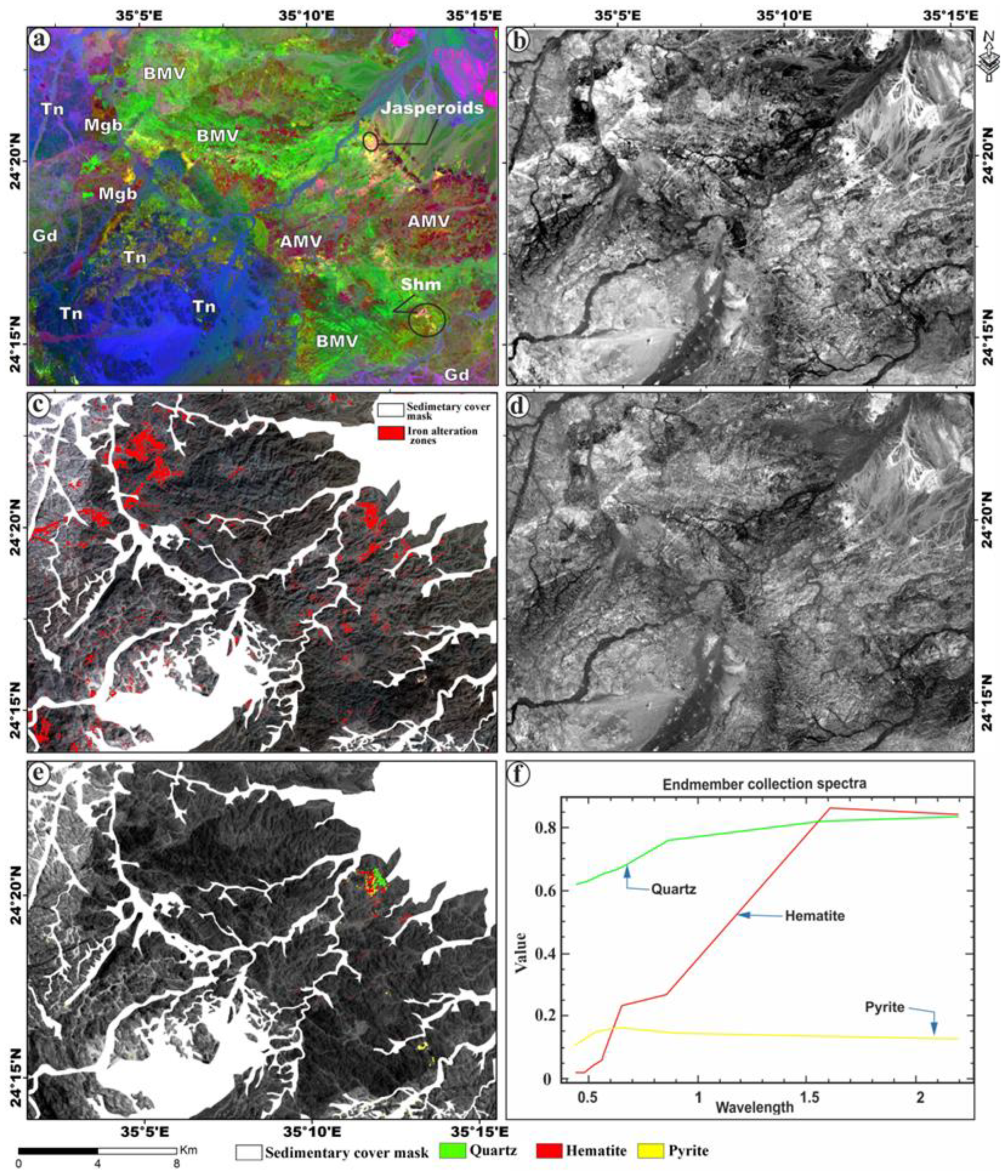
4.1. Remote Sensing Data
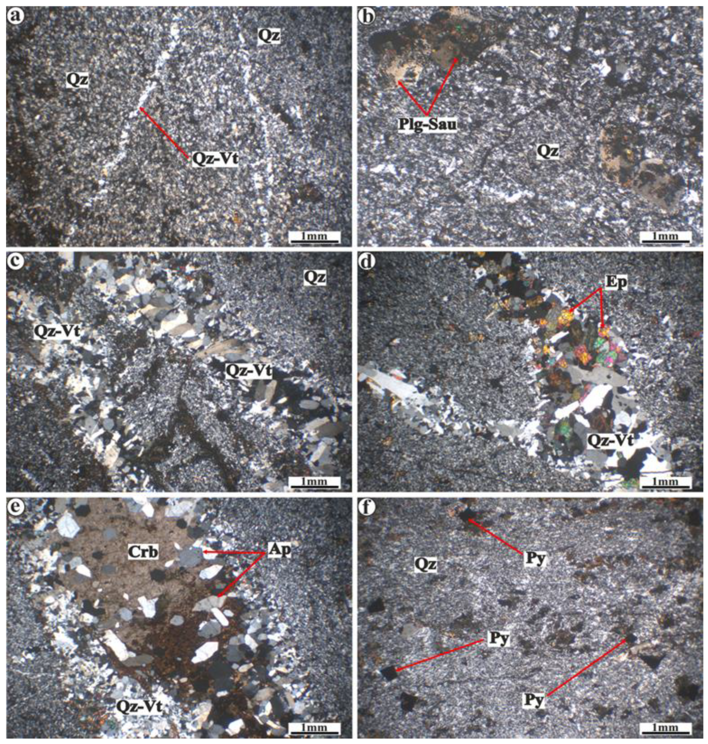
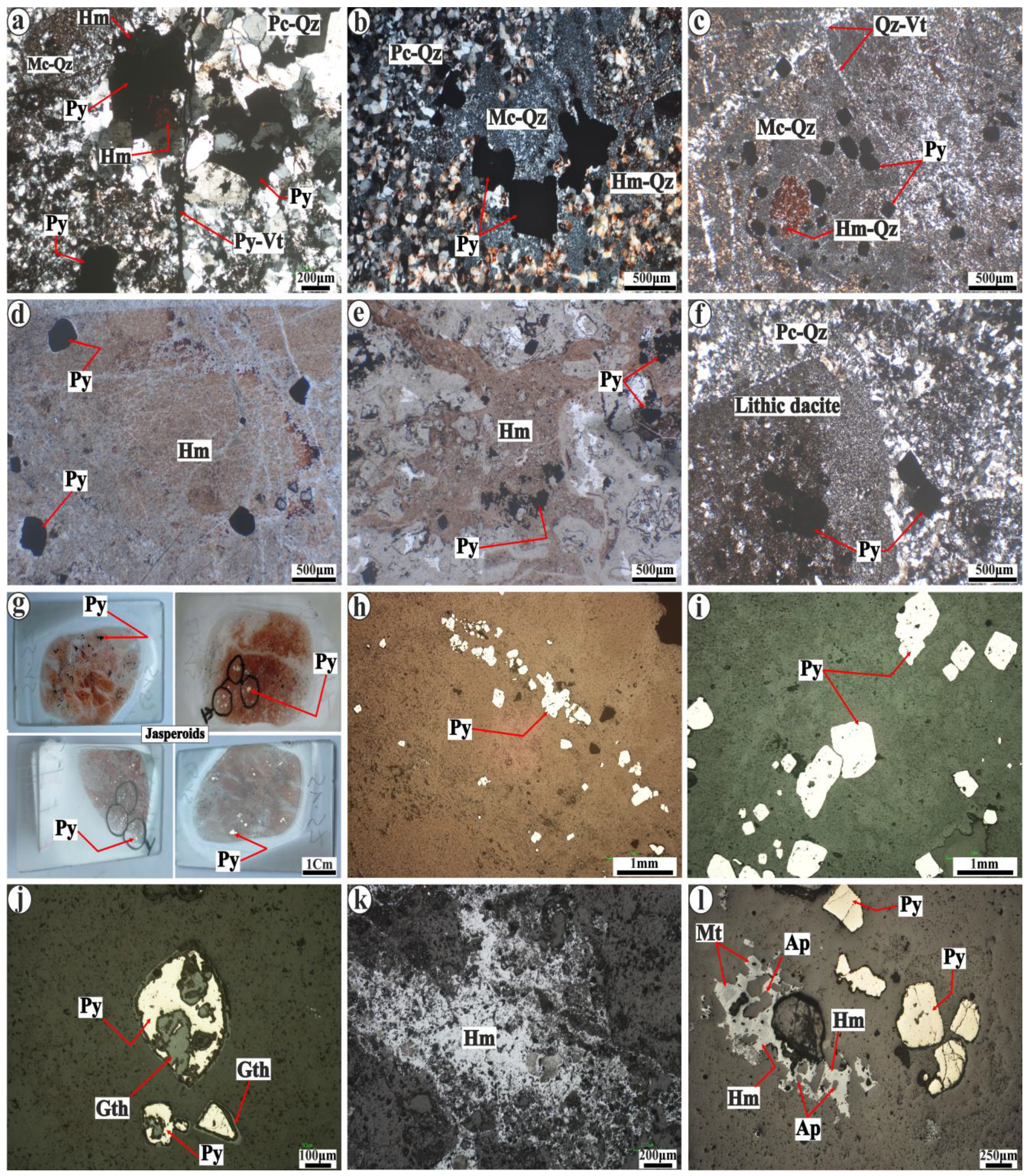
The remotely sensed data were used to distinguish various rock formations and hydrothermal alterations in the Wadi Ranga area, focusing on identifying the location of lenticular jasperoids. In the context of Landsat-8, the band ratios b6/b7, b6/b5 × b4/5, and b5 in RGB were employed to distinguish metavolcanics (AMV) with a dark red color and basic metavolcanics (BMV) with a green color. Granitic rocks were characterized by a greenish-violet color for granodiorites (Gd) and a bluish-violet for tonalites (Tn). Additionally, small bodies of metagabbro–diorite (Mgb) were marked with a reddish-brown color (Figure 3a).
The studied jasperoids appeared in yellow color at the south side of the Wadi Ranga entrance (Figure 3a). The application of the band ratio 4/2 on the Landsat-8 data proved to have a significant capability in distinguishing alteration zones characterized by iron-oxides [54] that led to the red tint of jasperoids. Therefore, these areas were visually represented with a white tone in Figure 3b. The utilization of the density slicing tool further contributed to enhancing the outcomes obtained from the band ratio of 4/2 with a red tint (Figure 3c). Similarly, Rowan et al. [60] developed the ASTER band 2/1 ratio to detect rocks influenced by significant ferric (Fe+3) alteration, manifested through minerals like hematite and goethite. This band ratio played a crucial role in identifying rocks stained with hematite, such as jasperoids. Consequently, rocks undergoing hematite alteration were visually represented by white pixels, as shown in Figure 3d.
Based on the fact that the studied jasperoids chiefly comprise silica and hematite, the Constrained Energy Minimization (CEM) method proved its worth in identifying the characteristics of these specific endmembers. The ASTER spectral library provides several mineral signatures with specific wavelengths, making the process of mineral identification easier without any need for more field observations; therefore, the CEM along with the ASTER spectral library were employed to identify the two minerals, silica in the form of quartz and hematite, which are associated with disseminated pyrite (Figure 3e,f).

4.2. Petrography

The felsic varieties of Ranga metavolcanic (MV)-hosted jasperoids are classified into porphyritic rhyolites, dacites, and related tuffs (Figure 4), while the basic ones, far away from Fe-Si-rich rocks, are mainly pillow lava flows and agglomerates. The Ranga felsic metatuffs comprise mainly microcrystalline groundmass (15–25 µm) of quartz (80–90 vol.%) associated with plagioclase (0.5–1.0 mm) saussurite phenocrysts (10–15 vol.%) (Figure 4a,b). They show replacement textures of polycrystalline quartz veinlets associated with epidote crosscuts in the examined samples (Figure 4c–e). Quartz veinlets appear in elongated shapes (1–1.5 mm) and intergrow with epidote. In quartz veinlets, there are mosaic-like patches (0.5–2.0 mm) of carbonate (5–10 vol.%), carrying hexagonal apatite between the crystals (Figure 4e). The Ranga felsic metatuffs are rich in disseminated pyrites (5–10 vol.%; Figure 4f), which occur as cubic crystals (0.5–1 mm) of magmatic origin; some pyrite crystals are locally altered to hematite that provides a red tint to the MV fine-grained quartz groundmass, especially at the shear zones around jasperoid lenses (Figure 4).
The jasperoids collected from the Ranga MV consist of quartz with subordinate pyrite and hematite–goethite (Figure 5a–f). Quartz occurs in two generations, either as micro-crystalline (Mc-Qz) or polycrystalline quartz (Pc-Qz) of mosaic appearance (Figure 5a,b), including disseminated pyrite grains up to 10 vol.% (Figure 5a–j,l). The polycrystalline quartz grains vary in size from 250 µm to 1 mm in the more recrystallized jasperoid samples (Figure 5a,b). Most of the jasperoids samples include two quartz generations, while few samples include only the microcrystalline quartz phase (Figure 5c). A network of veinlets filled with fine-grained quartz crosscut the whole jasperoids fabric (Figure 5c).
Hematite and goethite (10–15 vol.%) are the main iron oxide phases of jasperoids, manifesting as translucent, red to brown, and fine-grained aggregates. They stain quartz grains and quartz veinlets with a reddish or brownish appearance (Figure 5a–e). Hematite is more abundant than goethite and displays a variety of microscopic textures; it occurs either as a massive reddish-brown tint under the transmitted light and stains quartz spheroids (Figure 5d) or as a channeled-shaped growth over the microcrystalline quartz veinlets (Figure 5e). Hematite is distinguished by a light gray color under reflected light, either intergrown at the rims of the primary pyrite or replacing it completely, while goethite occurs as olive-gray with a colloform appearance. Massive anhedral hematite with a white-gray color was observed individually or intergrown with magnetite and apatite (Figure 5k–l). The existence of interstitial hexagonal apatite grains within hematite resembles those of Tyrone ironstones [61], suggesting its magmatic origin. Some jasperoids preserved some lithic clasts of dacitic composition from the hosted felsic metatuffs of jasperoids. These lithic clasts display an aphyric texture and comprise a very fine-grained quartz groundmass surrounded by coarser recrystallized quartz grains (Figure 5f).
The studied jasperoids in the Ranga area are rich in disseminated sulfides (up to 10 vol.%), similar to Tyrone ironstone hosting sulfides (up to 10 vol.%) in Ireland, where sulfidic jaspers were hosted in the felsic flows and metatuffs [61]. Pyrite is the dominant iron–sulfide mineral in the studied jasperoids (Figure 5e–l) as well as in the host metatuffs (Figure 4h). Pyrite occurs as euhedral to subhedral, light yellow crystals (0.5–1.0 mm) and is disseminated within the quartz groundmass and in quartz veinlets. Most of the studied pyrite crystals are replaced partially or completely with hematite and/or goethite, showing the red ochre (Figure 5g–j).

4.3. SEM-EDS Data and Mineral Chemistry

More than 50 photomicrographs were captured by Scanning Electron Microscopy (SEM) for the desired polished thin sections, revealing that hematite and goethite with subordinate magnetite are the iron oxides found in jasperoids, while pyrite is the sole sulfide mineral (Figure 6). The SEM microphotographs show the crystal forms and different relationships of the studied minerals, such as hematite and goethite, which occur either as sparse dissemination or replacement rims around the primary pyrite (Figure 6a–d). Most pyrites are partial to completely converted to secondary hematite and goethite pseudomorph after their cubic crystals (Figure 6a–d).
The colloform texture of goethite was recognized easily from SEM images and this texture can differentiate it from the massive hematite (Figure 6a–d). Hematite appears in two generations: the first-generation (Hm1) type is formed after pyrite, as evidenced by its cubic forms, while the second generation (Hm2) appears as coarse massive crystals carrying both the former cubic pyrite shape and the hexagonal apatite crystals (Figure 6e,f). The hematite generation (Hm1) was oxidized after the darker gray magnetite that only appears as sparse shapes within Hm1 (Figure 6e). Some pyrites appear to be fresh without any iron oxide replacements (Figure 6g,h).
A representative sampling of 32 analyzed points within minerals of jasperoids was conducted using EDS to determine their major oxide compositions (Supplementary Figure S1). The analyzed points were five points in hematite, two in goethite, eight in magnetite, two in apatite, and fifteen points in pyrites (Table 2 and Table 3). The repartition of Fe+3 (Fe2O3) and Fe+2 (FeO) wt.% contents from the EDS-normalized total iron (FeO total) was conducted using Droop’s [62] method (Table 2). Hematite is often considered an endmember of all other iron oxide transformations and mainly constitutes Fe2O3 ranging from 79.8 to 85.3 wt.% compared to goethite, which contains a lower Fe2O3 content (74–77.3 wt.%; Table 2). Both hematite and goethite have higher SiO2 (up to 6.97 wt.%) and Al2O3 contents (up to 1.59 wt.%) compared to magnetite (wt.%), i.e., SiO2 (up to 2.37 wt.%) and Al2O3 (up to 0.84 wt.%) (Table 2).
The low content of CuO (1.0 wt.%) that appeared in hematite may be due to the presence of micrometer-sized inclusions of Cu-rich sulfides. Magnetite is rich in FeO (61.76–67.32 wt.%) relative to Fe2O3 (27.87–30.29 wt.%), as shown in Table 2. The contents of SiO2 (up to 2.37 wt.%) and Al2O3 (up to 0.84 wt.%) in magnetite are lower than those in hematite and goethite. Magnetite also includes minor contents of Na2O, K2O, CaO, and MgO (<0.4 wt.%). The appearance of Co+2 (less than 1.0 wt.%) in the chemical structures of the studied iron oxides may be attributed to the substitution of Fe+2 by Co+2 (Table 2).
Apatite is used as a mineral indicator for interpreting geological environments since it can incorporate various ions into its structure [63]; this explains the minor contents of FeO (1.29–1.34 wt.%) and Na2O (0.36 wt.%) observed in the studied apatite (Table 2). CaO and P2O5 are considered the dominant oxides in the analyzed apatite with averages of 54.14 and 37.2 wt.%, respectively (Table 2). The saturation temperature at which apatite begins to recrystallize from the melt is about 850 °C based on Equation (1) of Piccoli and Candela [64].
Apatite (T) = ((26,400 × ClSiO2 − 4800)/(12.4 × ClSiO2 − In(ClP2O5) − 3.97))
where T is the apatite saturation temperature (in Kelvin), and ClSiO2 and ClP2O5 represent the concentration of silica and phosphorus in the melt (see Table 2), respectively; they are expressed as weight fractions (wt.%/100).
Pyrite has an average of 46.4 wt.% Fe and 52.9 wt.% S (Table 3). A trace amount (<0.2 wt.%) of Si was detected and could be attributed to the association of pyrite in a Si-rich groundmass (jasperoids). A minor concentration of Ag (up to 0.32 wt.%) was observed in some pyrites.

4.4. Bulk Compositions of Jasperoids and Host Rocks

The major and trace element compositions of some representative samples of both jasperoids and Ranga metavolcanics (MV) are shown in Table 4. The major element data of the Ranga MV hosted-jasperoids (Table 4) comprise mainly SiO2 (74.19 wt.% on average) for the more felsic samples (dacitic to rhyolitic metatuffs), except for one sample (Sr.53) that exhibited a lower SiO2 content (55.89 wt.%) and was plotted between dacites and andesites (named andesitic dacitic metatuffs). The more felsic metatuffs show small variations in Al2O3 (9.56–12.85 wt.%), Fe2O3 (≈3.53 wt.%), MgO (0.83–1.81 wt.%), CaO (0. 88–2.05 wt.%), K2O (0.03–1.0 wt.%), and Na2O (2.95–5.19 wt.%) compared to andesitic dacitic samples (Sr.53) that consist of higher Al2O3 (18.62 wt.%), Fe2O3 (≈6.27 wt.%), and CaO (9.52 wt.%). All analyzed samples have a low loss on ignition (LOI; 1.32–2.12 wt.%) reflecting the fresh nature of these rocks. The studied Ranga MV consisted primarily of felsic metatuffs with a subalkaline composition range from dacites to rhyolites according to Winchester and Floyd’s [65] classification diagram using the relationship between Nb/Y and Zr/Ti (Figure 7a).
The Wadi Ranga jasperoids are mainly composed of SiO2 (89.73–90.35 wt.%) due to the abundance of quartz and its microcrystalline forms and show wide ranges of Fe2O3t (2.73–6.63 wt.%) attributed to the significant amount of hematite, goethite, pyrite, and magnetite (Table 4). The Al2O3, CaO, and MgO concentrations are typically less than 0.4 wt.% on average, with minor contents of MnO and P2O5 (<0.04 wt.% on average) (Table 4). The remaining major elements (TiO2, Na2O, and K2O) are not detected in the analyzed jasperoids. A low loss on ignition (LOI) ranging from 1.08% to 1.49% (Table 4) reflects less H2O and alterations in the jasperoid and its host metatuffs. Bivariate geochemical plots of some major oxides and base metals for the studied jasperoids were conducted and compared to the literature data from Urals and Tyrone Si-rich ironstones associated with volcanogenic massive sulfide (VMS) deposits in Russia and Ireland, respectively [61,66,67]. Ferruginous deposits from the Urals are subdivided into jaspers/jasperites, gossanites, and Umbers [66,67]; however, their counterparts of the Tyrone igneous complex are mainly jaspers/jasperites [61]. The studied jasperoids are mainly plotted within jaspers/jasperites of Urals and coexist with the sulfidic jaspers of the Tyrone igneous complex that has a low total Fe2O3, MnO, CaO, and moderate base metals (Cu = 77.2 ppm, Pb = 26.6 ppm, and Zn = 30.9 ppm on average) alongside higher SiO2 (up to 93 wt.%) concentrations (Figure 7b–e and Table 4). The base metal contents of our jasperoids (Cu + Pb + Zn= 58.32–240.68 ppm) are generally lower than those of their host rocks (Cu + Pb + Zn = 43.96–1498.43 ppm), as shown in Table 4.
The Wadi Ranga felsic metatuffs have relatively high rare-earth element (REE) contents, varying from 37.32 to 89.66 ppm (Table 4). The chondrite (C1)-normalized REE pattern of the Ranga felsic metatuffs is flat in shape (La/Yb)N = 1.19–1.25); it also displays a nearly negative Eu anomaly (Eu/Eu* = 0.708–0.776; Table 4 and Figure 8a) and resembles REE patterns of the Shadli felsic MV [6,45,74]. The primitive mantle (PM)-normalized pattern of the Ranga felsic metatuffs generally shows enrichment in large-ion lithophile elements (LILEs; Ba, K, and Pb) and depletion in high-field strength elements (HFSEs: Nb, Ta, P, and Ti) (Figure 8b).
Moreover, the spider pattern is similar to that of the felsic metatuffs recorded in the Um Samuiki and the Wadi Ranga ([45,74]; Figure 8b). The Wadi Ranga jasperoids show a chondrite-normalized U-shaped REE pattern similar to Tyrone jaspers with a moderate (La/Yb)N ratio (1.8–6.1; Figure 8c) and are lower than both of their hosted calc-alkaline felsic metatuffs (1.19–1.25; Figure 8c) and the felsic Shadli MV after Maurice et al. [45] (1.3–1.9; Figure 8c). Furthermore, they display a slightly negative Eu anomaly (Eu/Eu* = 0.43–0.98; Table 4), which resembles that of both the hosted felsic metatuffs and Shadli MV after Maurice et al. [45] (Eu/Eu* = 0.708–0.776, 0.77–0.80, respectively). The primitive mantle-normalized patterns of the Ranga jasperoids exhibit enrichment in U and Zr, but depletion in Ti and Nb, similar to those of the Tyrone jasper PM patterns after Hollis et al. [61] (Figure 8d). The REE and trace element patterns of the Wadi Ranga jasperoids are considerably like those of the host MV rocks (Figure 8c,d), as both are high in LILEs and low in HFSEs. They have negative Eu, Nb, and Ti anomalies and positive K, Pb, and Sr anomalies resembling the Tyrone jasper and its host rhyolitic rocks [61] and Shadli MV [6,45,74].

5. Discussion

5.1. Petrogenesis and Tectonic Setting of Jasperoid-Bearing Metavolcanics

5.1.1. Magma Source and Affinity

The low loss on ignition values (LOI; 1.32–2.12 wt.%) and the absence of Ce anomalies (Table 4 and Figure 8a) of the Wadi Ranga felsic metatuffs support the fresh nature of these rocks and exclude the effect of any post-magmatic alteration [77]. The Wadi Ranga felsic metatuffs are classified as subalkaline metavolcanics by using the (Na2O + K2O) vs. SiO2 diagram (Figure 9a; [78]). Based on the AFM diagram [78], the Wadi Ranga felsic metatuffs fall in the calc-alkaline category and are far away from the tholeiitic rock field (Figure 9b), suggesting their generation in the island arc setting.
Similarly, the studied felsic metatuffs show calc-alkaline affinity in the Co vs. Th discrimination diagram (Figure 9c; [79]). This is consistent with the island arc metavolcanics (IMV or Shadli MV; Figure 9b) field, but different from the ophiolitic metavolcanics (OMV) based on the available chemical data from the SED of Egypt [6,9,72,73,74]. The felsic metatuffs of the Wadi Ranga can be classified as peraluminous types [80], exhibiting A/CNK values (Al2O3/[CaO + Na2O + K2O]) ranging from 1.37 to 2.03 (Figure 9d). Moreover, they show a magnesian character (Figure 9e) and some hydrous minerals, reflecting the origin from hydrous and oxidizing magmatic sources that likely originated in a subduction-related environment [81].
The Wadi Ranga felsic metatuffs show negative P and Ti anomalies that are likely attributed to the fractional crystallization of apatite and ilmenite, respectively (Figure 8b). This euhedral magmatic apatite in felsic metatuffs (Figure 4e) and jasperoids (Figure 5l) crystallized at a temperature of 850 °C [64]; this suggests the crystallization of metatuffs from the felsic melts at 850 °C, where some apatite crystals from the host rocks leached to the jasperoids (Figure 5l) that were generated after metatuffs at <850 °C. It is well known that the lithospheric magma is depleted in the HFSE relative to the LREE, and the HFSE/LREE ratio (Nb/La)N is useful for distinguishing the mantle sources of different rocks either lithospheric or asthenospheric mantle sources [82]. For example, low (Nb/La)N ratios (<0.7) suggest that the source is the lithospheric mantle, whereas higher ratios (>1.0) indicate an origin from the asthenospheric mantle; in addition, rocks have moderate (Nb/La)N ratios (0.7–1.0) derived from the mix of lithospheric and asthenospheric magma (Figure 9f). The Wadi Ranga metatuffs show a low (Nb/La)N ratio (0.27–0.23; Table 4), reflecting their magma source being the lithospheric mantle (Figure 9f).
There were lower (La/Yb)N (1.19–1.25; Table 4) and (La/Gd)N (1.06–1.17; Table 4) and the higher (Lu/Gd)N (0.13–0.14; Table 4) ratios of the studied metatuffs, excluding any contamination with continental crustal materials [76]. In addition, the Wadi Ranga felsic metatuffs possibly crystallized from fractionated melts that were derived from a depleted mantle source (Figure 9g,h) based on the Zr versus Nb and Y diagrams [83]. The negative anomalies of Nb and Ti, along with the positive anomalies at Pb, K, Rb, and Ba (Figure 8b), are attributed to the influence of fluids/melt derived from the subducted slab (oceanic crust) in a subduction-related setting [84,85,86,87]. Consequently, the influence of fluids/melts derived from the subducted slab on the lithospheric-derived melts (primary melts) and fractionation (by fractional crystallization) of these melts can yield more silicic rocks than those of andesitic composition, like the Wadi Ranga felsic metatuffs (55.89–76.49 wt.%; Table 4). The higher REE contents (ƩREE: 37.32–89.66 ppm; Table 4) of the Wadi Ranga felsic metatuffs relative to other mafic rocks in the area (ƩREE: 15.14–33.54 ppm [45]) suggest an origin during the late stage of the former fractionated basaltic rocks [11]. Therefore, the Wadi Ranga felsic metatuffs are thought to be derived from the partial melting of the calc-alkaline, peraluminous basaltic melts by fractional crystallization.

5.1.2. Tectonic Setting and Evolution

The HFSE and HREE are conservative elements that can be used for discriminating the tectonic environments of volcanic rocks [65,88,89]. The studied MV rocks and their jasperoids are rich in LILEs relative to HFSEs (Figure 8b,c), reflecting a supra-subduction zone (SSZ) setting [90]. The LILE enrichment relative to the HFSE one is attributed to the addition of fluids and/or melts derived from the subducted slab to the lithospheric mantle [91,92,93]. The Wadi Ranga felsic metatuffs plot in the field of volcanic arc granites (VAGs) and syn-collisional granites (Syn-COlGs) are close to oceanic ridge granites on the Nb vs. Y diagram (Figure 10a; [94]).
This is evidence of the island arc tectonic setting proposed for the generation of the Wadi Ranga MV rocks and their jasperoids, where these arc-related granites are considered the plutonic equivalent of the island arc metavolcanics.
The Wadi Ranga felsic metatuffs show characteristics similar to the arc magmatic rocks based on Y vs. Sr/Y discrimination diagram (Figure 10b; [95]). They also plot within the island arc basalts by using the Hf/3–Th–Nb/16 triangular diagram (Figure 10c; [96]). The arc-related signature of MV samples is proved by falling within the volcanic arc tholeiites field (VAT) and close to the back arc basin in the Y/15-La/10-Nb/8 diagram (Figure 10d; [97]). Based on the Th/Yb versus Nb/Yb diagram (Figure 10e; [98]), the Ranga felsic metatuffs have been likely formed from an N-MORB-depleted mantle source and are similar to those found in volcanic arcs of the SSZ setting [90]. Similarly, all studied samples plot on the field of arc volcanics are based on the Nb versus Ba/Nb diagram (Figure 10f), with a tendency for subduction enrichment [94,99,100,101]. The REE and trace element-normalized patterns of the Wadi Ranga MV closely resemble those of the Shadli MV (Figure 8a,b) [6,45,74], suggesting that they were likely formed in the island arc tectonic setting (Figure 11).
The sketch in Figure 11 summarizes the tectonic setting of the jasperoid lenses and their host metatuffs, where the subduction between two oceanic crusts produces an island arc volcano, such as the Shadli one. The volcanic vent of the Shadli volcano as a source of hydrothermal Si-rich fluids is surrounded by the felsic tuffs (later metamorphosed) as the host of the jasperoid lenses (Figure 11). Not only the volcanic vent but also the felsic dyke (Figure 2f) is the possible source of these fluids, where the felsic dyke is close to the shear zone and volcanic vent (Figure 11). Finally, the Ranga sulfidic jasperoids are hosted in Shadli metavolcanic rocks, which crystallized from peraluminous calc-alkaline magma.

5.2. Genesis of Jasperoid and Its Silica and Iron Sources

The sources of silica are various, but the most important sources are biogenic, detrital, and hydrothermal potential sources [102,103,104]. The remote sensing (Figure 3e,f), fieldwork (Figure 2g,h), textural (Figure 5), and geochemical characteristics (Figure 7b–e) suggest that the high silica (SiO2 up to 93.4 wt.%; Table 4) and iron (Fe2O3 up to 6.63 wt.%; Table 4) required for the formation of the studied sulfidic jasperoids are likely magmatic-derived hydrothermal fluids, circulated from a nearby volcanic vent [105,106,107] (Figure 11) and granitic dykes. For example, the existence of lithic clasts (Figure 5f) in the examined jasperoids is due to the nearby volcanic vent in the study area. The fieldwork demonstrates that the Hamata granitoid that intrudes the studied MV on the western side of the study area (Figure 1b) may be another source of Si-rich hydrothermal fluids, causing the hydrothermal alteration of the Shadli metavolcanics. For example, the investigated metavolcanic-hosted jasperoids are mainly crosscut by the felsic dyke, mainly alkali-feldspar granite related to the Hamata granitoid (Figure 2f).
The remote sensing data delineated the jasperoid areas and the highly enriched quartz and hematite bands in the Wadi Ranga metavolcanics based on the wavelength characteristics of the desired minerals (see Section 4.1; Figure 3e,f). The studied MV rocks around jasperoid lenses and veins (Figure 2d,e) are highly sheared and deformed due to fractures or faults, besides the occurrence of felsic dykes (Figure 2f). These sheared and fractured MV rocks reflect the occurrence of the shear zone that acts as pathways for the hydrothermal fluids possibly derived from the volcanic vent [61]; this was confirmed by the dominant major NW–SE faults conjugated with the NE–SW trend and the subordinate E–W trend in the Ranga MV rocks (Figure 1b). The magmatic-derived hydrothermal fluids are mainly of the Si-rich-type, circulating within the hosted sulfide-bearing MV (Figure 4f), leading to the leaching of Si, Fe, and S from the hosted MV (Figure 11) as sources of sulfidic jasperoids in the shear zones [108,109,110,111] (Figure 2h and Figure 5g), forming pyritization. The occurrence of a network of quartz veinlets within both jasperoids and their host rocks (Figure 4c–e and Figure 5c,d) supports that the Si-rich fluids are the main source of Si, while supergene fluids can leach Fe and S from the MV, forming silicification (Figure 11).
This explanation can elucidate the limited Fe content in the studied jasperoids (up to 6.63 wt.%; Table 4) relative to silica (up to 93.4 wt.%; Table 4). Additionally, the euhedral and compositionally homogeneous pyrites found in both jasperoids and their host felsic metatuffs (Table 3) suggest the same source and origin (Figure 4f and Figure 5h,i). Therefore, pyrite is of magmatic origin and formed in the early stage before the epigenetic Si-rich hydrothermal fluids derived from the nearby volcanic vent ([17]; Figure 2g,h). Under oxidizing conditions, the hydrothermal fluids altered primary pyrite, leaching S and adding OH. This leads to extensive silicification and hematization, as testified by goethite and hematite pseudomorphs after pyrite (Figure 6a–f). So, we think that pyritization is considered a magmatic–hydrothermal stage, where pyrite is of magmatic origin in the host metavolcanics, but its accumulation of up to 10 vol.% is attributed to magmatic-derived hydrothermal fluids. The second stage is silicification and then hematization, and these two processes are considered post-magmatic stages. So, euhedral hematite and goethite pseudomorph crystals after cubic pyrite are dominant in the Ranga jasperoid (Figure 6b,c,e).
Moreover, the variation diagrams of SiO2 contents (Figure 12a–c) in the Wadi Ranga jasperoids versus some representative mobile fluid elements (Rb, Th, Pb, Sr, Ba, and P) reflect that SiO2 (up to 93 wt.% in jasperoids) and these mobile elements come from an external source compared to the Fe2O3 (up to 6.63 wt.% in jasperoids; Table 4) that resulted from the internal leaching of Fe from the surrounding Shadli metavolcanics (Fe2O3: 3.28–6.27 wt.% in the host rocks; Table 4). The red staining of the Wadi Ranga jasperoids may be attributed to the secondary formation of iron oxides resulting from the oxidation of Fe+2 in pyrites to Fe+3, implying the red staining of the jasperoids in the form of hematite and goethite [12,108,112]. The increase in SiO2 (up to 6.97 wt.%) and Al2O3 (up to 1.59 wt.%) in hematite and goethite (Table 2) may be ascribed to the latter substitutions during high-grade metamorphism [34]. The trace contents of both TiO2 (up to 0.05 wt.%; Table 2) and MgO (up to 0.1 wt%) in magnetite indicate its secondary origin at low-temperature conditions [113,114]. Magnetite is possibly formed after pyrite and then altered into hematite in two stages, hematite I and hematite II (Hm1 and Hm2; Figure 6e). These iron oxides are of secondary origin and are considered a post-magmatic stage. The occurrence of apatite in the host rocks and jasperoids (Figure 4e and Figure 5l) reflects the possibility of apatite within the Si-rich sulfide ores, similar to that in the quartz–sulfide ores of Gaolong, China [115].
The Wadi Ranga jasperoids have geochemical characteristics that resemble sulfide-bearing jasperoids in the Pitcairn Islands. Both of them are rich in opaline silica (>35 wt. %) but are lower in Fe content (<10 wt.%) and base metals [116].
The Wadi Ranga jasperoids show enrichment in concentrations of typically mobile elements (like LILEs), such as Sr (up to 276.89 ppm), Ba (up to 232.27 ppm), Pb+2 (up to 54.7 ppm), and Rb (up to 13.78 ppm), in addition to Cu (up to 1333 ppm), Zr (up to 150 ppm), and Zn (up to 62.84 ppm) (Table 4 and Figure 8d), suggesting the role of hydrothermal sources. Moreover, the depletion of Al2O3 and TiO2 (immobile Ti) and the enrichment of Fe2O3 (mobile Fe) (Table 4) supports the role of hydrothermal fluids in the formation of the Ranga jasperoids [103,117,118]. The higher values of ([Fe + Mn]/Ti = 39,742–3,557,323 ppm) for the investigated jasperoids accompanied with lower values of (Al/[Al + Fe + Mn] = 211–585 ppm) are remarkable for the hydrothermal formation of jasperoids, in contrast with jasperoids after metalliferous sediments (clastic and biogenic materials) accumulated distally from the hydrothermal source [66,119,120,121]. The elemental ratios of Al, Fe, Mn, (Ni + Cu + Co), Si, and Co/Zn (Figure 13) are used in the classification diagrams to distinguish between hydrothermal, hydrogenous, and diagenetic Si-rich deposits. Based on the ternary diagram Fe–Mn–Al [122,123], the studied jasperoids plot within the hydrothermal field (Figure 13a). Moreover, the relation between Fe, Mn, and some base metals (Ni–Cu–Co) was used [124,125] to confirm the hydrothermal source of the studied jasperoids (Figure 13b). The relationship between Si and Al (Figure 13c) also supported the hydrothermal origin of the Ranga jasperoids and excluded the hydrogenous or diagenetic origin [126]. Additionally, the studied jasperoids fall within the hydrothermal field on the binary diagram of (Cu + Co + Ni) vs. (Co/Zn) (Figure 13d; [121,122]). In addition, the Ranga jasperoid U-shaped REE patterns (Figure 8c) are similar to those of Tyrone jasper in Northern Ireland, which formed in a similar island arc tectonic setting and is hosted in calc-alkaline tuffaceous rocks (Tanderagee member); these chemical characteristics (U-shaped REE patterns) confirm the arc-like setting and metasomatism by the slab-derived fluids [61,127].

5.3. Why Are Jasperiods Limited to a Specific Region within the Shadli Metavolcanics?

It is well known that Shadli metavolcanics are dominant in the SED of Egypt, and some of them are highly mineralized with copper, gold, talc, BIFs, and Ni-Fe-Co-sulfide deposits [8,9,10,11,29,31,32,33,72,128,129,130]. Shadli metavolcanics are completely free of jasperiods in most parts; however, a few places around the volcanic vent include jasperiods (Figure 11). The occurrence of jasperiods close to the volcanic vent is related to the source of Si, S, and gases from volcanoes [22,23]. In addition, the felsic dyke, mainly alkali-feldspar granites (Figure 2f), intruded into jasperoid-bearing metavolcanics. This is another possible source of highly Si-rich fluids. The source of Fe may be an internal source from the host metavolcanics (Fe2O3 ≈ 3.53; Table 4).
Structural features such as faults and shear zones represent localized regions of higher strain relative to the adjacent wall rocks [131]. They are characterized by intense deformation along specific planes; therefore, they often serve as pathways (Figure 11) for the migration and concentration of mineralizing fluids [132]. It is commonly accepted that shear zones play significant roles in the localization and evolution of hydrothermal flow on several scales. They control the transportation of magmatic, hydrothermal, and meteoric fluids during the mineralization process [131,132,133]. Shear zones or fractures enhance the permeability of rocks, thereby facilitating the transportation and precipitation of ore-forming fluids. This explains why the Ranga jasperoids are localized in the sheared and fractured MV rocks along the NW–SE, NE–SW, and E–W shear zones (Figure 1b) that acted as pathways for the circulation of the hydrothermal fluids from the volcanic vent and felsic dyke (Figure 2f). The distribution of jasperoid in a specific area of Shadli metavolcanics may have been influenced by the composition of the host metavolcanics, specifically the acidic type with high Si and Fe. The Ranga sulfidic jasperoids are hosted in Shadli metavolcanic rocks, which crystallized from the peraluminous calc-alkaline magma as a possible composition. These results may open the door for the future exploration of sulfide-rich jasperoids.

6. Conclusions

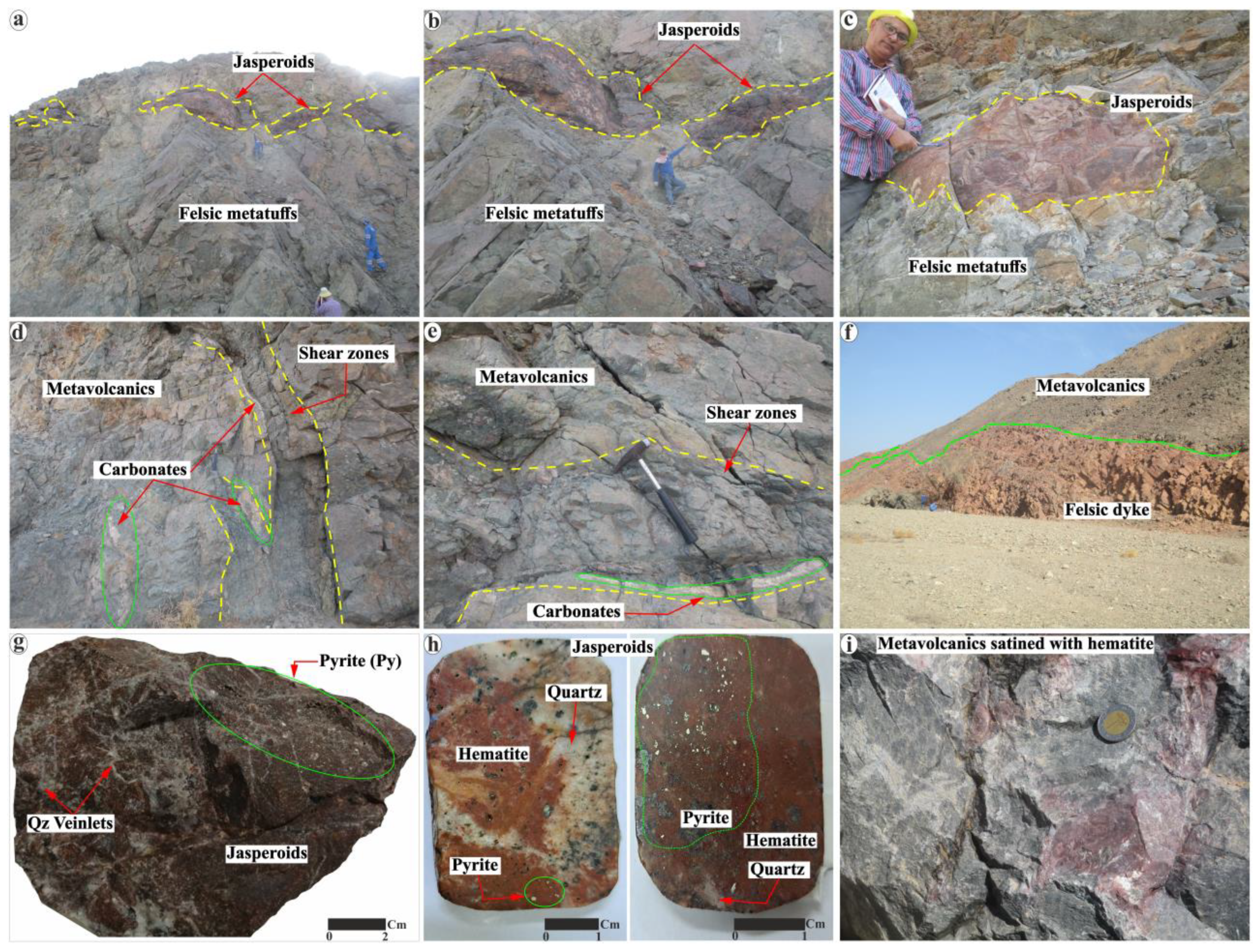
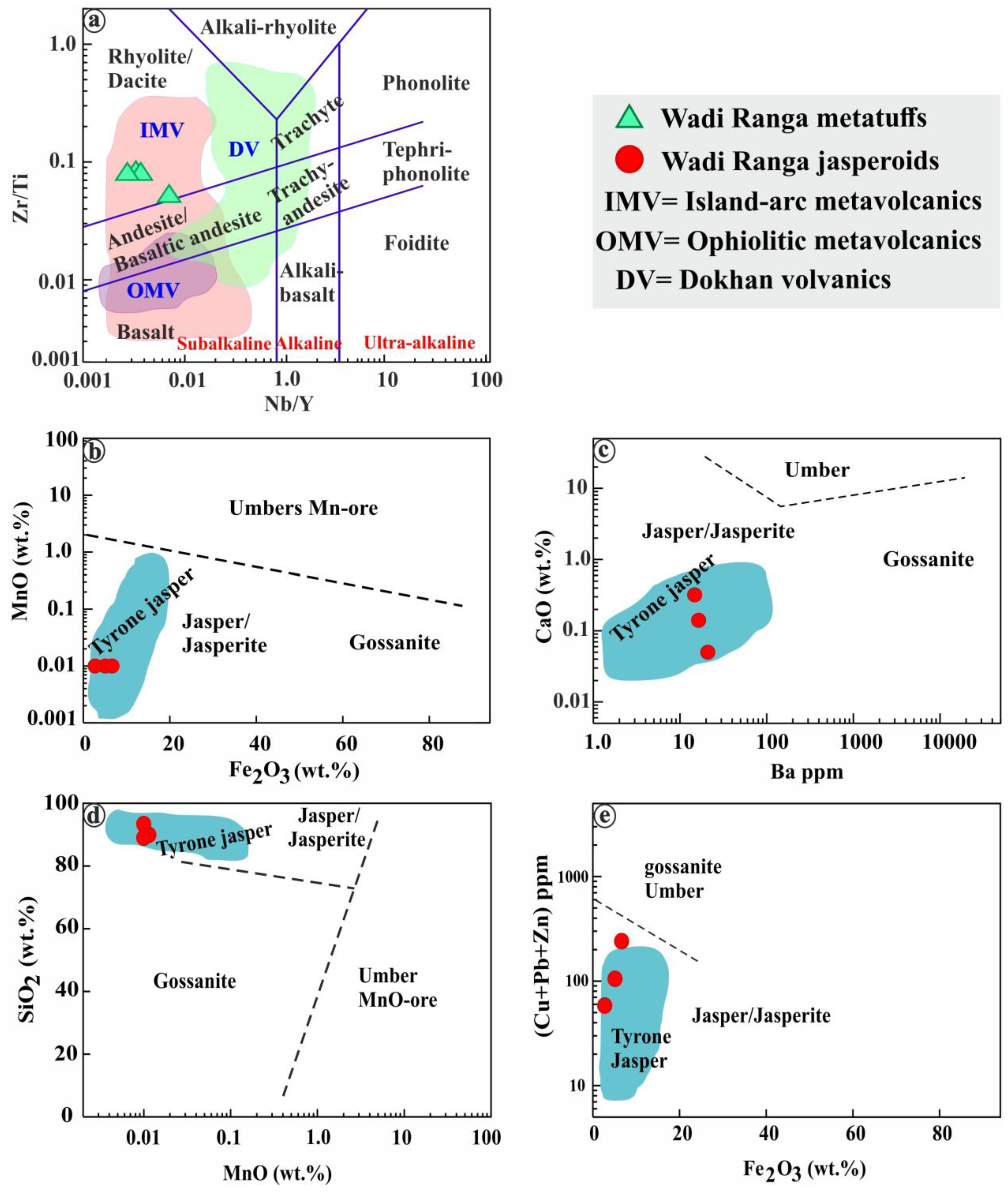
  • The Wadi Ranga jasperoids are hosted in Shadli metavolcanic rocks, which are mainly composed of rhyolitic and dacitic metatuffs.
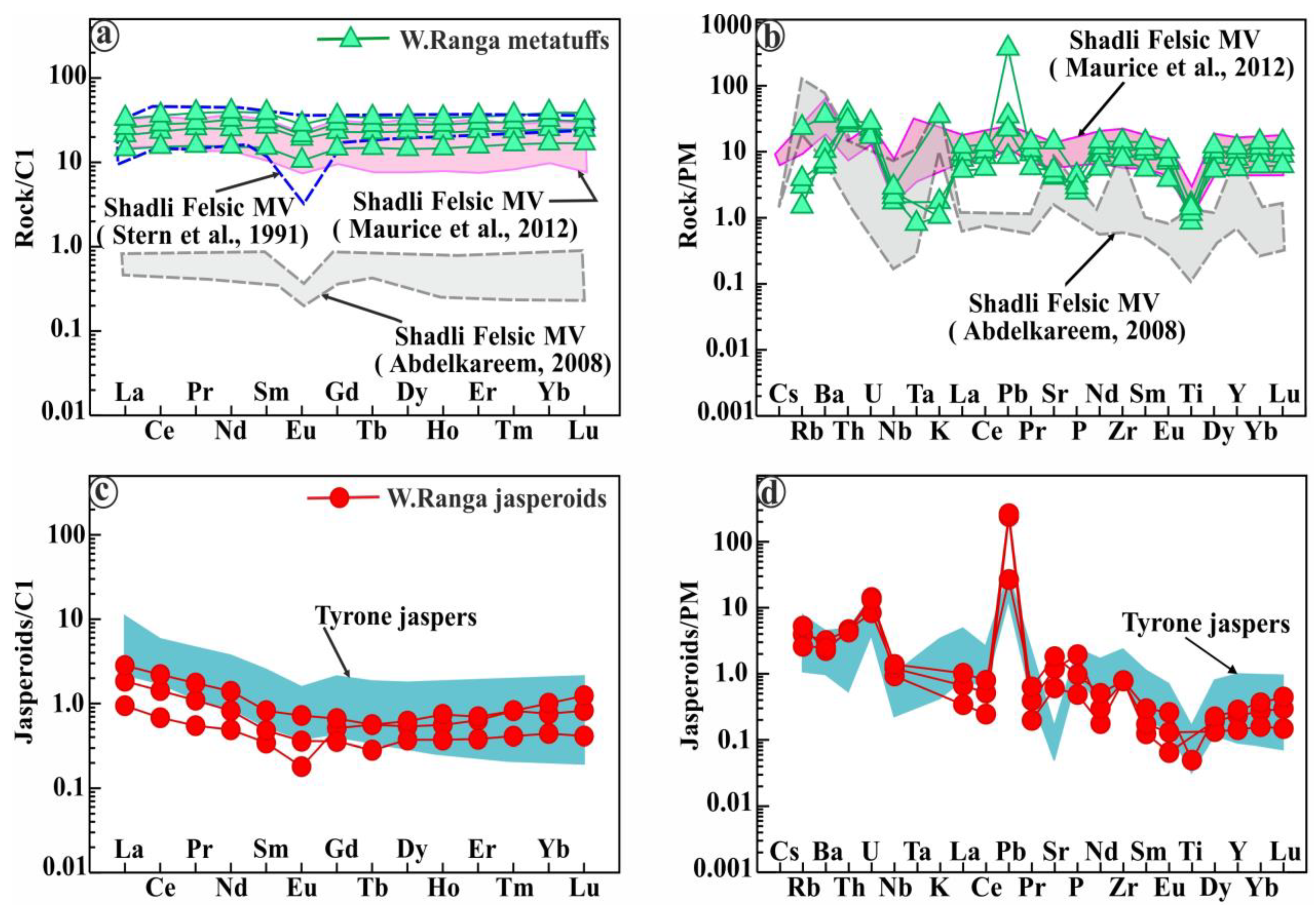
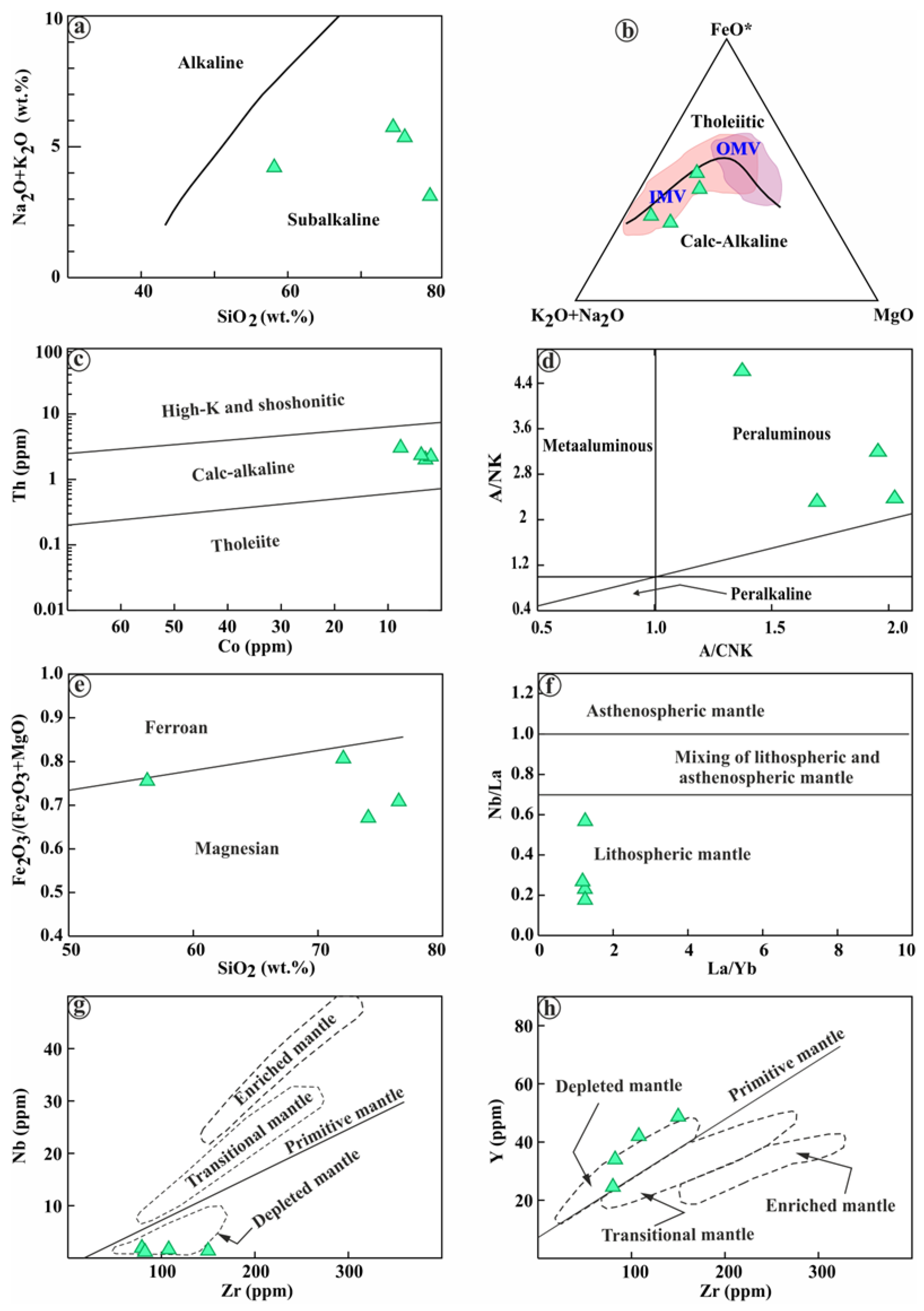
  • The studied metavolcanics (e.g., felsic metatuffs) crystallized from fractionated peraluminous calc-alkaline melts that were derived from a depleted lithospheric mantle source (N–MORB) in the island arc setting. They are rich in LILEs relative to HFSEs with a low (Nb/La)N ratio (<0.7), reflecting a high addition of fluids/melts from the subducted slab (oceanic crust) to the mantle-derived parent melts.
  • The Ranga jasperoids (SiO2: 89.73–90.35 wt.%; Fe2O3t: 2.73–6.63 wt.%) are considered the first documented Si–Fe–rich deposits hosted in the island arc metavolcanics. They are different from other jasperoids recorded in the Egyptian Nubian Shield as they are rich in pyrite (up to 10 vol.%), hematite, and goethite, with subordinate magnetite and Zn–Pb–Cu base metal sulfides. Their base metal content (Cu + Pb + Zn = 58.32–240.68 ppm) is generally lower than that of their host rocks (Cu + Pb + Zn = 43.96–1498.43 ppm).
  • The euhedral shape of pyrite associated with Wadi Ranga jasperoids reflects its magmatic origin and was accumulated by Si-rich fluids derived from the felsic dyke and volcanic vent. The low contents of iron within the studied jasperoids compared to silica reveal that the internal iron source (goethite and hematite) is a result of the secondary oxidation of the primary pyrite. So, massive pyritization occurred after the remobilization and accumulation of primary magmatic pyrite. Pyritization was followed by silicification and hematization, as suggested by hematite and goethite pseudomorphs.
  • The granitic dyke (felsic type) and volcanic vent likely represent the source of high silica in the Wadi Ranga jasperoids. The Si-rich hydrothermal fluids circulated along the NW–SE, NE–SW, and E–W major faults and shear zones in the Wadi Ranga MV rocks, forming the Ranga hydrothermal jasperoid veins and lenses. This is also evidenced by the existence of lithic clasts within the host metatuffs of jasperoids and the dominant major NW–SE faults conjugated with NE–SW and E–W trends in the Ranga MV rocks.
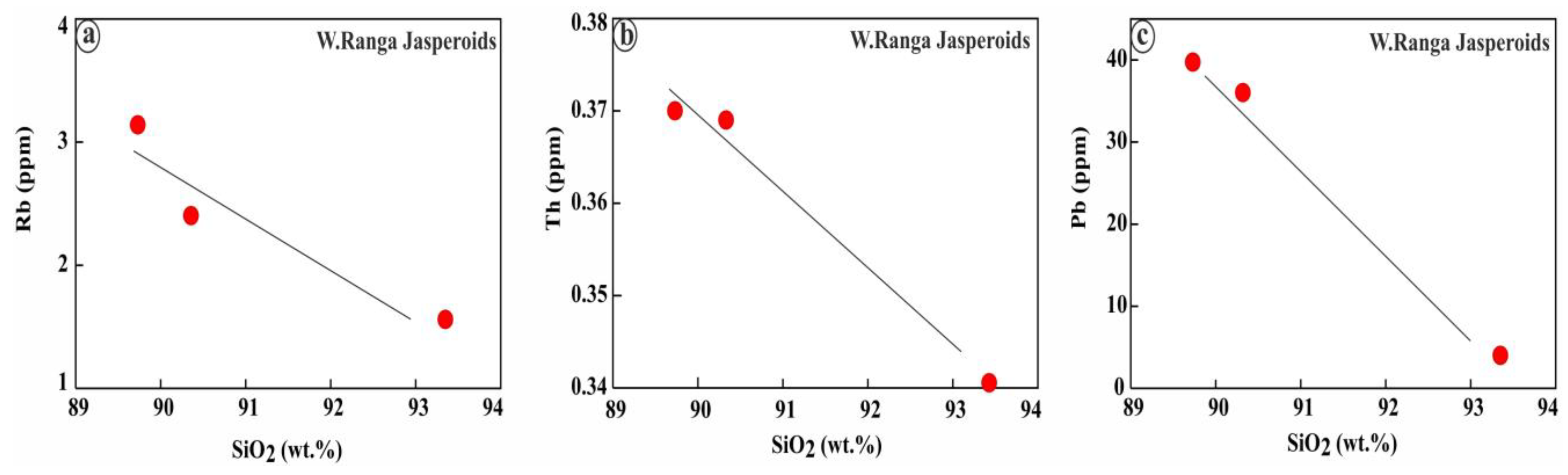
  • The enrichment in mobile elements (Rb, Ba, Th, Pb+2, Sr, P, Zn, Cu, and Mn) relative to the immobile elements (Ti, Nb, Zr, Hf, and Ta) alongside the relative abundance of Fe2O3 over Al2O3 and TiO2 supported the magmatic-derived hydrothermal Si-rich fluids that formed jasperoids. These fluids are rich in mobile elements (Rb, Ba, Th, Pb+2, Sr, Fe, P, Zn, Cu, and Mn) relative to immobile elements.
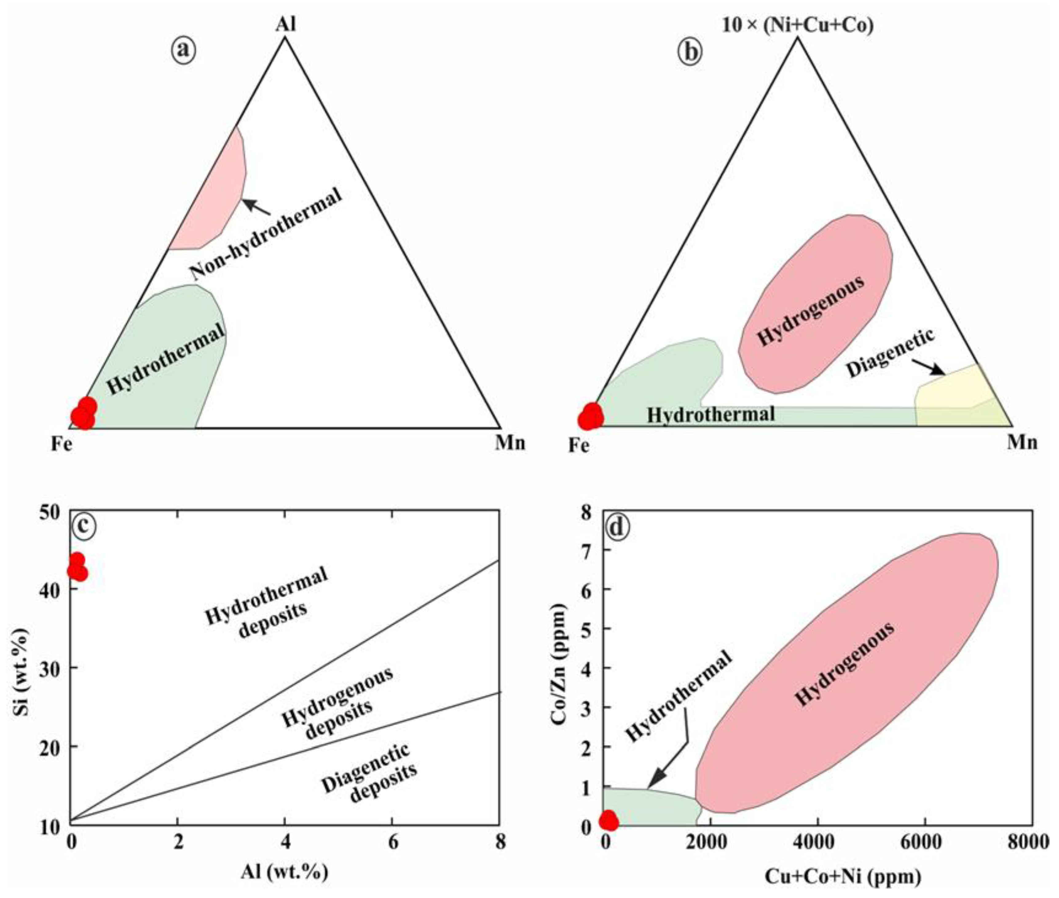
  • The Ranga jasperoids plot within the hydrothermal field supports their hydrothermal origin in the island arc setting and excludes the hydrogenous or diagenetic origin. This is confirmed by the jasperoid slightly U-shaped REE patterns, which are enriched in LILEs (e.g., K, Pb, and Sr) and depleted in HFSEs (e.g., Nb, Ta).

Supplementary Materials

The following supporting information can be downloaded at: https://www.mdpi.com/article/10.3390/min14070647/s1. Supplementary Figure S1. SEM images with EDS spectrum analyses of the dominant iron oxides, pyrite, and apatite in Wadi Ranga jasperoids. (a) Cubic pyrite. (b) Colloform goethite partially replaces cubic pyrite. (c) The first generation of hematite (Hm1) at the rims of former cubic pyrite. (d) The second generation of the massive hematite (Hm2). (e) Darker gray magnetite disperses within the former cubic pyrite and is intergrown with hematite (Hm1). (f) Hexagonal apatite. Abbreviations: Pyrite (Py), Goethite (Gth), Hematite1 (Hm1), Hematite2 (Hm2), Magnetite (Mt), and Apatite (Ap).

Author Contributions

Conceptualization, M.Z.K.; data curation, M.Z.K. and M.A.S.; formal analysis, M.Z.K., M.K.A., Y.I. and E.T.; funding acquisition, A.Y.K. and K.A.; investigation, M.Z.K., M.K.A., Y.I. and E.T.; methodology, M.Z.K. and M.A.S.; project administration, M.Z.K., M.A.S., S.A. and A.M.M.; resources, A.Y.K. and K.A.; supervision, M.Z.K., S.A. and A.M.M.; validation and visualization, M.Z.K. and M.A.S.; writing—original draft preparation, M.Z.K. and M.A.S.; and writing—review and editing, M.Z.K., S.A. and A.M.M. All authors have read and agreed to the published version of the manuscript.

Funding

This research was funded by the Researchers Supporting Project number (RSPD2024R546) at King Saud University in Riyadh, Saudi Arabia.

Data Availability Statement

All data derived from this research are presented in the enclosed figures and tables and Supplementary Figure S1, which can be downloaded at: https://www.mdpi.com/article/10.3390/min14070647/s1.

Acknowledgments

The first author is grateful to all colleagues in the Geology Department at both Niigata and Chiba Universities, Japan, for their support during his stay in August 2022 and December 2023, to polish thin sections, take petrographical photos, EDS–SEM, and conduct mineral chemistry (EPMA). We are grateful to the anonymous reviewers for their careful reading of our manuscript and their many insightful comments. The authors extend their sincere appreciation to the Researchers Supporting Project number (RSPD2024R546) at King Saud University in Riyadh, Saudi Arabia.

Conflicts of Interest

The authors declare no conflicts of interest.

References

  1. Stern, R.J.; Kröner, A. Late Precambrian crustal evolution in NE Sudan: Isotopic and geochronologic constraints. J. Geol. 1993, 101, 555–574. [Google Scholar] [CrossRef]
  2. Stern, R.J. Crustal evolution in the East African Orogen: A neodymium isotopic perspective. J. Afr. Earth Sci. 2002, 34, 109–117. [Google Scholar] [CrossRef]
  3. Stern, R.; Ali, K.; Liégeois, J.-P.; Johnson, P.; Kozdroj, W.; Kattan, F. Distribution and significance of pre-Neoproterozoic zircons in juvenile Neoproterozoic igneous rocks of the Arabian-Nubian Shield. Am. J. Sci. 2010, 310, 791–811. [Google Scholar] [CrossRef]
  4. Johnson, P.; Andresen, A.; Collins, A.; Fowler, A.; Fritz, H.; Ghebreab, W.; Kusky, T.; Stern, R. Late Cryogenian–Ediacaran history of the Arabian–Nubian Shield: A review of depositional, plutonic, structural, and tectonic events in the closing stages of the northern East African Orogen. J. Afr. Earth Sci. 2011, 61, 167–232. [Google Scholar] [CrossRef]
  5. Merdith, A.S.; Williams, S.E.; Müller, R.D.; Collins, A.S. Kinematic constraints on the Rodinia to Gondwana transition. Precambrian Res. 2017, 299, 132–150. [Google Scholar] [CrossRef]
  6. Stern, R.J.; Kröner, A.; Rashwan, A.A. A late Precambrian (∼710 Ma) high volcanicity rift in the southern Eastern Desert of Egypt. Geol. Rundsch. 1991, 80, 155–170. [Google Scholar] [CrossRef]
  7. Shukri, N.; Mansour, M.S. Lithostratigraphy of Um Samuiki District, Eastern Desert, Egypt. Evol. Miner. Arab. Nubian Shield 1980, 4, 83–93. [Google Scholar]
  8. Johnson, P.; Zoheir, B.; Ghebreab, W.; Stern, R.; Barrie, C.; Hamer, R. Gold-bearing volcanogenic massive sulfides and orogenic-gold deposits in the Nubian Shield. South Afr. J. Geol. 2017, 120, 63–76. [Google Scholar] [CrossRef]
  9. Faisal, M.; Yang, X.; Khalifa, I.H.; Amuda, A.K.; Sun, C. Geochronology and geochemistry of Neoproterozoic Hamamid metavolcanics hosting largest volcanogenic massive sulfide deposits in Eastern Desert of Egypt: Implications for petrogenesis and tectonic evolution. Precambrian Res. 2020, 344, 105751. [Google Scholar] [CrossRef]
  10. Abdel-Karim, A.-A.M.; El-Awady, A.; Khedr, M.Z.; El-Afandy, A.H.; Elwan, W.; Tamura, A.; Ali, S. Genesis of sulfide mineralization, Atshan and Darhib Areas, South Eastern Desert of Egypt: Evidence of fluid pathway effects along shear zones. Arab. J. Sci. Eng. 2022, 47, 641–665. [Google Scholar] [CrossRef]
  11. Khedr, M.Z.; Kamh, S.; Al Desouky, A.A.; Takazawa, E.; Hauzenberger, C.; Whattam, S.A.; El-Awady, A. Remote sensing and geochemical investigations of sulfide-bearing metavolcanic and gabbroic rocks (Egypt): Constraints on host-rock petrogenesis and sulfide genesis. Gondwana Res. 2023, 119, 282–312. [Google Scholar] [CrossRef]
  12. Cline, J.S.; Hofstra, A.H.; Muntean, J.L.; Tosdal, R.M.; Hickey, K.A. Carlin-Type Gold Deposits in Nevada: Critical Geologic Characteristics and Viable Models. Economic Geology 2005, 100th Anniversary Volume. Available online: https://pubs.geoscienceworld.org/segweb/books/edited-volume/1940/chapter-abstract/107715257/Carlin-Type-Gold-Deposits-in-NevadaCritical?redirectedFrom=fulltext (accessed on 21 January 2024).
  13. Kimberley, M.M. Geochemical distinctions among environmental types of iron formations. Chem. Geol. 1979, 25, 185–212. [Google Scholar] [CrossRef]
  14. Kimberley, M.M. Exhalative origins of iron formations. Ore Geol. Rev. 1989, 5, 13–145. [Google Scholar] [CrossRef]
  15. Fyon, J.; Breaks, F.; Heather, K.; Jackson, S.; Muir, T.; Stott, G.; Thurston, P.; Williams, H.; Sutcliffe, R. Metallogeny of metallic mineral deposits in the Superior Province of Ontario. Geology of Ontario. Edited by PC Thurston, HR Williams, RH Sutcliffe, and GM Stott. Ont. Geol. Surv. Spec. 1992, 4, 1091–1176. [Google Scholar]
  16. Simonson, B.M. Sedimentological constraints on the origins of Precambrian iron-formations. Geol. Soc. Am. Bull. 1985, 96, 244–252. [Google Scholar] [CrossRef]
  17. Phillips, G.; Groves, D.; Martyn, J. An epigenetic origin for Archean banded iron-formation-hosted gold deposits. Econ. Geol. 1984, 79, 162–171. [Google Scholar] [CrossRef]
  18. James, H.L. Sedimentary facies of iron-formation. Econ. Geol. 1954, 49, 235–293. [Google Scholar] [CrossRef]
  19. Fripp, R. Stratabound gold deposits in Archean banded iron-formation, Rhodesia. Econ. Geol. 1976, 71, 58–75. [Google Scholar] [CrossRef]
  20. Dimroth, E.; Chauvel, J.-J. Petrography of the Sokoman iron formation in part of the central Labrador trough, Quebec, Canada. Geol. Soc. Am. Bull. 1973, 84, 111–134. [Google Scholar] [CrossRef]
  21. Dimroth, E. Depositional environments and tectonic settings of the cherty iron-formations of the Canadian Shield. Geol. Soc. India 1986, 28, 239–250. [Google Scholar]
  22. Gross, G.A. A classification of iron formations based on depositional environments. Can. Mineral. 1980, 18, 215–222. [Google Scholar]
  23. Lydon, J.W. Ore deposit Models-8. Volcanogenic massive sulphide deposits Part I: A descriptive model. Geoscience Canada 1984, 11, 1–8. [Google Scholar]
  24. Kimberley, M.M. Debate about ironstone: Has solute supply been surficial weathering, hydrothermal convection, or exhalation of deep fluids? Terra Nova 1994, 6, 116–132. [Google Scholar] [CrossRef]
  25. Slack, J.; Grenne, T.; Bekker, A.; Rouxel, O.; Lindberg, P. Suboxic deep seawater in the late Paleoproterozoic: Evidence from hematitic chert and iron formation related to seafloor-hydrothermal sulfide deposits, central Arizona, USA. Earth Planet. Sci. Lett. 2007, 255, 243–256. [Google Scholar] [CrossRef]
  26. Bekker, A.; Slack, J.F.; Planavsky, N.; Krapez, B.; Hofmann, A.; Konhauser, K.O.; Rouxel, O.J. Iron formation: The sedimentary product of a complex interplay among mantle, tectonic, oceanic, and biospheric processes. Econ. Geol. 2010, 105, 467–508. [Google Scholar] [CrossRef]
  27. Grenne, T.; Slack, J.F. Mineralogy and geochemistry of silicate, sulfide, and oxide iron formations in Norway: Evidence for fluctuating redox states of early Paleozoic marine basins. Miner. Depos. 2019, 54, 829–848. [Google Scholar] [CrossRef]
  28. German, C.; Von Damm, K. Hydrothermal processes. In The Oceans and Marine Geochemistry; Elderfield, H., Ed.; Elsevier: Amsterdam, The Netherlands, 2003; Treatise on Geochemistry 6; pp. 181–222. [Google Scholar]
  29. El Habaak, G.H. Geology of Banded Iron Formation and Associated Metavolcanics at Wadi Kareim Area, Eastern Desert, Egypt. Ph.D. Thesis, Faculty of Science, Assiut University, Assiut, Egypt, 1992. [Google Scholar]
  30. El Habaak, G.H.; Mahmoud, M.S. Carbonaceous bodies of debatable organic provenance in the Banded Iron Formation of the Wadi Kareim area, Eastern Desert, Egypt. J. Afr. Earth Sci. 1994, 19, 125–133. [Google Scholar] [CrossRef]
  31. El Habaak, G.H.; Soliman, M. Rare earth element geochemistry of the Egyptian banded iron formations and the evolution of the Precambrian atmosphere and ocean. In Proceedings of the 4th International Conference on Geochemistry, Alexandria, Egypt, 1999; pp. 149–160. [Google Scholar]
  32. El Habaak, G.H. Pan-African skarn deposits related to banded iron formation, Um Nar area, central Eastern Desert, Egypt. J. Afr. Earth Sci. 2004, 38, 199–221. [Google Scholar] [CrossRef]
  33. Khudeir, A.; Ali, M.; El Habaak, G. The metavolcanics at Um Samiuki area, Egypt. Bull. Fac. Sci. Assiut. Univ. 1988, 17, 73–101. [Google Scholar]
  34. El Habaak, G.H. Ferruginous jasper and chert deposits associated with island-arc metavolcanics, Eastern Desert, Egypt. Acad. J. Sci. 2012, 1, 347–381. [Google Scholar]
  35. Aly, S.; Salem, I. Mineralogy and Chemistry of Jasper from Um Ghamis and El-Dabbah Metamorphic Iron Deposits, Central Eastern Desert, Egypt. Ann. Geol. Surv. Egypt 1985, 15, 223–232. [Google Scholar]
  36. Stern, R.J. Arc-assembly and continental collision in the Neoproterozoic African orogen: Implications for the consolidation of Gondwanaland. Annu. Rev. Earth Planet. Sci. 1994, 22, 319–351. [Google Scholar] [CrossRef]
  37. Khedr, M.Z.; Khashaba, S.M.A.; El-Shibiny, N.; Takazawa, E.; Hassan, S.M.; Azer, M.K.; Whattam, S.A.; El-Arafy, R.A.; Ichiyama, Y. Integration of remote sensing and geochemical data to characterize mineralized A-type granites, Egypt: Implications for origin and concentration of rare metals. Int. J. Earth Sci. 2023, 112, 1717–1745. [Google Scholar] [CrossRef]
  38. Hamimi, Z.; Abd El-Wahed, M.A. Suture (s) and major shear zones in the Neoproterozoic basement of Egypt. In The Geology of Egypt Book; Springer: Berlin/Heidelberg, Germany, 2020; pp. 153–189. [Google Scholar]
  39. Heikal, M.T.S.; Khedr, M.Z.; Abd El Monsef, M.; Gomaa, S.R. Petrogenesis and geodynamic evolution of neoproterozoic Abu Dabbab albite granite, central Eastern Desert of Egypt: Petrological and geochemical constraints. J. Afr. Earth Sci. 2019, 158, 103518. [Google Scholar] [CrossRef]
  40. Abdel-Khalek, M.; Takla, M.; Sehim, A.; Hamimi, Z.; El Manawi, A. Geology and tectonic evolution of Wadi Beitan area, southeastern Desert, Egypt. In Proceedings of the International Conference on Geology of the Arab World, Cairo, Egypt, 27–30 September 1992; pp. 369–394. [Google Scholar]
  41. Abdelsalam, M.G.; Abdeen, M.M.; Dowaidar, H.M.; Stern, R.J.; Abdelghaffar, A.A. Structural evolution of the Neoproterozoic western Allaqi–Heiani suture, southeastern Egypt. Precambrian Res. 2003, 124, 87–104. [Google Scholar] [CrossRef]
  42. Hagag, W.; Moustafa, R.; Hamimi, Z. Neoproterozoic Evolution and Najd-Related Transpressive Shear Deformations Along Nugrus Shear Zone, South Eastern Desert, Egypt (Implications from Field-Structural Data and AMS-Technique). Geotectonics 2018, 52, 114–133. [Google Scholar] [CrossRef]
  43. Hamimi, Z.; El-Fakharani, A.; Emam, A.; Barreiro, J.G.; Abdelrahman, E.; Abo-Soliman, M.Y. Reappraisal of the kinematic history of Nugrus shear zone using PALSAR and microstructural data: Implications for the tectonic evolution of the Eastern Desert tectonic terrane, northern Nubian Shield. Arab. J. Geosci. 2018, 11, 1–29. [Google Scholar] [CrossRef]
  44. Hussein, A.A.A.; Ali, M.M.; El Ramly, M. A proposed new classification of the granites of Egypt. J. Volcanol. Geotherm. Res. 1982, 14, 187–198. [Google Scholar] [CrossRef]
  45. Maurice, A.E.; Basta, F.F.; Khiamy, A.A. Neoproterozoic nascent island arc volcanism from the Nubian Shield of Egypt: Magma genesis and generation of continental crust in intra-oceanic arcs. Lithos 2012, 132, 1–20. [Google Scholar] [CrossRef]
  46. Gharib, M.E.; Ahmed, A.H. Late Neoproterozoic volcanics and associated granitoids at Wadi Ranga, south Eastern Desert, Egypt: A transition from subduction-related to intra-arc magmatism. Lithos 2012, 155, 236–255. [Google Scholar] [CrossRef]
  47. Conoco. Geological Maps of Egypt, NF 36 NE Bernice, Egypt, Scale 1: 500,000. 1987.Conoco Coral; Institut für Angewandte Geodäsie: Berlin, Germany, 1987. [Google Scholar]
  48. Conoco. Geological Maps of Egypt, NG 36 SE Gebel Hamata, Egypt, Scale 1: 500,000. 1987.Conoco Coral; Institut für Angewandte Geodäsie: Berlin, Germany, 1987. [Google Scholar]
  49. EGSMA. Geologic Map of Berence Quadrangle, Egypt, Scale 1:250,000; The Egyptian Geologic Survey and Mining Authority (EGSMA): Cairo, Egypt, 1997.
  50. EGSMA. Geologic Map of Hamata Quadrangle, Egypt, Scale 1:250,000; The Egyptian Geologic Survey and Mining Authority (EGSMA): Cairo, Egypt, 1997.
  51. Abrams, M.; Hook, S.; Ramachandran, B. ASTER User Handbook, Version 2: Advanced Spaceborne Thermal Emission and Reflection Radiometer; Jet Propulsion Laboratory: Pasadena, CA, USA, 2002. Available online: https://lpdaac.usgs.gov/documents/262/ASTER_User_Handbook_v2.pdf (accessed on 21 January 2024).
  52. NASA/METI/AIST. ASTER Level 1B Data Set Registered Radiance at the Sensor, NASA EOSDIS Land Processes Distributed Active Archive Center (LP DAAC), Japan Space Systems & U.S./Japan ASTER Science Team, 2001. Available online: https://lpdaac.usgs.gov/products/ast_l1bv003 (accessed on 21 January 2024).
  53. U.S. Geological Survey Distribution of European Space Agency’s Landsat-8 OLI data; Path = 173 and Row = 43, Collection Number 1, US. 2013. Available online: https://earthexplorer.usgs.gov/ (accessed on 21 January 2024).
  54. Sabins, F.F. Remote sensing for mineral exploration. Ore Geol. Rev. 1999, 14, 157–183. [Google Scholar] [CrossRef]
  55. Di Tommaso, I.; Rubinstein, N. Hydrothermal alteration mapping using ASTER data in the Infiernillo porphyry deposit, Argentina. Ore Geol. Rev. 2007, 32, 275–290. [Google Scholar] [CrossRef]
  56. Rockwell, B.W.; Hofstra, A.H. Identification of quartz and carbonate minerals across northern Nevada using ASTER thermal infrared emissivity data—Implications for geologic mapping and mineral resource investigations in well-studied and frontier areas. Geosphere 2008, 4, 218–246. [Google Scholar] [CrossRef]
  57. Amin, B.P.; Mazlan, H. Spectral transformation of ASTER data and the discrimination of hydrothermal alteration minerals in a semi-arid region, SE Iran. Int. J. Phys. Sci. 2011, 6, 2037–2059. [Google Scholar]
  58. Sabins, F.F. Remote Sensing. Principles and Interpretation; W. H. Freeman and Company: New York, NY, USA, 1997; pp. 366–371. [Google Scholar]
  59. Harsanyi, J.C. Detection and Classification of Subpixel Spectral Signatures in Hyperspectral Image Sequences; University of Maryland: Baltimore County, MD, USA, 1993. [Google Scholar]
  60. Rowan, L.C.; Hook, S.J.; Abrams, M.J.; Mars, J.C.J.E.G. Mapping hydrothermally altered rocks at Cuprite, Nevada, using the Advanced Spaceborne Thermal Emission and Reflection Radiometer (ASTER), a new satellite-imaging system. Econ. Geol. 2003, 98, 1019–1027. [Google Scholar] [CrossRef]
  61. Hollis, S.P.; Cooper, M.R.; Herrington, R.J.; Roberts, S.; Earls, G.; Verbeeten, A.; Piercey, S.J.; Archibald, S.M. Distribution, mineralogy and geochemistry of silica-iron exhalites and related rocks from the Tyrone Igneous Complex: Implications for VMS mineralization in Northern Ireland. J. Geochem. Explor. 2015, 159, 148–168. [Google Scholar] [CrossRef]
  62. Droop, G. A general equation for estimating Fe3+ concentrations in ferromagnesian silicates and oxides from microprobe analyses, using stoichiometric criteria. Mineral. Mag. 1987, 51, 431–435. [Google Scholar] [CrossRef]
  63. Taman, Z. Mineralogical and Geochemical Studies on Some Banded Iron Formations from the Eastern Desert of Egypt, and Their Industrial Uses. Ph.D. Thesis, Ain Shams University, Cairo, Egypt, 2005; p. 166. [Google Scholar]
  64. Piccoli, P.M.; Candela, P.A. Apatite in igneous systems. Reviews in Mineralogy and Geochemistry. Rev. Mineral. Geochem. 2002, 48, 255–292. [Google Scholar] [CrossRef]
  65. Winchester, J.A.; Floyd, P.A. Geochemical discrimination of different magma series and their differentiation products using immobile elements. Chem. Geol. 1977, 20, 325–343. [Google Scholar] [CrossRef]
  66. Brusnitsyn, A.; Zhukov, I. Manganese deposits of the Devonian Magnitogorsk palaeovolcanic belt (Southern Urals, Russia). Ore Geol. Rev. 2012, 47, 42–58. [Google Scholar] [CrossRef]
  67. Maslennikov, V.V.; Ayupova, N.; Herrington, R.; Danyushevskiy, L.; Large, R. Ferruginous and manganiferous haloes around massive sulphide deposits of the Urals. Ore Geol. Rev. 2012, 47, 5–41. [Google Scholar] [CrossRef]
  68. Moghazi, A. Geochemistry and petrogenesis of a high-K calc-alkaline Dokhan Volcanic suite, South Safaga area, Egypt: The role of late Neoproterozoic crustal extension. Precambrian Res. 2003, 125, 161–178. [Google Scholar] [CrossRef]
  69. Eliwa, H.; Kimura, J.-I.; Itaya, T. Late Neoproterozoic Dokhan Volcanics, North Eastern Desert, Egypt: Geochemistry and petrogenesis. Precambrian Res. 2006, 151, 31–52. [Google Scholar] [CrossRef]
  70. Abdel Wahed, A.A.; Ali, K.G.; Khalil, M.M.; Abdel Gawad, A.E. Dokhan volcanics of Gabal Monqul area, North Eastern Desert, Egypt: Geochemistry and petrogenesis. Arab. J. Geosci. 2012, 5, 29–44. [Google Scholar] [CrossRef]
  71. Dessouky, O.K.; Cai, M.; Ali, H.H.; Dardier, A.M.; Fowler, A.-R.; Ali, K.A.; Santosh, M.; Stüwe, K.; Hassan, M.M. A newly recorded Cryogenian-Ediacaran Dokhan Volcanic caldera with resurgent uplift in the Arabian-Nubian Shield, southwest Safaga, Egypt. Precambrian Res. 2023, 387, 106993. [Google Scholar] [CrossRef]
  72. Stern, R.J. Petrogenesis and tectonic setting of Late Precambrian ensimatic volcanic rocks, Central Eastern Desert of Egypt. Precambrian Res. 1981, 16, 195–230. [Google Scholar] [CrossRef]
  73. Abd El-Rahman, Y.; Polat, A.; Dilek, Y.; Fryer, B.J.; El-Sharkawy, M.; Sakran, S. Geochemistry and tectonic evolution of the Neoproterozoic incipient arc–forearc crust in the Fawakhir area, Central Eastern Desert of Egypt. Precambrian Res. 2009, 175, 116–134. [Google Scholar] [CrossRef]
  74. Abdelkareem, M. Massive Sulfide Deposit, and Associated Metavolcanics, Um Samuiki Area, South Eastern Desert, Egypt. Master’s Thesis, South Valley University, Qena, Egypt, 2008. [Google Scholar]
  75. McDonough, W.F.; Sun, S.-S. The composition of the Earth. Chem. Geol. 1995, 120, 223–253. [Google Scholar] [CrossRef]
  76. Rudnick, R.; Gao, S. Composition of the Continental Crust. Treatise Geochem. 2014, 4, 1–51. [Google Scholar] [CrossRef]
  77. Polat, A.; Hofmann, A. Alteration and geochemical patterns in the 3.7–3.8 Ga Isua greenstone belt, West Greenland. Precambrian Res. 2003, 126, 197–218. [Google Scholar] [CrossRef]
  78. Irvine, T.N.; Baragar, W. A guide to the chemical classification of the common volcanic rocks. Can. J. Earth Sci. 1971, 8, 523–548. [Google Scholar] [CrossRef]
  79. Hastie, A.R.; Kerr, A.C.; Pearce, J.A.; Mitchell, S. Classification of altered volcanic island arc rocks using immobile trace elements: Development of the Th–Co discrimination diagram. J. Petrol. 2007, 48, 2341–2357. [Google Scholar] [CrossRef]
  80. Maniar, P.D.; Piccoli, P.M. Tectonic discrimination of granitoids. Geological society of America bulletin 1989, 101, 635–643. [Google Scholar] [CrossRef]
  81. Frost, B.R.; Barnes, C.G.; Collins, W.J.; Arculus, R.J.; Ellis, D.J.; Frost, C.D. A geochemical classification for granitic rocks. J. Petrol. 2001, 42, 2033–2048. [Google Scholar] [CrossRef]
  82. Smith, E.I.; Sanchez, A.; Walker, J.D.; Wang, K. Geochemistry of mafic magmas in the Hurricane Volcanic field, Utah: Implications for small-and large-scale chemical variability of the lithospheric mantle. J. Geol. 1999, 107, 433–448. [Google Scholar] [CrossRef]
  83. Zhao, Z. Principles of Trace Element Geochemistry; Science Press: Beijing, China, 1997; pp. 1–495. [Google Scholar]
  84. Wilson, M. Igneous Petrogenesis: A Global Tectonic Approach; Unwin Hyman: London, UK, 1989; pp. 1–466. [Google Scholar]
  85. Rudnick, R.L.; Fountain, D.M. Nature and composition of the continental crust: A lower crustal perspective. Rev. Geophys. 1995, 33, 267–309. [Google Scholar] [CrossRef]
  86. Atherton, M.; Ghani, A. Slab breakoff: A model for Caledonian, Late Granite syn-collisional magmatism in the orthotectonic (metamorphic) zone of Scotland and Donegal, Ireland. Lithos 2002, 62, 65–85. [Google Scholar] [CrossRef]
  87. Kamber, B.S.; Ewart, A.; Collerson, K.D.; Bruce, M.C.; McDonald, G.D. Fluid-mobile trace element constraints on the role of slab melting and implications for Archaean crustal growth models. Contrib. Mineral. Petrol. 2002, 144, 38–56. [Google Scholar] [CrossRef]
  88. Pearce, J.A. Geochemical fingerprinting of oceanic basalts with applications to ophiolite classification and the search for Archean oceanic crust. Lithos 2008, 100, 14–48. [Google Scholar] [CrossRef]
  89. Pearce, J.A.; Peate, D.W. Tectonic implications of the composition of volcanic arc magmas. Annu. Rev. Earth Planet. Sci. 1995, 23, 251–285. [Google Scholar] [CrossRef]
  90. Pearce, J.A.; Lippard, S.; Roberts, S. Characteristics and tectonic significance of supra-subduction zone ophiolites. Geol. Soc. Lond. Spec. Publ. 1984, 16, 77–94. [Google Scholar] [CrossRef]
  91. Best, M. Amphibole-bearing cumulate inclusions, Grand Canyon, Arizona and their bearing on silica-undersaturated hydrous magmas in the upper mantle. J. Petrol. 1975, 16, 212–236. [Google Scholar] [CrossRef]
  92. Hawkesworth, C.; O’nions, R.; Pankhurst, R.; Hamilton, P.; Evensen, N. A geochemical study of island-arc and back-arc tholeiites from the Scotia Sea. Earth Planet. Sci. Lett. 1977, 36, 253–262. [Google Scholar] [CrossRef]
  93. Tarney, J.; Wood, D.; Saunders, A.; Cann, J.; Varet, J. Nature of mantle heterogeneity in the North Atlantic: Evidence from deep sea drilling. Philosophical Transactions of the Royal Society of London. Ser. A Math. Phys. Sci. 1980, 297, 179–202. [Google Scholar]
  94. Pearce, J.A.; Harris, N.B.; Tindle, A.G. Trace element discrimination diagrams for the tectonic interpretation of granitic rocks. J. Petrol. 1984, 25, 956–983. [Google Scholar] [CrossRef]
  95. Hou, Z.-Q.; Gao, Y.-F.; Qu, X.-M.; Rui, Z.-Y.; Mo, X.-X. Origin of adakitic intrusives generated during mid-Miocene east–west extension in southern Tibet. Earth Planet. Sci. Lett. 2004, 220, 139–155. [Google Scholar] [CrossRef]
  96. Wood, D.A. The application of a ThHfTa diagram to problems of tectonomagmatic classification and to establishing the nature of crustal contamination of basaltic lavas of the British Tertiary Volcanic Province. Earth Planet. Sci. Lett. 1980, 50, 11–30. [Google Scholar] [CrossRef]
  97. Cabanis, B. The La/10-Y/15-Nb/8 diagram-A tool for discriminating volcanic series and evidencing continental-crust magmatic mixtures and/or contamination. Comptes Rendus L’acaddmie Sci. 1989, 309, 2023–2029. [Google Scholar]
  98. Pearce, J.A. Immobile element fingerprinting of ophiolites. Elements 2014, 10, 101–108. [Google Scholar] [CrossRef]
  99. Taylor, S.R.; McLennan, S.M. The Continental Crust: Its Composition and Evolution; Blackwell Scientific Publications: Oxford, MA, USA, 1985; pp. 1–349. [Google Scholar]
  100. Sun, S.-S.; McDonough, W.F. Chemical and isotopic systematics of oceanic basalts: Implications for mantle composition and processes. Geol. Soc. Lond. Spec. Publ. 1989, 42, 313–345. [Google Scholar] [CrossRef]
  101. Condie, K.C. Chemical composition and evolution of the upper continental crust: Contrasting results from surface samples and shales. Chem. Geol. 1993, 104, 1–37. [Google Scholar] [CrossRef]
  102. Sugisaki, R.; Yamamoto, K.; Adachi, M. Triassic bedded cherts in central Japan are not pelagic. Nature 1982, 298, 644–647. [Google Scholar] [CrossRef]
  103. Adachi, M.; Yamamoto, K.; Sugisaki, R. Hydrothermal chert and associated siliceous rocks from the northern Pacific their geological significance as indication of ocean ridge activity. Sediment. Geol. 1986, 47, 125–148. [Google Scholar] [CrossRef]
  104. Yamamoto, S. Correlation between iron and magnesium and its significance on the distribution of heavy metals in deep-sea cherts. Sediment. Geol. 1986, 49, 261–280. [Google Scholar] [CrossRef]
  105. Grenne, T.; Slack, J.F. Geochemistry of jasper beds from the Ordovician Løkken ophiolite, Norway: Origin of proximal and distal siliceous exhalites. Econ. Geol. 2005, 100, 1511–1527. [Google Scholar] [CrossRef]
  106. Grenne, T.; Slack, J.F. Paleozoic and Mesozoic silica-rich seawater: Evidence from hematitic chert (jasper) deposits. Geology 2003, 31, 319–322. [Google Scholar] [CrossRef]
  107. Davidson, G.J.; Stolz, A.; Eggins, S. Geochemical anatomy of silica iron exhalites: Evidence for hydrothermal oxyanion cycling in response to vent fluid redox and thermal evolution (Mt. Windsor Subprovince, Australia). Econ. Geol. 2001, 96, 1201–1226. [Google Scholar] [CrossRef]
  108. Lovering, T.G. Jasperoid in the United States; Its Characteristics, Origin, and economic Significance; U.S. Geological Survey Professional Paper; USGS: Washington, DC, USA, 1972; pp. 1–176. [CrossRef]
  109. Binns, R.; Barriga, F.; Miller, D. Leg 193 synthesis: Anatomy of an active felsic-hosted hydrothermal system, eastern Manus Basin, Papua New Guinea. In Ocean Drilling Program, Scientific Results; ODP: College Station, TX, USA, 2007. [Google Scholar]
  110. Binns, R. Data report: Petrography and geochemistry of jasperoids from Site 1189. In Ocean Drilling Program, Scientific Results; ODP: College Station, TX, USA, 2006. [Google Scholar]
  111. Saitoh, M.; Shibuya, T.; Saito, T.; Torimoto, J.; Ueda, H.; Sato, T.; Suzuki, K. Experimental Hydrothermal Alteration of Rhyolite and Andesite at 325 °C and 300 Bar: Implications for a Potential Role of Volcanic Glass in the Fluid Composition in the Okinawa Trough. Minerals 2024, 14, 259. [Google Scholar] [CrossRef]
  112. Cline, J.S.; Hofstra, A.A. Ore-fluid evolution at the Getchell Carlin-type gold deposit, Nevada, USA. Eur. J. Mineral. 2000, 12, 195–212. [Google Scholar] [CrossRef]
  113. Buddington, A.F.; Lindsley, D. Iron-titanium oxide minerals and synthetic equivalents. J. Petrol. 1964, 5, 310–357. [Google Scholar] [CrossRef]
  114. Khedr, M.Z.; Arai, S. Composite origin of magnetite deposits hosted in Oman peridotites: Evidence for iron mobility during serpentinization. Ore Geol. Rev. 2018, 101, 180–198. [Google Scholar] [CrossRef]
  115. Lin, S.; Hu, K.; Cao, J.; Liu, Y.; Liu, S.; Zhang, B. Geochemistry and origin of hydrothermal apatite in Carlin-type Au ore deposits, southwestern China (Gaolong deposit). Ore Geol. Rev. 2023, 154, 105312. [Google Scholar] [CrossRef]
  116. Hekinian, R.; Hoffert, M.; Larque, P.; Cheminee, J.-L.; Stoffers, P.; Bideau, D. Hydrothermal Fe and Si oxyhydroxide deposits from South Pacific intraplate volcanoes and East Pacific Rise axial and off-axial regions. Econ. Geol. 1993, 88, 2099. [Google Scholar] [CrossRef]
  117. Yongzhang, Z.; Guangchi, T.; Chown, E.H.; Guha, J.; Huanzhang, L. Petrologic and geochemical characteristics and origin of Gusui cherts, Guangdong Province, China. Chin. J. Geochem. 1994, 13, 118–131. [Google Scholar] [CrossRef]
  118. Jim, P.; Haisheng, Y.; Wenjie, X. Geochemical Indication of Sinian Bedded Siliceous Rocks in the Hunan-Guizhou-Guangxi Area and Their Environmental Significance. Acta Geol. Sin. -Engl. Ed. 2000, 74, 46–53. [Google Scholar] [CrossRef]
  119. Boström, K.; Kraemer, T.; Gartner, S. Provenance and accumulation rates of opaline silica, Al, Ti, Fe, Mn, Cu, Ni and Co in Pacific pelagic sediments. Chem. Geol. 1973, 11, 123–148. [Google Scholar] [CrossRef]
  120. Brusnitsyn, A.; Zhukov, I. Geochemistry and formation model of manganiferous rocks in jaspers of the South Urals. Lithol. Miner. Resour. 2018, 53, 14–35. [Google Scholar] [CrossRef]
  121. Cronan, D.S. Metallogenesis at oceanic spreading centres. J. Geol. Soc. 1979, 136, 621–626. [Google Scholar] [CrossRef]
  122. Yamamoto, K. Geochemical study of Triassic bedded cherts from Kamiaso, Gifu Prefecture. J. Geol. Soc. Jpn 1983, 89, 143–162. [Google Scholar] [CrossRef]
  123. Sugisaki, R. Major element chemistry of the sediments on the central Pacific transect, Wake to Tahiti, GH80-1 cruise. Geol. Surv. Jpn. Cruise Rept. 1982, 18, 293–312. [Google Scholar]
  124. Toth, J.R. Deposition of submarine crusts rich in manganese and iron. Geol. Soc. Am. Bull. 1980, 91, 44–54. [Google Scholar] [CrossRef]
  125. Khan, M.A.; Kakar, M.I.; Ulrich, T.; Ali, L.; Kerr, A.C.; Mahmood, K.; Siddiqui, R.H. Genesis of manganese deposits in the Ali Khanzai block of the Zhob ophiolite, Pakistan: Inferences from geochemistry and mineralogy. J. Earth Sci. 2020, 31, 884–895. [Google Scholar] [CrossRef]
  126. Choi, J.H.; Hariya, Y. Geochemistry and depositional environment of Mn oxide deposits in the Tokoro Belt, northeastern Hokkaido, Japan. Econ. Geol. 1992, 87, 1265–1274. [Google Scholar] [CrossRef]
  127. Hollis, S.P.; Roberts, S.; Cooper, M.R.; Earls, G.; Herrington, R.; Condon, D.J.; Cooper, M.J.; Archibald, S.M.; Piercey, S.J. Episodic arc-ophiolite emplacement and the growth of continental margins: Late accretion in the Northern Irish sector of the Grampian-Taconic orogeny. Bulletin 2012, 124, 1702–1723. [Google Scholar] [CrossRef]
  128. Khedr, M.Z.; Abo Khashaba, S.M.; Takazawa, E.; Hassan, S.M.; Azer, M.K.; El-Shibiny, N.; Abdelrahman, K.; Ichiyama, Y. Genesis of Rare Metal Granites in the Nubian Shield: Tectonic Control and Magmatic and Metasomatic Processes. Minerals 2024, 14, 522. [Google Scholar] [CrossRef]
  129. Abdel-Karim, A.-A.M.; Ali, S.; El-Afandy, A.H.; El-Awady, A.; Khedr, M.Z.; Tamura, A.; Elwan, W. Hamama volcanogenic massive sulfide deposits, central Eastern Desert, Egypt: Mineralogical and tectonic implications. Euro-Mediterr. J. Environ. Integr. 2024, 9, 235–254. [Google Scholar] [CrossRef]
  130. Ghoneim, M.; Lebda, E.; Nasr, B.; Khedr, M. Geology and tectonic evolution of the area around Wadi Arais, Southeastern Desert, Egypt. In Proceedings of the 6th International Conference on Geology of the Arab World, Cairo, Egypt, 10–14 February 2002; Volume 1, pp. 45–66. [Google Scholar]
  131. Fossen, H.; Cavalcante, G.C.G. Shear zones–A review. Earth-Sci. Rev. 2017, 171, 434–455. [Google Scholar] [CrossRef]
  132. Tian, Z.; Liu, F.; Xiao, W.; Wang, H.; Zhu, Z.; Wang, D. Structure-controlled mineralization resulted from Paleoproterozoic orogenesis in the Dahenglu Cu-Co deposit, NE China. Ore Geol. Rev. 2024, 165, 105893. [Google Scholar] [CrossRef]
  133. Pirajno, F. Hydrothermal Processes, and Mineral Systems; Springer Science & Business Media: Berlin/Heidelberg, Germany, 2008. [Google Scholar]
Figure 2. Field photographs and hand specimens of jasperoids and their host metavolcanics. (a) Jasperoid lenses within felsic metatuffs. (b) Close-up view of jasperoid lenses in panel (a). (c) Small jasperoid lens with sharp contact with host metatuffs. (d) Shear zone with carbonate patches cutting through metavolcanics. (e) Shear zone with carbonate bands and cataclastic zone in metavolcanics. (f) Felsic dyke (e.g., alkali-feldspar granite) crosscutting. (g) the studied jasperoids are characterized by distinct silica-rich veinlets. (h) slabs of Wadi Ranga jasperoids. (i) hematitic staining also affects the surrounding metavolcanics.
Minerals 14 00647 g002
Figure 3. Composite Landsat-8 OLI and grayscale ASTER alteration indices of Shadli metavolcanics. (a) Composite Landsat–8 band ratio (b6/b7, b6/b5 × b4/b5, and b5) in RGB. (b) Grayscale Landsat-8 band ratio (b4/b2) image. (c) Grayscale density slicing the Landsat-8 band ratio (b4/b2) image after building a sedimentary cover mask. (d) Grayscale ASTER band ratio (b2/b1). (e) Constrained Energy Minimization (CEM) endmember of the quartz, hematite, and pyrite minerals using VNIR–SWIR Landsat-8 data and the ASTER-resampled spectral library. (f) Endmember collection spectra used in CEM using the ASTER spectral library for the VNIR–SWIR bands. The solid-colored lines symbolize the Landsat-8 bands’ wavelengths. Abbreviations: Acidic to intermediate metavolcanics (AMV), intermediate to basic metavolcanics (BMV), Metagabbro–diorite (Mgb), Granodiorite (Gd), Tonalite (Tn), and Atshan talc mine (Shm).
Minerals 14 00647 g003
Figure 4. Photomicrographs of Wadi Ranga jasperoid-bearing felsic metatuffs. (a) Microcrystalline quartz veinlets crosscut felsic metatuffs. (b) Plagioclase phenocrysts altered to saussurite in rhyolitic metatuffs. (c) Polycrystalline quartz groundmass cut by two quartz veinlets in felsic metatuffs. (d) Polycrystalline quartz cut by epidote veinlets. (e) Polycrystalline quartz cut by carbonate veinlets that are sieved with coarse quartz in felsic metatuffs. (f) Euhedral pyrite is disseminated within quartz groundmass in the felsic metatuffs. Abbreviations: Quartz (Qz), Quartz Veinlets (Qz–Vts), Plagioclase Saussurite (Plg–Sau), Epidote (Ep), Carbonate (Crb), Apatite (Ap), and Pyrite (Py).
Minerals 14 00647 g004
Figure 5. Photomicrographs of Wadi Ranga pyrite-rich jasperoids. (ac) Jasperoids comprise two quartz-size generations: polycrystalline and microcrystalline quartz associated with hematite and pyrite under transmitted light. (d,e) Massive and channel-shaped reddish-brown hematite textures, respectively, grown over the microcrystalline quartz grains and veinlets. (f) Lithic clast of dacite within jasperoids. (g) Thin polished sections of pyrite-rich jasperoids. Some pyrites appear with yellow color in reflected light, but the upper part of pyrite grains are black due to the absence of reflection of light. (h,i) Disseminated cubic pyrite. (j) Goethite partially replaces pyrite grains. (k) Massive hematite granule. (l) Interstitial apatite hosted within a hematite mosaic in the presence of pyrite and magnetite. Abbreviations: Microcrystalline Quartz (Mc–Qz), Polycrystalline Quartz (Pc–Qz), Hematite (Hm), Goethite (Gth), Hematite Growth over Quartz Grains (Hm–Qzs), Pyrite Veinlets (Py–Vts), Pyrite (Py), Magnetite (Mt), and Apatite (Ap).
Minerals 14 00647 g005
Figure 6. Scanning Electron Microscope (SEM) photomicrographs of Wadi Ranga pyrite-rich jasperoids. (ad) Goethite pseudomorphed former cubic pyrites. (e,f) Two generations of hematite (Hm1, Hm2) carrying hexagonal apatite with minor magnetite. (g,h) Fresh cubic pyrite.
Minerals 14 00647 g006
Figure 7. Whole-rock chemistry of Wadi Ranga metatuffs and their sulfidic jasperoids. (a) Zr/Ti vs. Nb/Y diagram [65]. (be) Bivariate geochemical plots of Wadi Ranga jasperoids show that jasperoids samples from Wadi Ranga are most like jaspers and jasperites from the Urals and Tyrone complex (data from [61,66,67]). The fields of Dokhan MV (DV) were drawn according to available data from Moghazi [68], Eliwa et al. [69], Abdel Wahed et al. [70], Dessouky et al. [71], ophiolitic MV (OMV) field from Stern [72], Abd El-Rahman et al. [73], island arc MV (IMV) field from Stern, et al. [6], Faisal et al. [9], and Abdelkareem [74]. The field of Tyrone jaspers in the bivariate geochemical plots was after Hollis et al. [61]. The studied felsic metatuffs show enrichment in base metals (Cu, Pb, and Zn), especially Cu (9.35–1333 ppm; Table 4) and Zn (31.35–110 ppm), because of the existence of sulfides observed during petrographical observation (Figure 4f). After comparing the chemical data of Ranga MV alongside their counterparts of island arc MV (IMV or Shadli MV; Figure 7a) and ophiolitic MV varieties (OMV; Figure 7a) distributed in the Centre and South Eastern Desert (SED), we concluded that the studied Ranga MV rocks resemble in composition the Shadli island arc MV [6,9,74]. The studied Ranga metavolcanics differ in chemical composition from OMV [72,73]. The incompatible trace element ratios of La/Yb (1.19–1.25; Table 4) and La/Gd (1.06–1.17; Table 4) of the studied felsic metatuffs are consistent with those of both chondrite (1.47 and 1.190 [75]) and the primitive mantle (1.46 and 1.191 [75]), but are lower than their averages in the upper continental crust (15.5 and 7.75 [76]). Moreover, the Lu/Gd ratio (0.13–0.14; Table 4) is higher than its average in the upper crust (0.078 [76]) and also consistent with chondrite and the primitive mantle (0.123 and 0.124 [75]).
Minerals 14 00647 g007
Figure 8. Trace element chemistry of Wadi Ranga metatuffs and their sulfidic jasperoids. (a) Chondrite (C1)-normalized REE pattern of Wadi Ranga metatuffs [75]. (b) The primitive mantle (PM)-normalized trace element patterns of Wadi Ranga metatuffs [75]. (c) Chondrite (C1)-normalized REE pattern of Wadi Ranga sulfidic jasperoids [75]. (d) The primitive mantle (PM)-normalized trace element patterns of Wadi Ranga sulfidic jasperoids [75]. The felsic metavolcanics fields used in the spider patterns (a,b) were adapted from Stern et al. [6], Maurice et al. [45], and Abdelkareem [74], while the field of Tyrone jaspers is after Hollis et al. [61].
Minerals 14 00647 g008
Figure 9. Magma type and source of Wadi Ranga metatuffs. (a) (Na2O + K2O) vs. SiO2 diagram [78]. (b) AFM diagram of Irvine and Baragar [78], showing a tholeiitic to calc-alkaline trend. (c) Co vs. Th diagram [79]. (d) A/CNK [Al2O3/(CaO + Na2O + K2O)] vs. A/NK [Al2O3/(Na2O + K2O)] diagram [80]. (e) SiO2 vs. Fe2O3/(Fe2O3 + MgO) diagram for magnesian character discrimination [81]. (f) La/Yb vs. Nb/La [82]. (g,h) Zr vs. Nb and Y diagrams, respectively. The field of ophiolitic MV (OMV) from Stern [72] and Abd El-Rahman et al. [73], while the island arc MV (IMV) field from Stern et al. [6], Faisal et al. [9], and Abdelkareem [74].
Minerals 14 00647 g009
Figure 10. The tectomagmatic discrimination diagrams of Wadi Ranga metatuffs. (a) Y vs. Nb diagram [94]. (b) Y vs. Sr/Y discrimination diagram [95]. (c) Hf/3–Th–Nb/16 ternary diagram [96]. (d) Y/15–La/10–Nb/8 ternary diagram [97]. (e) Nb/Yb vs. Th/Yb [98]. (f) Nb vs. Ba/Nb; fields of MORB, OIB, continental crust, and arc volcanics are after Pearce et al. [94], Taylor and McLennan [99], Sun and McDonough [100], and Condie [101]. Abbreviations: Volcanic Arc Granites (VAGs), Syn-Collisional Granites (Syn–COLGs), Within-Plate Granites (WPGs), Orogenic Granites (ORGs), Ocean Island Basalt (OIB), Normal Mid-Ocean Ridge Basalts (N–MORBs), Enriched Mid-Ocean Ridge Basalts (E–MORBs), Primitive Mantle (PM), and Volcanic Arc Tholeiites (VATs).
Minerals 14 00647 g010
Figure 11. Simplified tectonic sketch and ore genetic model of the Wadi Ranga jasperoids-bearing metavolcanics in an island arc setting.
Minerals 14 00647 g011
Figure 12. Variation diagrams of SiO2 and Fe2O3 versus some fluid mobile elements for the Wadi Ranga jasperoids, South Eastern Desert, Egypt. (a) SiO2 versus Rb. (b) SiO2 versus Th. (c) SiO2 versus Pb.
Minerals 14 00647 g012
Figure 13. Discriminating diagrams of the Wadi Ranga jasperoid samples. (a) Fe-Mn-Al ternary diagram [122,123]. (b) Fe–Mn–10 × (Ni + Cu + Co) ternary diagram [124,125]. (c) Al vs. Si diagram [126]. (d) (Cu + Co + Ni) vs. Co/Zn [124,125].
Minerals 14 00647 g013
Table 1. Spectral and spatial characteristics of the Landsat-8 OLI and ASTER bands used in the study.
CharacteristicsImagery System
Landsat-8 OLIASTER
Spectral and
spatial resolution of bands
SubsystemSpectral bandsWavelength (µm)Spatial resolution (m)SubsystemSpectral bandsWavelength (µm)Spatial resolution (m)
Coastal aerosol10.433–0.45330VNIR10.52–0.60 15
Visible20.450–0.51520.63–0.69
30.525–0.6003N0.78–0.86
40.630–0.6803B0.78–0.86
NIR50.845–0.885SWIR41.60–1.7030
SWIR61.560–0.16652.145–2.185
72.100–2.30062.185–2.225
panchromatic80.500–0.6801572.235–2.285
Cirrus91.360–1.3903082.295–2.365
92.360–2.430
TIR108.125–8.47590
118.475–8.825
128.925–9.275
1310.25–10.95
1410.95–11.65
Table 2. Representative EDS analyses of hematite, goethite, magnetite, and apatite in the Wadi Ranga jasperoids.
Sample No.Sr44Sr47Sr57Sr44Sr47Sr57Sr46Sr47
Point No.181531671631021391411710413814014514689160164
MineralHematiteGoethiteMagnetiteApatite
SiO21.861.696.826.971.603.193.352.371.681.732.000.420.302.332.15--
TiO2-------------0.050.03--
Al2O31.590.240.490.610.260.400.440.190.840.310.330.340.000.240.09--
Cr2O3-----------------
FeO*85.3879.8983.2282.8885.0377.3073.9894.4193.2590.2689.7896.6797.6189.8391.171.341.29
MnO-------------0.03---
MgO0.120.330.21-0.46--0.150.44----0.100.09--
CaO0.14-0.19----0.120.00----0.220.2753.8854.39
Na2O0.22---0.22--0.280.15----0.060.070.36-
K2O0.12------------0.030.01--
P2O5---------------36.3438.05
CoO-0.960.87--0.99-----0.830.93----
CuO1.04----------------
Total90.4683.1191.8090.4687.5881.8877.7797.5296.3692.3092.1198.2698.8492.8993.8791.9293.73
Mg#0.010.020.01-0.03--0.010.03----0.010.01--
FeO-------65.2164.4562.1061.7666.5167.3261.9662.94--
Fe2O385.3879.8983.2282.8885.0377.3073.9829.2028.8028.1628.0230.1630.2927.8728.23--
ClSiO2---------------0.90.9
ClP2O5---------------0.00.0
T (°C)---------------885.3885.3
Total iron as FeO*; Mg# = Mg/(Mg + Fe) atomic ratio; ClSiO2 and ClP2O5 are the concentrations of silica and phosphorus in the melt.
Table 3. Representative EDS analyses of pyrite in the Wadi Ranga jasperoids.
Sample No.Sr44Sr47Sr57
Point No.16192015115215415715898101103142143148149
Si0.150.090.08-4.42---0.080.110.08----
Al0.06---1.39-----0.07----
Ca----0.13----0.070.05----
Na----0.36----0.18-----
K----0.13----------
Ti----0.16----------
Mg----0.35----------
Fe46.3346.1246.2640.6149.3747.245.8446.2645.8945.4446.3347.3647.5648.6347.15
S53.4653.6153.3859.3943.752.854.1653.7453.7253.253.2852.6452.4451.3752.85
Ni--0.16------------
Cr--0.12------------
Ag-0.17------0.32-0.19----
La---------1-----
Total100100100100100100100100100.01100100100100100100
Table 4. Major (wt.%) and trace element (ppm) contents of Ranga MV (felsic metatuffs) and jasperoids.
Rock Name.Felsic MetatuffsJasperoids
Sample No.Sr52Sr55Sr53Sr58Sr47Sr56Sr57
SiO276.4974.0455.8972.0490.3289.7393.35
TiO20.170.230.320.280<d.l.0.01
Al2O39.5612.3918.6212.850.170.210.25
Fe2O33.843.286.273.465.16.632.73
MgO1.581.611.810.830.010.170.06
CaO1.900.889.522.050.050.320.14
Na2O2.955.194.014.56<d.l.<d.l.<d.l.
K2O0.050.030.031.00<d.l.<d.l.0
P2O50.050.070.090.060.010.040.02
MnO0.130.080.160.080.010.010.01
LOI1.771.322.121.391.121.491.08
Total98.5099.1398.8598.6196.7898.5997.66
A/CNK1.952.031.371.69---
A/NK3.192.374.612.31---
Mg#44.9249.3136.3932.220.394.834.17
Trace elements (ppm)
Rb1.780.892.3113.782.363.141.56
Ba39.5267.9147.23232.2720.9514.7916.42
Sr80.5484.74276.89103.4212.1423.6636.13
Nb1.131.611.361.900.610.790.91
Zr82.57107.83150.0181.158.258.138.24
Y33.9443.2449.6123.5410.611.21
Zn11065.08102.3631.3515.6462.8414.29
Cu133319.5593.189.3553.53138.140.02
Ni59.4431.7738.4830.074.279.671.03
Co2.971.957.623.772.120.521.39
Cr<d.l.<d.l.32.55<d.l.<d.l.<d.l.<d.l.
V33.0915.55158.7034.2818.7130.9712.66
Sc6.536.8111.0810.26<d.l.0.311.03
Ga5.915.6614.215.65<d.l.<d.l.<d.l.
Mo1.341.801.470.305.139.685.51
Sn<d.l.<d.l.12.05<d.l.<d.l.<d.l.<d.l.
Cs<d.l.<d.l.<d.l.<d.l.<d.l.<d.l.<d.l.
La4.916.017.723.350.220.430.66
Ce13.9717.0821.759.240.410.861.33
Pr2.332.693.531.440.050.10.16
Nd11.1614.5117.916.930.220.370.63
Sm3.874.495.642.180.050.070.12
Eu1.061.171.560.580.010.020.04
Gd4.515.686.862.870.10.070.13
Tb0.810.991.210.530.020.010.02
Dy5.646.818.163.490.130.090.15
Ho1.211.511.810.790.030.020.04
Er3.724.655.522.440.10.060.11
Tm0.570.730.880.400.020.010.02
Yb3.945.076.172.670.120.070.16
Lu0.590.740.940.410.020.010.03
Hf3.174.034.862.830.420.120.11
Ta<d.l.<d.l.0.030.00<d.l.<d.l.<d.l.
Pb54.695.161.253.2636.0339.744.01
Th2.002.223.022.330.370.370.34
U0.450.380.340.560.290.170.27
∑REE58.2972.1389.6637.321.52.193.6
Eu/Eu*0.7760.7080.7670.7090.430.870.98
(Nb/La)N0.230.270.180.57---
(La/Yb)N1.251.191.251.251.836.144.13
(La/Gd)N1.091.061.131.172.206.145.08
(Lu/Gd)N0.130.130.140.140.200.140.23
(Cu + Pb + Zn)1498.4389.79196.7943.96105.2240.6858.32
[Fe + Mn]/Ti----39,74251,6353,557,323
Al/[Al + Fe + Mn]----221211585
Total iron as Fe2O3; Mg# = Mg/(Mg + Fe) atomic ratio; A/CNK = (Al2O3/[CaO + Na2O + K2O]); A/NK = (Al2O3/[Na2O + K2O]). <d.l.: below the detection limit.
Disclaimer/Publisher’s Note: The statements, opinions and data contained in all publications are solely those of the individual author(s) and contributor(s) and not of MDPI and/or the editor(s). MDPI and/or the editor(s) disclaim responsibility for any injury to people or property resulting from any ideas, methods, instructions or products referred to in the content.

