Article

DOCK2 Mutation and Recurrent Hemophagocytic Lymphohistiocytosis

Department of Pediatrics, Division of Rheumatology, University of Alabama at Birmingham, Birmingham, AL 35233, USA
*
Author to whom correspondence should be addressed.
Life 2023, 13(2), 434; https://doi.org/10.3390/life13020434
Submission received: 23 December 2022 / Revised: 23 January 2023 / Accepted: 31 January 2023 / Published: 3 February 2023
(This article belongs to the Special Issue Natural Killer (NK) Cells: From Virology to Cancer Immunotherapy)

Abstract

:
Hemophagocytic lymphohistiocytosis (HLH) is a syndrome resulting from uncontrolled hyper-inflammation, excessive immune system activation, and elevated levels of inflammatory cytokines. HLH can be caused by the inability to downregulate activated macrophages by natural killer (NK) and CD8 cytotoxic T cells through a process reliant on perforin and granzyme B to initiate apoptosis. Homozygous genetic mutations in this process result in primary HLH (pHLH), a disorder that can lead to multi-system organ failure and death in infancy. Heterozygous, dominant-negative, or monoallelic hypomorphic mutations in these same genes can cause a similar syndrome in the presence of an immune trigger, leading to secondary HLH (sHLH). A genetic mutation in a potential novel HLH-associated gene, dedicator of cytokinesis 2 (DOCK2), was identified in a patient with recurrent episodes of sHLH and hyperinflammation in the setting of frequent central line infections. He required baseline immune suppression for the prevention of sHLH, with increased anti-cytokine therapies and corticosteroids in response to flares and infections. Using a foamy-virus approach, the patient’s DOCK2 mutation and wild-type (WT) control DOCK2 cDNA were separately transduced into a human NK-92 cell line. The NK-cell populations were stimulated with NK-sensitive K562 erythroleukemia target cells in vitro and degranulation and cytolysis were measured using CD107a expression and live/dead fixable cell dead reagent, respectively. Compared to WT, the patient’s DOCK2 mutation was found to cause significantly decreased NK cell function, degranulation, and cytotoxicity. This study speaks to the importance of DOCK2 and similar genes in the pathogenesis of sHLH, with implications for its diagnosis and treatment.

1. Introduction

Hemophagocytic lymphohistiocytosis (HLH) is a syndrome of disordered immune system function and regulation, resulting in hyperinflammation and elevated levels of pro-inflammatory cytokines. In response to an immune trigger, activated macrophages drive inflammation, secrete inflammatory cytokines, and phagocytose foreign antigens. However, when left unchecked, activated macrophages can cause tissue damage, hypotension, and organ failure. Natural killer (NK) cells and cytotoxic T lymphocytes function to eliminate activated macrophages, maintain a level of homeostasis, and prevent hyperinflammatory damage. The cytotoxic killing of activated macrophages occurs through a perforin and granzyme B dependent pathway, where a pore is created in the macrophage membrane and apoptotic granules are delivered to initiate cell death [1]. This pathway involves a multitude of genes involved in lysosomal packaging, trafficking, docking, fusion, pore creation, and delivery of cytotoxic granules [2]. Homozygous mutations in any one of these genes can cause ineffective killing of activated macrophages and result in unchecked inflammation. Primary HLH (pHLH) is a result of such homozygous mutations and absent immune regulation, leading to severe inflammation, multi-system organ failure, and frequently death in infancy and early childhood. Heterozygous, monoallelic hypomorphic, or dominant-negative mutations in these same genes can also cause similar hyperinflammatory effects, termed secondary HLH (sHLH). sHLH results from diminished, but not absent, immune regulation in conjunction with an immune trigger, such as an infection, malignancy, or rheumatic disease [3].
The dedicator of the cytokinesis (DOCK) family of proteins is an important intracellular signaling molecule involved in multiple pathways of immune system activation and function. DOCK proteins have been implicated in lymphocyte migration, cytoskeleton formation, differentiation of T cells, immunodeficiency syndromes, release of type 1 interferons, and many other important functions [4,5]. Specific to HLH pathogenesis, homozygous deficiency in certain DOCK2 proteins has been shown to result in impaired degranulation and release of perforin and granzyme B, leading to the uncontrolled proliferation of activated macrophages typified in p- and sHLH [6,7]. In the following report, we describe a patient suffering from recurrent bacterial infections in the setting of an implanted central line, many complicated by bouts of hyperinflammation consistent with sHLH. He was found to have a rare (GnomAD_exome 0.01%) mutation in DOCK2, a gene encoding a DOCK protein with important immune regulation functions. His mutation was transduced in vitro into a human NK-92 cell line and found to result in impaired NK cell degranulation and target cell lysis as compared to wild-type (WT) DOCK2. This report provides support of a potential role for DOCK2 genetic defects contributing to sHLH.

2. Materials and Methods

2.1. Patient Data

The patient’s electronic medical record was reviewed for hospitalizations, outpatient care, therapeutics, and pertinent laboratory values. All information was de-identified following an Institutional Review Board approved protocol. The patient’s DOCK2 mutation was identified via a commercially available (Invitae, San Francisco, CA, USA) genetic panel screening for pHLH gene variants and other genetic immunodeficiencies. This study using de-identified patient data was conducted using the UAB Institutional Review Board approved protocol, number 120907003.

2.2. DOCK2 Wild-Type and Mutant DNA Constructs

Wild-type (WT) DOCK2 copy DNA (cDNA) was generated from healthy donor peripheral blood mononuclear cells (PBMCs) using reverse transcription (ThermoFisher, Waltham, MA, USA). The WT cDNA sequence was confirmed using Sanger DNA sequence analysis. A human foamy virus (FV) expression system was utilized to insert WT DOCK2 cDNA into a plasmid vector as previously described [8]. Mutant DOCK2 cDNA was generated via site-directed mutagenesis, as previously described, based on the WT plasmid and confirmed with Sanger sequencing [9].

2.3. Viral Preparation and Transduction

HK293T cells were transfected with WT and mutant DOCK2 plasmids using FuGene HD (Promega, Madison, WI, USA). Supernatants were harvested after 72 h and concentrated with Lenti-X concentrator reagent (Takara, Kusatsu, Japan). Human NK-92 cell populations were separately infected with either WT- or mutant DOCK2-expressing FV and confirmed by flow cytometry for co-expressed green fluorescent protein (GFP) [8]. Transfected NK-92 cells were sorted based on GFP expression, and >98% of both WT- and mutant DOCK2-expressing NK-92 cells expressed GFP when studied.

2.4. Degranulation and Cytotoxicity Assays

NK-sensitive K562 erythroleukemia cells (provided by Dr. Olaf Kutch, Department of Microbiology, University of Alabama at Birmingham, Birmingham, AL, USA) and FV-infected NK-92 cells were co-incubated for 2–4 h, harvested, and stained with anti-CD56 (NK cell marker, Pacific Blue, Biolegend, San Diego, CA, USA). To study degranulation, the cells were incubated with fluorescein-conjugated anti-CD107a (LAMP1) antibodies (Allophycocyanin, Biolegend, San Diego, CA, USA). For cytotoxicity analysis, cells were stained with live/dead fixable cell dead reagent (Invitrogen, Waltham, MA, USA). In both cases, cells were analyzed by flow cytometry using FlowJo 10.2 software (FlowJo, Ashland, OR, USA) [8]. Statistical analyses were performed with GraphPad Prism 6 (GraphPad Software, La Jolla, CA, USA) software. Two-way ANOVA analysis was used to calculate p values (α = 0.05) for the NK-92 cell cytotoxicity assays [10].

3. Clinical History

This black male child was born at 33 weeks via twin gestation with a neonatal course complicated by necrotizing enterocolitis requiring significant bowel resection. After multiple surgeries and complications, including volvulus, he currently has 9% of his small bowel and his rectum remaining, with complete resection of his large intestine. Due to short-gut physiology, he has required parenteral nutrition via an implanted central line from very early in life. At age seven, he had his first episode of hyperinflammation in the setting of fever, emesis, and a negative infectious work-up. During this initial episode, he experienced pancytopenia, liver enzyme elevation, hyperferritinemia, and elevated LDH meeting HLH-2004 criteria [11]. Rheumatology was consulted regarding concern for HLH, and the patient improved substantially with three days of pulse-dose (30 mg/kg/dose) methylprednisolone and initiation of anakinra (recombinant human IL-1 receptor antagonist) via subcutaneous injection [12]. An HLH-specific work-up showed elevated sCD163 (a macrophage-specific scavenger receptor used as a biomarker for HLH) and soluble interleukin 2 receptor-α (sIL-2Ra or sCD25), but a 21-gene HLH panel (Supplemental Table S1) was negative for mutations in known HLH genes [13,14]. Since his initial episode of HLH, he has been hospitalized ten additional times over the last 5 years for various bloodstream infections secondary to his implanted central line, all of which have resulted in varying degrees of HLH flare, typified by fever, pancytopenia, elevated liver enzymes, and high ferritin levels. Initial and subsequent flares are shown below in Table 1 and Table 2.
During his admission at ten years of age for Bacillus licheniformis bacteremia, perforin and granzyme expression was noted to be elevated in his NK cells with perforin mean channel fluorescence (MCF) at 191% (normal 98–181%) and granzyme B MCF 1146% (152–835%) [15]. At his next hospitalization at age ten, he was found to have decreased CD107a expression with CD107a positive NK cells at 8% (normal 11–35%) and MCF of 183 (207–378). An immunodeficiency genetic panel during that same hospitalization revealed twelve variants of unknown significance, but only one in a gene potentially involved in HLH pathogenesis via disruption of NK cell function (Table 3). A rare (GnomAD_exome 0.01%) heterozygous missense variant in the DOCK2 gene c.1334A>G (p.Asn445Ser) was noted, which was thought to be involved in his recurrent bouts of HLH.
Since his initial episode of hyperinflammation, this patient has remained on anakinra at a baseline of 100 mg twice daily via subcutaneous injection. In between flares of disease, his laboratory values, with the exception of baseline diminished NK cell cytolytic function and degranulation, improve to normal and are closely monitored in the setting of chronic total parenteral nutrition [16]. When inflammation flares in the setting of infection, or with unknown trigger, anakinra is increased to 100 mg 2–3 times daily and oral vs. intravenous corticosteroids are added if needed. This regimen has been clinically effective in improving blood counts, decreasing hyperferritinemia, and resolving liver enzyme elevation in the acute setting, with corticosteroids and anakinra weaned slowly in the outpatient setting. Additional family education on safe and sterile access to his central line has diminished hospitalizations for sHLH.

4. Results

DOCK2 Mutation (p.Asn445Ser) Decreases NK Cell Degranulation and Cell Lysis

To further explore the effect of this patient’s DOCK2 on his recurrent episodes of hyperinflammation, WT DOCK2 and DOCK2 c.1334A>G (p.Asn445Ser) mutant cDNA were independently expressed into a human NK cell line NK-92. NK cell function was assessed through analysis of NK cell degranulation and cytotoxicity as detailed in Methods.
After lysosomal fusion with the cell membrane, CD107a cell surface expression occurs, and is, therefore, a reliable marker of NK cell degranulation [16]. At baseline and in the absence of K562 stimulatory cells, CD107a expression is low on both WT and mutant NK-92 cells (Figure 1A, left column). After NK-92 cell stimulation by K562 erythroleukemia cells, CD107a expression increased in both WT and DOCK2 mutant NK-92 cells (Figure 1A, right column). However, the DOCK2 mutant NK-92 cells exhibited significantly lower expression of CD107a, and therefore diminished degranulation capabilities than WT DOCK2 over-expressing NK-92 cells. DOCK2 mutant NK-92 cell CD107a expression was noted to be 65% of WT CD107a expression (n = 3, p = 0.0012), as seen in the estimation plot (Figure 1B).
The result of NK-cell degranulation and release of perforin and granzyme B is the lysis of pro-inflammatory target cells, ultimately calming the immune response. However, defects in this pathway can result in impaired cell lysis, leading to uncontrolled inflammation and HLH pathology. WT and DOCK2 p.Asn445Ser mutant NK-92 cells were again mixed with K562 erythroleukemia cells, stimulating degranulation of the NK cells. Using live/dead fixable cell dead reagent, target cell lysis was measured by flow cytometry (Figure 1C). 86% as many target cells were lysed by DOCK2 p.Asn445Ser mutant NK-92 cells as compared to WT NK-92 cells (n = 3, p = 0.426), meaning that the DOCK2 p.Asn445Ser mutation resulted in significantly decreased cell lysis as compared to wild-type.

5. Discussion

5.1. DOCK2 and Immunodeficiency

Homozygous DOCK2 deficiency has been implicated previously in immune dysfunction and immunodeficiencies. In a study of five patients with biallelic mutations in DOCK2, all were found to have invasive bacterial and viral infections early in life, and in vitro studies of patient immune cells showed multiple abnormalities [6]. Appropriate immune cell migration is important for trafficking cells to sites of infection and inflammation and in the development and maturation of lymphocytes. The DOCK2 protein has been shown to be influential in lymphocyte migration, chemotaxis, and actin cytoskeleton polymerization, and DOCK2 deficiency in mice results in both T and B lymphocyte migration abnormalities and T cell cytopenias [17]. In patients with DOCK2 mutations, similar T cell lymphopenia has been seen, along with impaired lymphocyte actin cytoskeleton polymerization [6]. Another pathway disrupted by DOCK2 deficiency is the formation of human interferons (IFNs), essential signaling proteins in defense against viral infections. Type 1 IFN induction loss was found in DOCK2 deficient mice, and production of IFN-α and IFN-λ was decreased in three patients with DOCK2 mutations [6,18]. Neutrophils can also be affected by DOCK2 deficiency. In a study of four siblings with a homozygous DOCK2 mutation, all were noted to have impaired actin polymerization in neutrophils, impaired shape change (important in chemotaxis), and decreased production of reactive oxygen species [19].

5.2. DOCK2 and HLH

In this study, the patient’s own NK cells exhibited diminished NK cell lysis and degranulation ex vivo both during and in between HLH episodes when clinically well. In vitro, his rare heterozygous DOCK2 mutation resulted in decreased NK cell degranulation and impaired cytotoxicity. This has been previously shown by Sakai et al., as although DOCK2−/− NK cells can effectively bind to target cells in vivo, cytotoxicity and synapse formation are significantly diminished [7]. Similar findings were seen in a study of five patients with DOCK2 mutations. These patients had normal circulating numbers of NK cells, but showed impaired degranulation after stimulation with human erythroleukemia K562 cells [6]. This impaired degranulation and cytotoxicity can lead to unchecked proliferation and pro-inflammatory activity of activated macrophages in patients with p- and sHLH. DOCK2 mutations have been implicated in prior cases of HLH and severe infection. A 12-month-old patient with a STAT2 pathogenic variant and unexplored heterozygous DOCK2 mutation developed life-threatening HLH in the setting of recent one-year-old immunizations and human herpes virus type 6 reactivation, which spontaneously resolved [20]. This same patient was noted to have additional serious infections with influenza A, varicella-zoster activation after vaccination, and febrile seizures with seasonal coronavirus infection [20]. Another patient with DOCK2 deficiency developed HLH in the setting of Epstein-Barr virus infection after hematopoietic stem cell transplantation [21]. Finally, a variant in DOCK2 was identified in a genome-wide association study for coronavirus disease-19 (COVID-19) in Japan and found to be associated with severe outcomes in patients under 65 years old [22]. In this study, postmortem lung samples of deceased COVID-19 patients were found to have decreased expression of DOCK2 in lung lymphocytes and macrophages, and inhibition of DOCK2 in a hamster model of COVID-19 resulted in increased severity of pulmonary edema, elevated viral loads, impaired macrophage response, and dysregulated IFN responses [22].
The rare missense DOCK2 mutation (c.1334A>G, p.Asn445Ser) reported herein likely functions as a partial dominant-negative disrupting WT DOCK2 function in both the patient and in the NK-92 cell line with endogenous WT DOCK2 expression where the DOCK2 missense mutation is over-expressed. This defect resulting in baseline diminished NK cell cytolytic activity is likely tolerated until a sufficient level of immune hyper-activation is present. For this child with a DOCK2 missense mutation, it was likely repeated episodes of bacterial sepsis that served as the triggering events. It is intriguing that during an sHLH episode, the child’s intracellular perforin and granzyme B levels were elevated, perhaps as a compensatory response to diminished NK cell lytic activity. Nonetheless, despite increased levels of perforin and granzyme B in the patient’s NK cells, NK cell dysfunction was still observed in the child with the DOCK2 missense mutation.
Similar to DOCK2, the related DOCK180 family member, DOCK8, is important for NK cell function [23]. Recently, including in the setting of the hyper-inflammatory (sHLH-like) post-COVID-19 multi-system inflammatory syndrome in children (MIS-C), patient-derived DOCK8 missense mutations have been shown to act in a partially dominant-negative manner to disrupt NK cell function [8,24]. Thus, both DOCK2 and DOCK8 missense mutations may contribute to sHLH in susceptible hosts by diminishing NK cell lytic function. Dominant-negative patient-derived missense mutations have been studied for several pHLH genes, including PRF1, STXBP2, and RAB27A [10,12,25,26,27,28,29]. While some missense variants in HLH genes can act as complete dominant-negative mutations, most have partial dominant-negative effects on perforin-mediated cytolysis, as is noted for the DOCK2 mutation reported herein [28]. In particular, the common PRF1 p.Ala91Val variant has repeatedly demonstrated modest effects on perforin-mediated cytolysis [25,26]. While the complete absence of PRF1 has been shown to result in prolonged engagement with the non-killed target cells yielding increased pro-inflammatory cytokines (e.g., IFN-γ) responsible for HLH, even partial disruption of perforin-mediated cytolysis by missense HLH gene mutations can delay cytolytic granule polarization yielding increased IFN-γ [29,30,31]. Interestingly, a parent with the same partial dominant-negative missense mutation in RAB27A contributing to CSS in his child has not experienced CSS despite having a similar decreased NK cell function as his child at baseline [29]. This can be explained using a threshold model of HLH disease development, such that genetically determined baseline partially disrupted NK cell dysfunction is tolerated until an insult (frequently infectious) overwhelms the immune regulatory system such that clinical HLH is evident [29,32,33]. Thus, partial dominant-negative mutations in DOCK2 (herein) and DOCK8 altering NK cell lytic function may contribute to sHLH development during excess inflammatory states such as sepsis [8,34].

6. Conclusions

HLH is a serious medical condition with the potential to cause severe morbidity and mortality in the pediatric population. DOCK2 and the related DOCK180 family protein, DOCK8, are increasingly recognized as important regulators of immune function, including NK cell cytolytic activity. Identification of sHLH patient-derived partial dominant-negative missense mutations in both DOCK2 (herein) and DOCK8 suggest their contributions to HLH pathogenesis. As genetic testing becomes more readily available at the bedside, with improving turn-around times, clinicians can identify patients at risk for primary and secondary HLH like never before. Still needed are larger pediatric population-based studies to identify patients with both known and novel mutations in HLH-related genes, combined with confirmatory laboratory investigation of the effects of the patient-derived mutations on HLH pathogenesis. With an increased understanding of HLH genetics, clinicians will have the opportunity to improve HLH diagnosis and treatment to mitigate the risk of serious morbidity and mortality.

Supplementary Materials

The following supporting information can be downloaded at: https://www.mdpi.com/article/10.3390/life13020434/s1, Table S1. Negative HLH genes.

Author Contributions

Conceptualization, M.Z. and R.Q.C.; methodology, M.Z. and R.Q.C.; software, M.Z. and R.Q.C.; validation, M.Z. and R.Q.C.; formal analysis, M.Z. and R.Q.C.; investigation, M.Z. and R.Q.C.; resources, M.Z. and R.Q.C.; data curation, M.Z. and R.Q.C.; writing—original draft preparation, D.D.R.; writing—review and editing, D.D.R., M.Z. and R.Q.C.; visualization, D.D.R., M.Z. and R.Q.C.; supervision, R.Q.C.; project administration, R.Q.C.; funding acquisition, R.Q.C. All authors have read and agreed to the published version of the manuscript.

Funding

D.D.R. was supported by a fellowship grant from Pfizer. R.Q.C. is supported by the Arthritis Foundation, Alabama Chapter endowed chair in Pediatric Rheumatology. M.Z. was funded by a Kaul Pediatric Research Institute grant to R.Q.C.

Institutional Review Board Statement

The study was approved by the Institutional Review Board of the University of Alabama at Birmingham (protocol code 120907003).

Informed Consent Statement

Patient consent was waived as the research involves no more than minimal risk, the waiver does not adversely affect the rights and welfare of the subject, and patient involvement is primarily a retrospective chart review.

Data Availability Statement

Data available upon request to the corresponding author.

Conflicts of Interest

D.D.R.’s fellowship position is funded in part by Pfizer. M.Z. has no conflict of interest to disclose. R.Q.C. discloses the following: work for SOBI on the advisory board for MAS, as a paid speaker/moderator for MEDSCAPE/WebMD, and investigator initiating a clinical trial of anakinra to treat MAS; consulting for Novartis and Sironax; Pfizer—clinical trial for MAS adjudication committee chair.

References

  1. De Saint Basile, G.; Ménasché, G.; Fischer, A. Molecular mechanisms of biogenesis and exocytosis of cytotoxic granules. Nat. Rev. Immunol. 2010, 8, 568–579. [Google Scholar] [CrossRef] [PubMed]
  2. Canna, S.W.; Marsh, R.A. Pediatric hemophagocytic lymphohistiocytosis. Blood 2020, 135, 1332–1343. [Google Scholar] [CrossRef] [PubMed]
  3. Carter, S.J.; Tattersall, R.S.; Ramanan, A.V. Macrophage activation syndrome in adults: Recent advances in pathophysiology, diagnosis and treatment. Rheumatology 2019, 58, 5–17. [Google Scholar] [CrossRef] [PubMed]
  4. Nishikimi, A.; Kukimoto-Niino, M.; Yokoyama, S.; Fukui, Y. Immune regulatory functions of DOCK family proteins in health and disease. Exp. Cell. Res. 2013, 319, 2343–2349. [Google Scholar] [CrossRef]
  5. Kunimura, K.; Uruno, T.; Fukui, Y. DOCK family proteins: Key players in immune surveillance mechanisms. Int. Immunol. 2020, 32, 5–15. [Google Scholar] [CrossRef]
  6. Dobbs, K.; Conde, C.D.; Zhang, S.-Y.; Parolini, S.; Audry, M.; Chou, J.; Haapaniemi, E.; Keles, S.; Bilic, I.; Okada, S.; et al. Inherited DOCK2 deficiency in patients with early-onset invasive infections. N. Engl. J. Med. 2015, 372, 2409–2422. [Google Scholar] [CrossRef]
  7. Sakai, Y.; Tanaka, Y.; Yanagihara, T.; Watanabe, M.; Duan, X.; Terasawa, M.; Nishikimi, A.; Sanematsu, F.; Fukui, Y. The Rac activator DOCK2 regulates natural killer cell-mediated cytotoxicity in mice through the lytic synapse formation. Blood 2013, 122, 386–393. [Google Scholar] [CrossRef]
  8. Vagrecha, A.; Zhang, M.; Acharya, S.; Lozinsky, S.; Singer, A.; Levine, C.; Al-Ghafry, M.; Levy, C.F.; Cron, R.Q. Hemophagocytic lymphohistiocytosis gene variants in multisystem inflammatory syndrome in children. Biology 2022, 11, 417. [Google Scholar] [CrossRef]
  9. Selliah, N.; Zhang, M.; White, S.; Zoltick, P.; Sawaya, B.E.; Finkel, T.H.; Cron, R.Q. FOXP3 inhibits HIV-1 infection of CD4 T-cells via inhibition of LTR transcriptional activity. Virology 2008, 381, 161–167. [Google Scholar] [CrossRef]
  10. Reiff, D.D.; Zhang, M.; Smitherman, E.A.; Mannion, M.L.; Stoll, M.L.; Weiser, P.; Cron, R.Q. A Rare STXBP2 mutation in severe COVID-19 and secondary cytokine storm syndrome. Life 2022, 12, 149. [Google Scholar] [CrossRef]
  11. Henter, J.-I.; Horne, A.; Aricó, M.; Egeler, R.M.; Filipovich, A.H.; Imashuku, S.; Ladisch, S.; McClain, K.; Webb, D.; Winiarski, J.; et al. HLH-2004: Diagnostic and therapeutic guidelines for hemophagocytic lymphohistiocytosis. Pediatr. Blood Cancer 2007, 48, 124–131. [Google Scholar] [CrossRef]
  12. Eloseily, E.M.; Weiser, P.; Crayne, C.B.; Haines, H.; Mannion, M.L.; Stoll, M.L.; Beukelman, T.; Atkinson, T.P.; Cron, R.Q. Benefit of anakinra in treating pediatric secondary hemophagocytic lymphohistiocytosis. Arthr. Rheumatol. 2020, 72, 326–334. [Google Scholar] [CrossRef]
  13. Henderson, L.A.; Cron, R.Q. Macrophage activation syndrome and secondary hemophagocytic lymphohistiocytosis in childhood inflammatory disorders: Diagnosis and management. Paediatr. Drugs 2020, 22, 29–44. [Google Scholar] [CrossRef]
  14. Schulert, G.S.; Cron, R.Q. The genetics of macrophage activation syndrome. Genes Immun. 2020, 21, 169–181. [Google Scholar] [CrossRef]
  15. Mellor-Heineke, S.; Villanueva, J.; Jordan, M.B.; Marsh, R.; Zhang, K.; Bleesing, J.J.; Filipovich, A.H.; Risma, K.A. Elevated granzyme B in cytotoxic lymphocytes is a signature of immune activation in hemophagocytic lymphohistiocytosis. Front. Immunol. 2013, 4, 72. [Google Scholar] [CrossRef]
  16. Rubin, T.S.; Zhang, K.; Gifford, C.; Lane, A.; Choo, S.; Bleesing, J.J.; Marsh, R.A. Perforin and CD107a testing is superior to NK cell function testing for screening patients for genetic HLH. Blood 2017, 129, 2993–2999. [Google Scholar] [CrossRef]
  17. Fukui, Y.; Hashimoto, O.; Sanui, T.; Oono, T.; Koga, H.; Abe, M.; Inayoshi, A.; Noda, M.; Oike, M.; Shirai, T.; et al. Haematopoietic cell-specific CDM family protein DOCK2 is essential for lymphocyte migration. Nature 2001, 412, 826–831. [Google Scholar] [CrossRef]
  18. Gotoh, K.; Tanaka, Y.; Nishikimi, A.; Nakamura, R.; Yamada, H.; Maeda, N.; Ishikawa, T.; Hoshino, K.; Uruno, T.; Cao, Q.; et al. Selective control of type I IFN induction by the Rac activator DOCK2 during TLR-mediated plasmacytoid dendritic cell activation. J. Exp. Med. 2010, 207, 721–730. [Google Scholar] [CrossRef]
  19. Moens, L.; Gouwy, M.; Bosch, B.; Pastukhov, O.; Nieto-Patlàn, A.; Siler, U.; Bucciol, G.; Mekahli, D.; Vermeulen, F.; Desmet, L.; et al. Human DOCK2 deficiency: Report of a novel mutation and evidence for neutrophil dysfunction. J. Clin. Immunol. 2019, 39, 298–308. [Google Scholar] [CrossRef]
  20. Freij, B.J.; Hanrath, A.T.; Chen, R.; Hambleton, S.; Duncan, C.J.A. Life-threatening influenza, hemophagocytic lymphohistiocytosis and probable vaccine-strain varicella in a novel case of homozygous STAT2 deficiency. Front. Immunol. 2021, 11, 624415. [Google Scholar] [CrossRef]
  21. Aytekin, E.S.; Çağdaş, D.; Tan, Ç.; Çavdarlı, B.; Bilgiç, I.; Tezcan, İ. Hematopoietic stem cell transplantation complicated with EBV associated hemophagocytic lymphohistiocytosis in a patient with DOCK2 deficiency. Turk. J. Pediatr. 2021, 63, 1072–1077. [Google Scholar] [CrossRef] [PubMed]
  22. Namkoong, H.; Edahiro, R.; Takano, T.; Nishihara, H.; Shirai, Y.; Sonehara, K.; Tanaka, H.; Azekawa, S.; Mikami, Y.; Lee, H.; et al. DOCK2 is involved in the host genetics and biology of severe COVID-19. Nature 2022, 609, 754–760. [Google Scholar] [CrossRef] [PubMed]
  23. Dimitrova, D.; Freeman, A.F. Current status of dedicator of cytokinesis-associated immunodeficiency: DOCK8 and DOCK2. Dermatol. Clin. 2017, 35, 11–19. [Google Scholar] [CrossRef] [PubMed]
  24. Reiff, D.D.; Cron, R.Q. Who would have predicted multisystem inflammatory syndrome in children? Curr. Rheumatol. Rep. 2022, 24, 1–11. [Google Scholar] [CrossRef]
  25. Schulert, G.S.; Zhang, M.; Fall, N.; Husami, A.; Kissell, D.; Hanosh, A.; Zhang, K.; Davis, K.; Jentzen, J.M.; Napolitano, L.; et al. Whole-exome sequencing reveals mutations in genes linked to hemophagocytic lymphohistiocytosis and macrophage activation syndrome in fatal cases of H1N1 influenza. J. Infect. Dis. 2016, 213, 1180–1188. [Google Scholar] [CrossRef]
  26. Zhang, K.; Johnson, J.A.; Biroschak, J.; Villanueva, J.; Lee, S.M.; Bleesing, J.J.; Risma, K.A.; Wenstrup, R.J.; Filipovich, A.H. Familial haemophagocytic lymphohistiocytosis in patients who are heterozygous for the A91V perforin variation is often associated with other genetic defects. Int. J. Immunogenet. 2007, 34, 231–233. [Google Scholar] [CrossRef]
  27. Zhang, M.; Behrens, E.M.; Atkinson, T.P.; Shakoory, B.; Grom, A.A.; Cron, R.Q. Genetic defects in cytolysis in macrophage activation syndrome. Curr. Rheumatol. Rep. 2014, 16, 439. [Google Scholar] [CrossRef]
  28. Spessott, W.A.; SanMillan, M.L.; McCormick, M.E.; Patel, N.; Villanueva, J.; Zhang, K.; Nichols, K.E.; Giraudo, C.G. Hemophagocytic lymphohistiocytosis caused by dominant-negative mutations in STXBP2 that inhibit SNARE-mediated membrane fusion. Blood 2015, 125, 1566–1577. [Google Scholar] [CrossRef]
  29. Zhang, M.; Bracaglia, C.; Prencipe, G.; Bemrich-Stolz, C.J.; Beukelman, T.; Dimmitt, R.A.; Chatham, W.W.; Zhang, K.; Li, H.; Walter, M.R.; et al. A heterozygous RAB27A mutation associated with delayed cytolytic granule polarization and hemophagocytic lymphohistiocytosis. J. Immunol. 2016, 196, 2492–2503. [Google Scholar] [CrossRef]
  30. Anft, M.; Netter, P.; Urlaub, D.; Prager, I.; Schaffner, S.; Watzl, C. NK cell detachment from target cells is regulated by successful cytotoxicity and influences cytokine production. Cell. Mol. Immunol. 2020, 17, 347–355. [Google Scholar] [CrossRef]
  31. Jenkins, M.R.; Rudd-Schmidt, J.A.; Lopez, J.A.; Ramsbottom, K.M.; Mannering, S.I.; Andrews, D.M.; Voskoboinik, I.; Trapani, J.A. Failed CTL/NK cell killing and cytokine hypersecretion are directly linked through prolonged synapse time. J. Exp. Med. 2015, 212, 307–317. [Google Scholar] [CrossRef]
  32. Strippoli, R.; Caiello, I.; De Benedetti, F. Reaching the threshold: A multilayer pathogenesis of macrophage activation syndrome. J. Rheumatol. 2013, 40, 761–767. [Google Scholar] [CrossRef]
  33. Brisse, E.; Wouters, C.H.; Matthys, P. Advances in the pathogenesis of primary and secondary haemophagocytic lymphohis-tiocytosis: Differences and similarities. Br. J. Haematol. 2016, 174, 203–217. [Google Scholar] [CrossRef]
  34. Kernan, K.F.; Ghaloul-Gonzalez, L.; Vockley, J.; Lamb, J.; Hollingshead, D.; Chandran, U.; Sethi, R.; Park, H.-J.; Berg, R.A.; Wessel, D.; et al. Prevalence of pathogenic and potentially pathogenic inborn error of immunity associated variants in children with severe sepsis. J. Clin. Immunol. 2022, 42, 350–364. [Google Scholar] [CrossRef]
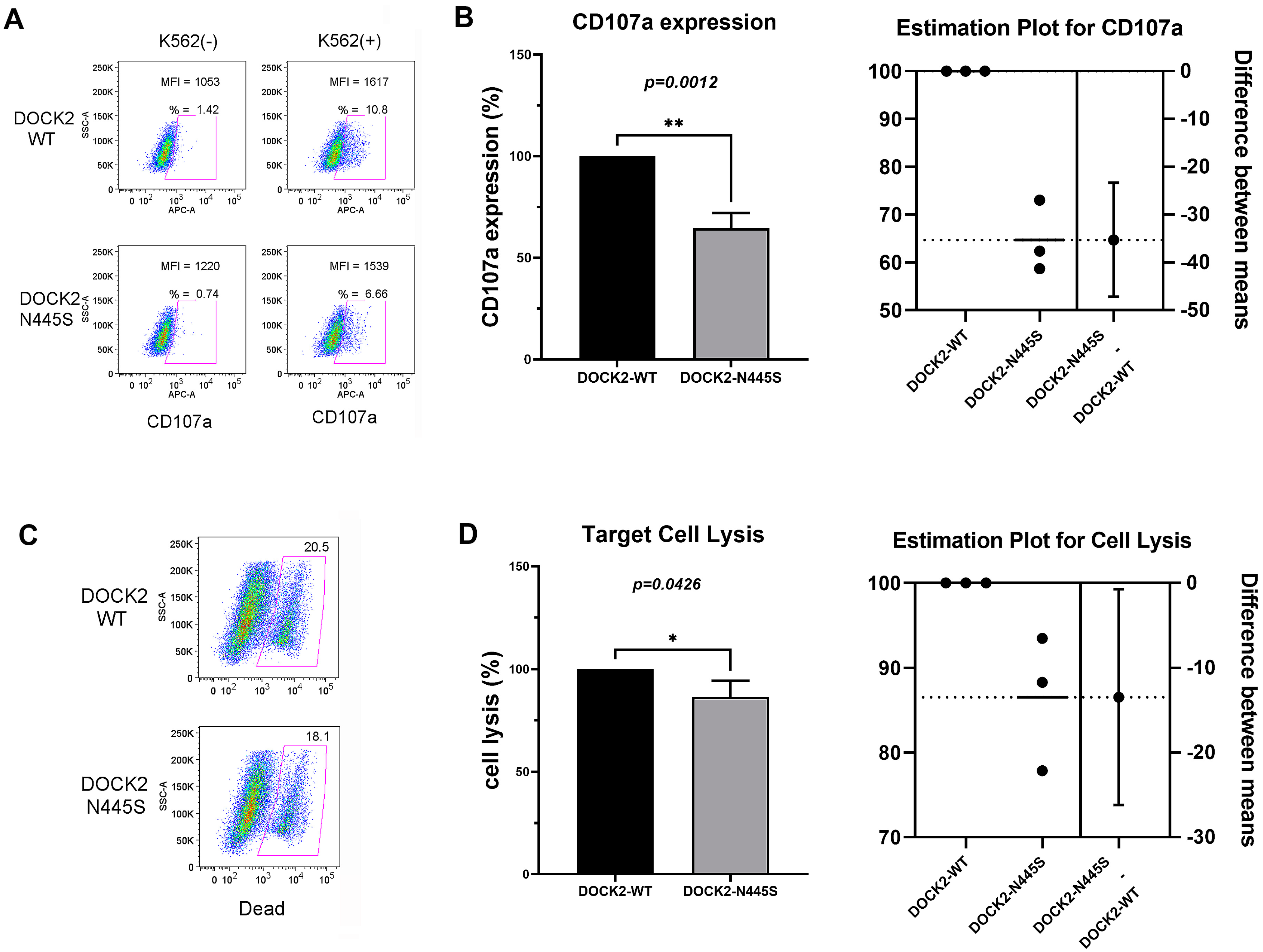
Figure 1. DOCK2 p.Asn445Ser mutation decreased NK-cell cytotoxicity and degranulation. (A) CD107a expression was analyzed by flow cytometry in WT NK-92 cells and DOCK2 p.Asn445Ser mutant NK-92 cells with and without K562 erythroleukemia cell co–stimulation. CD107a positive cells and mean fluorescence intensity are noted in each plot. (B) CD107a expression was significantly decreased in DOCK2 p.Asn445Ser mutant NK-92 cells after two hours of stimulation with K562 cells as compared to WT (** = p < 0.01). (C) WT NK-92 and DOCK2 p.Asn445Ser mutant NK-92 cells were mixed with K562 erythroleukemia cells for four hours at an effector-to-target cell ratio of 2:1 and stained with live/dead fixable cell dead reagent. Flow cytometry analysis of cell death is depicted along the x-axis. (D) DOCK2 p.Asn445Ser mutant NK-92 cells caused statistically significantly less (* = p < 0.05) cell lysis when stimulated as compared to WT NK-92 cells. The pictured results in (A,C) are one representative example of the three independent experiments summarized in (B,D).
Life 13 00434 g001
Table 1. Hospitalizations and blood counts.
Age (Years)AdmissionWBC CountANCALCHgbPlatelet Count
×103/μL×103/μL×103/μLg/dL×103/μL
7Fever, emesis
Negative infectious workup
Min: 1.53
Max: 7.24
(nml 4.31–11.0)
Min: 0.72
(nml 1.63–7.55)
Min: 0.34
(nml 0.97–3.96)
Min: 6.5
(nml 10.7–13.4)
Min: 108
Max: 426
(nml 140–440)
7Positive blood cultures:
Staphylococcus aureus
Candida albicans
Min: 1.82
Max: 8.57
(4.31–11.0)
Min: 1.09
(1.63–7.55)
Min: 0.43
(0.97–3.96)
Min: 6.3
(10.7–13.4)
Min: 55
Max: 471
(140–440)
7Fever, cough, congestion
Negative infectious workup
Min: 1.73
Max: 6.55
(4.31–11.0)
Min: 1.28
(1.63–7.55)
Min: 0.19
(0.97–3.96)
Min: 9.2
(10.7–13.4)
Min: 90
Max: 277
(140–440)
8Positive blood cultures:
Candida albicans
Min: 2.90
Max: 9.09
(4.31–11.0)
Min: 1.61
(1.63–7.55)
Min: 0.42
(0.97–3.96)
Min: 6.3
(10.7–13.4)
Min: 34
Max: 112
(140–440)
8Fever, lethargy
Negative infectious workup
Min: 0.39
Max: 2.47
(4.31–11.0)
Min: 0.23
(1.63–7.55)
Min: 0.14
(0.97–3.96)
Min: 6.9
(10.7–13.4)
Min: 94
Max: 400
(140–440)
10Positive blood cultures:
Bacillus licheniformis
Min: 0.73
Max: 4.82
(4.31–11.0)
Min: 0.45
(1.63–7.55)
Min: 0.24
(0.97–3.96)
Min: 5.8
(10.7–13.4)
Min: 25
Max: 394
(140–440)
10Fever, lethargy
Negative infectious workup
Min: 1.05
Max: 2.86
(4.31–11.0)
Min: 0.61
(1.63–7.55)
Min: 0.41
(0.97–3.96)
Min: 6.4
(10.7–13.4)
Min: 63
Max: 155
(140–440)
10Positive blood cultures:
Staphylococcus aureus
Min: 2.72
Max: 5.01
(4.31–11.0)
Min: 1.97
(1.63–7.55)
Min: 0.44
(0.97–3.96)
Min: 8.8
(10.7–13.4)
Min: 40
Max: 124
(140–440)
10Positive blood cultures:
Staphylococcus aureus
Min: 0.96
Max: 1.07
(4.31–11.0)
Min: 0.76
(1.63–7.55)
Min: 0.26
(0.97–3.96)
Min: 8.1
(10.7–13.4)
Min: 197
Max: 236
(140–440)
11Fever, lethargy
Negative infectious workup
Min: 2.40
Max: 3.81
(4.31–11.0)
Min: 1.26
(1.63–7.55)
Min: 0.59
(0.97–3.96)
Min: 9.6
(10.7–13.4)
Min: 109
Max: 262
(140–440)
11Positive blood cultures:
Klebsiella pneumoniae,
Staphylococcus aureus
Min: 1.98
Max: 5.93
(4.31–11.0)
Min: 1.04
(1.63–7.55)
Min: 0.26
(0.97–3.96)
Min: 7.5
(10.7–13.4)
Min: 51
Max: 239
(140–440)
Values outside of the normal ranges are bolded. Abbreviations: WBC—white blood cell; ANC—absolute neutrophil count; ALC—absolute lymphocyte count; Hgb—hemoglobin; nml—normal; min—minimum value; max—maximum value.
Table 2. Hospitalizations and inflammatory/liver markers.
Age (Years)AdmissionASTALTFerritinCRPESRFibrinogenLDH
U/LU/Lng/mLmg/dLmm/hrmg/dLU/L
7Fever, emesis
Negative infectious workup
Max: 399
(nml 15–40)
Max: 104
(nml 10–35)
Max: 52,650
(nml 22–340)
Max: 6.61
(nml < 0.5)
Min: 6
Max: 76
(nml 0–15)
Max/Min: 323
(nml 156–400)
Max: 10,884
(nml 420–750)
7Positive blood cultures:
Staphylococcus aureus
Candida albicans
Max: 392
(15–40)
Max: 118
(10–35)
Max: 36,972
(22–340)
Max: 21.12
(<0.5)
Min: 11
Max: 73
(0–15)
Min: 128
Max: 559
(156–400)
7Fever, cough, congestion
Negative infectious workup
Max: 229
(15–40)
Max: 316
(10–35)
Max: 13,194
(22–340)
Max: 8.84
(<0.5)
Min: 7
Max: 16
(0–15)
Max/Min: 195
(nml 156–400)
Max: 4272
(420–750)
8Positive blood cultures:
Candida albicans
Max: 76
(18–36)
Max: 45.5
(9.0–25.0)
Max: 1957
(13.7–78.8)
Max: 13.23
(<0.5)
Min: 14
Max: 52
(0–15)
Min: 309
Max: 532
(156–400)
8Fever, lethargy
Negative infectious workup
Max: 175
(18–36)
Max: 72.5
(9.0–25.0)
Max: 20,455.6
(13.7–78.8)
Max: 3.6
(<0.5)
Min: 11
Max: 30
(0–15)
10Positive blood cultures:
Bacillus licheniformis
Max: 127
(18–36)
Max: 121.3
(9.0–25.0)
Max: 4429.6
(13.7–78.8)
Max: 7.88
(<0.5)
Min: 9
Max: 30
(0–15)
Min: 84
Max: 197
(156–400)
Max: 1630
(170–283)
10Fever, lethargy
Negative infectious workup
Max: 225
(18–36)
Max: 168.9
(9.0–25.0)
Max: 2727.1
(13.7–78.8)
Max: 4.42
(<0.5)
Min: 9
Max: 24
(0–15)
Max/Min: 160
(nml 156–400)
10Positive blood cultures:
Staphylococcus aureus
Max: 100
(18–36)
Max: 100
(9.0–25.0)
Max: 896.1
(13.7–78.8)
Max: 15.52
(<0.5)
Min: 33
Max: 51
(0–15)
10Positive blood cultures:
Staphylococcus aureus
Max: 171
(18–36)
Max: 134.5
(9.0–25.0)
Max: 827.1
(13.7–78.8)
Max: 1.88
(<0.5)
Min: 15
Max: 28
(0–15)
11Fever, lethargy
Negative infectious workup
Max: 95
(18–36)
Max: 83.5
(9.0–25.0)
Max: 239.1
(13.7–78.8)
Max: 13.46
(<0.5)
Min/Max: 13
(0–15)
11Positive blood cultures:
Klebsiella pneumoniae,
Staphylococcus aureus
Max: 44
(18–36)
Max: 39
(9.0–25.0)
Max: 196
(13.7–78.8)
Max: 14.3
(<0.5)
Min: 48
Max: 77
(0–15)
Max: 241
(170–283)
Values outside of the normal ranges are bolded. Abbreviations: AST—aspartate aminotransferase; ALT—alanine transaminase; CRP—c-reactive protein; ESR—erythrocyte sedimentation rate; LDH—lactate dehydrogenase; nml—normal; min—minimum value; max—maximum value.
Table 3. Immunodeficiency genetic panel results.
GeneVariantZygosityVariant ClassificationDisease Association
G6PDc.[202G>A;376A>G] (p.[Val68Met;ASn126Asp])hemizygousPathogenicX-linked G6PD deficiency
PEPDDeletion (Exon 1)heterozygousPathogenicAutosomal recessive prolidase deficiency
PEPDc.932G>A (p.Arg311Gln)heterozygousUncertain SignificanceAutosomal recessive prolidase deficiency
DOCK2c.1334A>G (p.Asn445Ser)heterozygousUncertain SignificanceAutosomal recessive combined immunodeficiency due to DOCK2 deficiency
ERCC6L2c.4089T>G (p.Asn1363Lys)heterozygousUncertain SignificanceAutosomal recessive ERCC6L2 deficiency
FANCAc.753_755delinsAG (p.Asp252Ser)heterozygousUncertain SignificanceAutosomal recessive Fanconi anemia type A
FCHO1c.529C>T (p.Arg177Cys)heterozygousUncertain SignificanceAutosomal recessive combined immunodeficiency due to FCHO1 deficiency
G6PC3c.50A>C (p.Asn17Thr)heterozygousUncertain SignificanceAutosomal recessive severe congenital neutropenia
RBCK1c.1028C>T (p.Ala343Val)heterozygousUncertain SignificanceAutosomal recessive polyglucosan body myopathy with or without immunodeficiency
RTEL1c.3499+7C>T (intronic)heterozygousUncertain SignificanceAutosomal recessive dyskeratosis congenital spectrum disorders
SAMD9Lc.251dup (p.Asn84Lysfs * 3)heterozygousUncertain SignificanceAutosomal dominant ataxia-pancytopenia syndrome
UNC45Ac.2003C>A (p.Ser668Tr)heterozygousUncertain SignificanceNo well-established disease association
Disclaimer/Publisher’s Note: The statements, opinions and data contained in all publications are solely those of the individual author(s) and contributor(s) and not of MDPI and/or the editor(s). MDPI and/or the editor(s) disclaim responsibility for any injury to people or property resulting from any ideas, methods, instructions or products referred to in the content.