Article

Vitamin D3 Supplementation: Comparison of 1000 IU and 2000 IU Dose in Healthy Individuals

1
Department of Pharmacology and Toxicology, Faculty of Medicine in Pilsen, Charles University, 323 00 Pilsen, Czech Republic
2
Department of Immunochemistry, University Hospital and Faculty of Medicine in Pilsen, Charles University, 323 00 Pilsen, Czech Republic
3
Faculty of Science, Palacky University in Olomouc, 779 00 Olomouc, Czech Republic
4
Department of Clinical Biochemistry and Hematology, University Hospital and Faculty of Medicine in Pilsen, Charles University, 323 00 Pilsen, Czech Republic
*
Author to whom correspondence should be addressed.
Life 2023, 13(3), 808; https://doi.org/10.3390/life13030808
Submission received: 13 February 2023 / Revised: 10 March 2023 / Accepted: 13 March 2023 / Published: 16 March 2023
(This article belongs to the Special Issue Feature Papers in Medical Research)

Abstract

:
Background: Scientific studies point to a significant global vitamin D deficiency. The recommended dose of vitamin D for the adult population in Central Europe is 800–2000 IU/day. The aim of our study was to determine whether doses of 1000 IU or 2000 IU of vitamin D3 are adequate to achieve the sufficiency reference values of [25(OH)D]. Methods: Seventy-two healthy volunteers, average age twenty-two, took part in the study. The study was conducted from October to March in order to eliminate intra-dermal vitamin D production. Vitamin D3 in an oleaginous mixture was used. The participants used either 1000 IU or 2000 IU/daily for two 60-day periods with a 30-day break. Results: The dose of 1000 IU, taken for 60 days, increased vitamin D levels relatively little. Furthermore, serum vitamin D levels decreased in the 30 days following the cessation of supplementation. Taking 2000 IU daily led to a sharp increase in serum levels which plateaued 30 days after the subjects stopped using vitamin D3 drops. Conclusions: Both doses, taken daily, can help maintain adequate vitamin D levels during the winter months. A daily dose of 2000 IU, however, maintained the desired levels of vitamin D for a longer period.

1. Introduction

There has been a huge interest in vitamin D in recent years, not only in the fields of science and medicine but among the general public as well. This interest in vitamin D was sparked owing to a number of publications that have highlighted the worldwide vitamin D deficiency [1,2,3,4,5]. Vitamin D deficiency has been reported in 40% of the European population, 24% of the population in the USA and 37% of the Canadian population. Pregnant women, obese children and adults, populations with darker skin tone and people who rarely expose themselves to sunlight are all groups at risk of developing hypovitaminosis D [6,7].
Vitamin D belongs to the group of fat-soluble vitamins that are essential for human health. There are three ways for an individual to obtain vitamin D: exposure to ultraviolet light (UV), consuming vitamin D in food, or via supplementation [8].
We distinguish two basic forms of vitamin D: ergocalciferol and cholecalciferol. The chemical structures of ergocalciferol and cholecalciferol are not identical, but their metabolites show similar biological effects. Ergocalciferol, also known as a vitamin D2, is formed in the presence of UV radiation from its precursor, ergosterol. This occurs mainly in fungi, plants and yeast [9]. The dominant form in the animal world, and thus in humans, is cholecalciferol, or vitamin D3. Vitamin D3 is synthesized de novo in the skin from 7-dehydrocholesterol under the action of UV radiation. Depending on the wavelength, we divide UV radiation into UVA (315–400 nm), UVB (280–315 nm) and UVC (100–280 nm). UVB rays are responsible for the conversion of vitamin D3 from its precursors [10,11].
The axial tilt of the earth determines the amount of UVB that reaches the earth’s surface. In the winter months, when the angle of the sun on the horizon is lower than 45°, the rays take longer to go through the atmosphere and this results in an increased absorption of UVB radiation [12]. The amount of UVB radiation, and thus the amount of vitamin D3 that our bodies can produce, depends significantly on the latitude, season and time of day [13]. Only minimal or no vitamin D3 synthesis occurs at higher latitudes (above 33°) during the autumn and winter months and in the morning and late afternoon [14,15]. The synthesis of vitamin D3 is affected by a number of other external factors such as the weather, environmental pollution, layers of clothing and the use of sunscreen. Personal factors including genetic polymorphisms, age and skin colour also play a significant role since melanin in the epidermis significantly reduces the efficiency of UV radiation absorption [16].
Vitamin D3, having been synthesized in the skin, binds to a vitamin D-binding protein (VDBP) and is transported through the blood into the liver, where it undergoes hydroxylation and becomes hydroxyvitamin D3 [25(OH)D3], or calcidiol [17].
The biologically inactive form [25(OH)D3] is transported to the kidneys, where it undergoes further hydroxylation to its active form: 1α,25-d-hydroxyvitamin D3 [1,25(OH)2D3], or calcitriol [18]. The liver and kidneys are the main tissues responsible for vitamin D3 metabolism; however hydroxylases necessary for the synthesis of calcitriol are also present in various other tissues, including the skin.
Vitamin D itself has no significant biological activity. The effects of vitamin D are mediated through its active metabolite [1,25(OH)2D], which has the properties of a hormone and therefore vitamin D can be considered a prohormone [19]. The biological effect of [1,25(OH)2D] is manifested by the regulation of gene expression through binding to the vitamin D receptor (VDR) in the target tissue [20].
VDR can be found in almost every cell and tissue of the human body and this results in vitamin D having a very wide spectrum of effect [21]. The role of vitamin D in the regulation of calcium and phosphorus, a process that supports most metabolic neuromuscular transmission functions as well as the mineralization of bones, is widely acknowledged [22,23]. Vitamin D deficiency is associated with an increased risk of osteopenia, osteoporosis, fractures and rickets in childhood [24,25,26]. In recent years, there have been many studies that point to the extra-skeletal effects of vitamin D. Cell proliferation, differentiation and immune modulation are all affected by vitamin D levels [27,28]. Furthermore, vitamin D plays a role in a number of diseases [21,29], such as: type 1 and 2 diabetes [30,31,32], hypertension [33,34,35,36], cardiovascular disease [37,38,39], autoimmune diseases [40,41,42] and cancer [43,44,45,46].
Vitamin D levels are commonly determined based on a measurement of [25(OH)D], the main stock form of vitamin D. There is no global consensus on the [25(OH)D] concentration that defines vitamin D deficiency [6]. In Europe, a concentration of [25(OH)D] < 25 nmol/L has been considered critical for several decades now and a patient presenting with these levels is considered at risk of metabolic bone disease. According to the Endocrinology Society, blood serum levels of [25(OH)D] < 50 nmol/L are commonly considered a vitamin D deficiency, while concentrations of [25(OH)D] > 75 nmol/L are considered sufficient by most authors [15,47]. Various studies point to the fact that parathyroid hormone levels are only reduced in patients presenting with levels below 75 nmol/L of [25(OH)D] and thus concentrations of [25(OH)D] in a range of 75–125 nmol/L are commonly considered optimal for reducing the risk of developing secondary hyperparathyroidism and for maximizing musculoskeletal health [48,49,50,51]. Overall, values of [25(OH)D] up to 250 nmol/L are considered safe for children and adults [26].
Historically, vitamin D was mainly prescribed to prevent rickets. A dose of 200 IU/day of vitamin D3 was considered adequate for people under the age of fifty. However, in 2011, the US Institute of Medicine (IOM) established that a dose of 200 IU/day is insufficient. While this low dose addresses bone metabolism issues, it does not support the other important physiological functions of vitamin D [23]. The IOM then adjusted the daily vitamin D3 allowances to 400 IU for infants, 600 IU for children and 800 IU for adults; doses which are adequate for maintaining a concentration of 50 nmol/L of [25(OH)D] [52].
Nevertheless, a number of other studies have evaluated values of [25(OH)D] 50 nmol/L as insufficient [53,54,55,56]. The Endocrine Society in the USA recommends supplementing with a daily dose of 1000 IU for children who are over a year old and 1500–2000 IU for adults, so as to achieve a concentration of at least [25(OH)D] 75 nmol/L [48]. Daily vitamin D3 recommendations for Central Europe were published in 2013. The recommended dose for neonates and infants is 400–600 IU/day, for children and adolescents 600–1000 IU/day and for adults 800–2000 IU/day [57]. The aim of our study was to determine whether doses of 1000 IU or 2000 IU of vitamin D3 are sufficient for achieving and maintaining the reference values 75–200 nmol/L of [25(OH)D] in a sample of healthy young people during the winter months in the Czech Republic, a time when there is no natural synthesis of vitamin D.

2. Materials and Methods

2.1. Participants

A total of seventy-two healthy volunteers (fifty female and twenty-two male medical students), whose mean age was 22 years (20–27 years range). All participants filled in a questionnaire aimed at gathering information on the state of their health. Additional information about their type of diet, or use of hormonal contraception in women, was also collected as diet and hormonal contraceptives are known to influence changes in serum vitamin D levels.
The characteristics of the participants are summarized in Table 1. No additional supplementation of vitamin D in the form of medication or food supplements was allowed.
None of the participants was excluded based on the state of their health. No metabolic or endocrine diseases were reported.
This study was conducted in the period of October to March in order to eliminate the interference of intradermal production of vitamin D3.

2.2. Dosing Regimen

Participants were requested to take a vitamin D3 supplement according to the dosing regimen depicted in Table 2. An oral solution containing 0.5 mg/mL (20,000 IU) of vitamin D3 dissolved in an oleaginous mixture in a drip container was used. One drop of the solution is equivalent to 12.5 μg (500 IU) of vitamin D3 and the participants were instructed to use two to four drops daily for 60 days with a 30-day break after each 60-day period.

2.3. Blood Samples

The blood drawing schedule is depicted in Table 3. Blood was drawn five times in total over the course of six months.
Samples of peripheral blood were acquired using a VACUETTE® CAT Serum Separator Clot Activator (Greiner Bio-One, Kremsmünster, Austria) serum collection tube. After cooling down the samples to room temperature, the tubes were centrifuged at 1700 rpm for 10 min in order to separate the serum from the whole blood sample. Every serum sample was then divided into three aliquots of an equal volume of 500 µL and stored at −80 °C before further analysis.

2.4. Sample Analysis

Measurement of total vitamin D levels was performed using the Access 25(OH) Vitamin D Total chemiluminescent assay in a Unicel® DxI 800 (Beckman Coulter, Brea, CA, USA). This assay meets the Ghent University 25(OH) standard [58] and detects both 25(OH) vitamin D2 and 25(OH) vitamin D3. All samples were thawed at the same time and analyzed in one batch. All measurements were performed in accordance with the instructions provided by the manufacturers.
Analyses of calcium, magnesium and phosphate concentrations were performed using the Cobas system (Cobas 8000 Analyzer, Cobas c702 module, Roche Diagnostics, Basel, Switzerland).

2.5. Statistical Analysis

Statistical analysis was performed using SAS, V. 9.4. (SAS Institute Inc., Cary, NC, USA) and MATLAB, V. R2007b (The Math-Works, Inc., Natick, MA, USA).
Discrete characteristics are expressed as frequency and percentages; continuous characteristics are expressed as mean, median, minimum, maximum, lower and upper quartile of serum vitamin D levels. The Chi-square test and Wilcoxon two-sample test were used to compare subgroups and determine statistical significance for categorical and continuous data, respectively.
Correlations were analyzed using Spearman rank correlation coefficient.
p-value < 0.05 was considered to be statistically significant.
Graphical statistical displays in the form of box–whisker plots and spaghetti plots are presented below.
For the power analysis, we assumed an increase in vitamin D from a mean of 75 nmol/L to a mean of 80 nmol/L after supplementation.
In accordance with previous studies, the standard deviation was assumed as 15 nmol/L. The correlation between the value before and after supplementation is relatively strong (r = 0.60 also according to previous experiences).
One-sided testing was assumed, and therefore one-sided Alpha = 0.025 (the same as two-sided Alpha = 0.0) and a power of 80% were assumed, due to the explanatory character of all the statistical analysis performed.
The sample size estimation carried out prior to the study resulted in 71 subjects.

3. Results

The summarized results of measured vitamin D levels are shown in Table 4.
The mean initial concentration of serum [25(OH)D] was 75.50 nmol/L with a standard deviation of 20.30 nmol/L. The lowest recorded value was 28.18 nmol/L and the highest 134.47 nmol/L. One participant had deficient [25(OH)D] concentrations (28.18 nmol/L), six (8.3%) participants had insufficient [25(OH)D] concentrations (37.08–48.20 nmol/L), thirty-eight (51.4%) participants had [25(OH)D] levels lower than 75 nmol/L (50.21–74.58 nmol/L). Twenty-seven (37.5%) participants had adequate serum [25(OH)D] concentrations (higher than 75 nmol/L).
Following the 1000 IU vitamin D3 supplementation, the mean concentration rose to 83.43 nmol/L with a standard deviation of 20.77 nmol/L. The lowest recorded value was 51.89 nmol/L and the highest was 157.68 nmol/L. No participants had [25(OH)D] concentrations in the range of deficiency or insufficiency. Thirty (41.7%) participants had [25(OH)D] levels higher than 50 nmol/L but lower than 75 nmol/L (51.89–73.20 nmol/L). Forty-two (58.3%) participants had adequate [25(OH)D] concentrations ranging from 75.21 to 157 nmol/L.
After the 30-day break in supplementation, the mean concentration fell to 66.54 nmol/L with a standard deviation of 15.70 nmol/L. One participant had deficient [25(OH)D] concentrations (34.25 nmol/L). Seven (9.7%) participants had insufficient concentrations (43.65–49.49 nmol/L). Concentrations lower than 75 nmol/L were found in forty-two (58.3%) participants (51.44–74.78 nmol/L). Twenty-two (30.5%) participants had adequate serum [25(OH)D] concentrations (76.63–117.15 nmol/L).
The mean concentrations of serum [25(OH)D] levels after 60 days of 2000 IU supplementation rose to 81.36 nmol/L with a standard deviation of 16.30 nmol/L. No participant had [25(OH)D] levels indicative of deficiency. One participant had insufficient [25(OH)D] levels (46.04 nmol/L). Twenty-seven (37.5%) participants had concentrations below 75 nmol/L (59.89 nmol/L–74.11 nmol/L). Forty-four (61.1%) participants had serum [25(OH)D] levels higher than 75 nmol/L (75.12–151.76 nmol/L).
Following the second 30-day break, the mean concentration of serum [25(OH)D] was 82.21 nmol/L with a standard deviation of 17.91 nmol/L. No participant had deficient [25(OH)D] concentrations. Two (2.8%) participants had insufficient [25(OH)D] concentrations (42.63–49.29 nmol/L). Twenty-four (33.3%) participants had concentrations higher than 50 nmol/L but lower than 75 nmol/L (54.21–74.46 nmol/L). Forty-six (63.9%) participants had sufficient [25(OH)D] levels (76.69–126.94 nmol/L).
A visual representation of the results is given in box plot Figure 1.
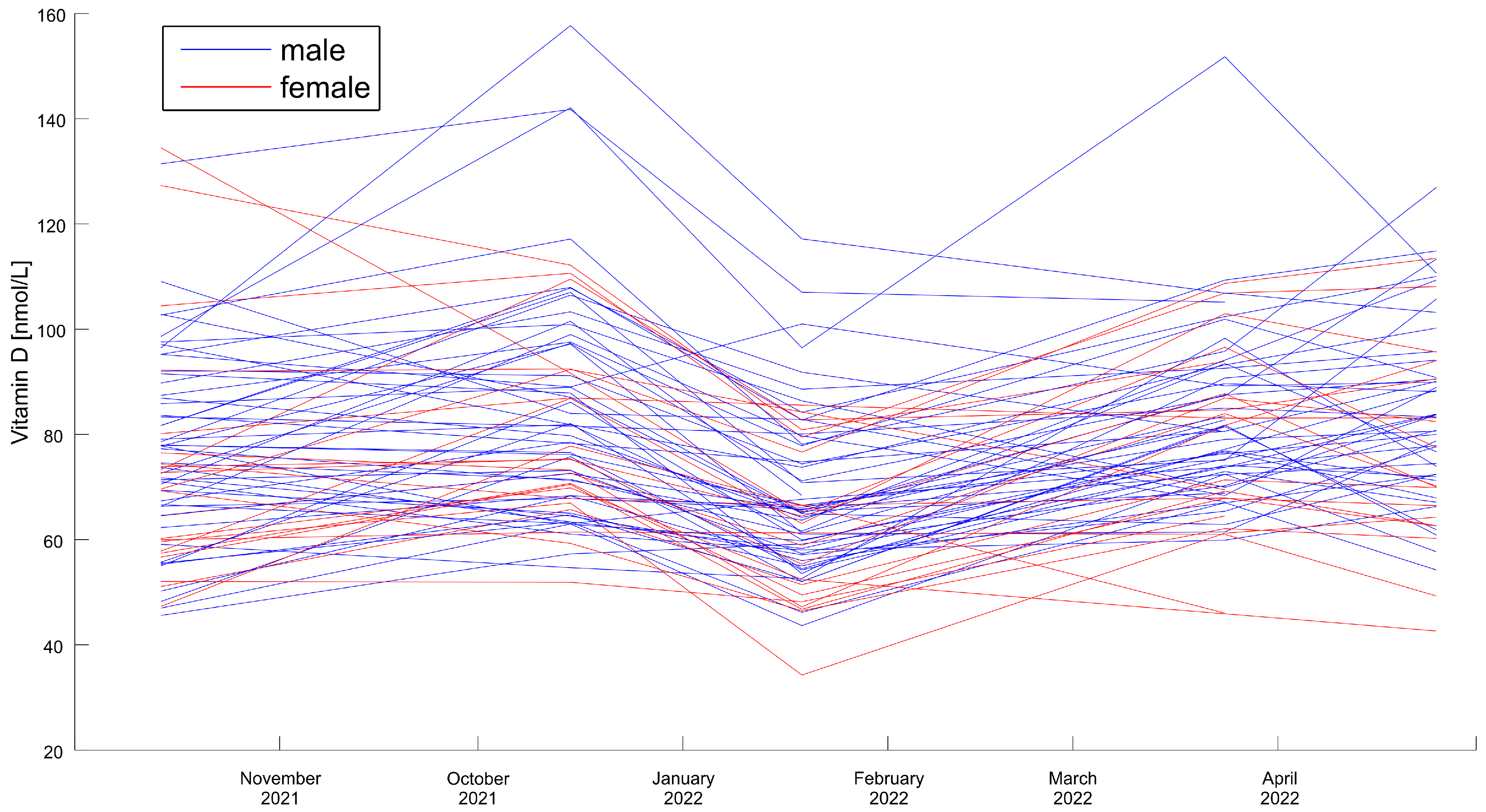
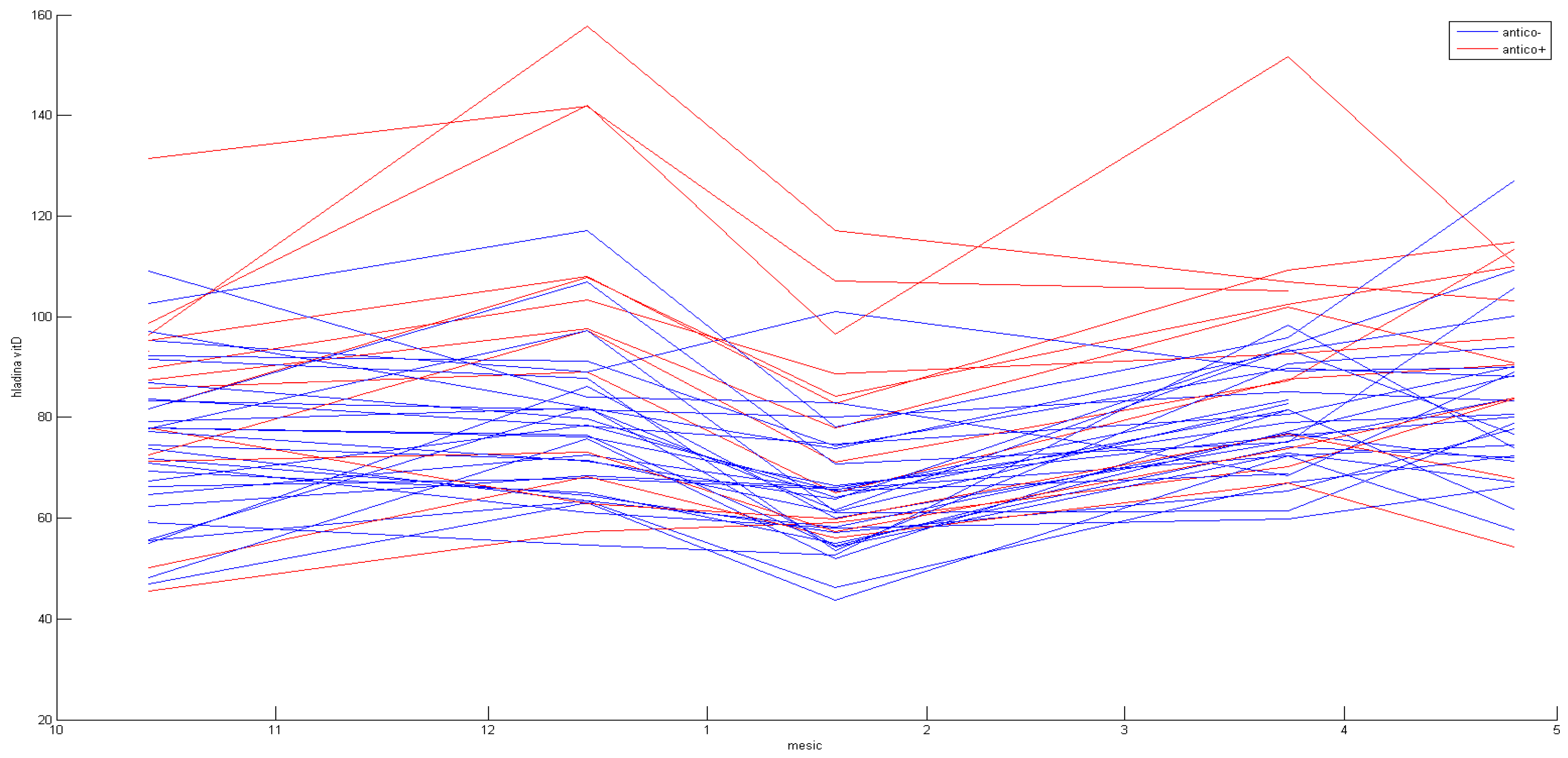
The general trends in vitamin D concentrations in all individual participants are presented in a spaghetti plot in Figure 2.
We did not observe statistically significant differences between the sexes: the initial levels and the levels of vitamin D during the course of supplementation did not differ significantly. The results in men are slightly lower in each measurement, but without statistical significance.
[25(OH)D] concentrations were higher in female participants using hormonal contraceptives. There was a statistically significant difference in the increase between the first and second sampling (p < 0.0308), and then vitamin D levels stayed higher with varying statistical significance (p < 0.0308; p < 0.0242; p < 0.0501 non-significant, respectively). The results are presented visually in Figure 3. Detailed results can be seen in Table 5.
There was no statistically significant difference in [25(OH)D] concentrations in participants on vegan or vegetarian diets.
Mean concentration of serum calcium, magnesium and phosphates and their maximums and minimums are presented in Table 6.

4. Discussion

Vitamin D is an important micronutrient involved in a variety of physiological functions. There is, however, no consensus as to the vitamin D levels necessary for optimal health. The concentration of 50 nmol/L of [25(OH)D] is considered essential for maintaining adequate skeletal health [59,60], but no optimal concentration level has been established for extra-skeletal health. Concentrations of [25(OH)D] higher than 120 nmol/L have been shown to significantly decrease parathyroid hormone concentrations [61], significantly decrease adverse pregnancy outcomes [62] and enable the transfer of cholecalciferol into breast milk in breast-feeding women [63]. Concentrations of 120 nmol/L or above are common in individuals who are often exposed to sunlight: individuals who work outdoors and live in tropical and subtropical regions [64].
A group of participants, aged 22–27, was selected to describe changes in vitamin D levels in healthy subjects. A young cohort was chosen as older people are known to be more prone to vitamin D insufficiency, regardless of latitude [65]. Several changes in vitamin D metabolism are age-dependent. The absorption of calcium from the intestines decreases with age [66], the number of vitamin D receptors is lower in the elderly [67] and the hydroxylation of [25(OH)D] in the kidneys is reduced with aging as renal function decreases [68]. An oil-based oral solution was chosen for its assumed higher bioavailability in comparison to solid dosage forms [69].
The effectiveness of both supplementation dosage regimens was visible in our study. The study started with twenty-seven (37.5%) participants who had adequate serum [25(OH)D] concentrations (higher than 75 nmol/L). The first supplementation dose of 1000 IU, taken for 60 days, increased the mean vitamin D concentrations relatively little, with median values that ranged from 73.8 nmol/L to 78.4 nmol/L (6.2%). The change was, however, statistically significant, as p < 0.0288. Forty-two (58.3%) participants reached adequate [25(OH)D] concentrations ranging from 75.21 to 157 nmol/L. After the first dosage regimen, the participants took no supplements for 30 days, which explains the sharp drop in the median values from 78.4 nmol/L to 64.7 nmol/L; i.e., a drop of 13.7 nmol/L (17.5%) between the second and third blood sampling. This decrease was highly statistically significant: p < 0.0001. The second supplementation dose of 2000 IU, taken for 60 days, increased the mean vitamin D concentrations sharply. Between the third and fourth blood sampling the median increased sharply: from 64.7 nmol/L to 80.5 nmol/L; i.e., by 15.8 nmol/L (24.4%). This increase was highly statistically significant: p < 0.0001. Forty-four (61.1%) participants had serum [25(OH)D] levels higher than 75 nmol/L (75.12–151.76 nmol/L).
The value of vitamin D remained stable between the fourth and fifth sampling, with a negligible increase in the median from 81.4 nmol/L to 82.2 nmol/L; p = 0.5118. This stood in contrast to the first supplementation dose of 1000 IU, after which a significant decrease in [25(OH)D] concentration was observed during the first 30-day break from vitamin D3 drop administration.
While the daily dosing of 1000 IU cholecalciferol successfully increased the levels of serum vitamin D during winter months, a significant decrease in serum vitamin D was observed thirty days after discontinuation. A dose of 2000 IU increased vitamin D levels in a manner similar to the lower dose. There was, however, a minimal decrease in vitamin D levels 30 days after the supplementation ceased. Both 1000 IU and 2000 IU doses were therefore shown to be safe for substitution therapy. There were no cases of levels higher than 200 nmol/L, which is the upper limit of the laboratory reference values. Calcium and phosphate concentrations remained stable throughout this study.
There are very few other studies evaluating the effect of 1000 IU and 2000 IU vitamin D3 supplementation in a healthy population in the winter months at a similar latitude.
In 2017, Piltz et al. compared the effectiveness of vitamin D3 supplementation in a sample of young German women. Their study, which took place in the winter period and included two groups of patients, used multi-micronutrient supplements along with vitamin D3. In the first group of patients the daily dose of vitamin D3 was 200 IU, whereas in the second group the daily dose of vitamin D3 was 800 IU. In the 200 IU group, the required concentration of 75 nmol/L was reached in only 15.3% of patients, and in the 800 IU group, the required concentration of 75 nmol/L was reached in a mere 66.3% of patients [70].
In 2008, Holick et al. conducted a study on a sample of healthy adults in which they reported that even a daily dose of 1000 IU of vitamin D3 is not sufficient in order to achieve a concentration of 75 nmol/L [25(OH)D] in the blood during the winter months [71].
Diamond et al. published a 2012 study in which they provided vitamin D deficient patients with supplements. These patients were given a dose of either 2000 IU or 5000 IU of vitamin D3 for three months: from February to April. The study reported that only 43% of the patients taking the 2000 IU dose achieved the required concentration of 75 nmol/L, whereas 93% of the patients who were given 5000 IU reached the level of sufficiency. It is important to note that the patients were selected based on a pre-existing deficiency. The doses used in our study reflected a seasonal decrease in vitamin D in otherwise healthy individuals [72].
The available literature supports our observation that use of hormonal contraception proved to be associated with statistically significant higher [25(OH)D] plasma concentrations [73,74].
Vegetarians and vegans might be at risk of low vitamin D intake [75,76,77]. Our study, however, showed no statistically significant difference in [25(OH)D] levels between omnivores and vegetarians or vegans at any time point. Such results can be explained by the quality and adequacy of the participants’ vegan and vegetarian diet, since individual nutrient intake is known to vary significantly [78]. Furthermore, we had a relatively small sample size of vegans and vegetarians (8; 11.1% of all participants) and a majority of omnivores.

5. Conclusions

Our study is unique in its utilization of a group of healthy participants. The study thus puts forward the supplementation regimens that are suitable for a young healthy population during the winter months; a time when vitamin D production in the skin is low. The study is limited by its use of a relatively simple model of supplementation, without a cross-over or placebo control. It should, however, be noted that studies with a more complicated design are not common in vitamin D research.
Our results show that a daily dose of either 1000 IU or 2000 IU of vitamin D3 is an adequate choice for maintaining sufficient vitamin D levels during the winter months. A daily dose of 2000 IU was, however, observed to maintain the desired levels of serum vitamin D concentration for a longer period than a daily dose of 1000 IU and can thus be recommended for supplementing young healthy individuals during the winter months.

Author Contributions

Conceptualization, R.K., E.D. and M.J.; methodology, R.K. and M.K.; investigation, R.V., M.J., E.D., P.B. and M.K.; data curation, L.P.; writing—original draft preparation, R.V., M.J., T.F. and E.D.; writing—review and editing, O.T., L.P. and R.K.; visualization, T.F.; supervision, R.K.; project administration, E.D. All authors have read and agreed to the published version of the manuscript.

Funding

This study was supported by the Ministry of Health, Czech Republic-conceptual development of research organization (Faculty Hospital in Pilsen-FNPl, 00669806), BBMRI-CZ: Biobank net-work—a versatile platform for the research of the etiopathogenesis of diseases CZ.02.1.01/0.0/0.0/16_013/000167 and LM2015089, and by the Cooperation Program, research area Pharmaceutical Sciences.

Institutional Review Board Statement

The study was conducted in accordance with the Declaration of Helsinki, and approved by the Ethics Committee of University Hospital Pilsen and the Faculty of Medicine in Pilsen, Charles University (protocol code 374/2020, date of approval 6 August 2020).

Informed Consent Statement

Informed consent was obtained from all subjects involved in this study.

Data Availability Statement

Data are available from the corresponding author.

Conflicts of Interest

The authors declare no conflict of interest.

References

  1. Zhu, Z.; Zhan, J.; Shao, J.; Chen, W.; Chen, L.; Li, W.; Ji, C.; Zhao, Z. High prevalence of vitamin D deficiency among children aged 1 month to 16 years in Hangzhou, China. BMC Public Health 2012, 12, 126. [Google Scholar] [CrossRef] [PubMed] [Green Version]
  2. Merewood, A.; Mehta, S.D.; Grossman, X.; Chen, T.C.; Mathieu, J.S.; Holick, M.F.; Bauchner, H. Widespread Vitamin D Deficiency in Urban Massachusetts Newborns and Their Mothers. Pediatrics 2010, 125, 640–647. [Google Scholar] [CrossRef]
  3. Shivane, V.K.; Sarathi, V.; Bandgar, T.; Menon, P.; Shah, N.S. High prevalence of hypovitaminosis D in young healthy adults from the western part of India. Postgrad. Med. J. 2011, 87, 514–518. [Google Scholar] [CrossRef] [PubMed]
  4. Greene-Finestone, L.S.; Berger, C.; de Groh, M.; Hanley, D.A.; Hidiroglou, N.; Sarafin, K.; Poliquin, S.; Krieger, J.; Richards, J.B.; Goltzman, D.; et al. 25-Hydroxyvitamin D in Canadian adults: Biological, environmental, and behavioral correlates. Osteoporos. Int. 2011, 22, 1389–1399. [Google Scholar] [CrossRef]
  5. Nesby-O’Dell, S.; Scanlon, K.S.; Cogswell, M.E.; Gillespie, C.; Hollis, B.W.; Looker, A.C.; Allen, C.; Doughertly, C.; Gunter, E.W.; Bowman, B.A. Hypovitaminosis D prevalence and determinants among African American and white women of reproductive age: Third National Health and Nutrition Examination Survey, 1988–1994. Am. J. Clin. Nutr. 2002, 76, 187–192. [Google Scholar] [CrossRef] [PubMed] [Green Version]
  6. Cashman, K.D.; Dowling, K.G.; Škrabáková, Z.; Gonzalez-Gross, M.; Valtueña, J.; De Henauw, S.; Moreno, L.; Damsgaard, C.T.; Michaelsen, K.F.; Mølgaard, C.; et al. Vitamin D deficiency in Europe: Pandemic? Am. J. Clin. Nutr. 2016, 103, 1033–1044. [Google Scholar] [CrossRef] [Green Version]
  7. Amrein, K.; Scherkl, M.; Hoffmann, M.; Neuwersch-Sommeregger, S.; Köstenberger, M.; Tmava Berisha, A.; Martucci, G.; Pilz, S.; Malle, O. Vitamin D deficiency 2.0: An update on the current status worldwide. Eur. J. Clin. Nutr. 2020, 74, 1498–1513. [Google Scholar] [CrossRef]
  8. Schmidt, D.R.; Holmstrom, S.R.; Tacer, K.F.; Bookout, A.L.; Kliewer, S.A.; Mangelsdorf, D.J. Regulation of Bile Acid Synthesis by Fat-Soluble Vitamins A and D. J. Biol. Chem. 2010, 285, 14486–14494. [Google Scholar] [CrossRef] [Green Version]
  9. Tripkovic, L.; Lambert, H.; Hart, K.; Smith, C.P.; Bucca, G.; Penson, S.; Chope, G.; Hyppönen, E.; Berry, J.; Vieth, R.; et al. Comparison of vitamin D2 and vitamin D3 supplementation in raising serum 25-hydroxyvitamin D status: A systematic review and meta-analysis. Am. J. Clin. Nutr. 2012, 95, 1357–1364. [Google Scholar] [CrossRef] [Green Version]
  10. Kulie, T.; Groff, A.; Redmer, J.; Hounshell, J.; Schrager, S. Vitamin D: An Evidence-Based Review. J. Am. Board Fam. Med. 2009, 22, 698–706. [Google Scholar] [CrossRef] [Green Version]
  11. Passeron, T.; Bouillon, R.; Callender, V.; Cestari, T.; Diepgen, T.l.; Green, A.C.; van der Pols, J.C.; Bernard, B.A.; Ly, F.; Bernerd, F.; et al. Sunscreen photoprotection and vitamin D status. Br. J. Dermatol. 2019, 181, 916–931. [Google Scholar] [CrossRef] [PubMed] [Green Version]
  12. Kantheti, R.B.; Kantheti, K.R. Smart Watch to Track the Levels of Vitamin D. Int. Res. J. Eng. Technol. 2020, 7, 4880–4883. [Google Scholar]
  13. Aguilar-Shea, A.L. Vitamin D, the natural way. Clin. Nutr. ESPEN 2021, 41, 10–12. [Google Scholar] [CrossRef] [PubMed]
  14. Webb, A.R.; Kline, L.; Holick, M.F. Influence of Season and Latitude on the Cutaneous Synthesis of Vitamin D3: Exposure to Winter Sunlight in Boston and Edmonton Will Not Promote Vitamin D3 Synthesis in Human Skin. J. Clin. Endocrinol. Metab. 1988, 67, 373–378. [Google Scholar] [CrossRef] [Green Version]
  15. Holick, M.F.; Chen, T.C. Vitamin D deficiency: A worldwide problem with health consequences. Am. J. Clin. Nutr. 2008, 87, S1080–S1086. [Google Scholar] [CrossRef] [Green Version]
  16. Bikle, D.D. Vitamin D metabolism and function in the skin. Mol. Cell. Endocrinol. 2011, 347, 80–89. [Google Scholar] [CrossRef] [Green Version]
  17. Christakos, S.; Dhawan, P.; Verstuyf, A.; Verlinden, L.; Carmeliet, G. Vitamin D: Metabolism, Molecular Mechanism of Action, and Pleiotropic Effects. Physiol. Rev. 2016, 96, 365–408. [Google Scholar] [CrossRef] [Green Version]
  18. Holick, M.F. The vitamin D deficiency pandemic: Approaches for diagnosis, treatment and prevention. Rev. Endocr. Metab. Disord. 2017, 18, 153–165. [Google Scholar] [CrossRef]
  19. Bikle, D.D. Vitamin D Metabolism, Mechanism of Action, and Clinical Applications. Chem. Biol. 2014, 21, 319–329. [Google Scholar] [CrossRef] [Green Version]
  20. Meyer, M.B.; Goetsch, P.D.; Pike, J.W. Genome-wide analysis of the VDR/RXR cistrome in osteoblast cells provides new mechanistic insight into the actions of the vitamin D hormone. J. Steroid Biochem. Mol. Biol. 2010, 121, 136–141. [Google Scholar] [CrossRef] [Green Version]
  21. Nair, R.; Maseeh, A. Vitamin D: The “sunshine” vitamin. J. Pharmacol. Pharmacother. 2012, 3, 118–126. [Google Scholar] [PubMed]
  22. Meyer, M.B.; Goetsch, P.D.; Pike, J.W. VDR/RXR and TCF4/β-Catenin Cistromes in Colonic Cells of Colorectal Tumor Origin: Impact on c-FOS and c-MYC Gene Expression. Mol. Endocrinol. 2012, 26, 37–51. [Google Scholar] [CrossRef] [PubMed]
  23. Holick, M.F. Resurrection of vitamin D deficiency and rickets. J. Clin. Investig. 2006, 116, 2062–2072. [Google Scholar] [CrossRef] [PubMed] [Green Version]
  24. Holick, M.F. Vitamin D Deficiency. N. Engl. J. Med. 2007, 357, 266–281. [Google Scholar] [CrossRef]
  25. DeLuca, H.F. Overview of general physiologic features and functions of vitamin D. Am. J. Clin. Nutr. 2004, 80, S1689–S1696. [Google Scholar] [CrossRef] [Green Version]
  26. Pludowski, P.; Holick, M.F.; Grant, W.B.; Konstantynowicz, J.; Mascarenhas, M.R.; Haq, A.; Povoroznyuk, V.; Balatska, N.; Barbosa, A.P.; Karonova, T.; et al. Vitamin D supplementation guidelines. J. Steroid Biochem. Mol. Biol. 2018, 175, 125–135. [Google Scholar] [CrossRef] [Green Version]
  27. Umar, M.; Sastry, K.S.; Chouchane, A.I. Role of Vitamin D Beyond the Skeletal Function: A Review of the Molecular and Clinical Studies. Int. J. Mol. Sci. 2018, 19, 1618. [Google Scholar] [CrossRef] [Green Version]
  28. Aranow, C. Vitamin D and the Immune System. J. Investig. Med. 2011, 59, 881–886. [Google Scholar] [CrossRef] [Green Version]
  29. Pludowski, P.; Holick, M.F.; Pilz, S.; Wagner, C.L.; Hollis, B.W.; Grant, W.B.; Shoenfeld, Y.; Lerchbaum, E.; Llewellyn, D.J.; Kienreich, K.; et al. Vitamin D effects on musculoskeletal health, immunity, autoimmunity, cardiovascular disease, cancer, fertility, pregnancy, dementia and mortality—A review of recent evidence. Autoimmun. Rev. 2013, 12, 976–989. [Google Scholar] [CrossRef]
  30. Aljabri, K.S.; Bokhari, S.A.; Khan, M.J. Glycemic changes after vitamin D supplementation in patients with type 1 diabetes mellitus and vitamin D deficiency. Ann. Saudi Med. 2010, 30, 454–458. [Google Scholar] [CrossRef]
  31. Song, Y.; Wang, L.; Pittas, A.G.; Del Gobbo, L.C.; Zhang, C.; Manson, J.E.; Hu, F.B. Blood 25-Hydroxy Vitamin D Levels and Incident Type 2 Diabetes: A meta-analysis of prospective studies. Diabetes Care 2013, 36, 1422–1428. [Google Scholar] [CrossRef] [PubMed] [Green Version]
  32. Chiu, K.C.; Chu, A.; Go, V.L.W.; Saad, M.F. Hypovitaminosis D is associated with insulin resistance and β cell dysfunction. Am. J. Clin. Nutr. 2004, 79, 820–825. [Google Scholar] [CrossRef] [Green Version]
  33. Scragg, R.; Sowers, M.; Bell, C. Serum 25-hydroxyvitamin D, Ethnicity, and Blood Pressure in the Third National Health and Nutrition Examination Survey. Am. J. Hypertens. 2007, 20, 713–719. [Google Scholar] [CrossRef] [PubMed]
  34. Judd, S.E.; Nanes, M.S.; Ziegler, T.R.; Wilson, P.W.F.; Tangpricha, V. Optimal vitamin D status attenuates the age-associated increase in systolic blood pressure in white Americans: Results from the third National Health and Nutrition Examination Survey. Am. J. Clin. Nutr. 2008, 87, 136–141. [Google Scholar] [CrossRef] [Green Version]
  35. Dong, J.; Wong, S.L.; Lau, C.W.; Lee, H.K.; Ng, C.F.; Zhang, L.; Yao, X.; Chen, Z.Y.; Vanhoutte, P.M.; Huang, Y. Calcitriol protects renovascular function in hypertension by down-regulating angiotensin II type 1 receptors and reducing oxidative stress. Eur. Heart J. 2012, 33, 2980–2990. [Google Scholar] [CrossRef] [PubMed] [Green Version]
  36. Li, Y.C.; Kong, J.; Wei, M.; Chen, Z.-F.; Liu, S.Q.; Cao, L.-P. 1,25-Dihydroxyvitamin D3 is a negative endocrine regulator of the renin-angiotensin system. J. Clin. Investig. 2002, 110, 229–238. [Google Scholar] [CrossRef]
  37. Pilz, S.; Tomaschitz, A.; März, W.; Drechsler, C.; Ritz, E.; Zittermann, A.; Cavalier, E.; Pieber, T.R.; Lappe, J.M.; Grant, W.B.; et al. Vitamin D, cardiovascular disease and mortality. Clin. Endocrinol. 2011, 75, 575–584. [Google Scholar] [CrossRef]
  38. Chen, S.; Glenn, D.J.; Ni, W.; Grigsby, C.L.; Olsen, K.; Nishimoto, M.; Law, C.S.; Gardner, D.G. Expression of the Vitamin D Receptor Is Increased in the Hypertrophic Heart. Hypertension 2008, 52, 1106–1112. [Google Scholar] [CrossRef] [Green Version]
  39. Brøndum-Jacobsen, P.; Benn, M.; Jensen, G.B.; Nordestgaard, B.G. 25-Hydroxyvitamin D Levels and Risk of Ischemic Heart Disease, Myocardial Infarction, and Early Death. Arterioscler. Thromb. Vasc. Biol. 2012, 32, 2794–2802. [Google Scholar] [CrossRef] [Green Version]
  40. Arnson, Y.; Amital, H.; Shoenfeld, Y. Vitamin D and autoimmunity: New aetiological and therapeutic considerations. Ann. Rheum. Dis. 2007, 66, 1137–1142. [Google Scholar] [CrossRef] [Green Version]
  41. Shoenfeld, N.; Amital, H.; Shoenfeld, Y. The effect of melanism and vitamin D synthesis on the incidence of autoimmune disease. Nat. Rev. Rheumatol. 2009, 5, 99–105. [Google Scholar] [CrossRef] [PubMed]
  42. Shapira, Y.; Agmon-Levin, N.; Shoenfeld, Y. Mycobacterium Tuberculosis, Autoimmunity, and Vitamin D. Clin. Rev. Allerg. Immunol. 2010, 38, 169–177. [Google Scholar] [CrossRef] [PubMed]
  43. Manson, J.E.; Mayne, S.T.; Clinton, S.K. Vitamin D and Prevention of Cancer: Ready for Prime Time? Obstet. Gynecol. Surv. 2011, 66, 494–495. [Google Scholar] [CrossRef]
  44. Li, M.; Li, L.; Zhang, L.; Hu, W.; Shen, J.; Xiao, Z.; Wu, X.; Chan, F.L.; Cho, C.H. 1,25-Dihydroxyvitamin D3 suppresses gastric cancer cell growth through VDR- and mutant p53-mediated induction of p21. Life Sci. 2017, 179, 88–97. [Google Scholar] [CrossRef] [PubMed]
  45. McDonnell, S.L.; Baggerly, C.; French, C.B.; Baggerly, L.L.; Garland, C.F.; Gorham, E.D.; Lappe, J.M.; Heaney, R.P. Serum 25-Hydroxyvitamin D Concentrations ≥40 ng/mL Are Associated with >65% Lower Cancer Risk: Pooled Analysis of Randomized Trial and Prospective Cohort Study. PLoS ONE 2016, 11, e0152441. [Google Scholar] [CrossRef] [PubMed] [Green Version]
  46. Ben-Shoshan, M.; Amir, S.; Dang, D.T.; Dang, L.H.; Weisman, Y.; Mabjeesh, N.J. 1α,25-dihydroxyvitamin D3 (Calcitriol) inhibits hypoxia-inducible factor-1/vascular endothelial growth factor pathway in human cancer cells. Mol. Cancer Ther. 2007, 6, 1433–1439. [Google Scholar] [CrossRef] [PubMed] [Green Version]
  47. Holick, M.F.; Binkley, N.C.; Bischoff-Ferrari, H.A.; Gordon, C.M.; Hanley, D.A.; Heaney, R.P.; Murad, M.H.; Weaver, C.M. Evaluation, Treatment, and Prevention of Vitamin D Deficiency: An Endocrine Society Clinical Practice Guideline. J. Clin. Endocrinol. Metab. 2011, 96, 1911–1930. [Google Scholar] [CrossRef] [Green Version]
  48. Fischer, P.R.; Thacher, T.D.; Pettifor, J.M. Pediatric vitamin D and calcium nutrition in developing countries. Rev. Endocr. Metab. Disord. 2008, 9, 181. [Google Scholar] [CrossRef]
  49. Holick, M.F.; Siris, E.S.; Binkley, N.; Beard, M.K.; Khan, A.; Katzer, J.T.; Petruschke, R.A.; Chen, E.; de Papp, A.E. Prevalence of Vitamin D Inadequacy among Postmenopausal North American Women Receiving Osteoporosis Therapy. J. Clin. Endocrinol. Metab. 2005, 90, 3215–3224. [Google Scholar] [CrossRef] [Green Version]
  50. American Geriatrics Society Workgroup on Vitamin D Supplementation for Older Adults. Recommendations Abstracted from the American Geriatrics Society Consensus Statement on Vitamin D for Prevention of Falls and Their Consequences. J. Am. Geriatr. Soc. 2014, 62, 147–152. [Google Scholar] [CrossRef]
  51. Valcour, A.; Blocki, F.; Hawkins, D.M.; Rao, S.D. Effects of Age and Serum 25-OH-Vitamin D on Serum Parathyroid Hormone Levels. J. Clin. Endocrinol. Metab. 2012, 97, 3989–3995. [Google Scholar] [CrossRef] [PubMed]
  52. Ross, A.C.; Manson, J.E.; Abrams, S.A.; Aloia, J.F.; Brannon, P.M.; Clinton, S.K.; Durazo-Arvizu, R.A.; Gallagher, J.C.; Gallo, R.L.; Jones, G.; et al. The 2011 Report on Dietary Reference Intakes for Calcium and Vitamin D from the Institute of Medicine: What Clinicians Need to Know. J. Clin. Endocrinol. Metab. 2011, 96, 53–58. [Google Scholar] [CrossRef] [PubMed]
  53. Souberbielle, J.-C.; Body, J.-J.; Lappe, J.M.; Plebani, M.; Shoenfeld, Y.; Wang, T.J.; Bischoff-Ferrari, H.A.; Cavalier, E.; Ebeling, P.R.; Fardellone, P.; et al. Vitamin D and musculoskeletal health, cardiovascular disease, autoimmunity and cancer: Recommendations for clinical practice. Autoimmun. Rev. 2010, 9, 709–715. [Google Scholar] [CrossRef] [PubMed] [Green Version]
  54. Wimalawansa, S.J. Non-musculoskeletal benefits of vitamin D. J. Steroid Biochem. Mol. Biol. 2018, 175, 60–81. [Google Scholar] [CrossRef]
  55. Grant, W.B.; Wimalawansa, S.J.; Holick, M.F.; Cannell, J.J.; Pludowski, P.; Lappe, J.M.; Pittaway, M.; May, P. Emphasizing the Health Benefits of Vitamin D for Those with Neurodevelopmental Disorders and Intellectual Disabilities. Nutrients 2015, 7, 1538–1564. [Google Scholar] [CrossRef] [Green Version]
  56. Garland, C.F.; Kim, J.J.; Mohr, S.B.; Gorham, E.D.; Grant, W.B.; Giovannucci, E.L.; Baggerly, L.; Hofflich, H.; Ramsdell, J.W.; Zeng, K.; et al. Meta-Analysis of All-Cause Mortality According to Serum 25-Hydroxyvitamin D. Am. J. Public Health 2014, 104, e43–e50. [Google Scholar] [CrossRef]
  57. Płudowski, P.; Karczmarewicz, E.; Bayer, M.; Carter, G.; Chlebna-Sokół, D.; Czech-Kowalska, J.; Dębski, R.; Decsi, T.; Dobrzańska, A.; Franek, E.; et al. Practical guidelines for the supplementation of vitamin D and the treatment of deficits in Central Europe—Recommended vitamin D intakes in the general population and groups at risk of vitamin D deficiency. Endokrynol. Pol. 2013, 64, 319–327. [Google Scholar] [CrossRef] [Green Version]
  58. Thienpont, L.M.; Stepman, H.C.; Vesper, H.W. Standardization of measurements of 25-hydroxyvitamin D3 and D2. Scand. J. Clin. Lab. Investig. Suppl. 2012, 243, 41–49. [Google Scholar]
  59. Dawson-Hughes, B.; Heaney, R.P.; Holick, M.F.; Lips, P.; Meunier, P.J.; Vieth, R. Estimates of optimal vitamin D status. Osteoporos. Int. 2005, 16, 713–716. [Google Scholar] [CrossRef]
  60. Vieth, R. What is the optimal vitamin D status for health? Prog. Biophys. Mol. Biol. 2006, 92, 26–32. [Google Scholar] [CrossRef]
  61. Ginde, A.A.; Wolfe, P.; Camargo, C.A.; Schwartz, R.S. Defining vitamin D status by secondary hyperparathyroidism in the U.S. population. J. Endocrinol. Investig. 2012, 35, 42–48. [Google Scholar] [CrossRef]
  62. Heaney, R.P. Assessing vitamin D status. Curr. Opin. Clin. Nutr. 2011, 14, 440–444. [Google Scholar] [CrossRef] [PubMed]
  63. Hollis, B.W.; Pittard, W.B.; Reinhardt, T.A. Relationships among Vitamin-D, 25-Hydroxyvitamin-D, and Vitamin-D-Binding Protein Concentrations in the Plasma and Milk of Human-Subjects. J. Clin. Endocr. Metab. 1986, 62, 41–44. [Google Scholar] [CrossRef]
  64. Barger-Lux, M.J.; Heaney, R.P. Effects of above average summer sun exposure on serum 25-hydroxyvitamin D and calcium absorption. J. Clin. Endocr. Metab. 2002, 87, 4952–4956. [Google Scholar] [CrossRef]
  65. Lips, P. Vitamin D deficiency and secondary hyperparathyroidism in the elderly: Consequences for bone loss and fractures and therapeutic implications. Endocr. Rev. 2001, 22, 477–501. [Google Scholar] [CrossRef] [PubMed]
  66. Pattanaungkul, S.; Riggs, B.L.; Yergey, A.L.; Vieira, N.E.; O’Fallon, W.M.; Khosla, S. Relationship of intestinal calcium absorption to 1,25-dihydroxyvitamin D [1,25(OH)2D] levels in young versus elderly women: Evidence for age-related intestinal resistance to 1,25(OH)2D action. J. Clin. Endocrinol. Metab. 2000, 85, 4023–4027. [Google Scholar] [CrossRef] [PubMed]
  67. Bischoff-Ferrari, H.A.; Borchers, M.; Gudat, F.; Durmuller, U.; Stahelin, H.B.; Dick, W. Vitamin D receptor expression in human muscle tissue decreases with age. J. Bone Miner. Res. 2004, 19, 265–269. [Google Scholar] [CrossRef] [PubMed]
  68. Tsai, K.S.; Heath, H., 3rd; Kumar, R.; Riggs, B.L. Impaired vitamin D metabolism with aging in women. Possible role in pathogenesis of senile osteoporosis. J. Clin. Investig. 1984, 73, 1668–1672. [Google Scholar] [CrossRef] [Green Version]
  69. Grossmann, R.E.; Tangpricha, V. Evaluation of vehicle substances on vitamin D bioavailability: A systematic review. Mol. Nutr. Food Res. 2010, 54, 1055–1061. [Google Scholar] [CrossRef] [Green Version]
  70. Pilz, S.; Hahn, A.; Schon, C.; Wilhelm, M.; Obeid, R. Effect of Two Different Multimicronutrient Supplements on Vitamin D Status in Women of Childbearing Age: A Randomized Trial. Nutrients 2017, 9, 30. [Google Scholar] [CrossRef] [Green Version]
  71. Holick, M.F.; Biancuzzo, R.M.; Chen, T.C.; Klein, E.K.; Young, A.; Bibuld, D.; Reitz, R.; Salameh, W.; Ameri, A.; Tannenbaum, A.D. Vitamin D2 is as effective as vitamin D3 in maintaining circulating concentrations of 25-hydroxyvitamin D. J. Clin. Endocr. Metab. 2008, 93, 677–681. [Google Scholar] [CrossRef] [PubMed]
  72. Diamond, T.; Wong, Y.K.; Golombick, T. Effect of oral cholecalciferol 2,000 versus 5,000 IU on serum vitamin D, PTH, bone and muscle strength in patients with vitamin D deficiency. Osteoporos. Int. 2013, 24, 1101–1105. [Google Scholar] [CrossRef] [PubMed]
  73. Moller, U.K.; Streym, S.; Jensen, L.T.; Mosekilde, L.; Schoenmakers, I.; Nigdikar, S.; Rejnmark, L. Increased plasma concentrations of vitamin D metabolites and vitamin D binding protein in women using hormonal contraceptives: A cross-sectional study. Nutrients 2013, 5, 3470–3480. [Google Scholar] [CrossRef]
  74. Harmon, Q.E.; Umbach, D.M.; Baird, D.D. Use of Estrogen-Containing Contraception Is Associated with Increased Concentrations of 25-Hydroxy Vitamin D. J. Clin. Endocr. Metab. 2016, 101, 3370–3377. [Google Scholar] [CrossRef] [Green Version]
  75. Crowe, F.L.; Steur, M.; Allen, N.E.; Appleby, P.N.; Travis, R.C.; Key, T.J. Plasma concentrations of 25-hydroxyvitamin D in meat eaters, fish eaters, vegetarians and vegans: Results from the EPIC-Oxford study. Public Health Nutr. 2011, 14, 340–346. [Google Scholar] [CrossRef] [PubMed] [Green Version]
  76. Elorinne, A.L.; Alfthan, G.; Erlund, I.; Kivimaki, H.; Paju, A.; Salminen, I.; Turpeinen, U.; Voutilainen, S.; Laakso, J. Food and Nutrient Intake and Nutritional Status of Finnish Vegans and Non-Vegetarians. PLoS ONE 2016, 11, e0148235. [Google Scholar] [CrossRef] [Green Version]
  77. Hansen, T.H.; Madsen, M.T.B.; Jorgensen, N.R.; Cohen, A.S.; Hansen, T.; Vestergaard, H.; Pedersen, O.; Allin, K.H. Bone turnover, calcium homeostasis, and vitamin D status in Danish vegans. Eur. J. Clin. Nutr. 2018, 72, 1046–1054. [Google Scholar] [CrossRef]
  78. Craig, W.J.; Mangels, A.R. Position of the American Dietetic Association: Vegetarian Diets. J. Am. Diet. Assoc. 2009, 109, 1266–1282. [Google Scholar] [CrossRef]
Figure 1. Distribution of vitamin D serum concentrations during the supplementation study.
Life 13 00808 g001
Figure 2. Visualization of individual changes in vitamin D concentrations in each participant.
Life 13 00808 g002
Figure 3. Distribution of vitamin D serum concentrations during the supplementation study: the effects of hormonal contraception.
Life 13 00808 g003
Table 1. Characteristics of participants in this study.
VariablesNumberPercentage
Male2230.6
Female5069.4
All72100.0
Median of age23 (20–27) years
Hormonal contraception1419.4
Vegan/vegetarian811.1
Table 2. Dosing regimen.
MonthDoseNumber of Days
October1000 IU (2 drops)60
November1000 IU (2 drops)
DecemberBreak30
January2000 IU (4 drops)60
February2000 IU (4 drops)
MarchBreak30
Table 3. Blood draws.
No. of Blood DrawsDateRelation to Vitamin D Supplementation
114 October 2021Before the start of the supplementation
215 December 2021After 60 days of 1000 IU supplementation
319 January 2022After the first 30-day break
424 March 2022After 60 days of 2000 IU supplementation
525 April 2022After the second 30-day break
Table 4. Serum vitamin D concentrations before and after supplementation. All values are given in nmol/L.
VariableMeanMinimumMaximumMedianLower QuartileUpper Quartile
Before supplementation75.528.2134.573.860.090.1
After 60 days of 1000 IU supplementation83.451.9157.778.468.194.8
After the first 30-day break66.534.3117.264.755.277.2
After 60 days of 2000 IU supplementation81.446.0151.880.569.589.7
After the second 30-day break82.242.6126.980.769.992.4
Table 5. Serum vitamin D concentrations in female participants with and without hormonal contraception. Concentration values are given in nmol/L.
Hormonal ContraceptionNSamplingMeanMinimumMaximumMedianLower QuartileUpper Quartile
w/141st84.145.6131.486.672.695.2
2nd100.557.3157.797.573.1107.9
3rd78.756.0117.277.959.888.6
4th94.867.0151.892.676.7105.1
5th93.254.2114.993.383.7110.3
w/o311st74.747.0109.174.662.383.6
2nd77.954.7117.177.467.884.0
3rd64.143.7101.062.655.070.8
4th78.959.998.377.272.585.0
5th83.157.7126.980.372.489.9
Table 6. Serum concentrations of calcium, magnesium and phosphates. Values are presented as mean concentration with maximum and minimum given in brackets. All values are in mmol/L.
VariableCalciumMagnesiumPhosphates
Before supplementation2.37
(2.11–2.60)
0.84
(0.75–1.06)
1.17
(0.67–1.54)
After 60 days of 1000 IU supplementation2.34
(2.11–2.52)
0.83
(0.71–0.94)
1.11
(0.59–1.46)
After the first 30-day break2.38
(2.16–2.58)
0.84
(0.70–0.99)
1.12
(0.68–1.53)
After 60 days of 2000 IU supplementation2.37
(2.13–2.60)
0.84
(0.70–1.01)
1.08
(0.70–1.48)
After the second 30-day break2.36
(2.14–2.55)
0.85
(0.73–0.98)
1.17
(0.73–1.68)
Disclaimer/Publisher’s Note: The statements, opinions and data contained in all publications are solely those of the individual author(s) and contributor(s) and not of MDPI and/or the editor(s). MDPI and/or the editor(s) disclaim responsibility for any injury to people or property resulting from any ideas, methods, instructions or products referred to in the content.