Article

The Roles of Coenzyme A Binding Pocket Residues in Short and Medium Chain Acyl-CoA Synthetases

1
Department of Genetics and Biochemistry, Clemson University, Clemson, SC 29634, USA
2
College of Science and Technology, Wenzhou-Kean University, Wenzhou 325060, China
*
Authors to whom correspondence should be addressed.
These authors contributed equally to this work.
Life 2023, 13(8), 1643; https://doi.org/10.3390/life13081643
Submission received: 6 June 2023 / Revised: 20 July 2023 / Accepted: 26 July 2023 / Published: 28 July 2023
(This article belongs to the Special Issue Structure, Function, Diversity and Evolution of Archaeal Proteins)

Abstract

:
Short- and medium-chain acyl-CoA synthetases catalyze similar two-step reactions in which acyl substrate and ATP bind to form an enzyme-bound acyl-adenylate, then CoA binds for formation of the acyl-CoA product. We investigated the roles of active site residues in CoA binding in acetyl-CoA synthetase (Acs) and a medium-chain acyl-CoA synthetase (Macs) that uses 2-methylbutyryl-CoA. Three highly conserved residues, Arg193, Arg528, and Arg586 of Methanothermobacter thermautotrophicus Acs (AcsMt), are predicted to form important interactions with the 5′- and 3′-phosphate groups of CoA. Kinetic characterization of AcsMt variants altered at each of these positions indicates these Arg residues play a critical role in CoA binding and catalysis. The predicted CoA binding site of Methanosarcina acetivorans Macs (MacsMa) is structurally more closely related to that of 4-chlorobenzoate:coenzyme A ligase (CBAL) than Acs. Alteration of MacsMa residues Tyr460, Arg490, Tyr525, and Tyr527, which correspond to CoA binding pocket residues in CBAL, strongly affected CoA binding and catalysis without substantially affecting acyl-adenylate formation. Both enzymes discriminate between 3′-dephospho-CoA and CoA, indicating interaction between the enzyme and the 3′-phosphate group is important. Alteration of MacsMa residues Lys461 and Lys519, located at positions equivalent to AcsMt Arg528 and Arg586, respectively, had only a moderate effect on CoA binding and catalysis. Overall, our results indicate the active site architecture in AcsMt and MacsMa differs even though these enzymes catalyze mechanistically similar reactions. The significance of this study is that we have delineated the active site architecture with respect to CoA binding and catalysis in this important enzyme superfamily.

1. Introduction

Acetyl-CoA synthetase (Acs) plays fundamental roles in the metabolism and physiology of cells from all three domains of life [1,2], and its regulation by acetylation is well studied [3]. Acs and other short and medium chain acyl-CoA synthetases catalyze a two-step reaction in which the first step Equation (1) requires acyl substrate and ATP but not CoA for formation of an enzyme-bound acyl-AMP intermediate with release of inorganic pyrophosphate (PPi) as a product. In the second step Equation (2), the acyl group is transferred to the sulfhydryl group of CoA and the acyl-CoA and AMP products are released.
E + acyl substrate + ATP ⇆ E•acyl-AMP + PPi
E•acyl-AMP + HSCoA ⇆ E + acyl-CoA + AMP
Structures of Acs from Saccharomyces cerevisiae (AcsSc; PDB ID 1RY2) and Salmonella enterica (AcsSe; PDB ID 2P2F) [4,5] represent conformations of the enzyme in the acyl-adenylate-forming Equation (1) and thioester-forming Equation (2) steps, respectively. These structures indicate that the C-terminal domain rotates 140° toward the N-terminal domain in the transition between the two steps of the reaction. This domain alternation has been proposed to form the complete active site for proper positioning of CoA for nucleophilic attack on the acyl group of the intermediate during catalysis of the second half-reaction Equation (2) [5,6].
The 2.1 Å crystal structure of MacsMa (PDB ID 3ETC), a medium chain acyl-CoA synthetase from Methanosarcina acetivorans [7], revealed that in the absence of substrate this enzyme is in a similar conformation to that for thioester formation. This was surprising, given that the AcsSe structure in this same conformation was obtained from enzyme crystallized in the presence of adenosine 5′propylphosphate, which mimics the acetyl-adenylate intermediate and CoA [5]. Recently, the structure of the Lathyrus sativus oxalyl-CoA synthetase was solved in the presence of ATP and oxalate but not CoA and was also found to adopt the thioester forming conformation [8].
Our characterization of the Methanothermobacter thermautotrophicus Acs (AcsMt) and Archaeoglobus fulgidus Acs (AcsAf) revealed that these enzymes are more diverse in substrate utilization than previously thought [9]. Whereas the acyl substrate range for AcsMt is limited to acetate and propionate with a strong preference for acetate, AcsAf has a broader acyl substrate range that includes butyrate, valerate, and the branched-chain isobutyrate, and has only a slight preference for acetate over propionate. The Pyrobaculum aerophilum Acs likewise has an expanded acyl substrate range [10].
Characterization of MacsMa revealed that the preferred acyl substrate is the branched chain 2-methylbutyrate [11]. The enzyme has a broad acyl substrate range for the acyl-adenylate forming step of the reaction, with the ability to utilize propionate (C3) to octanoate (C8) as well as certain branched chain substrates; however, the acyl-adenylate formed with many of these substrates was not suitable for the thioester-forming second step of the reaction and was released in the absence of CoA. CoA inhibited acyl-AMP release and instead promoted its breakdown to AMP and the acyl group, which were released along with PPi [11]. In the presence of 2-methylbutyrate, MacsMa did not release the acyl-AMP intermediate in the absence of CoA and in the presence of CoA completed the two-step reaction and released 2-methylbutyryl-CoA, AMP, and PPi as products [11].
As Acs and Macs catalyze similar two-step reactions that differ only in the acyl substrate, it was expected that these enzymes would have similar active site architecture in which the acyl substrate binding pocket is expanded to accommodate larger substrates. We have shown that Trp416 in AcsMt (Trp414 in AcsSe) plays an essential role in determining acyl substrate range and preference [12]. This Trp in almost completely conserved among Acs sequences but is replaced by Gly in medium chain acyl-CoA synthetases. Based on our results, other labs have engineered the acyl substrate pocket of Acs to utilize novel substrates to generate alternative acyl-CoA substrates for metabolic engineering [13,14,15].
Inspection of the AcsSe and MacsMa crystal structures [5,7] and our analysis of site-directed variants altered in the acyl substrate pocket of MacsMa and AcsMt [11,12] indicate fundamental differences in the active site architecture of the two enzymes. Trp416 of AcsMt is replaced by Gly in MacsMa, as would be expected, and an alternate Trp residue, Trp259, occupies a position similar to that of Trp416 and was shown to be critical for substrate binding and catalysis [11,12].
ATP binding site determinants have been investigated in Acs [16,17] but not Macs. However, signature motif III (YXXGD) of the acyl-adenylate-forming enzyme superfamily [18], shown by Ingram-Smith et al. [16] to play a key role in ATP binding and catalysis in Acs, is well conserved in MacsMa as 431YHTGD435. The Asp at the last position in motif III is invariant among superfamily members and interacts with one or both hydroxyl groups of the ribose moiety of ATP in all of the structures available thus far, including that of MacsMa [7], suggesting that residues in this motif may serve similar roles in ATP binding in both Acs and Macs.
Short- and medium-chain acyl-CoA synthetases are widespread in the archaea [9] and have provided a rich background for studying the structural and biochemical diversity within this family. Here we report our investigation of the CoA binding sites of MacsMa and AcsMt. As previously shown for acyl substrate binding and catalysis of the first step of the reaction, our results indicate that key residues involved in CoA binding and catalysis of the second step of the reaction in AcsMt are dispensable in MacsMa. Instead the CoA binding site of MacsMa more closely resembles that of 4-chlorobenzoate CoA ligase (CBAL), which catalyzes the formation of 4-chlorobenzoyl-CoA [19,20,21,22,23].

2. Materials and Methods

2.1. Site-Directed Mutagenesis

Site-directed alteration of the MacsMa and AcsMt gene was accomplished with the QuickChange kit (Stratagene, cat. 200519) and the altered sequences were confirmed by sequencing. Oligonucleotides for site-directed mutagenesis were purchased from Integrated DNA Technologies (www.idtdna.com).

2.2. Purification of MacsMa and AcsMt Enzymes

The MacsMa and AcsMt enzymes were heterologously produced in Escherichia coli Rosetta Blue (DE3) placI (EMD Millipore) as described previously [11,12]. Clarified cell lysate was applied to a 5 mL His-Trap column and purified protein was eluted using a linear gradient of increasing imidazole concentration in buffer. The purified enzymes were dialyzed against 25 mM Tris, 10% glycerol [pH 7.5], aliquoted, and stored at −20 °C. Protein concentrations were determined by the Bradford method [24] using Bio-Rad Protein Assay Kit II (Bio-Rad, cat. 5000002) according to the manufacturer’s instructions.

2.3. Assay for Acyl-CoA and Acyl-Adenylate Production

The hydroxamate assay [25,26] measures production of activated acyl groups, including both acyl-CoA and acyl-adenylate. Reaction mixtures (0.3 mL containing 100 mM Tris-HCl [pH 7.5] (Fisher Scientific, cat. BP152-5) and 600 mM hydroxylamine-HCl (Acros, cat. 270100010) [pH 7.0]) with varied concentrations of acyl substrate, MgATP (Fisher Scientific, cat. BP413-25), and CoA (Fisher Scientific, cat. BP25101). Reactions were stopped by the addition of two volumes (0.6 mL) stop solution [1 N HCl, 5% trichloroacetic acid (Acros, cat. 152130010), 1.25% FeCl3 (Fisher Scientific, I88-500)]. The change in absorbance at 540 nm was measured and product formation was calculated by comparison to a standard curve. Reactions were performed at the optimal temperature for each enzyme (55 °C for MacsMa and 65 °C for AcsMt). For ethanol-soluble acyl substrates, the concentration of the stock solutions were adjusted such that the final ethanol concentration in the reaction was kept constant at 2%. All reactions were performed in triplicate.
For determination of apparent kinetic parameters, the concentration of each substrate was varied individually while the concentrations of the other substrates were held constant at a saturating level (~5–10 times the Km for that substrate). The apparent kinetic parameters with their standard errors were calculated using non-linear regression to fit the data to the Michaelis-Menten equation. All reactions were performed in triplicate. Values are the mean ± standard deviation.

2.4. Assay for Inorganic Pyrophosphate Production

Pyrophosphate production by MacsMa was determined by the molybdate assay as described in Meng et al. [11]. Briefly, reactions (0.3 mL) were performed at 55 °C and reaction mixtures contained 50 mM Tris-HCl [pH 7.5], 4 mM MgCl2, 10 mM dithiothreitol (Fisher Scientific, cat. BP172-25). The concentrations of ATP, CoA, and acyl substrate were varied for determination of kinetic parameters. Reactions were terminated at 10 min by addition of 0.08 mL H2O, 0.05 mL 0.5 M 2-mercaptoethanol (Fisher Scientific, cat. O34461-100), 0.05 mL molybdate reagent [2.5% ammonium molybdate (Fisher Scientific, cat. A674-500) in 5 N H2SO4], and 0.02 mL Eikonogen reagent [25 mM sodium sulfite (Fisher Scientific, cat. S447-500), 13 mM 1-amino-2-naphthol-4-sulfonic acid (Themo Scientific, cat. B24339-22), 963 mM sodium meta-bisulfite (Fisher Scientific S244-500)]. The absorbance at 580 nm was measured after 10 min and compared to a PPi standard curve. All reactions were performed in triplicate. Values are the mean ± standard deviation.

2.5. Assay for Acyl-CoA Thioester Bond Formation

Acyl-CoA thioester bond formation was measured as previously described [27]. Briefly, reactions (0.5 mL) were performed at 55 °C in 100 mM Tris-HCl (pH 7.5) with a range of substrate concentrations. Acyl-CoA thioester bond formation was measured spectroscopically at 233 nm. All reactions were performed in triplicate. Values are the mean ± standard deviation.

3. Results

3.1. Conserved Arg Residues in Acs Interact with CoA

Inspection of the AcsSe structure reveals interaction between the negatively charged phosphate groups of CoA and two conserved Arg residues, Arg191 and Arg584, with Arg191 interacting with both the 5′-diphosphate and 3′-phosphate groups and Arg 584 interacting with just the 3′-phosphate of CoA [5]. An additional highly conserved Arg residue, Arg526, interacts with the phosphate group of the acyl-adenylate intermediate and has been predicted to play a role in stabilizing the thioester-forming conformation [5]. These three Arg residues are conserved in AcsMt as Arg193, Arg528, and Arg586, respectively, and occupy similar positions relative to CoA (Figure 1).
Each of these Arg residues was individually altered to Ala, Lys, and Gln in AcsMt and kinetic parameters were determined for the purified enzyme variants. Overall, alterations at Arg193 had the most severe effect on the Km value for CoA. The Km values for CoA for the Arg193Lys and Arg193Gln variants increased 18.9- and 41.0-fold, respectively, and the Arg193 Ala variant was unsaturable for CoA (Table 1). The Arg586 and the Arg528 variants generally showed much less of an effect on the Km for CoA, with increases ranging from less than two-fold up to 8.8-fold except for the Arg528Ala variant, which was rendered unsaturable for CoA (Table 1).
The turnover rates for all the Arg variants were significantly impaired (Table 1), with 34- to 38-fold reductions in kcat observed for the Arg586Lys and Arg586Ala variants, 160- to 291-fold reductions for the Arg193Lys and Arg193Gln variants, and 130- to 326-fold reductions for the Arg528Gln and Arg528Lys variants. The most severe reduction in catalysis was observed for the Arg586Gln variant, which displayed a 680-fold reduced kcat. The effects on the overall catalytic efficiency with CoA ranged from a 58-fold reduction for the Arg586Lys variant to a nearly 12,000-fold reduction for the Arg193Gln variant. Even the more conservative Arg193Lys alteration resulted in ~3000-fold reduced catalytic efficiency, suggesting Arg193 plays a critical role in catalysis as well as CoA binding.
The Km values for ATP and acetate were also determined for each variant. Alterations at the targeted Arg residues had only minor effects on the Km for ATP (Supplemental Table S1). The Km for acetate for most of the variants was similar to that for the wild-type enzyme with the exception of the Arg193Ala, Arg528Ala, and Arg586Gln variants which were unsaturable for acetate even at concentrations as high as 800 mM (Supplemental Table S1).

3.2. Interaction between Arg586 and the 3′-Phosphate Group of CoA Is Important for Substrate Binding and Catalysis

Based on the AcsSe structure, Arg586 of AcsMt is predicted to interact with the 3′ phosphate group of CoA. To examine the contribution and nature of this interaction in CoA binding and catalysis, we examined whether the unaltered enzyme and the Arg586Ala and Arg586Lys variants could discriminate between CoA and 3′-dephospho CoA. The wild-type enzyme had over 10-fold higher Km for 3′-dephospho CoA than for CoA but catalysis was not greatly reduced. The resulting 26.5-fold higher catalytic efficiency with CoA versus 3′-dephospho CoA (Table 2) indicates that the interaction between the enzyme and the 3′-phosphate group plays an important role in CoA binding.
The Arg586Ala variant had a 6-fold higher Km value for CoA but similar Km value for 3′-dephospho CoA as the wild-type enzyme. Catalysis was greatly reduced with either substrate, resulting in just 2.2-fold difference in catalytic efficiency with CoA versus 3′-dephospho CoA (Table 2), indicating this variant can no longer discriminate well between the presence and absence of the 3′-phosphate group. Retention of a positive charge at position 586 in the Arg586Lys variant was not sufficient to restore discrimination between CoA and 3′-dephospho CoA. The Km for CoA was less than 2-fold increased versus that of the wild-type enzyme. This variant had a lower Km for 3′-dephospho CoA than the wild-type enzyme or the Arg586Ala variant, but kcat was still greatly reduced resulting in only 3.7-fold preference for CoA versus 3′-dephospho CoA (Table 2).

3.3. Electrostatic Interaction between MacsMa and the 3′-Phosphate Group of CoA Is Important

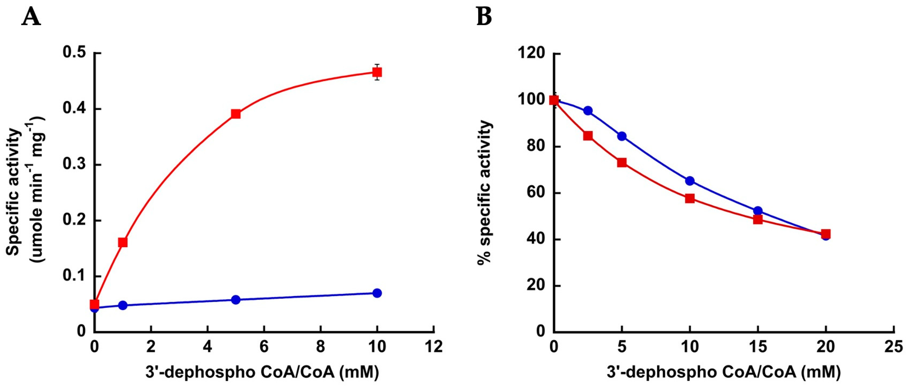
To examine whether MacsMa also makes an electrostatic interaction with the 3′-phosphate group of CoA, the ability of wild-type enzyme to discriminate between CoA and 3′-dephospho CoA was determined. The enzyme displayed very low 2-methylbutyryl-CoA synthetase activity with 3′-dephospho CoA even at a concentration of 10 mM, whereas the activity observed with 10 mM CoA was over 5-fold higher (Figure 2A). Kinetic parameters could not be determined with 3′-dephospho CoA, so the level of discrimination could not be ascertained.
In the absence of CoA, wild-type MacsMa catalyzes synthesis and release of an acyl-adenylate when less favorable acyl substrates such as propionate are used, and the presence of CoA inhibits this activity [11]. Inhibition of the acyl-adenylate synthetase activity by CoA versus 3′-dephospho CoA was examined as another means for determining whether interaction between the enzyme and the 3′-phosphate group of CoA is important. The acyl-adenylate synthetase activity was inhibited by both CoA and 3′-dephospho CoA to a similar extent (Figure 2B), suggesting that interaction with the 3′-phosphate group is important for CoA binding for the second step of the reaction but does not play a role in interaction between CoA and the enzyme for the first step of the reaction or when the second step cannot occur.

3.4. The CoA Binding Pocket in MacsMa Resembles That in CBAL

The CoA nucleotide binding pocket in the AcsSe and CBAL structures differs but the pantetheine tunnel is similar [5,20]. In CBAL, the aromatic residues Phe473 and Trp440 play key roles in CoA binding and catalysis by accommodating the adenine moiety of CoA. Alterations of these residues greatly reduced catalytic efficiency for the second step of the reaction while having little effect on first step [22]. Arg475 interacts with the CoA 3’-phosphate [5,20] and alteration reduced catalytic efficiency [22].
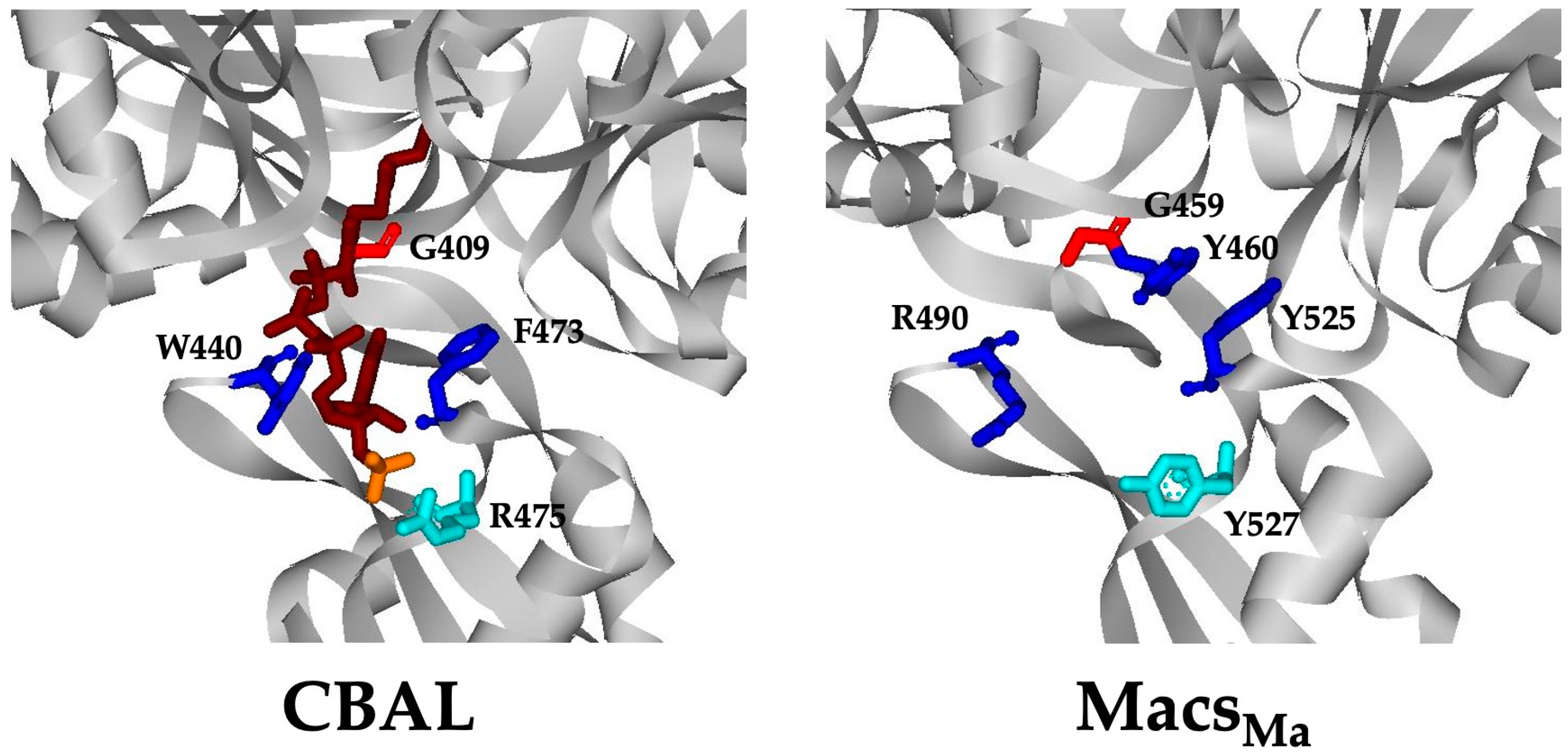
Comparison of the MacsMa, AcsSe, and CBAL structures revealed that the CoA binding site of MacsMa more closely resembles that of CBAL [21]. In MacsMa, Tyr525 and Arg490 replace Phe473 and Trp440 of CBAL, respectively (Figure 3), although Tyr460 of MacsMa is also positioned such that it could function similarly to Trp440 of CBAL, which interacts with the adenine moiety of CoA [5,20]. Tyr527 of MacsMa occupies a similar location to Arg475 of CBAL but the side chain is positioned away from the 3′-phosphate and may instead interact with the ribose group of CoA via its benzoyl group [21]. Gly459, located in close proximity to the putative CoA binding site of MacsMa, is highly conserved among all members of the adenylate-forming enzyme superfamily and has been proposed to be necessary to open the pantetheine tunnel in the thioester-forming conformation [21].
Based on these structural comparisons, we investigated the role of MacsMa residues Gly459, Tyr525, Tyr460, Arg490 and Tyr527 in CoA binding and catalysis. Alterations were made at each of these residues and the recombinant enzyme variants were produced and purified. The Tyr460Ala and Arg490Ala variants were insoluble and were not characterized. Kinetic parameters were determined for the purified enzyme variants to examine the impact of the alterations on acyl-CoA synthetase activity. Alteration of Gly459 to Ala had little effect on enzymatic activity. The Km and kcat values showed just slight changes from those for the wild-type enzyme for the 2-methylbutyryl-CoA synthetase activity (Table 3 and Supplemental Table S2).
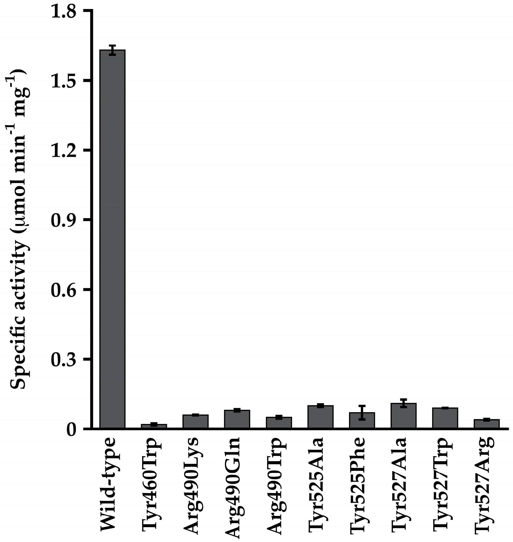
Alterations at Tyr460, Tyr525, Tyr527, and Arg490 proved to be very deleterious to the acyl-CoA synthetase activity of MacsMa, with little 2-methylbutyryl-CoA synthetase activity observed even at high CoA concentrations. These variants displayed 15- to 80-fold reduced specific activity relative to the wild-type enzyme (Figure 4), and kinetic parameters could not be determined due to the low activity. These variants also had reduced propionyl-adenylate synthetase activity, with kcat values reduced 4.5- to 21-fold (Supplemental Table S3). The Km values for propionate and ATP were not substantially affected in these variants except for the Tyr525Ala variant for which the Km value for propionate increased 6.0-fold and that for ATP decreased 14.7-fold (Supplemental Table S3).
To examine whether these alterations affected just the second step of the reaction in which CoA binding occurs or affected catalysis of the first step of the reaction as well, kinetic parameters were determined for the CoA-independent propionyl-adenylate synthetase activity of the enzyme. Except for the Tyr525Ala variant, the Km values for propionate and ATP showed ~2-fold or less change from the values observed for the unaltered enzyme although the kcat values were ~2–10 fold decreased (Supplementary Table S3). These results suggest that although the first step of the reaction is affected, the impact is not enough to account for the near lack of acyl-CoA synthetase activity and that CoA binding and/or catalysis of the second step of the reaction are specifically affected.
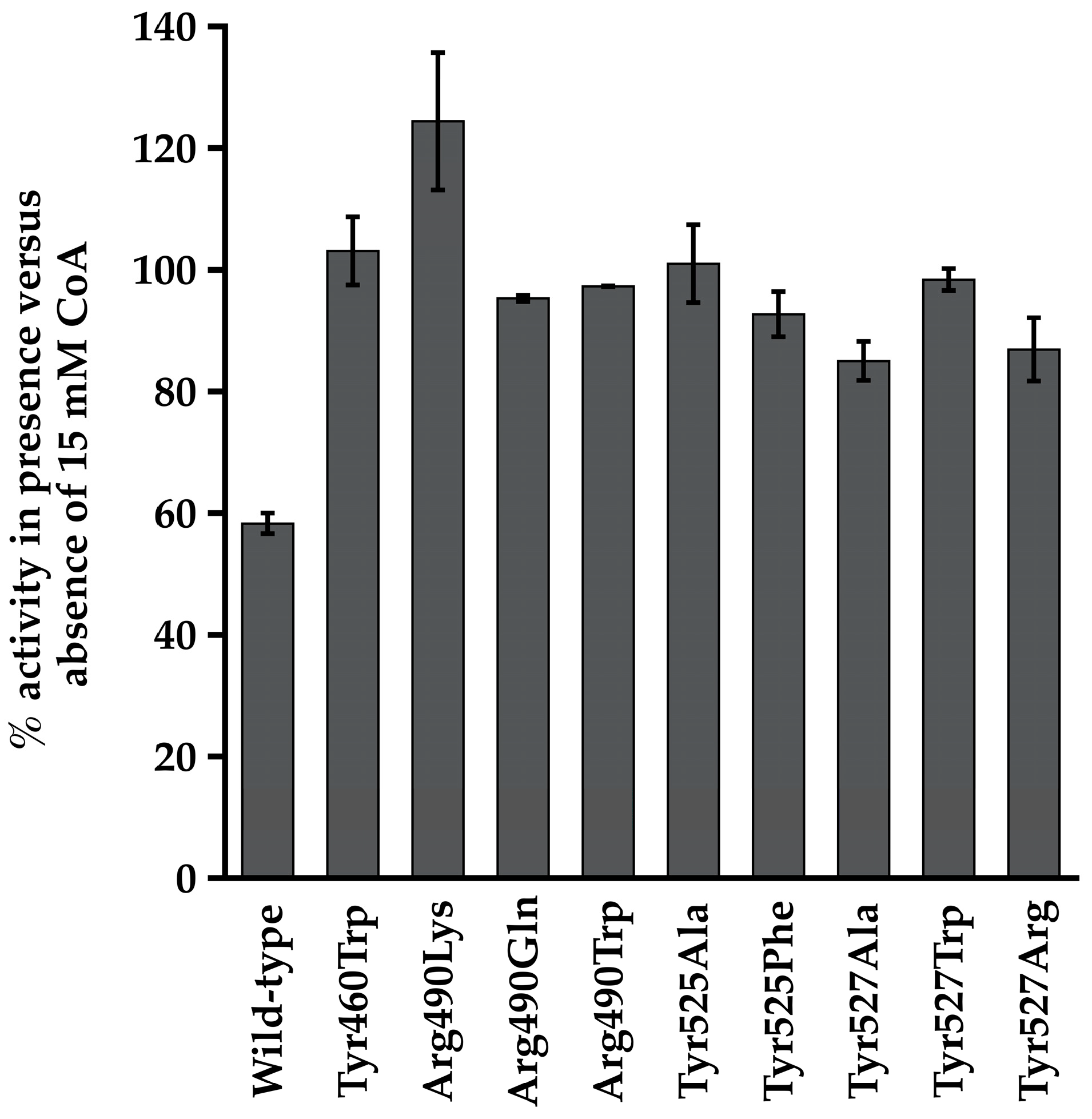
Because CoA inhibits the propionyl-adenylate synthetase activity of the wild-type enzyme [11], we examined the effect of a high concentration of CoA on this activity in the variants as a means for determining whether CoA can still bind even though the variants cannot catalyze the second step of the reaction. The presence of 15 mM CoA reduced activity of the wild-type enzyme by nearly 40% but had little to no inhibitory effect on activity of the variants (Figure 5). The presence of 15 mM CoA stimulated propionyl-adenylate synthetase activity of the Arg490Lys variant by nearly 25%. The reason for this is unknown and was not investigated further.

3.5. The Corresponding Lys Residues in MacsMa Do Not Play a Major Role in CoA Binding

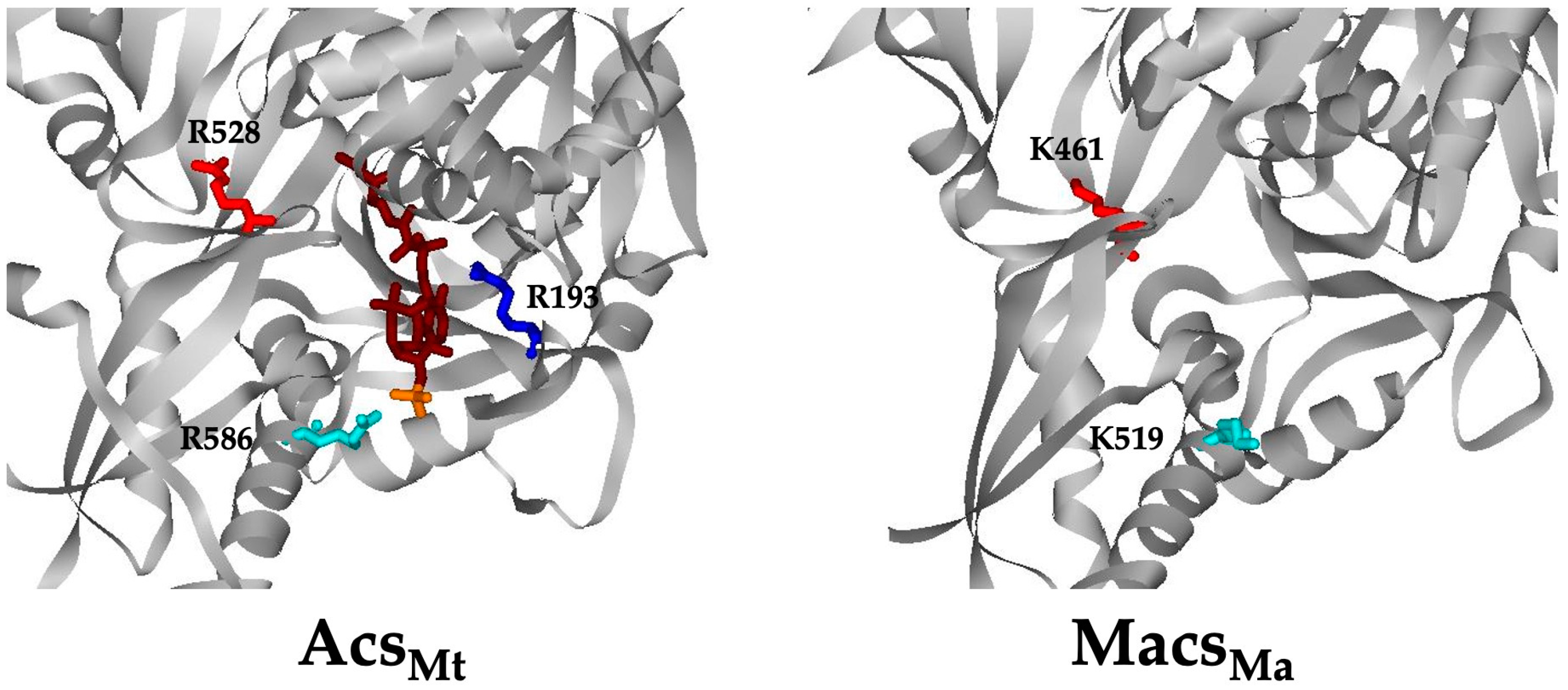
To provide further confirmation that CoA binding in MacsMa more closely resembles that in CBAL than Acs, we also examined the role of Lys461 and Lys519, which are positioned similarly to Arg528 and Arg586 of AcsMt (Figure 6). These Lys residues were individually altered to Arg and Ala and the purified variants were characterized. Kinetic parameters were determined using 2-methylbutyrate, the preferred substrate for the acyl-CoA synthetase activity of these enzyme variants. The Lys461Ala and Lys461Arg variants showed just 1.9-fold and 3.0-fold decrease, respectively, in the Km for CoA and a modest (less than 10-fold) decrease in the Km value for 2-methylbutyrate (Table 3). The Km values for 2-methylbutyrate and ATP were not substantially affected (Supplementary Table S2). These results suggest that Lys461 in MacsMa does not play a role similar to the corresponding Arg in Acs as there was little impact on CoA binding and catalysis.
The Lys519Arg alteration resulted in less than 2-fold change in the Km for any substrate or the turnover rate for the 2-methylbutyryl-CoA synthetase (Table 3 and Supplementary Table S2) or the propionyl-adenylate synthetase (Supplementary Table S3) activities. In contrast, the Lys519Ala variant had too little activity to determine kinetic parameters for either activity (Table 3 and Supplementary Tables S2 and S3).

4. Discussion

We have previously investigated substrate binding and catalysis in the short- and medium-chain acyl-CoA synthetases and identified residues important for acyl substrate binding in Acs and Macs and ATP binding in Acs. Here we examined CoA binding in AcsMt and MacsMa.
Inspection of the AcsSc and AcsSe structures [4,5] revealed two conformations for the enzyme. In the first step of the reaction, the C-terminal domain is positioned out and away from the active site but then swings in toward the N-terminal domain for catalysis of the second step of the reaction. Three Arg residues, Arg191, Arg526, and Arg584 (Arg193, Arg528, and Arg586 of AcsMt, respectively) were proposed to play an important role in CoA binding and catalysis of the second step. Arg191 interacts with both the 5′-diphosphate and the 3′-diphosphate groups of CoA. As this residue is on the N-terminal domain and already present in the active site before domain alternation, it may play an important role in initial binding of CoA. Arg584 enters the active site after domain alternation to interact with the 3′-phosphate group, and Arg526, also on the C-terminal domain, was proposed to stabilize the thioester-forming conformation through interaction with the phosphate group of the acyl-adenylate intermediate [5]. Although this residue was not proposed to directly interact with CoA, it may influence CoA binding and catalysis by locking the enzyme in the thioester-forming conformation and thus encasing CoA in the active site.
We altered each of these Arg residues individually in AcsMt and assessed each variant’s kinetic abilities. All the variants were impaired in catalysis, with kcat values reduced by 34 to 680-fold. The effect of these alterations on the Km for CoA varied, with alterations at Arg193 being the most detrimental and replacements at Arg528 and Arg586 having more variable effects. As might be expected, substitution with Ala at each position was the most deleterious, likely due to loss of both side chain charge and size. In fact, the Arg193Ala and Arg528Ala variants were not saturable for CoA or, surprisingly, for acetate.
Replacement of Arg586 had a lesser effect than replacement at Arg193, most likely because Arg586 only contacts CoA at the 3′-phosphate rather than at both the 5′-diphosphate and the 3′-phosphate as for Arg193. However, this single point of contact between Arg586 and CoA is important in CoA recognition and/or binding as shown by the fact that the Arg586 variants were unable to distinguish between CoA and 3′-dephosphoCoA.
Alteration of Arg528 increased the Km for CoA and decreased kcat despite Arg528 appearing to contact the phosphate group of the acyl-adenylate intermediate rather than direct contact with CoA, thus stabilizing the thioester-forming conformation of the enzyme. Substitution at this position would then be expected to reduce the ability of the enzyme to maintain proper positioning of the acyl-adenylate intermediate in the active site, thus affecting catalysis and influencing the ability to bind CoA as well. Our results suggest these three Arg residues are essential for CoA binding and catalysis, directly or indirectly. These residues may also influence acetate binding in the first step of the reaction, perhaps through an inability to fully control domain alternation.
In contrast to our results, Reger et al. [6] reported that alteration of Arg526 and Arg584 of AcsSe resulted in just a 2-fold decrease in catalysis. The Km for CoA increased for each of the variants, ranging from 4-fold for the Arg526Ala variant to 7 to 8-fold for the Arg584Ala and Arg584Glu variants [6]. However, no alterations were made at Arg191, the equivalent to Arg193 of AcsMt. Reger et al. [6] determined the kinetic parameters for CoA and ATP using 20 mM acetate in the reaction mixture for all enzyme variants. Given that the wild-type enzyme has a reported Km for acetate of 6.05 mM, the kinetic constants for ATP and CoA may have been determined at subsaturating acetate concentrations. These inconsistencies between our results and those of Reger et al. [6] may thus reflect differences between the two Acs enzymes, which share only 49% sequence identity at the amino acid level, or the experimental conditions. Such differences among Acs enzymes were already noted for acyl substrate selection [9,10].
In the MacsMa structure, the enzyme was found to be in a similar conformation to that observed for AcsSe, as if poised for the second step of the reaction even in the absence of substrates [7]. Comparison of the CBAL [21] and AcsSe structures [5] in the conformation for the second step of the reaction revealed that the binding pockets for CoA nucleotide moiety in these enzymes are significantly different [21], with more interactions with the N-terminal domain in Acs but with the C-terminal domain for CBAL. Superposition of the MacsMa structure with the CBAL and AcsSe structures indicates that the CoA binding site more closely resembles that of CBAL [7]. The recent structure of a 2-hydroxyisobutyric acid CoA ligase shows CoA binding in a substantially bent conformation in the thioester conformation [28]. This contrasts with the more stretched conformations of CoA observed in the thioester forming conformations of CBAL, Macs, and Acs.
We investigated five residues in MacsMa predicted to interact with CoA based on comparison with the CBAL structure. Tyr460, Arg590, Tyr525, or Tyr527 variants displayed greatly reduced 2-methylbutyryl-CoA synthetase activity, and propionyl-adenylate synthetase activity was also reduced but to a much lesser extent. Alteration at Gly459, which is strictly conserved in the acyl-adenylate-forming superfamily [7] reduced the turnover rate for both enzymatic activities but did not substantially affect the Km values for substrates.
In order to examine whether the alterations in the putative CoA binding pocket residues affected just catalysis or also affect CoA binding, we took advantage of the fact that CoA inhibits the propionyl-adenylate synthetase activity of MacsMa [11]. In each case, the variant showed less inhibition of the propionyl-adenylate synthetase activity by CoA than for the wild-type enzyme, suggesting that CoA cannot bind as well and supporting that these four residues play a key role in CoA binding as well as catalysis by MacsMa.
MacsMa lacks each of the three Arg residues investigated in AcsMt. However, Arg528 and Arg586 of AcsMt are replaced by Lys residues at the corresponding positions (residues 461 and 519) in MacsMa. Structurally, although these residues are in the vicinity of the predicted CoA binding pocket of MacsMa, they are more remote from CoA than the Arg residues of Acs. Our kinetics results for MacsMa variants altered at these Lys residues suggest that Lys461 does not play a role in CoA binding or catalysis. Although Lys519 may play some role, maintenance of the positive charge at this position is sufficient. Alterations at these positions (with the exception of a Lys519Ala alteration) resulted in only mild reductions in kcat (5-fold or less) for either the acyl-CoA synthetase activity or the propionyl-adenylate synthetase activity. Per residue binding free energy decomposition had previously identified Lys461 as a residue important in 2-methyl butyrate binding and catalysis [29]. Our alteration of Lys461 to alanine and arginine had only 7.5-fold and 9-fold reduction on Km, respectively.
Overall, although Acs and Macs have similarities in active site architecture for substrate binding and catalysis of the first step of the reaction, our results strongly suggest that the active site architecture for CoA binding and catalysis of the second step has diverged greatly. Although structural comparison between AcsSe and MacsMa revealed distinct differences in the CoA binding pocket [7], it appears that electrostatic interaction with the 3′-phosphate group of CoA is important for both enzymes; however, this interaction occurs with disparate residues in each enzyme. Differences in acyl substrate binding sites among acyl-CoA synthetase family members is not surprising as the enzymes must accommodate substrates of different lengths that may be branched or unbranched. However, the diversity in CoA binding sites among family members was unexpected.

Supplementary Materials

The following supporting information can be downloaded at: https://www.mdpi.com/article/10.3390/life13081643/s1, Table S1: Km values for acetate and ATP for AcsMt wild-type and variant enzymes; Table S2: Km values for 2-methylbutyrate and ATP for wild-type MacsMa and the Lys461, Lys519, and Gly459 variants; Table S3: Kinetic parameters for the propionyl-adenylate synthetase activity of wild-type MacsMa and the Lys461, Lys519, Gly459, Tyr460, Tyr525, Tyr527, and Arg490 variants.

Author Contributions

Conceptualization, C.I.-S. and K.S.; investigation, Y.M., C.I.-S. and O.A.; writing—original draft preparation, Y.M., C.I.-S. and K.S.; writing—review and editing, C.I.-S. and K.S. All authors have read and agreed to the published version of the manuscript.

Funding

This research was funded by NIH (Award GM69374-01A1) and the South Carolina Experiment Station (Project SC-1700198). This work represents technical contribution number 6242 of the Clemson University Experiment Station.

Institutional Review Board Statement

Not applicable.

Informed Consent Statement

Not applicable.

Data Availability Statement

All data relevant to this study are reported within.

Acknowledgments

Parts of this manuscript are based on a Ph.D. dissertation by Yu Meng (Meng, Y. 2010. Investigation of Biochemistry and Enzymology of Acyl-Coenzyme A Synthetase. https://tigerprints.clemson.edu/all_dissertations/516) (accessed on 6 June 2023).

Conflicts of Interest

The authors declare no conflict of interest.

References

  1. Moffett, J.R.; Puthillathu, N.; Vengilote, R.; Jaworski, D.M.; Namboodiri, A.M. Acetate revisited: A key biomolecule at the nexus of metabolism, epigenetics, and oncogenesis—Part 2: Acetate and ACSS2 in health and disease. Front. Physiol. 2020, 11, 580171. [Google Scholar] [CrossRef]
  2. Moffett, J.R.; Puthillathu, N.; Vengilote, R.; Jaworski, D.M.; Namboodiri, A.M. Acetate revisited: A key biomolecule at the nexus of metabolism, epigenetics and oncogenesis—Part 1: Acetyl-CoA, acetogenesis and acyl-CoA short-chain synthetases. Front. Physiol. 2020, 11, 580167. [Google Scholar] [CrossRef] [PubMed]
  3. VanDrisse, C.M.; Escalante-Semerena, J.C. Protein acetylation in bacteria. Annu. Rev. Microbiol. 2019, 73, 111–132. [Google Scholar] [CrossRef]
  4. Jogl, G.; Tong, L. Crystal structure of yeast acetyl-coenzyme A synthetase in complex with AMP. Biochemistry 2004, 43, 1425–1431. [Google Scholar] [CrossRef]
  5. Gulick, A.M.; Starai, V.J.; Horswill, A.R.; Homick, K.M.; Escalante-Semerena, J.C. The 1.75 Å crystal structure of acetyl-CoA synthetase bound to adenosine-5′-propylphosphate and coenzyme A. Biochemistry 2003, 42, 2866–2873. [Google Scholar] [CrossRef] [PubMed]
  6. Reger, A.S.; Carney, J.M.; Gulick, A.M. Biochemical and crystallographic analysis of substrate binding and conformational changes in acetyl-CoA synthetase. Biochemistry 2007, 46, 6536–6546. [Google Scholar] [CrossRef] [Green Version]
  7. Shah, M.B.; Ingram-Smith, C.; Cooper, L.L.; Qu, J.; Meng, Y.; Smith, K.S.; Gulick, A.M. The 2.1 Å crystal structure of an acyl-CoA synthetase from Methanosarcina acetivorans reveals an alternate acyl-binding pocket for small branched acyl substrates. Proteins 2009, 77, 685–698. [Google Scholar] [CrossRef] [PubMed]
  8. Goldsmith, M.; Barad, S.; Peleg, Y.; Albeck, S.; Dym, O.; Brandis, A.; Mehlman, T.; Reich, Z. The identification and characterization of an oxalyl-CoA synthetase from grass pea (Lathyrus sativus L.). RSC Chem. Biol. 2022, 3, 320–333. [Google Scholar] [CrossRef]
  9. Ingram-Smith, C.; Smith, K.S. AMP-forming acetyl-CoA synthetases in Archaea show unexpected diversity in substrate utilization. Archaea 2007, 2, 95–107. [Google Scholar] [CrossRef] [Green Version]
  10. Brasen, C.; Urbanke, C.; Schonheit, P. A novel octameric AMP-forming acetyl-CoA synthetase from the hyperthermophilic crenarchaeon Pyrobaculum aerophilum. FEBS Lett. 2005, 579, 477–482. [Google Scholar] [CrossRef] [Green Version]
  11. Meng, Y.; Ingram-Smith, C.; Cooper, L.L.; Smith, K.S. Characterization of an archaeal medium-chain acyl coenzyme A synthetase from Methanosarcina acetivorans. J. Bacteriol. 2010, 192, 5982–5990. [Google Scholar] [CrossRef] [PubMed] [Green Version]
  12. Ingram-Smith, C.; Woods, B.I.; Smith, K.S. Characterization of the acyl substrate binding pocket of acetyl-CoA synthetase. Biochemistry 2006, 45, 11482–11490. [Google Scholar] [CrossRef] [PubMed]
  13. D’Ambrosio, H.K.; Derbyshire, E.R. Investigating the role of class I adenylate-forming enzymes in natural product biosynthesis. ACS Chem. Biol. 2020, 15, 17–27. [Google Scholar] [CrossRef] [PubMed]
  14. Mouterde, L.M.M.; Stewart, J.D. Application of acetyl-CoA synthetase from Methanothermobacter thermautotrophicus to non-native substrates. Enzym. Microb. Technol. 2019, 128, 67–71. [Google Scholar] [CrossRef]
  15. Sofeo, N.; Hart, J.H.; Butler, B.; Oliver, D.J.; Yandeau-Nelson, M.D.; Nikolau, B.J. Altering the substrate specificity of acetyl-CoA synthetase by rational mutagenesis of the carboxylate binding pocket. ACS Synth. Biol. 2019, 8, 1325–1336. [Google Scholar] [CrossRef] [Green Version]
  16. Ingram-Smith, C.; Thurman, J.L., Jr.; Zimowski, K.; Smith, K.S. Role of motif III in catalysis by acetyl-CoA synthetase. Archaea 2012, 2012, 509579. [Google Scholar] [CrossRef] [Green Version]
  17. Gallego-Jara, J.; Terol, G.L.; Ecija Conesa, A.; Zambelli, B.; Canovas Diaz, M.; de Diego Puente, T. Characterization of acetyl-CoA synthetase kinetics and ATP-binding. Biochim. Biophys. Acta Gen. Subj. 2019, 1863, 1040–1049. [Google Scholar] [CrossRef]
  18. Chang, K.H.; Xiang, H.; Dunaway-Mariano, D. Acyl-adenylate motif of the acyl-adenylate/thioester-forming enzyme superfamily: A site-directed mutagenesis study with the Pseudomonas sp. strain CBS3 4-chlorobenzoate:coenzyme A ligase. Biochemistry 1997, 36, 15650–15659. [Google Scholar] [CrossRef]
  19. Chang, K.H.; Liang, P.H.; Beck, W.; Scholten, J.D.; Dunaway-Mariano, D. Isolation and characterization of the three polypeptide components of 4-chlorobenzoate dehalogenase from Pseudomonas sp. strain CBS-3. Biochemistry 1992, 31, 5605–5610. [Google Scholar] [CrossRef]
  20. Gulick, A.M.; Lu, X.; Dunaway-Mariano, D. Crystal structure of 4-chlorobenzoate:CoA ligase/synthetase in the unliganded and aryl substrate-bound states. Biochemistry 2004, 43, 8670–8679. [Google Scholar] [CrossRef]
  21. Reger, A.S.; Wu, R.; Dunaway-Mariano, D.; Gulick, A.M. Structural characterization of a 140 degrees domain movement in the two-step reaction catalyzed by 4-chlorobenzoate:CoA ligase. Biochemistry 2008, 47, 8016–8025. [Google Scholar] [CrossRef] [Green Version]
  22. Wu, R.; Cao, J.; Lu, X.; Reger, A.S.; Gulick, A.M.; Dunaway-Mariano, D. Mechanism of 4-chlorobenzoate:coenzyme a ligase catalysis. Biochemistry 2008, 47, 8026–8039. [Google Scholar] [CrossRef] [Green Version]
  23. Wu, R.; Reger, A.S.; Lu, X.; Gulick, A.M.; Dunaway-Mariano, D. The mechanism of domain alternation in the acyl-adenylate forming ligase superfamily member 4-chlorobenzoate: Coenzyme A ligase. Biochemistry 2009, 48, 4115–4125. [Google Scholar] [CrossRef] [PubMed] [Green Version]
  24. Bradford, M.M. A rapid and sensitive method for the quantitation of microgram quantities of protein utilizing the principle of protein-dye binding. Anal. Biochem. 1976, 72, 248–254. [Google Scholar] [CrossRef] [PubMed]
  25. Lipmann, F.; Tuttle, L.C. A specific micromethod for determination of acyl phosphates. J. Biol. Chem. 1945, 159, 21–28. [Google Scholar] [CrossRef]
  26. Rose, I.A.; Grunberg-Manago, M.; Korey, S.F.; Ochoa, S. Enzymatic phosphorylation of acetate. J. Biol. Chem. 1954, 211, 737–756. [Google Scholar] [CrossRef] [PubMed]
  27. Lee, H.Y.; Na, K.B.; Koo, H.M.; Kim, Y.S. Identification of active site residues in Bradyrhizobium japonicum acetyl-CoA synthetase. J. Biochem. 2001, 130, 807–813. [Google Scholar] [CrossRef] [PubMed]
  28. Zahn, M.; Kurteva-Yaneva, N.; Schuster, J.; Krug, U.; Georgi, T.; Muller, R.H.; Rohwerder, T.; Strater, N. Structures of 2-hydroxyisobutyric acid-CoA ligase reveal determinants of substrate specificity and describe a multi-conformational catalytic cycle. J. Mol. Biol. 2019, 431, 2747–2761. [Google Scholar] [CrossRef]
  29. Du, J.; Wang, X.; Nie, Q.; Yang, J.; Yao, X. Computational study of the binding mechanism of medium chain acyl-CoA synthetase with substrate in Methanosarcina acetivorans. J. Biotechnol. 2017, 259, 160–167. [Google Scholar] [CrossRef] [PubMed]
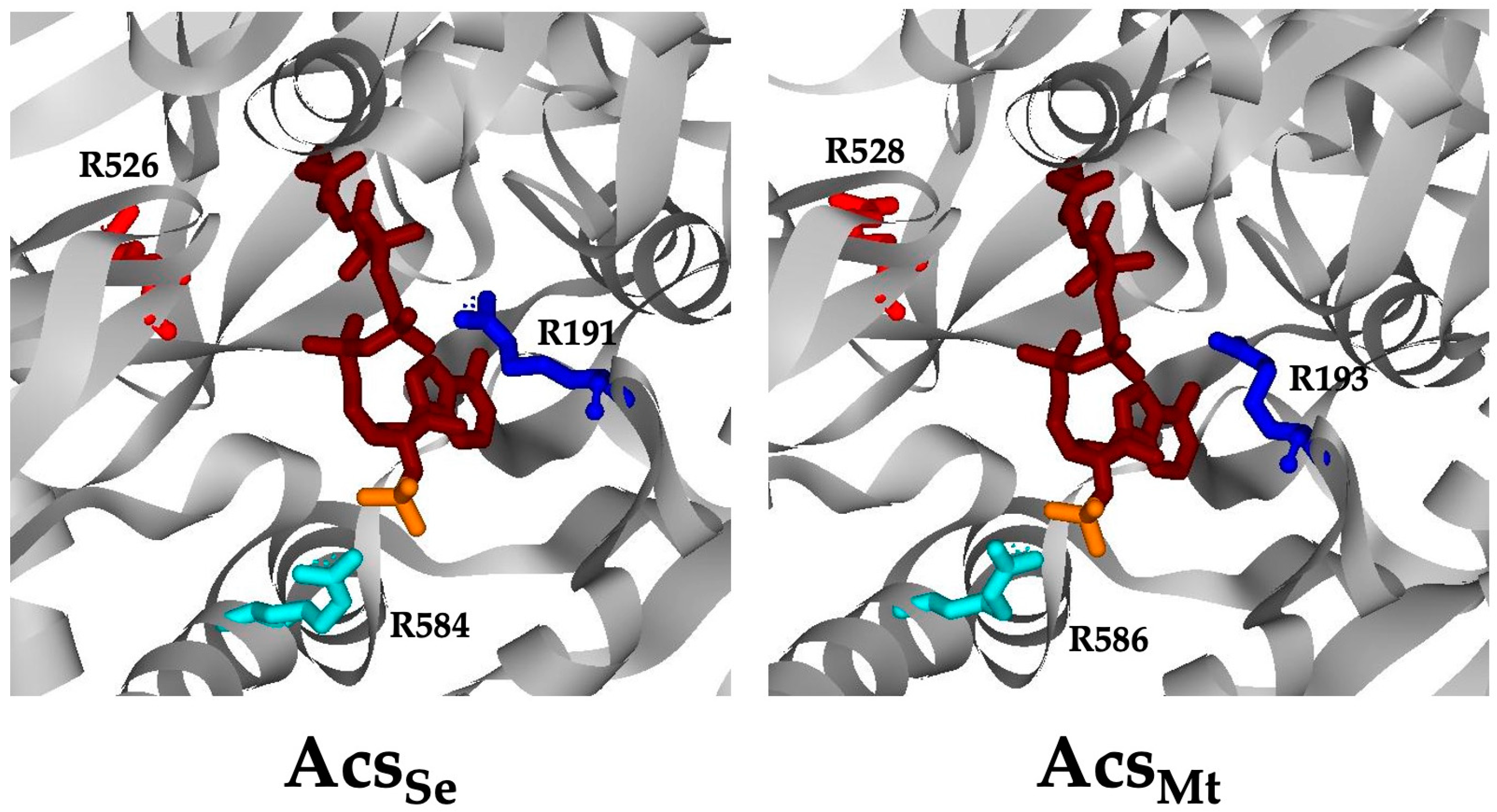
Figure 1. CoA binding region of AcsSe and AcsMt. The AcsMt structure (right) was modeled on AcsSe (left; PDB ID 2P2F). CoA is shown in magenta, with the 3′-phosphate group in orange. Corresponding Arg residues in each structure (AcsSe/AcsMt) are displayed as follows: Arg526/528 in red, Arg191/193 in blue, and Arg584/586 in aqua.
Life 13 01643 g001
Figure 2. Effect of CoA and 3′-dephospho CoA on the acyl-CoA synthetase and propionyl-adenylate synthetase activities of MacsMa. (A) Acyl-CoA synthetase activity of MacsMa with CoA or 3′-dephospho CoA. Activity was measured at increasing concentrations of either CoA (red) or 3’-dephospho CoA (blue). Specific activities shown are the mean ± standard deviation of three replicates. (B) Inhibition of the propionyl-CoA synthetase activity of MacsMa by CoA (red) versus 3′-dephospho CoA (blue). Activities shown are the percent activity measured in the absence of CoA (100%) versus presence of CoA and are the mean ± standard deviation of three replicates.
Life 13 01643 g002
Figure 3. The CoA binding region of CBAL and MacsMa. The CBAL structure (left; PDB ID 3CW9) has CoA bound (in magenta with the 3′-phosphate group in orange). Residues shown to play an important role in CoA binding and catalysis are indicated. Residues predicted to be important for CoA binding in MacsMa (right; PDB ID 3ETC) are shown in the same color as the corresponding residues in CBAL.
Life 13 01643 g003
Figure 4. 2-methylbutyryl-CoA synthetase specific activity of wild-type MacsMa and the Tyr460, Arg490, Tyr525, and Tyr527 variants determined in the presence of 15 mM CoA.
Life 13 01643 g004
Figure 5. Effect of CoA on propionyl-adenylate synthetase activity of MacsMa wild-type and variants. Activities in the presence of 15 mM CoA were normalized as percentages relative to the specific activity observed for each enzyme in the absence of CoA. Reactions were performed in triplicate and values are the mean ± SD.
Life 13 01643 g005
Figure 6. Positioning of corresponding residues Arg528/Lys461 (red) and Arg586/Lys519 (blue) in AcsMt (left) and MacsMa (right).
Life 13 01643 g006
Table 1. Kinetics parameters for AcsMt wild-type and variant enzymes.
EnzymeKm CoA
(mM)
kcat
(sec−1)
kcat/Km
(sec−1 mM−1)
Wild-type a0.19 ± 0.00381.6 ± 0.7423.7 ± 4.7
Arg193AlaUnsaturable b
Arg193Lys3.6 ± 0.370.51 ± 0.010.14 ± 0.01
Arg193Gln7.8 ± 0.400.28 ± 0.0040.036 ± 0.002
Arg528AlaUnsaturable b
Arg528Lys0.45 ± 0.030.25 ± 0.010.54 ± 0.02
Arg528Gln1.67 ± 0.050.63 ± 0.040.38 ± 0.06
Arg586Ala1.19 ± 0.082.13 ± 0.091.80 ± 0.06
Arg586Lys0.33 ± 0.0132.39 ± 0.0167.34 ± 0.24
Arg586Gln0.28 ± 0.0090.12 ± 0.0030.45 ± 0.028
a Values are taken from [9]. b The enzyme was not saturable for CoA at concentrations up to 25 mM and kinetic parameters could not be determined.
Table 2. Discrimination between CoA and 3′-dephospho CoA (deCoA) for wild-type AcsMt and the Arg586 variants.
EnzymeSubstrateKm
(mM)
kcat
(sec−1)
kcat/Km
(sec−1 mM−1)
(kcat/Km CoA)/
(kcat/Km deCoA)
Wild-typeCoA0.19 ± 0.00381.6 ± 0.7423.7 ± 4.726.5
deCoA2.16 ± 0.5034.5 ± 2.916.0 ± 2.5
Arg586AlaCoA1.19 ± 0.082.13 ± 0.091.80 ± 0.062.2
deCoA2.05 ± 0.401.66 ± 0.110.81 ± 0.11
Arg586LysCoA0.33 ± 0.012.39 ± 0.027.34 ± 0.203.7
deCoA0.86 ± 0.041.72 ± 0.012.01 ± 0.09
Table 3. Kinetic parameters for the 2-methylbutyryl-CoA synthetase activity for wild-type MacsMa and the Gly459, Lys461, and Lys519 variants.
Enzymekcat
(sec−1)
Km CoA
(mM)
Wild-type2.15 ± 0.104.09 ± 0.45
Gly459Ala0.41 ± 0.012.09 ± 0.06
Lys461Ala0.55 ± 0.032.19 ± 0.18
Lys461Arg0.46 ± 0.101.38 ± 0.14
Lys519Ala**
Lys519Arg1.07 ± 0.027.18 ± 0.29
* Activity was too low for determination of kinetic parameters.
Disclaimer/Publisher’s Note: The statements, opinions and data contained in all publications are solely those of the individual author(s) and contributor(s) and not of MDPI and/or the editor(s). MDPI and/or the editor(s) disclaim responsibility for any injury to people or property resulting from any ideas, methods, instructions or products referred to in the content.