Review

Bidirectional Regulation of GABAA Reversal Potential in the Adult Brain: Physiological and Pathological Implications

by
Haram R. Kim
1 and
Marco Martina
1,2,*
1
Department of Neuroscience, Feinberg School of Medicine, Northwestern University, 300 E. Superior, Chicago, IL 60611, USA
2
Department of Psychiatry, Feinberg School of Medicine, Northwestern University, 300 E. Superior, Chicago, IL 60611, USA
*
Author to whom correspondence should be addressed.
Life 2024, 14(1), 143; https://doi.org/10.3390/life14010143
Submission received: 8 December 2023 / Revised: 16 January 2024 / Accepted: 17 January 2024 / Published: 19 January 2024
(This article belongs to the Special Issue Ion Channels and Neurological Disease)

Abstract

:
In physiological conditions, the intracellular chloride concentration is much lower than the extracellular. As GABAA channels are permeable to anions, the reversal potential of GABAA is very close to that of Cl, which is the most abundant free anion in the intra- and extracellular spaces. Intracellular chloride is regulated by the activity ratio of NKCC1 and KCC2, two chloride-cation cotransporters that import and export Cl, respectively. Due to the closeness between GABAA reversal potential and the value of the resting membrane potential in most neurons, small changes in intracellular chloride have a major functional impact, which makes GABAA a uniquely flexible signaling system. In most neurons of the adult brain, the GABAA reversal potential is slightly more negative than the resting membrane potential, which makes GABAA hyperpolarizing. Alterations in GABAA reversal potential are a common feature in numerous conditions as they are the consequence of an imbalance in the NKCC1-KCC2 activity ratio. In most conditions (including Alzheimer’s disease, schizophrenia, and Down’s syndrome), GABAA becomes depolarizing, which causes network desynchronization and behavioral impairment. In other conditions (neonatal inflammation and neuropathic pain), however, GABAA reversal potential becomes hypernegative, which affects behavior through a potent circuit deactivation.

1. Introduction

The balance between excitation and inhibition (E/I balance) is critical for maintaining optimal function of the brain [1,2,3,4]. At the cellular level, a range of inhibitory mechanisms are designed to precisely control the excitation so that the neurons convey accurate information through the network [5,6,7,8]. In the brain, GABA (γ-aminobutyric acid) is the primary inhibitory neurotransmitter for both long-range and local inhibitory transmission. E/I imbalance can be caused by alterations of excitation, inhibition, or both. Altered E/I balance has been suggested as a causal factor for many symptoms associated with neuropsychiatric diseases, including fragile X syndrome [9], Alzheimer’s disease [10], and schizophrenia [11,12], and in most cases, it is attributable to altered GABAergic signaling [13,14].
In the brain, GABA exercises its effects by acting on two major types of receptors, the ionotropic (GABAAR) receptor and the metabotropic (GABABR) receptor. In contrast to the GABAB receptors, which set the basal tone of inhibition through indirectly opening potassium channels via G-protein activation, GABAA receptors immediately alter the ion permeability of the membrane. When the GABAAR is activated, monovalent anions (mostly chloride but also nitrate and bicarbonate ions [15]) pass through the transmembrane channel structure of receptors. This movement of anions generates the postsynaptic current (PSC). The PSC amplitude depends on the number and availability of receptors and the intracellular concentration of anions, as well as the amount of GABA released from presynaptic terminals. Although multiple mechanisms regulate signaling through GABAA receptors, including the modulation of the subunit composition of GABAA receptors and the alteration in GABA metabolism, an increasing amount of work suggests that in most pathological conditions, the main alterations in GABAA function are caused by changes in the relative activity of NKCC1 and KCC2, two chloride/potassium cotransporters that import and export chloride, respectively, and, thus, set the reversal potential of the current.
Over the past decades, it has become clear that despite its primary role as an inhibitory signal, the GABAA current can also be excitatory. Apart from the well-known role of depolarizing GABAA in developmental stages, which has been extensively investigated, numerous pathological states are associated with the reappearance of a depolarizing effect of GABA in the adult brain. Additionally, we recently showed that alterations leading to hypernegative GABAA reversal potential also have an important functional impact on the adult cortex [16]. Here, we review the bidirectional regulation of GABAA reversal potential in pathological conditions of the adult brain and discuss how the altered GABAA function may impact network function.

2. GABAA Reversal Potential

2.1. GABAA Reversal Potential and Intracellular Chloride

In physiological conditions, the concentration of intracellular chloride ([Cl]i = 5~30 mM) is considerably lower compared to extracellular chloride ([Cl]o = ~140 mM) [17]. As GABAA channels are permeable to anions, the reversal potential of GABAA is very close to that of Cl, which is the most abundant free anion in physiological solutions. A few studies have measured [Cl]i directly in physiological conditions using the visualization of chloride ions [18,19], but their reliability in assessing absolute chloride concentrations remains questionable. The most reliable method to estimate [Cl]i remains the measurement of GABAA current reversal potential. The reversal potential of GABAA can be obtained by measuring the GABAA currents at different membrane voltages. Using the perforated patch clamp recording technique, which preserves the native concentration of anions, [Cl]i is calculated using the Nernst equation from the measured reversal potential.
At reversal potential, the net Cl flux across the membrane is zero, and the effect of GABAA activation is entirely due to shunting inhibition (the conductance increase that diminishes cellular excitability due to Ohm’s law). In most neurons of the adult brain, the GABAA reversal potential is slightly more negative than the resting membrane potential, which makes GABAA hyperpolarizing. It is important to stress that because, in most cases, the GABAA reversal potential is close to the resting potential, even small changes in GABAA reversal can shift the effect of GABAA activation from hyperpolarizing to depolarizing.
Thus, small increases in [Cl]i can cause the GABAA current reversal potential to become depolarized relative to the resting potential. However, depolarizing reversal potential is not synonymous with an excitatory effect. It is only when this depolarizing effect exceeds the inhibitory shunting effect that GABAA becomes excitatory. Due to the vicinity, in most neurons, between the value of GABAA reversal potential and resting membrane potential, the opposite case is also true, and decreased [Cl]i boosts the influx of negatively charged ions and, therefore, strengthens the inhibitory effect of the current.
Interestingly, the internal chloride concentration in neurons is differentially regulated across developmental stages and cell types, even in the normal brain. This sets the GABAA current clearly apart from the currents mediated by ionotropic glutamate receptors (AMPAR and NMDAR), which have reversal potentials (~0 mV) that are much less sensitive to changes in sodium (Na+) or potassium (K+) concentration and never change from excitatory to inhibitory. This “flexibility” of the GABAA current, which is regulated by the level of [Cl]I, sets GABAA apart from other signaling systems.

2.2. Chloride/Potassium Cotransporters

Neuronal [Cl]i is primarily set by the activity of NKCC1 and KCC2, two chloride/potassium cotransporters that act in opposite directions. While NKCC1 increases [Cl]i, KCC2 promotes Cl extrusion [20]. The actions of these two transporters are differently balanced in different locations and conditions and depend on transporter density and distribution as well as modulation (such as phosphorylation).
NKCC1 is widely expressed in cells within and outside the brain. NKCC1 transcripts and proteins are also present in non-neuronal cells like oligodendrocytes, microglia, and astrocytes [21]. KCC2 expression, on the contrary, is neuron-selective across numerous CNS structures, including the spinal cord, retina, neocortex, and most subcortical structures [22,23,24].
The structures of both transporters were well described [25,26,27,28,29]. NKCC1 cotransports Na+ and K+ inwardly together with two Cl, thus increasing [Cl]i. In contrast, KCC2 exports K+ and Cl and thus decreases [Cl]i. As in mature neurons of the adult brain the activity of KCC2 exceeds that of NKCC1, [Cl]i is maintained at low levels, which results in the GABAA current being inhibitory and hyperpolarizing.
Increased NKCC1 activity, on the other hand, results in a depolarizing shift in the GABAA current reversal potential by elevating [Cl]i. In this case, the reversal potential of the GABAA current becomes depolarized compared with the resting membrane potential (see Section 2.1). In contrast, increased KCC2 activity lowers [Cl]i and hyperpolarizes GABAA reversal potential to values considerably lower than the resting membrane potential, producing a stronger inhibitory effect.

3. Regulation of GABAA Reversal Potential in Physiological Conditions

Depolarizing GABAA has been discovered and intensively studied in immature neurons for over 30 years, and it is evolutionally conserved across vertebrate species and in Caenorhabditis elegans [30]. Ben-Ari (2002) suggested that there are several potential biological advantages to having GABA mediate excitation in immature neurons because, compared with the excitatory glutamatergic current, the depolarizing GABA current could be less excitotoxic for postsynaptic neurons and more energetically efficient as it does not require pumping ions against large transmembrane gradients. Strong evidence also supports the idea that the reversal potential of the GABAA current is heavily regulated, even in a normally functioning adult brain, which suggests the idea that the flexibility of the GABAA current effect is exploited as a general mechanism to that allows precise regulation of synaptic transmission. As there are excellent published reviews on the GABAA shift in developmental stages [17,31,32], in this section, we will only briefly summarize it and focus on the GABAA reversal potential regulation in the adult brain.

3.1. Developmental Regulation

The presence of depolarizing GABAA current is the norm in the brain during early development when GABAA is the main fast excitatory neurotransmitter and key to establishing normal circuitry [33,34]. Neonatal neurons express a substantial amount of NKCC1, while KCC2 expression is very low. At this stage, GABAA is depolarizing, as [Cl]i is 20–30 mM, which implies a GABAA reversal potential of −40 to −50 mV [17], while the resting potential is similar to that of mature neurons (~−70 mV) [35]. This depolarizing GABAA has a critical role in synaptic formation and in the establishment of the neuronal network. GABAA currents become inhibitory around postnatal Day 12 in rodents [36] and around 2 years of age in humans, largely due to increased KCC2 expression leading to lower intracellular Cl [37,38,39,40].

3.2. Cell Type and Spatial Heterogeneity of Chloride Transporters

In the adult brain, NKCC1 is strongly expressed in all types of non-neuronal cells—oligodendrocytes, microglia, and astrocytes [21,41,42]. It is suggested that its splice variants (NKCC1a/1b) are involved distinctively in the localization of NKCC1. NKCC1a is predominant in glial cells, and NKCC1b is found in neurons [21].
The presence of NKCC1 in glial cells increases the complexity of GABAA signaling. Astrocytes affect the activity of transporters by changing the extracellular potassium and chloride concentration around the neurons [43,44]. Additionally, astrocytes directly contribute to synaptic transmission by releasing glutamate and other modulators in an activity-regulated calcium-dependent or -independent fashion [45,46,47]. Beyond the effect on membrane potential, astrocytic NKCC1 has been shown to regulate mitochondrial calcium levels in astrocytes [48]. So, NKCC1 regulation in non-neuronal cells has a major impact on brain function. A discussion of these effects is beyond the scope of this paper; interested readers can find detailed reviews on this topic [49,50].
Even when restricting the analysis to neuronal expression, changes in NKCC1/KCC2 activity are typically confined to select brain areas. For example, in a pharmacological model of cognitive impairment associated with schizophrenia, NKCC1 is increased in the ventral but not in the dorsal subregion of the medial prefrontal cortex (mPFC) [51], and in a rodent model of neuropathic pain, KCC2 expression is increased in the mPFC but not in the neighboring anterior cingulate cortex [16]. NKCC1 and KCC2 can also be differentially regulated in different layers within the same brain areas, as shown in mouse granule cells and outer and middle molecular layers following entorhinal denervation [52].
Interestingly, in the adult mouse hippocampus, the reversal potential of the GABAA current in parvalbumin-positive interneurons is sensitive to KCC2 and NKCC1 blockades in a similar way to in pyramidal neurons, yet it is ~7 mV more depolarized than in pyramidal neurons [53], suggesting the differential expression of transporters between these cell types. Together with the observation that non-neuronal cells abundantly express NKCC1, these data point to the necessity of single-cell resolution studies when evaluating NKCC1/KCC2 and show that using tissue-level approaches, such as Western blot analysis of homogenized brain tissue, may easily miss cell-type selective alterations.

3.3. Compartmental Distribution of Chloride Transporters in Neurons

3.3.1. Somatodendritic Compartment

Both NKCC1 and KCC2 are distributed differently across cellular compartments. NKCC1 mRNA is detected and abundant in most cells of the adult brain [21], but protein expression in mature neurons is low, which has hampered studying the subcellular distribution of this transporter.
Recently, a super-resolution microscopy study in dissociated neurons took advantage of hemagglutinin-tagged transporters in hippocampal neurons and showed that NKCC1 isoforms are detected in the somatodendritic compartment [54]. This study also found that the NKCC1-KCC2 ratio is higher in axon initial segments of hippocampal neurons (see Section 3.3.2). Another study found that in the adult brain, two KCC2 isoforms (KCC2a and KCC2b) are expressed, but their expression differs between different cellular compartments (E.G. soma and proximal dendrites versus distal neuronal dendrite [22]). Both dentate gyrus granule cells and CA1 pyramidal cells of the adult hippocampus show different patterns of KCC2 expression from distal/apical dendrites to the soma [55]. This observation also suggests that the NKCC1-KCC2 ratio is differentially regulated along dendrites, possibly providing local control over the efficacy of synaptic activation. Indeed, spatially nonuniform [Cl]i shifts have been described in dendrites [56]. In keeping with the idea of spatial segregation in [Cl]i gradients, KCC2 is enriched in the vicinity of synapses [57,58,59] by a gephyrin-dependent mechanism, suggesting that GABAA is hyperpolarizing in these structures. In particular, KCC2 is clustered at the spine periphery rather than at the postsynaptic density (PSD) region [60]. NKCC1 is expressed in dendrites both at perisynaptic and extrasynaptic sites, where, in most cases, it colocalizes with KCC2 [54]. Intriguingly, these authors also showed that, in cultured neurons, NKCC1 and, to a lesser extent, KCC2 proteins are mobile, suggesting that their distribution can be rapidly modified in response to neuronal activity and the activation of individual synapses. This observation suggests that activity-dependent alterations in NKCC1/KCC2 activity contribute to synaptic plasticity. In agreement with this suggestion, a study showed that in hippocampal neurons (both cultured and in acute slices), KCC2 activity is regulated on a timescale of minutes by coincident postsynaptic spiking and synaptic activity by a calcium-dependent mechanism, which leads to persistent changes in synaptic strength [61].

3.3.2. Axonal Structures

In mature neurons, NKCC1 appears to be more abundantly expressed in the axon initial segment (AIS) compared with the somatodendritic compartment, which creates an axo-somatodendritic gradient in intracellular chloride [62]. Additionally, KCC2 expression in the AIS is low [55,63] or absent [54]. This high NKCC1-to-KCC2 ratio maintains high [Cl]i and, therefore, a depolarizing effect of GABAA in these structures. Accordingly, elegant work showed that the GABAergic input from axo-axonic cells to cortical pyramidal neuron axons is excitatory and elicits action potential firing in postsynaptic axons [63], although it may become hyperpolarizing in older animals [64].

3.4. Circadian Regulation of GABAA Reversal Potential

A very recent paper reported another intriguing aspect of [Cl]i homeostasis in the adult brain, circadian regulation [65]. These authors showed that in the pyramidal neurons of the adult mouse neocortex baseline, [Cl]i undergoes a two-fold increase from day to night and that such an increase is due to changes in the surface expression and phosphorylation of NKCC1 and KCC2. These findings add another layer of complexity to the understanding of GABAergic signaling in the brain and provide further proof of the remarkable flexibility of this system.

4. Regulation of Neuronal GABAA Reversal Potential in Disease

4.1. Depolarizing GABAA Current

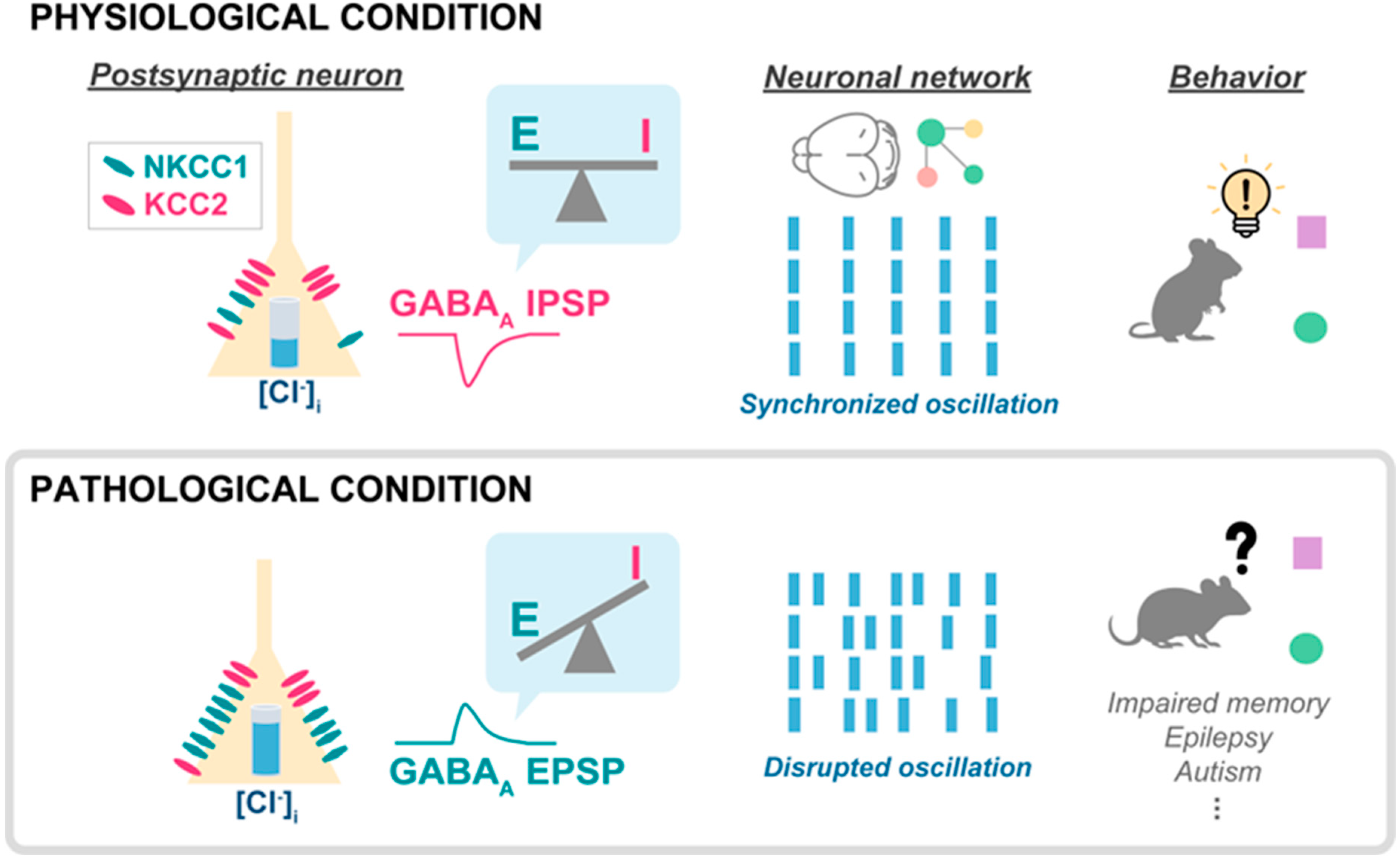
An altered NKCC1-KCC2 activity ratio has been linked with various neuropathological disorders and discussed in previous reviews [66,67]. Depolarizing GABAA currents in the adult brain have been reported in many pathological conditions such as epilepsy [37,68], Down syndrome [69], schizophrenia [51], and severe stress [70]. The mechanism by which depolarizing GABA currents interferes with brain activity is likely caused by a loss of synchronization in network oscillations (Figure 1). The cellular mechanisms leading to the altered reversal potential include alterations in transporter expression and in their regulation. For example, increased activity of kinase, such as OXSR1 and WNK3, which regulate NKCC1 and KCC2 was reported in the prefrontal cortex of schizophrenia patients [71]. On the other hand, in subchronic phencyclidine (scPCP)-treated mice, an animal model that mimics the cognitive symptoms associated with schizophrenia [72,73], depolarizing GABAA appears in the prefrontal cortex as a consequence of increased NKCC1 mRNA expression [51]. In line with these data, a gain-of-function NKCC1 mutation has been identified in human schizophrenia patients [74]. Thus, similar to the scenario in Down syndrome [69] and in the spinal cord of neuropathic pain models [75], NKCC1 is the transporter whose activity is mostly altered in schizophrenia. Therefore, the cellular mechanism leading to altered GABAA reversal potential in the diseased adult brain appears to be different than in the neonatal brain, where the depolarizing GABAA current is due to low KCC2 expression [39,40]. Importantly, the pharmacological inhibition of NKCC1 rescues cognitive impairment in both scPCP and Down syndrome mice. Additionally, depolarizing GABA has also been suggested as a mechanism leading to a loss of the synapse specificity of LTP in aging animals [76]. Finally, depolarizing GABA may also play a role in Alzheimer’s disease, as it was recently shown that NKCC1 expression is increased in the hippocampus of mice injected with amyloid β-peptide [77]. These authors further showed that treatment with beta-amyloid (Aβ1–42) also increases NKCC1 expression in hippocampal cultures. Another group [78] obtained complementary results showing KCC2 downregulation in 5xFAD mice, a widely used animal model of Alzheimer’s disease. Importantly, these authors also showed that treating the mice with the KCC2 activator CLP290 rescued cognitive performance.

4.2. Hypernegative GABAA Current

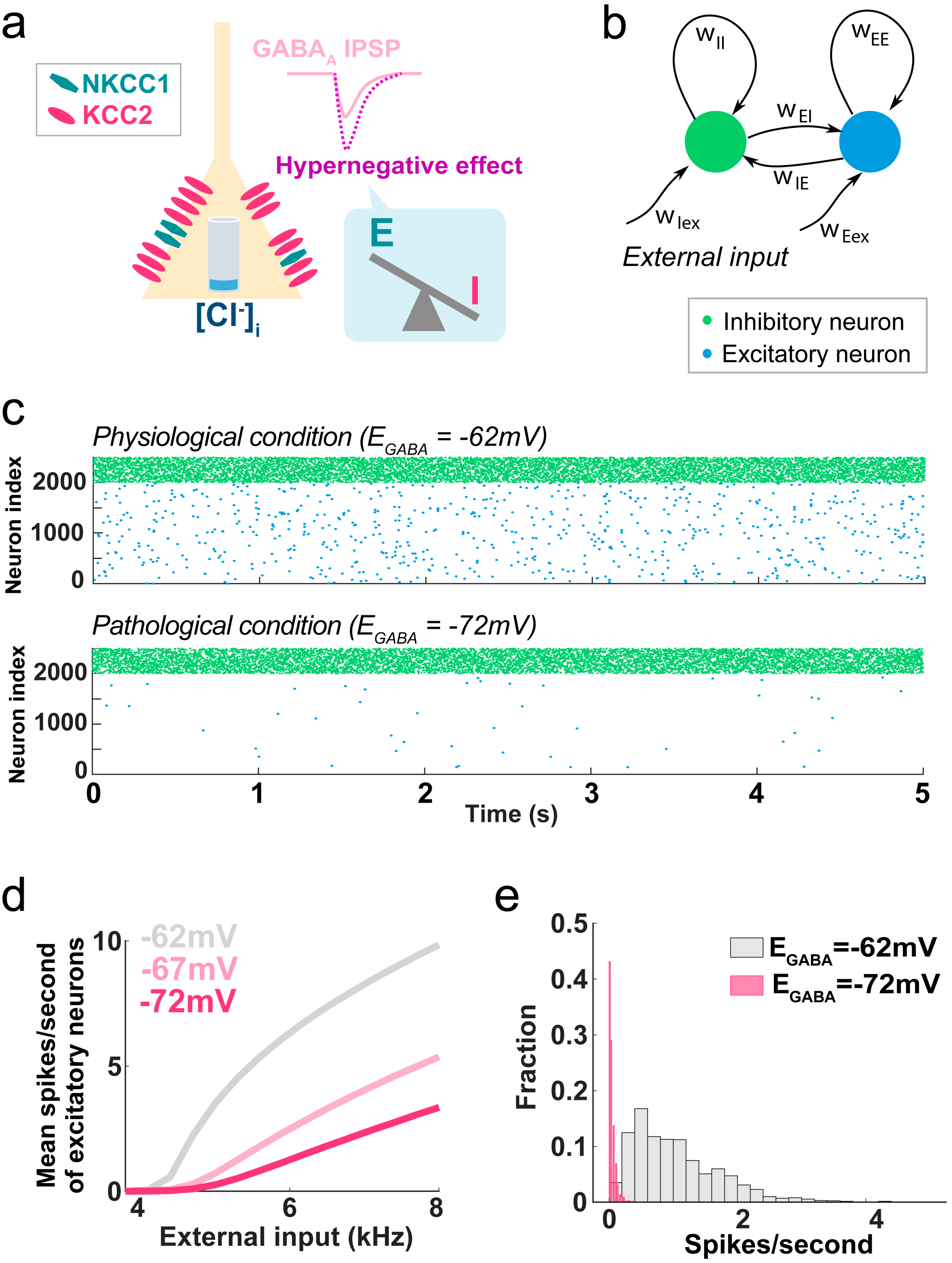
So far, the study of GABAA current reversal potential alterations in pathological conditions has been largely focused on the (re)appearance of excitatory GABAA in adult animals. However, recent investigations have shown that the opposite regulation can also be detrimental and that excessive GABAA-mediated inhibition due to hypernegative current reversal potential represents another potential mechanism of pathology. For example, adult mice that were exposed to neonatal inflammation (using an LPS injection) show cognitive impairments associated with hypernegative GABAA reversal potential and increased KCC2 activity in the hippocampus [79]. Interestingly, these authors showed a complex TGF-β1-dependent regulation, suggesting the alteration is not simply due to an overdrive of the normal developmental KCC2 upregulation. Accordingly, we recently showed that a hypernegative GABAA current can appear in the adult brain independently of any developmental dysregulation. We found that in adult mice 7 days after peripheral nerve injury (the Spared Nerve Injury model of neuropathic pain), the reversal potential of the GABAA current in the pyramidal neurons of the medial prefrontal cortex is ~10 mV more negative than in sham-operated control mice. This change is due to a large overexpression of KCC2 transcripts and, to a minor extent, to decreased NKCC1 expression [16]. Computational modeling of a cortical network demonstrated that the ~10 mV shift in GABAA reversal potential is by itself sufficient to dramatically reduce the overall activity of the network, resulting in over 60%-decreased activity of excitatory neurons in the range of natural external inputs (Figure 2). Thus, hypernegative GABA has a major impact on neuronal activity and should be considered whenever increased inhibition is present and no changes in the activity and/or number of interneurons are detected.

4.3. GABAA Flexibility and Pathology

Why are changes in GABAA current reversal potential detected in so many different conditions? The proximity of the value of the GABAA current reversal potential and the resting membrane potential of most neurons is probably the critical factor. Such proximity means that even small changes in the intracellular chloride concentration can switch the current effect from hyperpolarizing to depolarizing; this is a unique property of the GABAA channel and sets it apart from other synaptic receptors. Another reason might be the intrinsic complexity and plasticity of the regulatory mechanisms. Intracellular chloride concentration changes significantly during development because the regulating machinery is heavily modulated by numerous mechanisms (including epigenetic [80]); this makes this process particularly sensitive to even small functional abnormalities. Therefore, we hypothesize that alterations in GABAA reversal potential (either in the depolarizing or hyperpolarizing direction) in the cerebral cortex represent a common maladaptive mechanism leading to cognitive impairment in multiple pathological conditions with different etiologies.

5. Network Effects and Clinical Implications of Flexible GABAA Reversal Potential

Numerous studies show that neuronal oscillations in the prefrontal cortex and hippocampus are closely associated with cognitive tasks. Theta rhythms (4–8 Hz) in the cortex are enhanced during memory tasks in humans and animals [81,82]. Gamma oscillations (25–100 Hz) are also critical for cognitive function, and aberrant gamma oscillations have been observed in multiple cognitive disorders. Additionally, changes in brain oscillations can predict cognitive symptoms in individuals with autism spectrum disorders, and increasing evidence shows that long-range gamma synchronization is reduced in patients with schizophrenia [83] and other conditions characterized by cognitive disability [84]. GABAergic inhibition is essential to the generation of oscillatory networks [85,86]. Accordingly, E/I balance in the PFC and hippocampus is critical for cognitive and social functions [87,88]. However, despite the centrality of E/I imbalance in numerous brain disorders, the cellular mechanisms of E/I imbalance are mostly unknown. As oscillations emerge from reciprocal interactions between populations of excitatory (glutamatergic) and inhibitory (GABAergic) neurons [89], E/I imbalance may be due to multiple types of abnormalities in GABAergic and/or glutamatergic signaling. Consequently, it is possible that differences in the mechanisms underlying E/I imbalance lay behind the large variety and diverse severity of cognitive impairments in different neurodevelopmental disorders. While abundant evidence has been collected over the past several years supporting the role of depolarizing GABA as an important pathogenic mechanism in various diseases, it has only become clear very recently that the hypernegative reversal potential of the GABAA current also has a major functional impact, although the network mechanisms are likely different. We suggest that the main functional impact of depolarizing GABA is a loss of network synchronization, which results in the disruption of network oscillations (Figure 1). On the other hand, a hypernegative GABAA current seems to cause a general inhibition of the circuit, roughly equivalent to an increased number of inhibitory synaptic inputs (Figure 2).
Additionally, while both these conditions represent dysfunctions in GABAergic signaling, their potential treatment requires completely different approaches, each with potential drawbacks. The case of hypernegative GABAA is theoretically straightforward because GABA antagonists are likely beneficial to reduce the effects of the hypernegative GABAA. The use of such drugs, however, is limited by their toxicity and epileptogenicity. The situation is more complex in the case of depolarizing GABA, where inhibition is insufficient but cannot be improved by the common approach of using GABA agonists such as benzodiazepines or GABA analogs such as valproic acid or gabapentin. The most direct treatment for this condition would obviously be the restoration of the inhibitory effect of GABAA, which can be achieved either pharmacologically or genetically [51,69]. Both these approaches, however, also have major limitations. As depolarizing GABA in disease conditions is, by and large, the consequence of NKCC1 upregulation, NKCC1 antagonists, such as bumetanide, may provide a treatment opportunity. However, these drugs have very poor BBB permeability [90], which limits their effective concentration in the brain parenchyma. Additionally, these drugs do not have any cellular selectivity and not only affect excitatory and inhibitory neurons but also non-neuronal cells, which often express NKCC1 at a higher level than neurons. Thus, the network effects are necessarily broad and hard to predict. The genetic approach may help as NKCC1 expression could be selectively repressed in select cellular populations, but this approach is still far from viable for clinical treatment. An alternative approach may take advantage of KCC2 agonists, which would be neuron-selective, but effective agonists are still missing. However, KCC2 activity is regulated by numerous signaling pathways such as WNK, PKC, and BDNF [20,71,91,92,93]; therefore, these might also represent interesting pharmacological targets. Even in this case, however, cell-type selectivity would help considerably.

6. Conclusions

The GABAA current represents a particularly vulnerable target in many neurological conditions. This is mainly driven by two facts: 1—The net effect of GABAAR activation may be inhibitory or excitatory, depending on the electrochemical gradient for the permeant anions across the membrane of the postsynaptic neurons. Additionally, even when inhibitory, the GABAA current may dramatically change its efficacy depending on its reversal potential. Consequently, this highly plastic and regulated system is sensitive to even small changes in transmembrane ionic gradients. 2—This functional balance is regulated by mechanisms that are highly plastic because they are designed to change during physiological development. Taken together, these factors explain both the particular vulnerability of this system to even small cellular alterations and the oversized functional effects that can derive from these alterations. In keeping with such strong vulnerability, alterations in GABAA reversal potential have been recently implicated in numerous psychiatric and neurological conditions, suggesting that regulation of chloride-cation cotransporters represents a viable therapeutic target.

Author Contributions

H.R.K.: literature search, conceptualization of the paper and writing. M.M.: literature search, conceptualization of the paper and writing. All authors have read and agreed to the published version of the manuscript.

Funding

This work was supported by NIH grants NS112292 and DA044121 to M.M.

Conflicts of Interest

The authors declare no conflicts of interest.

References

  1. Atallah, B.V.; Scanziani, M. Instantaneous modulation of gamma oscillation frequency by balancing excitation with inhibition. Neuron 2009, 62, 566–577. [Google Scholar] [CrossRef] [PubMed]
  2. Yizhar, O.; Fenno, L.E.; Prigge, M.; Schneider, F.; Davidson, T.J.; O’Shea, D.J.; Sohal, V.S.; Goshen, I.; Finkelstein, J.; Paz, J.T.; et al. Neocortical excitation/inhibition balance in information processing and social dysfunction. Nature 2011, 477, 171–178. [Google Scholar] [CrossRef] [PubMed]
  3. Dehghani, N.; Peyrache, A.; Telenczuk, B.; Le Van Quyen, M.; Halgren, E.; Cash, S.S.; Hatsopoulos, N.G.; Destexhe, A. Dynamic Balance of Excitation and Inhibition in Human and Monkey Neocortex. Sci. Rep. 2016, 6, 23176. [Google Scholar] [CrossRef]
  4. Eichler, S.A.; Meier, J.C. E-I balance and human diseases—From molecules to networking. Front. Mol. Neurosci. 2008, 1, 2. [Google Scholar] [CrossRef] [PubMed]
  5. D’Amour, J.A.; Froemke, R.C. Inhibitory and excitatory spike-timing-dependent plasticity in the auditory cortex. Neuron 2015, 86, 514–528. [Google Scholar] [CrossRef] [PubMed]
  6. Isaacson, J.S.; Scanziani, M. How inhibition shapes cortical activity. Neuron 2011, 72, 231–243. [Google Scholar] [CrossRef] [PubMed]
  7. Liu, B.H.; Li, Y.T.; Ma, W.P.; Pan, C.J.; Zhang, L.I.; Tao, H.W. Broad inhibition sharpens orientation selectivity by expanding input dynamic range in mouse simple cells. Neuron 2011, 71, 542–554. [Google Scholar] [CrossRef]
  8. Haider, B.; Hausser, M.; Carandini, M. Inhibition dominates sensory responses in the awake cortex. Nature 2013, 493, 97–100. [Google Scholar] [CrossRef]
  9. Bassell, G.J.; Warren, S.T. Fragile X syndrome: Loss of local mRNA regulation alters synaptic development and function. Neuron 2008, 60, 201–214. [Google Scholar] [CrossRef]
  10. Snowden, S.G.; Ebshiana, A.A.; Hye, A.; Pletnikova, O.; O’Brien, R.; Yang, A.; Troncoso, J.; Legido-Quigley, C.; Thambisetty, M. Neurotransmitter Imbalance in the Brain and Alzheimer’s Disease Pathology. J. Alzheimers Dis. 2019, 72, 35–43. [Google Scholar] [CrossRef]
  11. Yang, G.J.; Murray, J.D.; Wang, X.J.; Glahn, D.C.; Pearlson, G.D.; Repovs, G.; Krystal, J.H.; Anticevic, A. Functional hierarchy underlies preferential connectivity disturbances in schizophrenia. Proc. Natl. Acad. Sci. USA 2016, 113, E219–E228. [Google Scholar] [CrossRef] [PubMed]
  12. Kehrer, C.; Maziashvili, N.; Dugladze, T.; Gloveli, T. Altered Excitatory-Inhibitory Balance in the NMDA-Hypofunction Model of Schizophrenia. Front. Mol. Neurosci. 2008, 1, 6. [Google Scholar] [CrossRef] [PubMed]
  13. Lozano, R.; Hare, E.B.; Hagerman, R.J. Modulation of the GABAergic pathway for the treatment of fragile X syndrome. Neuropsychiatr. Dis. Treat. 2014, 10, 1769–1779. [Google Scholar] [CrossRef] [PubMed]
  14. Gonzalez-Burgos, G.; Cho, R.Y.; Lewis, D.A. Alterations in cortical network oscillations and parvalbumin neurons in schizophrenia. Biol. Psychiatry 2015, 77, 1031–1040. [Google Scholar] [CrossRef] [PubMed]
  15. Bormann, J.; Hamill, O.P.; Sakmann, B. Mechanism of anion permeation through channels gated by glycine and gamma-aminobutyric acid in mouse cultured spinal neurones. J. Physiol. 1987, 385, 243–286. [Google Scholar] [CrossRef]
  16. Kim, H.R.; Long, M.; Sekerkova, G.; Maes, A.; Kennedy, A.; Martina, M. Hypernegative GABA(A) Reversal Potential in Pyramidal Cells Contributes to Medial Prefrontal Cortex Deactivation in a Mouse Model of Neuropathic Pain. J. Pain, 2023; ahead of print. [Google Scholar] [CrossRef]
  17. Ben-Ari, Y. Excitatory actions of gaba during development: The nature of the nurture. Nat. Rev. Neurosci. 2002, 3, 728–739. [Google Scholar] [CrossRef]
  18. Fukuda, A.; Tanaka, M.; Yamada, Y.; Muramatsu, K.; Shimano, Y.; Nishino, H. Simultaneous optical imaging of intracellular Cl- in neurons in different layers of rat neocortical slices: Advantages and limitations. Neurosci. Res. 1998, 32, 363–371. [Google Scholar] [CrossRef]
  19. Kuner, T.; Augustine, G.J. A genetically encoded ratiometric indicator for chloride: Capturing chloride transients in cultured hippocampal neurons. Neuron 2000, 27, 447–459. [Google Scholar] [CrossRef]
  20. Kahle, K.T.; Delpire, E. Kinase-KCC2 coupling: Cl- rheostasis, disease susceptibility, therapeutic target. J. Neurophysiol. 2016, 115, 8–18. [Google Scholar] [CrossRef]
  21. Kurki, S.N.; Uvarov, P.; Pospelov, A.S.; Trontti, K.; Hubner, A.K.; Srinivasan, R.; Watanabe, M.; Hovatta, I.; Hubner, C.A.; Kaila, K.; et al. Expression patterns of NKCC1 in neurons and non-neuronal cells during cortico-hippocampal development. Cereb. Cortex 2023, 33, 5906–5923. [Google Scholar] [CrossRef]
  22. Markkanen, M.; Karhunen, T.; Llano, O.; Ludwig, A.; Rivera, C.; Uvarov, P.; Airaksinen, M.S. Distribution of neuronal KCC2a and KCC2b isoforms in mouse CNS. J. Comp. Neurol. 2014, 522, 1897–1914. [Google Scholar] [CrossRef] [PubMed]
  23. Vu, T.Q.; Payne, J.A.; Copenhagen, D.R. Localization and developmental expression patterns of the neuronal K-Cl cotransporter (KCC2) in the rat retina. J. Neurosci. 2000, 20, 1414–1423. [Google Scholar] [CrossRef] [PubMed]
  24. Hubner, C.A.; Stein, V.; Hermans-Borgmeyer, I.; Meyer, T.; Ballanyi, K.; Jentsch, T.J. Disruption of KCC2 reveals an essential role of K-Cl cotransport already in early synaptic inhibition. Neuron 2001, 30, 515–524. [Google Scholar] [CrossRef] [PubMed]
  25. Chew, T.A.; Orlando, B.J.; Zhang, J.; Latorraca, N.R.; Wang, A.; Hollingsworth, S.A.; Chen, D.H.; Dror, R.O.; Liao, M.; Feng, L. Structure and mechanism of the cation-chloride cotransporter NKCC1. Nature 2019, 572, 488–492. [Google Scholar] [CrossRef]
  26. Neumann, C.; Rosenbaek, L.L.; Flygaard, R.K.; Habeck, M.; Karlsen, J.L.; Wang, Y.; Lindorff-Larsen, K.; Gad, H.H.; Hartmann, R.; Lyons, J.A.; et al. Cryo-EM structure of the human NKCC1 transporter reveals mechanisms of ion coupling and specificity. EMBO J. 2022, 41, e110169. [Google Scholar] [CrossRef] [PubMed]
  27. Agez, M.; Schultz, P.; Medina, I.; Baker, D.J.; Burnham, M.P.; Cardarelli, R.A.; Conway, L.C.; Garnier, K.; Geschwindner, S.; Gunnarsson, A.; et al. Molecular architecture of potassium chloride co-transporter KCC2. Sci. Rep. 2017, 7, 16452. [Google Scholar] [CrossRef] [PubMed]
  28. Xie, Y.; Chang, S.; Zhao, C.; Wang, F.; Liu, S.; Wang, J.; Delpire, E.; Ye, S.; Guo, J. Structures and an activation mechanism of human potassium-chloride cotransporters. Sci. Adv. 2020, 6, eabc5883. [Google Scholar] [CrossRef]
  29. Hartmann, A.M.; Nothwang, H.G. NKCC1 and KCC2: Structural insights into phospho-regulation. Front. Mol. Neurosci. 2022, 15, 964488. [Google Scholar] [CrossRef]
  30. Han, B.; Bellemer, A.; Koelle, M.R. An evolutionarily conserved switch in response to GABA affects development and behavior of the locomotor circuit of Caenorhabditis elegans. Genetics 2015, 199, 1159–1172. [Google Scholar] [CrossRef]
  31. Peerboom, C.; Wierenga, C.J. The postnatal GABA shift: A developmental perspective. Neurosci. Biobehav. Rev. 2021, 124, 179–192. [Google Scholar] [CrossRef]
  32. Cherubini, E.; Gaiarsa, J.L.; Ben-Ari, Y. GABA: An excitatory transmitter in early postnatal life. Trends Neurosci. 1991, 14, 515–519. [Google Scholar] [CrossRef] [PubMed]
  33. Leinekugel, X.; Khalilov, I.; McLean, H.; Caillard, O.; Gaiarsa, J.L.; Ben-Ari, Y.; Khazipov, R. GABA is the principal fast-acting excitatory transmitter in the neonatal brain. Adv. Neurol. 1999, 79, 189–201. [Google Scholar] [PubMed]
  34. Ben-Ari, Y.; Cherubini, E.; Corradetti, R.; Gaiarsa, J.L. Giant synaptic potentials in immature rat CA3 hippocampal neurones. J. Physiol. 1989, 416, 303–325. [Google Scholar] [CrossRef] [PubMed]
  35. Verheugen, J.A.; Fricker, D.; Miles, R. Noninvasive measurements of the membrane potential and GABAergic action in hippocampal interneurons. J. Neurosci. 1999, 19, 2546–2555. [Google Scholar] [CrossRef] [PubMed]
  36. Dehorter, N.; Vinay, L.; Hammond, C.; Ben-Ari, Y. Timing of developmental sequences in different brain structures: Physiological and pathological implications. Eur. J. Neurosci. 2012, 35, 1846–1856. [Google Scholar] [CrossRef]
  37. Dzhala, V.I.; Talos, D.M.; Sdrulla, D.A.; Brumback, A.C.; Mathews, G.C.; Benke, T.A.; Delpire, E.; Jensen, F.E.; Staley, K.J. NKCC1 transporter facilitates seizures in the developing brain. Nat. Med. 2005, 11, 1205–1213. [Google Scholar] [CrossRef] [PubMed]
  38. Hyde, T.M.; Lipska, B.K.; Ali, T.; Mathew, S.V.; Law, A.J.; Metitiri, O.E.; Straub, R.E.; Ye, T.; Colantuoni, C.; Herman, M.M.; et al. Expression of GABA signaling molecules KCC2, NKCC1, and GAD1 in cortical development and schizophrenia. J. Neurosci. 2011, 31, 11088–11095. [Google Scholar] [CrossRef]
  39. Rivera, C.; Voipio, J.; Kaila, K. Two developmental switches in GABAergic signalling: The K+-Cl cotransporter KCC2 and carbonic anhydrase CAVII. J. Physiol. 2005, 562 Pt 1, 27–36. [Google Scholar] [CrossRef]
  40. Yamada, J.; Okabe, A.; Toyoda, H.; Kilb, W.; Luhmann, H.J.; Fukuda, A. Cl uptake promoting depolarizing GABA actions in immature rat neocortical neurones is mediated by NKCC1. J. Physiol. 2004, 557 Pt 3, 829–841. [Google Scholar] [CrossRef]
  41. Su, G.; Kintner, D.B.; Flagella, M.; Shull, G.E.; Sun, D. Astrocytes from Na(+)-K(+)-Cl() cotransporter-null mice exhibit absence of swelling and decrease in EAA release. Am. J. Physiol. Cell Physiol. 2002, 282, C1147–C1160. [Google Scholar] [CrossRef]
  42. Kanaka, C.; Ohno, K.; Okabe, A.; Kuriyama, K.; Itoh, T.; Fukuda, A.; Sato, K. The differential expression patterns of messenger RNAs encoding K-Cl cotransporters (KCC1,2) and Na-K-2Cl cotransporter (NKCC1) in the rat nervous system. Neuroscience 2001, 104, 933–946. [Google Scholar] [CrossRef] [PubMed]
  43. Kitayama, T. The Role of Astrocytes in the Modulation of K(+)-Cl()-Cotransporter-2 Function. Int. J. Mol. Sci. 2020, 21, 9539. [Google Scholar] [CrossRef]
  44. Nguyen, T.D.; Ishibashi, M.; Sinha, A.S.; Watanabe, M.; Kato, D.; Horiuchi, H.; Wake, H.; Fukuda, A. Astrocytic NKCC1 inhibits seizures by buffering Cl() and antagonizing neuronal NKCC1 at GABAergic synapses. Epilepsia 2023, 64, 3389–3403. [Google Scholar] [CrossRef]
  45. Fellin, T.; Pascual, O.; Gobbo, S.; Pozzan, T.; Haydon, P.G.; Carmignoto, G. Neuronal synchrony mediated by astrocytic glutamate through activation of extrasynaptic NMDA receptors. Neuron 2004, 43, 729–743. [Google Scholar] [CrossRef] [PubMed]
  46. Covelo, A.; Araque, A. Neuronal activity determines distinct gliotransmitter release from a single astrocyte. eLife 2018, 7, e32237. [Google Scholar] [CrossRef] [PubMed]
  47. Huda, R.; Chang, Z.; Do, J.; McCrimmon, D.R.; Martina, M. Activation of astrocytic PAR1 receptors in the rat nucleus of the solitary tract regulates breathing through modulation of presynaptic TRPV1. J. Physiol. 2018, 596, 497–513. [Google Scholar] [CrossRef] [PubMed]
  48. Kintner, D.B.; Luo, J.; Gerdts, J.; Ballard, A.J.; Shull, G.E.; Sun, D. Role of Na+-K+-Cl cotransport and Na+/Ca2+ exchange in mitochondrial dysfunction in astrocytes following in vitro ischemia. Am. J. Physiol. Cell Physiol. 2007, 292, C1113–C1122. [Google Scholar] [CrossRef] [PubMed]
  49. Rose, C.R.; Verkhratsky, A. Principles of sodium homeostasis and sodium signalling in astroglia. Glia 2016, 64, 1611–1627. [Google Scholar] [CrossRef]
  50. Kahle, K.T.; Simard, J.M.; Staley, K.J.; Nahed, B.V.; Jones, P.S.; Sun, D. Molecular mechanisms of ischemic cerebral edema: Role of electroneutral ion transport. Physiology 2009, 24, 257–265. [Google Scholar] [CrossRef]
  51. Kim, H.R.; Rajagopal, L.; Meltzer, H.Y.; Martina, M. Depolarizing GABA(A) current in the prefrontal cortex is linked with cognitive impairment in a mouse model relevant for schizophrenia. Sci. Adv. 2021, 7, eaba5032. [Google Scholar] [CrossRef]
  52. Del Turco, D.; Paul, M.H.; Schlaudraff, J.; Muellerleile, J.; Bozic, F.; Vuksic, M.; Jedlicka, P.; Deller, T. Layer-specific changes of KCC2 and NKCC1 in the mouse dentate gyrus after entorhinal denervation. Front. Mol. Neurosci. 2023, 16, 1118746. [Google Scholar] [CrossRef] [PubMed]
  53. Otsu, Y.; Donneger, F.; Schwartz, E.J.; Poncer, J.C. Cation-chloride cotransporters and the polarity of GABA signalling in mouse hippocampal parvalbumin interneurons. J. Physiol. 2020, 598, 1865–1880. [Google Scholar] [CrossRef] [PubMed]
  54. Pol, E.; Come, E.; Merlaud, Z.; Gouhier, J.; Russeau, M.; Scotto-Lomassese, S.; Moutkine, I.; Marques, X.; Levi, S. NKCC1 and KCC2 Chloride Transporters Have Different Membrane Dynamics on the Surface of Hippocampal Neurons. Cells 2023, 12, 2363. [Google Scholar] [CrossRef] [PubMed]
  55. Baldi, R.; Varga, C.; Tamas, G. Differential distribution of KCC2 along the axo-somato-dendritic axis of hippocampal principal cells. Eur. J. Neurosci. 2010, 32, 1319–1325. [Google Scholar] [CrossRef] [PubMed]
  56. Weilinger, N.L.; Wicki-Stordeur, L.E.; Groten, C.J.; LeDue, J.M.; Kahle, K.T.; MacVicar, B.A. KCC2 drives chloride microdomain formation in dendritic blebbing. Cell Rep. 2022, 41, 111556. [Google Scholar] [CrossRef] [PubMed]
  57. Al Awabdh, S.; Donneger, F.; Goutierre, M.; Seveno, M.; Vigy, O.; Weinzettl, P.; Russeau, M.; Moutkine, I.; Levi, S.; Marin, P.; et al. Gephyrin Interacts with the K-Cl Cotransporter KCC2 to Regulate Its Surface Expression and Function in Cortical Neurons. J. Neurosci. 2022, 42, 166–182. [Google Scholar] [CrossRef] [PubMed]
  58. Gulyas, A.I.; Sik, A.; Payne, J.A.; Kaila, K.; Freund, T.F. The KCl cotransporter, KCC2, is highly expressed in the vicinity of excitatory synapses in the rat hippocampus. Eur. J. Neurosci. 2001, 13, 2205–2217. [Google Scholar] [CrossRef] [PubMed]
  59. Chamma, I.; Heubl, M.; Chevy, Q.; Renner, M.; Moutkine, I.; Eugene, E.; Poncer, J.C.; Levi, S. Activity-dependent regulation of the K/Cl transporter KCC2 membrane diffusion, clustering, and function in hippocampal neurons. J. Neurosci. 2013, 33, 15488–15503. [Google Scholar] [CrossRef]
  60. Chamma, I.; Chevy, Q.; Poncer, J.C.; Levi, S. Role of the neuronal K-Cl co-transporter KCC2 in inhibitory and excitatory neurotransmission. Front. Cell Neurosci. 2012, 6, 5. [Google Scholar] [CrossRef]
  61. Woodin, M.A.; Ganguly, K.; Poo, M.M. Coincident pre- and postsynaptic activity modifies GABAergic synapses by postsynaptic changes in Cl- transporter activity. Neuron 2003, 39, 807–820. [Google Scholar] [CrossRef]
  62. Khirug, S.; Yamada, J.; Afzalov, R.; Voipio, J.; Khiroug, L.; Kaila, K. GABAergic depolarization of the axon initial segment in cortical principal neurons is caused by the Na-K-2Cl cotransporter NKCC1. J. Neurosci. 2008, 28, 4635–4639. [Google Scholar] [CrossRef] [PubMed]
  63. Szabadics, J.; Varga, C.; Molnar, G.; Olah, S.; Barzo, P.; Tamas, G. Excitatory effect of GABAergic axo-axonic cells in cortical microcircuits. Science 2006, 311, 233–235. [Google Scholar] [CrossRef] [PubMed]
  64. Rinetti-Vargas, G.; Phamluong, K.; Ron, D.; Bender, K.J. Periadolescent Maturation of GABAergic Hyperpolarization at the Axon Initial Segment. Cell Rep. 2017, 20, 21–29. [Google Scholar] [CrossRef] [PubMed]
  65. Pracucci, E.; Graham, R.T.; Alberio, L.; Nardi, G.; Cozzolino, O.; Pillai, V.; Pasquini, G.; Saieva, L.; Walsh, D.; Landi, S.; et al. Daily rhythm in cortical chloride homeostasis underpins functional changes in visual cortex excitability. Nat. Commun. 2023, 14, 7108. [Google Scholar] [CrossRef] [PubMed]
  66. Cherubini, E.; Di Cristo, G.; Avoli, M. Dysregulation of GABAergic Signaling in Neurodevelomental Disorders: Targeting Cation-Chloride Co-transporters to Re-establish a Proper E/I Balance. Front. Cell Neurosci. 2021, 15, 813441. [Google Scholar] [CrossRef] [PubMed]
  67. Savardi, A.; Borgogno, M.; De Vivo, M.; Cancedda, L. Pharmacological tools to target NKCC1 in brain disorders. Trends Pharmacol. Sci. 2021, 42, 1009–1034. [Google Scholar] [CrossRef] [PubMed]
  68. Cohen, I.; Navarro, V.; Clemenceau, S.; Baulac, M.; Miles, R. On the origin of interictal activity in human temporal lobe epilepsy in vitro. Science 2002, 298, 1418–1421. [Google Scholar] [CrossRef] [PubMed]
  69. Deidda, G.; Parrini, M.; Naskar, S.; Bozarth, I.F.; Contestabile, A.; Cancedda, L. Reversing excitatory GABAAR signaling restores synaptic plasticity and memory in a mouse model of Down syndrome. Nat. Med. 2015, 21, 318–326. [Google Scholar] [CrossRef]
  70. MacKenzie, G.; Maguire, J. Chronic stress shifts the GABA reversal potential in the hippocampus and increases seizure susceptibility. Epilepsy Res. 2015, 109, 13–27. [Google Scholar] [CrossRef]
  71. Arion, D.; Lewis, D.A. Altered expression of regulators of the cortical chloride transporters NKCC1 and KCC2 in schizophrenia. Arch. Gen. Psychiatry 2011, 68, 21–31. [Google Scholar] [CrossRef]
  72. Jentsch, J.D.; Roth, R.H. The neuropsychopharmacology of phencyclidine: From NMDA receptor hypofunction to the dopamine hypothesis of schizophrenia. Neuropsychopharmacology 1999, 20, 201–225. [Google Scholar] [CrossRef] [PubMed]
  73. Steeds, H.; Carhart-Harris, R.L.; Stone, J.M. Drug models of schizophrenia. Ther. Adv. Psychopharmacol. 2015, 5, 43–58. [Google Scholar] [CrossRef] [PubMed]
  74. Merner, N.D.; Mercado, A.; Khanna, A.R.; Hodgkinson, A.; Bruat, V.; Awadalla, P.; Gamba, G.; Rouleau, G.A.; Kahle, K.T. Gain-of-function missense variant in SLC12A2, encoding the bumetanide-sensitive NKCC1 cotransporter, identified in human schizophrenia. J. Psychiatr. Res. 2016, 77, 22–26. [Google Scholar] [CrossRef]
  75. Chen, S.R.; Zhu, L.; Chen, H.; Wen, L.; Laumet, G.; Pan, H.L. Increased spinal cord Na(+)-K(+)-2Cl() cotransporter-1 (NKCC1) activity contributes to impairment of synaptic inhibition in paclitaxel-induced neuropathic pain. J. Biol. Chem. 2014, 289, 31111–31120. [Google Scholar] [CrossRef] [PubMed]
  76. Ferando, I.; Faas, G.C.; Mody, I. Diminished KCC2 confounds synapse specificity of LTP during senescence. Nat. Neurosci. 2016, 19, 1197–1200. [Google Scholar] [CrossRef]
  77. Lam, P.; Vinnakota, C.; Guzmán, B.C.F.; Newland, J.; Peppercorn, K.; Tate, W.P.; Waldvogel, H.J.; Faull, R.L.M.; Kwakowsky, A. Beta-Amyloid (Aβ) Increases the Expression of NKCC1 in the Mouse Hippocampus. Molecules 2022, 27, 2440. [Google Scholar] [CrossRef]
  78. Keramidis, I.; McAllister, B.B.; Bourbonnais, J.; Wang, F.; Isabel, D.; Rezaei, E.; Sansonetti, R.; Degagne, P.; Hamel, J.P.; Nazari, M.; et al. Restoring neuronal chloride extrusion reverses cognitive decline linked to Alzheimer’s disease mutations. Brain 2023, 146, 4903–4915. [Google Scholar] [CrossRef]
  79. Rong, J.; Yang, Y.; Liang, M.; Zhong, H.; Li, Y.; Zhu, Y.; Sha, S.; Chen, L.; Zhou, R. Neonatal inflammation increases hippocampal KCC2 expression through methylation-mediated TGF-beta1 downregulation leading to impaired hippocampal cognitive function and synaptic plasticity in adult mice. J. Neuroinflamm. 2023, 20, 15. [Google Scholar] [CrossRef]
  80. Orlov, S.N. NKCC1 as an epigenetically regulated transporter involved in blood pressure elevation with age. Am. J. Hypertens. 2011, 24, 1264. [Google Scholar] [CrossRef]
  81. Raghavachari, S.; Kahana, M.J.; Rizzuto, D.S.; Caplan, J.B.; Kirschen, M.P.; Bourgeois, B.; Madsen, J.R.; Lisman, J.E. Gating of human theta oscillations by a working memory task. J. Neurosci. 2001, 21, 3175–3183. [Google Scholar] [CrossRef]
  82. Lee, H.; Simpson, G.V.; Logothetis, N.K.; Rainer, G. Phase locking of single neuron activity to theta oscillations during working memory in monkey extrastriate visual cortex. Neuron 2005, 45, 147–156. [Google Scholar] [CrossRef] [PubMed]
  83. Mulert, C.; Kirsch, V.; Pascual-Marqui, R.; McCarley, R.W.; Spencer, K.M. Long-range synchrony of gamma oscillations and auditory hallucination symptoms in schizophrenia. Int. J. Psychophysiol. 2011, 79, 55–63. [Google Scholar] [CrossRef] [PubMed]
  84. Alemany-Gonzalez, M.; Gener, T.; Nebot, P.; Vilademunt, M.; Dierssen, M.; Puig, M.V. Prefrontal-hippocampal functional connectivity encodes recognition memory and is impaired in intellectual disability. Proc. Natl. Acad. Sci. USA 2020, 117, 11788–11798. [Google Scholar] [CrossRef] [PubMed]
  85. Fricker, D.; Miles, R. EPSP amplification and the precision of spike timing in hippocampal neurons. Neuron 2000, 28, 559–569. [Google Scholar] [CrossRef] [PubMed]
  86. Cardin, J.A. Inhibitory Interneurons Regulate Temporal Precision and Correlations in Cortical Circuits. Trends Neurosci. 2018, 41, 689–700. [Google Scholar] [CrossRef] [PubMed]
  87. Ferguson, B.R.; Gao, W.J. PV Interneurons: Critical Regulators of E/I Balance for Prefrontal Cortex-Dependent Behavior and Psychiatric Disorders. Front. Neural Circuits 2018, 12, 37. [Google Scholar] [CrossRef] [PubMed]
  88. Sohal, V.S.; Rubenstein, J.L.R. Excitation-inhibition balance as a framework for investigating mechanisms in neuropsychiatric disorders. Mol. Psychiatry 2019, 24, 1248–1257. [Google Scholar] [CrossRef] [PubMed]
  89. Wilson, H.R.; Cowan, J.D. Excitatory and inhibitory interactions in localized populations of model neurons. Biophys. J. 1972, 12, 1–24. [Google Scholar] [CrossRef]
  90. Loscher, W.; Kaila, K. CNS pharmacology of NKCC1 inhibitors. Neuropharmacology 2022, 205, 108910. [Google Scholar] [CrossRef]
  91. Kahle, K.T.; Deeb, T.Z.; Puskarjov, M.; Silayeva, L.; Liang, B.; Kaila, K.; Moss, S.J. Modulation of neuronal activity by phosphorylation of the K-Cl cotransporter KCC2. Trends Neurosci. 2013, 36, 726–737. [Google Scholar] [CrossRef]
  92. Porcher, C.; Medina, I.; Gaiarsa, J.L. Mechanism of BDNF Modulation in GABAergic Synaptic Transmission in Healthy and Disease Brains. Front. Cell Neurosci. 2018, 12, 273. [Google Scholar] [CrossRef] [PubMed]
  93. Rivera, C.; Li, H.; Thomas-Crusells, J.; Lahtinen, H.; Viitanen, T.; Nanobashvili, A.; Kokaia, Z.; Airaksinen, M.S.; Voipio, J.; Kaila, K.; et al. BDNF-induced TrkB activation down-regulates the K+-Cl cotransporter KCC2 and impairs neuronal Cl extrusion. J. Cell Biol. 2002, 159, 747–752. [Google Scholar] [CrossRef] [PubMed]
Figure 1. Schematic of the effects of depolarizing GABAA in disease. In postsynaptic neurons, two types of chloride/potassium cotransporters (NKCC1 and KCC2) regulate intracellular chloride concentration ([Cl]i). The polarity of the GABAA current, either depolarizing or hyperpolarizing, is largely determined by the transmembrane chloride concentration gradient. When activation of GABAA receptors pathologically evokes excitatory (EPSP) instead of inhibitory (IPSP) synaptic potentials due to increased NKCC1-KCC2 activity ratio, the excitatory/inhibitory (E/I) balance is altered, which leads to a disruption of network oscillations and behavioral impairment.
Life 14 00143 g001
Figure 2. Effect of hypernegative GABAA current on network function. (a) When the neuronal KCC2-NKCC1 activity ratio is increased, intracellular chloride concentration ([Cl]i) is lower than the physiological level, and the inhibitory effect of GABAA current is strengthened, disrupting the E/I balance. (be) Simulated effects of hypernegative GABAA current on cortical network activity. In pathological conditions (GABAA reversal potentials = −72 mV), the spike frequency of the postsynaptic excitatory neurons is severely decreased, even when the frequency of the inhibitory inputs is unaffected. (b) Computational model of a cortical network based on local microcircuit of excitatory–inhibitory recurrent architecture. (c) Simulated spontaneous spiking activity of excitatory (blue) and inhibitory (green) cortical neurons with physiological (−62 mV, top panel) or pathologically hyperpolarized (−72 mV, bottom) GABAA reversal potentials. (d) Average firing rate of excitatory neurons with three different GABAA reversal potentials in response to variable external input frequencies. (e) Histogram of firing rates of excitatory neurons when the external input was set at 4.5 kHz. (Adapted with permission from [16]).
Life 14 00143 g002
Disclaimer/Publisher’s Note: The statements, opinions and data contained in all publications are solely those of the individual author(s) and contributor(s) and not of MDPI and/or the editor(s). MDPI and/or the editor(s) disclaim responsibility for any injury to people or property resulting from any ideas, methods, instructions or products referred to in the content.