Review

Are Gender Differences Important for Autoimmune Liver Diseases?

1
Scientific Consultant IRCCS Negrar, 37024 Verona, Italy
2
University of Padova, 35122 Padova, Italy
3
Department of Pharmaceutical and Pharmacological Sciences, University of Padova, 35122 Padova, Italy
*
Author to whom correspondence should be addressed.
Life 2024, 14(4), 500; https://doi.org/10.3390/life14040500
Submission received: 12 March 2024 / Revised: 4 April 2024 / Accepted: 11 April 2024 / Published: 12 April 2024
(This article belongs to the Special Issue Gender Medicine: Are Gender Differences Important for Medicine?)

Abstract

:
Gender Medicine has had an enormous expansion over the last ten years. Autoimmune liver diseases include several conditions, i.e., autoimmune hepatitis (AIH), primary biliary cholangitis (PBC), primary sclerosing cholangitis (PSC), and conditions involving the liver or biliary tree overlapping with AIH, as well as IgG4-related disease. However, little is known about the impact of sex in the pathogenesis and natural history of these conditions. The purpose of this review is to provide an update of the gender disparities among the autoimmune liver diseases by reviewing the data published from 1999 to 2023. The epidemiology of these diseases has been changing over the last years, due to the amelioration of knowledge in their diagnosis, pathogenesis, and treatment. The clinical data collected so far support the existence of sex differences in the natural history of autoimmune liver diseases. Notably, their history could be longer than that which is now known, with problems being initiated even at a pediatric age. Moreover, gender disparity has been observed during the onset of complications related to end-stage liver disease, including cancer incidence. However, there is still an important debate among researchers about the impact of sex and the pathogenesis of these conditions. With this review, we would like to emphasize the urgency of basic science and clinical research to increase our understanding of the sex differences in autoimmune liver diseases.

1. Introduction

The disease burden of autoimmune liver disease includes autoimmune hepatitis (AIH), primary biliary cholangitis (PBC), primary sclerosing cholangitis (PSC), and overlapping conditions between AIH and PBC or PSC. A complete review on gender disparities in liver disease, including autoimmune liver disease, has recently been published [1]. Indeed, these disparities imply a wide contrast, not only regarding the medical point of view, but also social policies, employment, housing, health care, behavior factors, and cultural and social values [2]. In fact, the terms sex and gender are usually referred to as biological or socio-cultural issues, respectively. Actually, all the approaches often comprise both biological and social dimensions [3]. As already outlined [1], most studies describing autoimmune liver diseases have focused on one sex only and less on the comparative aspects. Men and women are differently affected by diseases in general and also by liver diseases; thus, sex-stratified medicine is an extremely important, although understudied, facet of medical care [4]. In general, clinical data suggest that men and women exhibit differences regarding the epidemiology and the progression of many diseases, including liver diseases [5,6], and there is an urgent need for basic science and clinical research to increase our understanding of gender differences in these contexts [7].
The aim of this paper is to review the current knowledge regarding the influence of sex on the setting of autoimmune liver diseases, underlining the relevance of sex-specific analysis in the prognosis and management of these diseases. To provide a state-of-the-art analysis of this topic, a review of the literature in PubMed, Scopus, and Science Direct was conducted (included dates: 1999–2023).

2. Autoimmune Hepatitis

Autoimmune hepatitis (AIH) is a rare inflammatory liver disease which can progress to end-stage liver disease and liver failure [8]. The point of prevalence in Northern Europe is estimated at 10–20 per 100,000 subjects, with an incidence rate of 1.2 per 100,000 person-years [9] and a 1:3.5 male–female ratio [5]. In a retrospective cohort of adults in the Optum Clinformatics Data Mart (2009–2018) in the USA, AIH incidence was 4 per 100,000 person-years and the prevalence was 26.6 per 100,000 [10]. Male sex, black race, and Hispanic ethnicity were associated with AIH with cirrhosis [10].
An exhaustive review of the pathogenesis of AIH has recently been published [11]. It has been estimated that approximately 9–12% of patients with AIH experienced a drug-induced hypersensitization process, and, of these, most were women [12]. In a preclinical in vivo study, Thomas and collaborators [13] immunized mice with an epitope of CYP2E1 and showed that IL-4 activated CD4+ T cells, thereby inducing the recruitment and influx of neutrophils, macrophages, and mast cells into the liver. They also showed that IL-33-induced FOXP3+ regulatory T cells to confer protection against drug-induced autoimmune hepatitis in female and male mice. This work also confirms the previous results implicating female sex, by demonstrating that female BALB/C mice develop a more severe drug-induced autoimmune hepatitis [14].

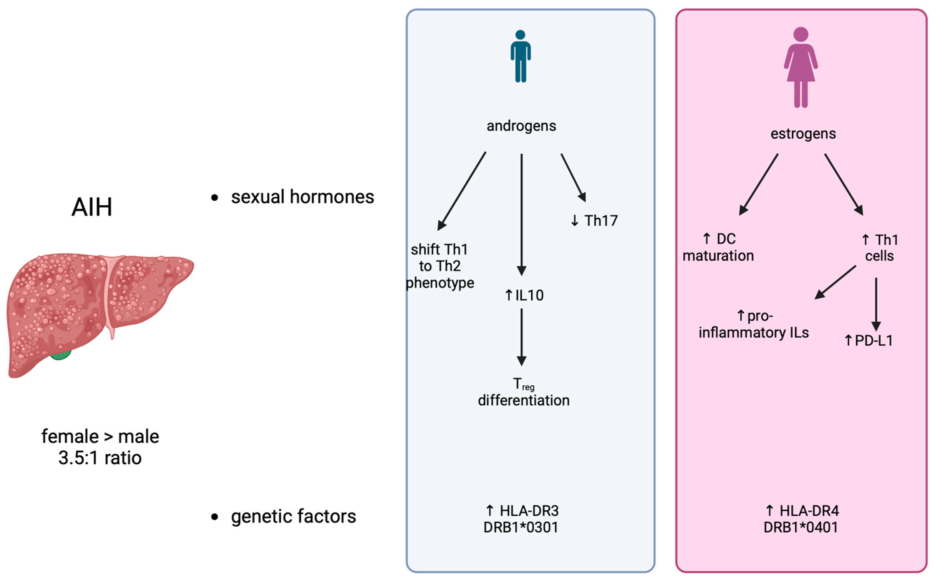
2.1. Factors Responsible for Gender Differences

The most important factors responsible for gender differences in AIH include: (1) sexual hormones; (2) genetic factors; (3) environmental factors; and (4) microbiome (Figure 1).
Sexual hormones. Insight about the role of sex hormones in AIH has been obtained by observations during pregnancy. Interestingly, pregnancy in AIH patients presents an increased risk of complications, both fetal and maternal [15], with respect to healthy women, although the successful delivery of healthy newborns has been reported in most AIH patients. On the other hand, it should be noticed that pregnancy may have a significant impact on the history and outcome of AIH. For example, up to 50% of patients encounter flares in AIH activity after delivery, or flares or AIH manifestations during or immediately after pregnancy [16,17]. Furthermore, the onset of de novo AIH has also been observed after delivery [18]. On the contrary, in many AIH patients, disease severity is reduced during pregnancy, so that the therapy’s dosage can be reduced [16,19]. From the mechanistic point of view, estrogens influence immune system activity, mainly acting on dendritic cells (DC). Evidence shows that these hormones can either promote DC maturation, leading to an increase in T helper 1 (Th1) cell activity, due to the increased production of proinflammatory interleukins (ILs) [20,21,22], or they can establish a tolerogenic immune environment by upregulating the expression of checkpoint inhibitors, such as PD-L1 [23,24]. On the other hand, androgens may also influence the pathogenesis of AIH by shifting the Th-1 to the Th-2 phenotype [25], thus reducing Th-17 cells in the liver [26] and increasing the secretion of IL-10, which drives the differentiation of regulatory T cells (Tregs) [27]. Notably, human and mouse T cells express cytosolic androgens and membrane-bound androgen receptors (mAR); androgens led to changes in cytokine expression in T cells either directly or indirectly via antigen presenting cells, with a shift toward a decrease in pro-inflammatory cytokine expression [28].
Genetic factors. Genes related to immune system functions are encoded on the sexual X and Y chromosomes [29]. AIH has been significantly associated with human histocompatibility complex (HLA) haplotypes. The most significant associations emerged in the alleles encoding for HLA-DR3 and DR4 [30]. A sex dimorphism was evident, since male AIH patients were characterized by an increased expression of HLA-DR3 (DRB1*0301) [31], whereas HLA-DR4 (DRB1*0401) was increased in female patients [32]. Furthermore, DRB1*0301 patients show an earlier onset of AIH, which also has more severe biochemical and histological features than those observed in the other haplotypes, as well as worse outcomes [32,33]. On the other hand, DRB1*0401 patients often have other immunological comorbidities but also higher response rates to the pharmacological treatments [32,33].
Environmental factors. Environmental factors, such as chemicals or viruses, can represent the triggers for AIH development [34]. Environmental factors can lead to the initialization of the autoimmune response by four different mechanisms. (1) The massive non-specific activation of resting T cells, which can be exerted either by viral infections, such as, for example, the Epstein–Barr virus (EBV) [35], or xenobiotics, like Concavalin A, is able to induce the secretion of proinflammatory cytokines [36]. (2) The creation of a neoantigen by the binding of a self-protein with a chemical compound, which has been reported both for natural products, such as black cohosh [37], and drugs, like statins [38]. (3) Molecular mimicry, i.e., the cross-reactivity between a viral and a self-protein, which has been proposed for HCV and SARS-CoV2 patients [39]. (4) The modulation of gene expression, exerted by molecules triggering an autoimmune reaction due to the creation of a pro-inflammatory microenvironment, ideal for autoantigen presentation [34].
Microbiome. It is well known that dysbiosis, i.e., the pathological changes of the gut microbiome, plays a pivotal role in driving aberrant immune responses and is fundamental in the development of autoimmune diseases [40], including AIH [41], although no information about the possible effect of sex is available so far for these patients. However, it is well known that AIH patients are characterized by a significant reduction in gut microbiome diversity, with increased relative abundance of aerobic or partially anaerobic microorganisms [42]. Mechanistic findings obtained in animal models demonstrated that the dysregulated interaction between the gut microbiome and mucosal immune system might be pivotal in AIH pathogenesis, but the molecular mechanisms linking AIH development and the microbiome needs to be further investigated [43]. Recent studies have confirmed that processes driven by the gut microbiome, including the alteration of gut permeability, the migration of gut microbes or their byproducts, and the disruption of immune homeostasis, are main actors in AIH development and progression [44,45]. In this context, the modulation of the microbiome can be exploited as a therapeutic target. For example, the genus_Clostridium_innocuum_group has recently been identified as protective against AIH [46], suggesting a possible role for microbiome modulation in the therapy of this disease, and autoimmune liver disease in general.

2.2. Clinical Course

Male gender was identified as a risk factor for adverse outcomes, including the development of hepatocellular carcinoma (HCC) [9]. Furthermore, a nationwide study of Danish patients with AIH showed that mortality was increased in patients with multiple extra-hepatic diseases [47].
The impact of gender and race on the outcome of patients with AIH has been analyzed using a National Hospital Registry in the USA [48]. Using the propensity score method, a total of 9218 patients were analyzed, of which 4609 were females and 4609 were males. For the race comparison, 3688 African American patients and 3173 Hispanics were analyzed, with an equal number of whites. In multivariate analysis, females were less likely to develop complications of portal hypertension or acute liver failure (ALF). When comparing races, black patients had higher rates of ALF and hepato-renal syndrome, but lower rates of encephalopathy. Hispanics also exhibited higher rates of hepatic complications, including ascites, variceal bleeding, spontaneous bacterial peritonitis, and encephalopathy.
Patients with AIH have been shown to carry a 1.5 times higher 10-year risk of cancer than matched controls from the general population [49]. The absolute 10-year risk of any cancer was highest in the patients with cirrhosis and in those with older age. A recent meta-analysis of 39 studies showed that the overall incidence of HCC was 3.53 per 1000 person-years [50]. The risk of HCC was significantly higher among males that among females, with an over two-fold higher risk as a whole. Further analyses revealed that the risk of HCC was also correlated with the presence of cirrhosis, especially in Asian populations [48,49,50].
It is not known whether a gender disparity exists in terms of response to immunosuppressive treatment, but it is well known that many factors, including sex, age [51], and liver dysfunction [52], can influence the pharmacokinetics and pharmacodynamics of drugs. However, about 10% of patients with AIH present normal levels of gamma-globulins. Those patients are indistinguishable from patients with typical AIH, based on the prevalence of concomitant autoimmune conditions, histological changes, laboratory markers, and response to treatment. It has been hypothesized that this subgroup has a higher chance of drug withdrawal [53].

3. Primary Biliary Cholangitis

Primary biliary cholangitis (PBC) is a chronic autoimmune liver disease characterized by immune-mediated destruction of small and medium intrahepatic bile ducts that lead to cholestasis [54] and eventually progresses to cirrhosis and liver failure [55,56], characterized by a complex and not completely defined pathogenetic mechanism [57]. The incidence and prevalence of PBC differ according to region, with increased prevalence in Northern Europe [58]. In the United States, the prevalence of PBC has been shown to be variable according to age, sex, and race, as demonstrated in a study from the Fibrotic Liver Disease Consortium that reports the highest prevalence in white women aged 60–70 years [59]. The predominant population affected by PBC are middle-aged women, with an estimated 1 in 1000 women over the age of 40 affected globally [60].
The specific female preponderance is well known [61]. The F/M ratio of 9:1 has been described in several series of patients; however, more recently, more incident cases of males with PBC have been observed. Unless most of the recent studies have been performed with administrative data, the F/M ratio tends to be lower than previously reported.

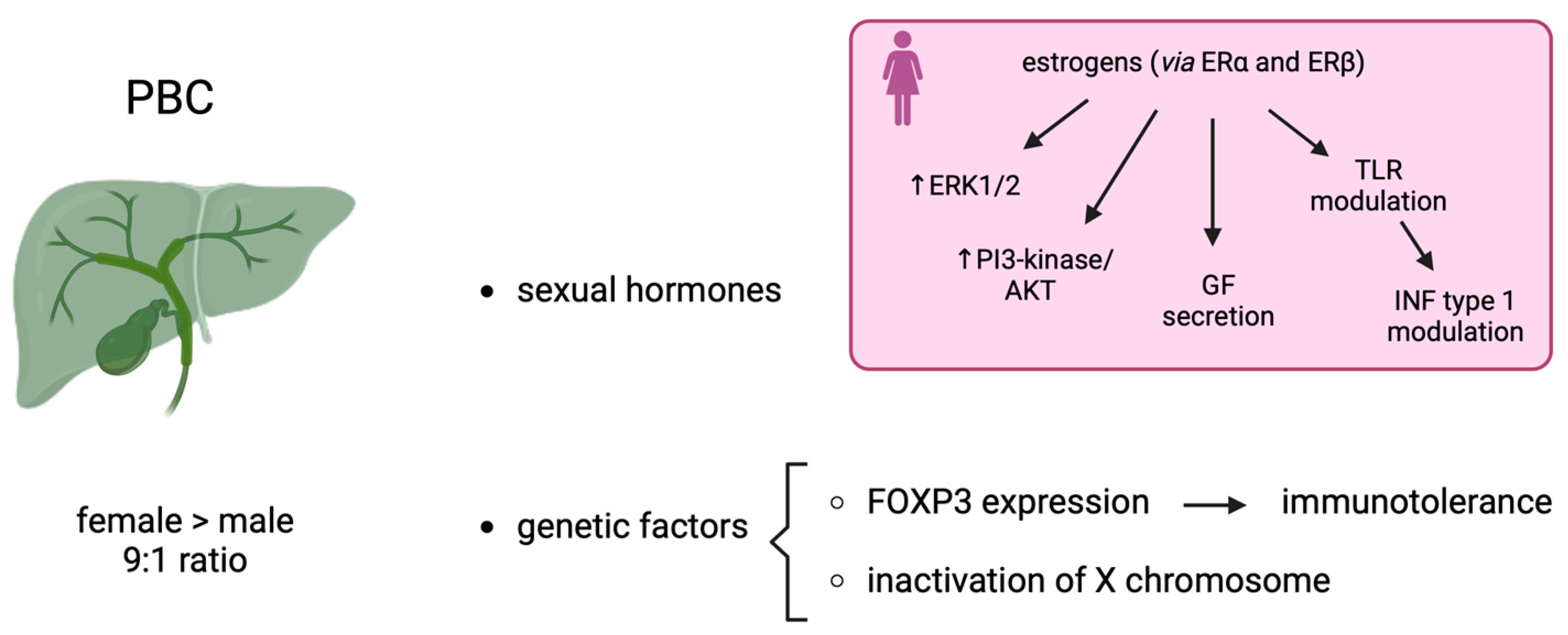
3.1. Factors Responsible for Gender Differences

Factors responsible for gender differences in PBC include sexual hormones and genetic factors (Figure 2).
Sexual hormones. Estrogen receptor alpha (ERa), which may play a significant role in cholestasis, is highly expressed in PBC [62]. However, both ERa and ERb modulate the response of cholangiocyte to damage activating intracellular cascades involving ERK1/2 (extracellular regulated kinase 1/2) and PI3-kinase/AKT (phosphatidylinositol-3′kinase), which are involved in the signaling pathways of growth factors, such as nerve growth factor and endothelial factor. Moreover, estrogen stimulates the secretion of different growth factors related to the proliferation of cholangiopathies [63].
Furthermore, high levels of estrogens drive the immune response from the Th1 to the Th2 phenotype [64]. T helper cells are critical for the progression of PBC. This has been demonstrated in a mouse model of autoimmune cholangitis, which has a notably female predominance [65]. Mechanistically, the knockout of interferon type I signaling prevents this female-prevalent autoimmune cholangitis phenotype [66]. Although an effect directly exerted by estrogens on IFN-γ expression has not yet been demonstrated, it is well known that sexual hormones can modify the expression of pattern of recognition receptors, such as Toll-like receptors (TLR) [67]. Therefore, this modulation can have an impact on the levels of type 1 IFN, indirectly. Furthermore, preclinical in vivo data indicate that estrogens can have, per se, a homeostatic positive effect on cholangiocytes [3]. This observation is corroborated by the loss of ER expression in patients with PBC of a severe grade [68]. Little is known about the effect of testosterone on the growth and survival of cholangiocytes. In male rats, androgen receptors (AR) are expressed by cholangiocytes, and testosterone stimulates their proliferation in a rodent model of cholestasis [69].
Genetic factors. An important issue for understating the higher prevalence of PBC in females was the inactivation of the X chromosome [70]. During embryonic development, one of the two X chromosomes is randomly inactivated in females; this process, under normal conditions, results in a cellular mosaicism where about one-half of the cells in a given tissue express either the maternal X or the paternal chromosome. However, since X chromosome inactivation is not complete, with 15–23% of genes escaping inactivation, a female-specific heterogeneous population of cells with biallelic expression of some X-linked genes is emerging. A recent elegant chromosome X-wide association study analyzed 5.244 case patients with PBC and 11,875 controls [71]. Indeed, a significant locus has been identified; this locus is characterized by a super-enhancer targeting all the genes of the region, including FOXP3. FOXP3 is a specific marker of Tregs, which are critical for the maintenance of immune tolerance [72]. Moreover, FOXP3 RNA expression levels in the whole blood show a significant differential expression between males and females.

3.2. Clinical Course

The largest cohort of hospitalized patients with PBC (between 2007 and 2014) was recently published in the USA using the National Inpatient Sample records [73]. The median age was 62 ± 14.8 years, and the ample was made up of 81% females. Males were younger than females (58.6 vs. 62.7 years). The clinical features of chronic liver disease were similar in both genders.
A retrospective study conducted in Taiwan on 75 PBC patients [74] showed that 84% of cases were females with a median age of 64.6 ± 1.78 years, whereas males had a median age of 46.6 ± 5.6 years. Male patients showed fewer extrahepatic autoimmune disorders and more severe liver injuries than females. Interestingly, this study analyzed the relationship between androgen response expression and systemic inflammation using the blood transcriptome of 90 PBC patients. The gene set variation analysis (FSVA) showed a positive significant correlation between peripheral mononuclear cells (PMNCs), the androgen receptor response, and the inflammatory response. It can be argued that this factor can explain the more severe course of PBC in males than in females, but more data are needed to further explain this hypothesis.
In PBC there is also a strong association between male gender, advanced disease, lack of response to ursodeoxycholic acid (UDCA), and the risk of HCC [75,76,77]. Moreover, it is known that the incidence and mortality rates of HCC are 2–5 times higher among men than women [78]. It has been hypothesized that the sex disparity between males and females is due to the effect of estrogens that protect hepatocytes from malignant transformation throughout the downregulation of Interleukin 6 (IL-6) and its release from Kupffer cells [79]. Although the modulation of the microbiome has been suggested as a possible therapeutic target [80], the role of sex in this context still needs to be studied and understood.

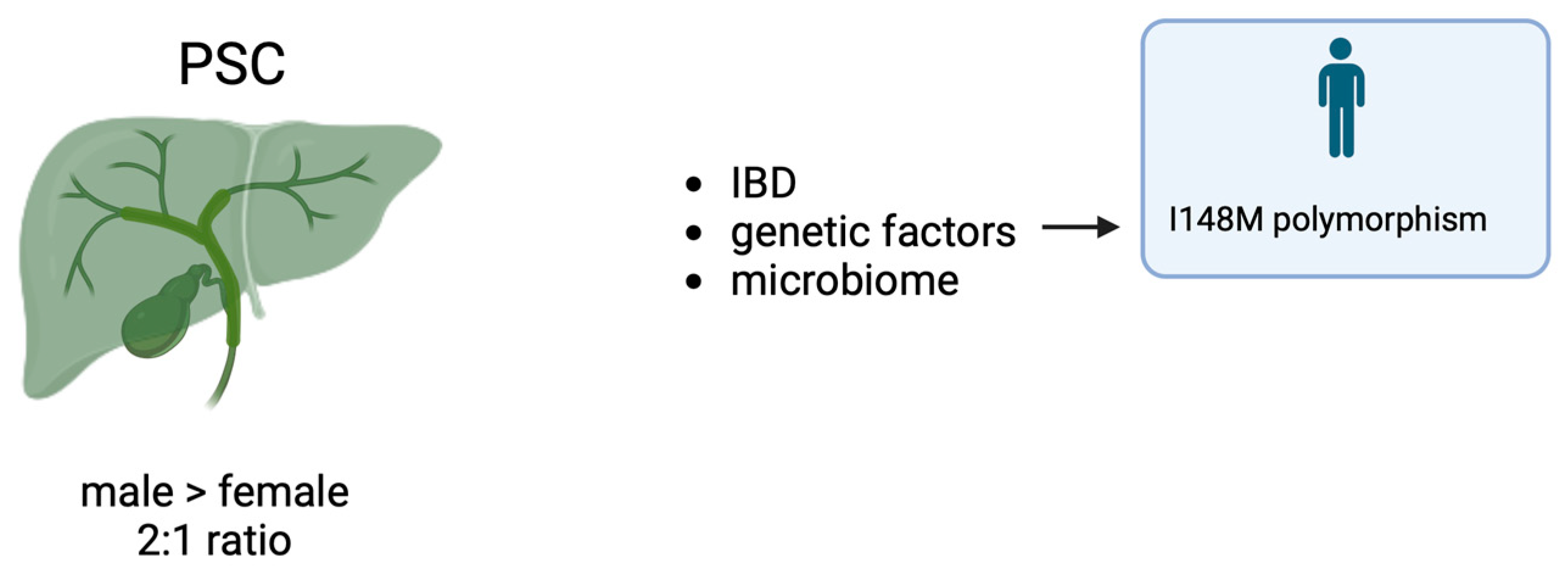
4. Primary Sclerosing Cholangitis

Primary sclerosing cholangitis (PSC) is a rare disease characterized by progressive cholestasis and by intrahepatic and extrahepatic fibro-inflammatory biliary strictures [81,82]. PSC mainly affects relatively young males and is strongly associated with inflammatory bowel disease (IBD). The prevalence of IBD in a recent systematic meta-analysis was 71.1%, most commonly found in ulcerative colitis [40]. On the contrary, a systematic meta-analysis, including 64 studies for a total of 776,700 patients, showed that the pooled prevalence of PSC in IBD was 2.16% [83]. The prevalence was higher in men with IBD (2.09%) compared with women (1.79%), p < 0.001. Overall, the highest pooled prevalence was observed in South America and the lowest in South East Asia [84].
Different phenotype variants can be recognized: classic large-duct PBC characterized by multifocal intra- and extrahepatic strictures with ductal dilatation with or without association with IBD, small duct PSC characterized by typical histological changes in absence of radiographic abnormalities and overlap syndrome with AIH and PSC associated with elevated levels of IgG4. In PSC, the male–female ratio is 2:1. A systemic review [85], including eight studies from North America and European countries, estimated a PSC incidence rate of 0.77 per 100,000 person-years. However, when excluding the two non-population-based studies, the incidence rate increased to 1.00 [85]. Similar rates were reported by Boonstra et al. in a systematic review of 11 studies from North America and Europe [58]. Furthermore, a recent systematic review of population-based studies reported the highest incidence in Northern Europe (Finland, 1.58, and Norway, 1.3, per 100,000 subjects, respectively) and Minnesota (1.47), with the lowest rate observed in the Mediterranean area (Italy, 0.1) [86]. Using the database from the Italian Epidemiological Rare Disease Registry, the crude annual incidence of PSC in females increased from 0.05 per 100,000 in 2012 to 0.09 per 100,000 in 2014, reaching a rate similar to that of males [67].

Role of Gender in PSC

Aside from the detailed reports on the epidemiology, the role of gender and sex hormones in PSC is virtually ignored [87]. However, an international study, which included 7121 patients with PSC from Europe, North America, and Australia, showed that female sex was associated with a lower risk of liver transplant or death and with a lower risk of hepatobiliary malignancies [88]. Moreover, the I148M variant polymorphism was investigated in two different cohorts of PSC: a German cohort (including 121 prospective subjects) and in a Norwegian cohort with 347 subjects (234 of whom received ERC and 123 of whom had a dominant stenosis) [89]. The results showed that male carriers of the I148M variant showed significantly reduced actuarial survival free of liver transplantation (p = 0.013) compared to male wildtype patients. Again, the I148M polymorphism did not affect female PSC patients with bile duct stenosis.
Microbiome. RNA sequencing studies demonstrated that PSC patients have an altered gut microbiome [90,91,92]. In particular, the gut microbiome in PSC patients is significantly different than that of healthy controls, being characterized by a drop of microbial diversity and differences in the amounts of specific bacteria, especially Enterococcus and Veillonella [90,91]. The interesting study by Rühlemann and collaborators pointed out that the peculiar and reproducible microbiota composition of PSC patients is modelled by the disease itself and not influenced by environmental factors [90] or inflammatory bowel disease (IBD), which is frequently present in PSC patients and vice versa [91]. Since Enterococcus positively correlates with ALP levels, it has been suggested that a link might exist between these bacteria and PSC severity [91] (Figure 3).
The risk factors for AIH, PBC, and PSC displaying sex dimorphism are reported in Table 1.

5. Overlap Syndromes

Overlap syndromes between AIH and PBC or PSC have been recognized since the 1980s. However, the concept of overlapping has been changed over the years. As far as PBC is concerned, the term AIH/PBC overlap syndrome has been considered as the coexistence of the two separate diseases with a variable onset during their natural history: (1) development of cholestatic changes during the course of AIH; (2) development of autoimmune features during the course of PBC; (3) simultaneous diagnosis of AIH and PBC. The diagnostic approach of this variant has been generally adopted according to the “Paris criteria” based on the simultaneous or consecutive presence of at least two out of three biochemical, serological, and histological criteria for both PBC and AIH [93]. However, the diagnostic criteria imply a morphologic approach through liver biopsy, thus unanswered questions remain, including the correlation between the clinical aspects and the morphology [94]. In a systematic review, which included 17 studies of PBC/AIH comprising a total of 402 patients, female gender was present in 87–100% of either retrospective or prospective studies [95].
The AIH/PSC overlap syndrome leads to some interesting observations. This condition has been described in both children and adults. In a large pediatric cohort, which included 781 subjects with PSC, 33% of cases had an overlap syndrome with AIH [96]. Patients with overlap were more likely to be female and more likely to have an association with IBD. Moreover, age, gender, and AIH overlap did not impact long-term outcome.
In childhood, the term “autoimmune sclerosing cholangitis” was preferred to AIH/PSC overlap syndrome [97]. However, quite recently, a new concept has arisen based on studies on the natural history of PSC and its variants. As hypothesized [98], autoimmune sclerosing cholangitis and PSC/AIH overlap syndrome represent temporal phases along a fundamental PSC continuum. In this view, there is a need for implementing prospective studies on clinical trials involving immunosuppressant and new molecules, and prospective studies on the natural history of PSC as well.

6. IgG4-Related Disease

IgG4-related disease (IgG4-RD) potentially involves all organs of the body, presenting with mass forming lesions and characterized by IgG4-positive plasma cells infiltrating the tissues [99]. A Chinese study evaluated the sex disparities in clinical characteristics and prognoses of IgG4-RD [100]. Four hundred and three patients (150 females and 253 males) were included, with a male–female ratio of 1.69. Male sex was significantly predominant compared to females; the significance was more pronounced in male patients older than 60 years. This point is particularly important, because in a previous report, which included a group of 25 patients ≤17 years of age, a slight female predominance was found [101]. In general, female patients presented Mikulicz’s disease and thyroiditis more frequently, while, in male patients, autoimmune pancreatitis, sclerosing cholangitis, and retroperitoneal fibrosis were more frequent. Moreover, in the group treated with steroids, male sex was independently associated with a worse prognosis.
In another multicenter study, which included 184 Latin Americans with IgG4-RD, men and women were equally affected by this condition [102]. However, male sex was significantly associated with the biliary tract phenotype with kidney and retroperitoneal involvement.
IgG4-related cholangitis (IgG4-RC) is the major hepatobiliary manifestation of IgG4-RD and involves mainly men with a median age of >55 years [103]. It is noteworthy, however, that the clinical presentation of IgG4-RC often mimics other biliary diseases, such as PSC and cholangiocarcinoma [103].

7. Sex Disparities in Autoimmune Liver Diseases in Pediatrics

A comprehensive review of juvenile AIH has recently been published [104]. Sixty to eighty percent of patients with AIH are females, and 40% have a family history of autoimmune disorders. In general, children with AIH, similarly to adults, have the same prevalence of female sex, as summarized in Table 2.
It should be stressed that mortality is still present, reaching 28.5% in Ghana [105].
More recently, a 30-year follow-up of 159 patients with childhood onset autoimmune liver disease has been published [112]. One hundred and nineteen children presented with AIH. Female gender was predominant in both type 1 AIH (63%) and type 2 AIH (67%). Interestingly, during follow-up, the following biliary disease progression was observed: 19.8% of patients with type1 AIH developed biliary features by adulthood and 50% of them developed a classical PSC [112].
Moreover, a cohort of 117 children diagnosed with AIH between 1973 and 2002 underwent a median follow-up of 20 years [113]. Type 2 AIH showed a higher prevalence of females than the group with type 1 AIH (45/52 vs. 41/65, p = 0058). A sustained remission after treatment withdrawal was recorded in 24%, and, in terms of liver outcome, there were no differences between type 1 and type 2 AIH.
PBC is virtually absent in childhood. There are only anecdotal cases so far described in the literature of pediatric-onset PBC. The first report described two cases of PBC diagnosed at 16 and 15 years of age in 2 girls in Canada [114]. The third case reported in the world was seen in a 17-year-old young woman who suffered from a previous infection of Borrelia burgdorferi [115]. One further case of type 2 AIH associated with an unexpected and transient presence of PBC-specific AMA has been described by Invernizzi et al. [116]. Finally, the youngest case of PBC has been described in a 5-year-old female child presented with jaundice and encephalopathy [117]. All cases described in the literature of PBC-onset in childhood are females.
There is a very high number of reports on PSC in childhood. A review by Giorgina Mieli-Vergani and Diego Vergani focused on the unique features of PSC in children [118]. Young patients with PSC frequently displayed features of autoimmunity (one third of whom having AIH/PSC overlap syndrome). Moreover, they experienced a beneficial effect from immunosuppressive therapy, which is in contrast with adults who have disappointing effects with such treatment. In another review, the same authors observed that the mode of presentation of autoimmune cholangitis was similar to type 1 AIH, but the bile duct disease progressed in half of them, leading to liver transplant [119]. Furthermore, the authors observed that cholangiocarcinoma rarely develops in children compared to adults, in whom it is a frequent complication of PSC [119].
Indeed, it has been stressed that PSC is rare in pediatrics, whereas sclerosing cholangitis is associated with strong autoimmune features [120]. In a retrospective cohort from the King’s College Hospital, which included 83 children with autoimmune sclerosing cholangitis, female sex accounted for 42% of cases, while in a prospective cohort, which included 27 patients, female sex was present in 55% of cases [97]. According to the more relevant studies on juvenile sclerosing cholangitis, the prevalence of males ranged between 57% and 65% [97]. Unfortunately, none of the reports on autoimmune sclerosing cholangitis in pediatrics focuses on gender disparities in clinical features or outcomes. Prospective studies on large cohorts of children with sclerosing cholangitis are needed in order to better understand gender disparities in this condition. Moreover, many open questions need to be addressed with the collaboration of many experts in the field of both pediatrics and adults [121].

8. Conclusions and Future Perspectives

Factors contributing to sex-related and gender-related modulation of autoimmune liver diseases and their outcomes in women and men are extremely important. Sex-related factors, including genetic, epigenetic, sex hormones, and gene–hormone interaction, are not fully understood. The best paradigm is either AIH or PBC. Genetic factors, similarly to other complex diseases, are not linked to a specific gene; HLA-linked genes are important for disease susceptibility. Concerning AIH, in Northern Central America, an association with DRB1*0301 and DRB1*0401 alleles has been described, while in Brazilians, DRB1*13 and DRB1*03 seem to be the more prevalent [122]. However, associations with single nucleotide polymorphisms within HLA and non-HLA genes have been assessed in autoimmune liver diseases but do not seem to explain gender disparities.
The interaction between genes and sexual hormones do represent a clue factor in the pathogenesis of these diseases. Estrogens and their fluctuations during the reproductive and non-reproductive life may modulate the course of the disease. It is well known that AIH is less invasive in pregnancy, and, during this phase, there is a reduced need for immunosuppressive therapy. In the case of PBC, its early onset in a young woman causes a more progressive liver disease with higher risk of complications. On the other hand, a late onset in a post-menopause woman frequently leads to a mild disease. PBC in men is rather infrequent, although more cases are being diagnosed in recent years. A man with PBC has a higher risk of developing HCC, mainly in cases of advanced disease or if they do not respond to UDCA. This observation implies a different strategy for optimizing the follow-up of the disease. Female patients with mild disease can undergo a physical examination every 8–12 months, with an ultrasound every year. Female patients with advanced disease should have a visit every six months and an ultrasound every six months as well. Male patients with PBC should have a regular follow-up every six months, optimizing a program for the prevention of HCC every six months.
In regard to PSC, although male sex is more frequent, the follow-up strategies should be the same in both sexes, because of the double neoplastic risk: cholangiocarcinoma and colorectal cancer for those with associated IBD.
Future directions for research mainly concern the eventual gender disparities in responding to treatment and the consequent need to well differentiate the response to treatment according to gender in clinical trials. More urgent perspectives regarding the importance of gender disparities among autoimmune liver diseases are included within the concept of “rare disease”, according to the Reference Network for Hepatological Disease (ERN RARE LIVER). It is well known that AIH, PBC, and PSC are classified within the regulation for ERN RARE LIVER in Europe, whereas in Italy only PSC is considered a rare disease. New expectations for autoimmune liver diseases concern a dramatic change in their epidemiology, due to an increase in new diagnoses all over the world. Thus, it could be speculated that, in a few years, these diseases will no longer fall within the group of rare diseases. However, among the disease groups, male patients with PBC will continue to be regarded as a subgroup which needs special attention and timely referral to a specialist. Moreover, future interventions for PSC patients will be planned in both sexes for improving symptom control, preventing a terminal stage of liver disease, and preventing neoplastic complications. Finally, male sex is an important risk factor in all autoimmune liver diseases for disease progression and development of HCC. Therefore, in these cases, liver transplant is particularly important for survival.

Funding

This research received no external funding.

Institutional Review Board Statement

Not applicable.

Data Availability Statement

Not applicable.

Conflicts of Interest

The authors declare no conflicts of interest.

References

  1. Kardashian, A.; Serper, M.; Terrault, N.; Nephew, L.D. Health disparities in chronic liver disease. Hepatology 2023, 77, 1382–1403. [Google Scholar] [CrossRef] [PubMed]
  2. Castrucci, B.C.; Auerbach, J. Meeting Individual Social Needs Falls Short of Addressing Social Determinants of Health. Health Aff. Forefr. 2019, 77, 25–28. [Google Scholar]
  3. Ortona, E.; Delunardo, F.; Baggio, G.; Malorni, W. A sex and gender perspective in medicine: A new mandatory challenge for human health. Preface. Ann. Ist. Super. Sanita 2016, 52, 146–148. [Google Scholar] [CrossRef] [PubMed]
  4. Westergaard, D.; Moseley, P.; Sørup, F.K.H.; Baldi, P.; Brunak, S. Population-wide analysis of differences in disease progression patterns in men and women. Nat. Commun. 2019, 10, 666. [Google Scholar] [CrossRef]
  5. Sayaf, K.; Gabbia, D.; Russo, F.P.; De Martin, S. The Role of Sex in Acute and Chronic Liver Damage. Int. J. Mol. Sci. 2022, 23, 10654. [Google Scholar] [CrossRef]
  6. Sayaf, K.; Zanotto, I.; Gabbia, D.; Alberti, D.; Pasqual, G.; Zaramella, A.; Fantin, A.; De Martin, S.; Russo, F.P. Sex Drives Functional Changes in the Progression and Regression of Liver Fibrosis. Int. J. Mol. Sci. 2023, 24, 16452. [Google Scholar] [CrossRef]
  7. Baggio, G.; Corsini, A.; Floreani, A.; Giannini, S.; Zagonel, V. Gender medicine: A task for the third millennium. Clin. Chem. Lab. Med. CCLM 2013, 51, 713–727. [Google Scholar] [CrossRef]
  8. Manns, M.P.; Lohse, A.W.; Vergani, D. Autoimmune hepatitis—Update 2015. J. Hepatol. 2015, 62, S100–S111. [Google Scholar] [CrossRef]
  9. Grønbæk, L.; Vilstrup, H.; Jepsen, P. Autoimmune hepatitis in Denmark: Incidence, prevalence, prognosis, and causes of death. A nationwide registry-based cohort study. J. Hepatol. 2014, 60, 612–617. [Google Scholar] [CrossRef]
  10. Bittermann, T.; Lewis, J.D.; Levy, C.; Goldberg, D.S. Sociodemographic and geographic differences in the US epidemiology of autoimmune hepatitis with and without cirrhosis. Hepatology 2023, 77, 367–378. [Google Scholar] [CrossRef]
  11. Yuming, Z.; Ruqi, T.; Gershwin, M.E.; Xiong, M. Autoimmune Hepatitis. Clin. Liver Dis. 2024, 28, 15–35. [Google Scholar] [CrossRef] [PubMed]
  12. Castiella, A. Drug-induced autoimmune liver disease: A diagnostic dilemma of an increasingly reported disease. World J. Hepatol. 2014, 6, 160. [Google Scholar] [CrossRef] [PubMed]
  13. Thomas, D.; Wu, T.Y.; Cottagiri, M.; Nyandjo, M.; Njoku, D.B. Induction of Drug-Induced, Autoimmune Hepatitis in BALB/c Mice for the Study of Its Pathogenic Mechanisms. J. Vis. Exp. 2020, 159, 59174. [Google Scholar] [CrossRef]
  14. Cho, J.; Kim, L.; Li, Z.; Rose, N.R.; Talor, M.V.; Njoku, D.B. Sex bias in experimental immune-mediated, drug-induced liver injury in BALB/c mice: Suggested roles for Tregs, estrogen, and IL-6. PLoS ONE 2013, 8, e61186. [Google Scholar] [CrossRef] [PubMed]
  15. Schramm, C.; Herkel, J.; Beuers, U.; Kanzler, S.; Galle, P.R.; Lohse, A.W. Pregnancy in Autoimmune Hepatitis: Outcome and Risk Factors. Am. J. Gastroenterol. 2006, 101, 556–560. [Google Scholar] [CrossRef] [PubMed]
  16. Buchel, E.; Van Steenbergen, W.; Nevens, F.; Fevery, J. Improvement of Autoimmune Hepatitis During Pregnancy Followed by Flare-Up After Delivery. Am. J. Gastroenterol. 2002, 97, 3160–3165. [Google Scholar] [CrossRef] [PubMed]
  17. Heneghan, M.A. Management and outcome of pregnancy in autoimmune hepatitis. Gut 2001, 48, 97–102. [Google Scholar] [CrossRef] [PubMed]
  18. Izumi, Y.; Kaneko, A.; Oku, K.; Kimura, M.; Tanaka, S.; Tada, H.; Tatsumi, K.; Takano, T.; Hidaka, Y.; Amino, N. Development of liver dysfunction after delivery is possibly due to postpartum autoimmune hepatitis. A report of three cases. J. Intern. Med. 2002, 252, 361–367. [Google Scholar] [CrossRef]
  19. Westbrook, R.H.; Yeoman, A.D.; Kriese, S.; Heneghan, M.A. Outcomes of pregnancy in women with autoimmune hepatitis. J. Autoimmun. 2012, 38, J239–J244. [Google Scholar] [CrossRef]
  20. Delpy, L.; Douin-Echinard, V.; Garidou, L.; Bruand, C.; Saoudi, A.; Guéry, J.-C. Estrogen Enhances Susceptibility to Experimental Autoimmune Myasthenia Gravis by Promoting Type 1-Polarized Immune Responses. J. Immunol. 2005, 175, 5050–5057. [Google Scholar] [CrossRef]
  21. Seillet, C.; Rouquié, N.; Foulon, E.; Douin-Echinard, V.; Krust, A.; Chambon, P.; Arnal, J.-F.; Guéry, J.-C.; Laffont, S. Estradiol Promotes Functional Responses in Inflammatory and Steady-State Dendritic Cells through Differential Requirement for Activation Function-1 of Estrogen Receptor α. J. Immunol. 2013, 190, 5459–5470. [Google Scholar] [CrossRef]
  22. Siracusa, M.C.; Overstreet, M.G.; Housseau, F.; Scott, A.L.; Klein, S.L. 17β-Estradiol Alters the Activity of Conventional and IFN-Producing Killer Dendritic Cells. J. Immunol. 2008, 180, 1423–1431. [Google Scholar] [CrossRef] [PubMed]
  23. Liu, H.Y.; Buenafe, A.C.; Matejuk, A.; Ito, A.; Zamora, A.; Dwyer, J.; Vandenbark, A.A.; Offner, H. Estrogen inhibition of EAE involves effects on dendritic cell function. J. Neurosci. Res. 2002, 70, 238–248. [Google Scholar] [CrossRef] [PubMed]
  24. Papenfuss, T.L.; Powell, N.D.; McClain, M.A.; Bedarf, A.; Singh, A.; Gienapp, I.E.; Shawler, T.; Whitacre, C.C. Estriol Generates Tolerogenic Dendritic Cells In Vivo That Protect against Autoimmunity. J. Immunol. 2011, 186, 3346–3355. [Google Scholar] [CrossRef]
  25. Bebo, B.F.; Schuster, J.C.; Vandenbark, A.A.; Offner, H. Androgens alter the cytokine profile and reduce encephalitogenicity of myelin-reactive T cells. J. Immunol. 1999, 162, 35–40. [Google Scholar] [CrossRef]
  26. Schwinge, D.; Carambia, A.; Quaas, A.; Krech, T.; Wegscheid, C.; Tiegs, G.; Prinz, I.; Lohse, A.W.; Herkel, J.; Schramm, C. Testosterone Suppresses Hepatic Inflammation by the Downregulation of IL-17, CXCL-9, and CXCL-10 in a Mouse Model of Experimental Acute Cholangitis. J. Immunol. 2015, 194, 2522–2530. [Google Scholar] [CrossRef]
  27. Liva, S.M.; Voskuhl, R.R. Testosterone Acts Directly on CD4+ T Lymphocytes to Increase IL-10 Production. J. Immunol. 2001, 167, 2060–2067. [Google Scholar] [CrossRef]
  28. Henze, L.; Schwinge, D.; Schramm, C. The Effects of Androgens on T Cells: Clues to Female Predominance in Autoimmune Liver Diseases? Front. Immunol. 2020, 11, 1567. [Google Scholar] [CrossRef] [PubMed]
  29. Meester, I.; Manilla-Muñoz, E.; León-Cachón, R.B.R.; Paniagua-Frausto, G.A.; Carrión-Alvarez, D.; Ruiz-Rodríguez, C.O.; Rodríguez-Rangel, X.; García-Martínez, J.M. SeXY chromosomes and the immune system: Reflections after a comparative study. Biol. Sex Differ. 2020, 11, 3. [Google Scholar] [CrossRef]
  30. Liberal, R.; Grant, C.R.; Mieli-Vergani, G.; Vergani, D. Autoimmune hepatitis: A comprehensive review. J. Autoimmun. 2013, 41, 126–139. [Google Scholar] [CrossRef]
  31. Al-Chalabi, T.; Underhill, J.A.; Portmann, B.C.; McFarlane, I.G.; Heneghan, M.A. Impact of gender on the long-term outcome and survival of patients with autoimmune hepatitis. J. Hepatol. 2008, 48, 140–147. [Google Scholar] [CrossRef]
  32. Czaja, A.J.; Donaldson, P.T. Gender effects and synergisms with histocompatibility leukocyte antigens in type 1 autoimmune hepatitis. Am. J. Gastroenterol. 2002, 97, 2051–2057. [Google Scholar] [CrossRef] [PubMed]
  33. Czaja, A.J. Genetic Factors Affecting the Occurrence, Clinical Phenotype, and Outcome of Autoimmune Hepatitis. Clin. Gastroenterol. Hepatol. 2008, 6, 379–388. [Google Scholar] [CrossRef] [PubMed]
  34. Béland, K.; Lapierre, P.; Alvarez, F. Influence of genes, sex, age and environment on the onset of autoimmune hepatitis. World J. Gastroenterol. 2009, 15, 1025. [Google Scholar] [CrossRef] [PubMed]
  35. Vento, S.; Guella, L.; Mirandola, F.; Cainelli, F.; Di Perri, G.; Solbiati, M.; Concia, E.; Ferraro, T. Epstein-Barr virus as a trigger for autoimmune hepatitis in susceptible individuals. Lancet 1995, 346, 608–609. [Google Scholar] [CrossRef] [PubMed]
  36. Tiegs, G.; Hentschel, J.; Wendel, A. A T cell-dependent experimental liver injury in mice inducible by concanavalin A. J. Clin. Investig. 1992, 90, 196–203. [Google Scholar] [CrossRef] [PubMed]
  37. Cohen, S.M.; O’Connor, A.M.; Hart, J.; Merel, N.H.; Te, H.S. Autoimmune hepatitis associated with the use of black cohosh: A case study. Menopause 2004, 11, 575–577. [Google Scholar] [CrossRef] [PubMed]
  38. Alla, V.; Abraham, J.; Siddiqui, J.; Raina, D.; Wu, G.Y.; Chalasani, N.P.; Bonkovsky, H.L. Autoimmune Hepatitis Triggered by Statins. J. Clin. Gastroenterol. 2006, 40, 757–761. [Google Scholar] [CrossRef] [PubMed]
  39. Czaja, A.J. Incorporating the Molecular Mimicry of Environmental Antigens into the Causality of Autoimmune Hepatitis. Dig. Dis. Sci. 2023, 68, 2824–2842. [Google Scholar] [CrossRef]
  40. Zhang, X.; Chen, B.; Zhao, L.; Li, H. The Gut Microbiota: Emerging Evidence in Autoimmune Diseases. Trends Mol. Med. 2020, 26, 862–873. [Google Scholar] [CrossRef]
  41. Rosser, E.C.; De Gruijter, N.M.; Matei, D.E. Mini-Review: Gut-Microbiota and the Sex-Bias in Autoimmunity—Lessons Learnt From Animal Models. Front. Med. 2022, 9, 910561. [Google Scholar] [CrossRef] [PubMed]
  42. Cheng, Z.; Yang, L.; Chu, H. The Gut Microbiota: A Novel Player in Autoimmune Hepatitis. Front. Cell. Infect. Microbiol. 2022, 12, 947382. [Google Scholar] [CrossRef] [PubMed]
  43. Yuksel, M.; Wang, Y.; Tai, N.; Peng, J.; Guo, J.; Beland, K.; Lapierre, P.; David, C.; Alvarez, F.; Colle, I.; et al. A novel “humanized mouse” model for autoimmune hepatitis and the association of gut microbiota with liver inflammation. Hepatology 2015, 62, 1536–1550. [Google Scholar] [CrossRef] [PubMed]
  44. Wang, J.; Zhu, N.; Su, X.; Gao, Y.; Yang, R. Gut-Microbiota-Derived Metabolites Maintain Gut and Systemic Immune Homeostasis. Cells 2023, 12, 793. [Google Scholar] [CrossRef] [PubMed]
  45. Terziroli Beretta-Piccoli, B.; Mieli-Vergani, G.; Vergani, D. HLA, gut microbiome and hepatic autoimmunity. Front. Immunol. 2022, 13, 980768. [Google Scholar] [CrossRef] [PubMed]
  46. Fu, Y.; Li, J.; Zhu, Y.; Chen, C.; Liu, J.; Gu, S.; Zheng, Y.; Li, Y. Causal effects of gut microbiome on autoimmune liver disease: A two-sample Mendelian randomization study. BMC Med. Genom. 2023, 16, 232. [Google Scholar] [CrossRef] [PubMed]
  47. Birn-Rydder, R.; Jensen, M.D.; Jepsen, P.; Grønbæk, L. Extrahepatic autoimmune diseases in autoimmune hepatitis: Effect on mortality. Liver Int. 2022, 42, 2466–2472. [Google Scholar] [CrossRef]
  48. Lee, D.U.; Kwon, J.; Koo, C.; Han, J.; Fan, G.H.; Jung, D.; Addonizio, E.A.; Chang, K.; Urrunaga, N.H. Clinical implications of gender and race in patients admitted with autoimmune hepatitis: Updated analysis of US hospitals. Frontline Gastroenterol. 2023, 14, 111–123. [Google Scholar] [CrossRef] [PubMed]
  49. Jensen, M.D.; Jepsen, P.; Vilstrup, H.; Grønbæk, L. Increased Cancer Risk in Autoimmune Hepatitis: A Danish Nationwide Cohort Study. Am. J. Gastroenterol. 2022, 117, 129–137. [Google Scholar] [CrossRef]
  50. Yan, L.-J.; Yao, S.-Y.; Meng, G.-X.; Liu, K.-X.; Li, H.-C.; Ding, Z.-N.; Dong, Z.-R.; Hong, J.-G.; Chen, Z.-Q.; Li, T. Sex and regional disparities in incidence of hepatocellular carcinoma in autoimmune hepatitis: A systematic review and meta-analysis. Hepatol. Int. 2021, 15, 1413–1420. [Google Scholar] [CrossRef]
  51. Orlando, R.; De Martin, S.; Andrighetto, L.; Floreani, M.; Palatini, P. Fluvoxamine pharmacokinetics in healthy elderly subjects and elderly patients with chronic heart failure. Br. J. Clin. Pharmacol. 2010, 69, 279–286. [Google Scholar] [CrossRef] [PubMed]
  52. Palatini, P.; De Martin, S. Pharmacokinetic drug interactions in liver disease: An update. World J. Gastroenterol. 2016, 22, 1260–1278. [Google Scholar] [CrossRef] [PubMed]
  53. Hartl, J.; Miquel, R.; Zachou, K.; Wong, G.-W.; Asghar, A.; Pape, S.; Sebode, M.; Peiseler, M.; Zenouzi, R.; Ehlken, H.; et al. Features and outcome of AIH patients without elevation of IgG. JHEP Rep. 2020, 2, 100094. [Google Scholar] [CrossRef]
  54. Gabbia, D.; Dalla Pozza, A.; Albertoni, L.; Lazzari, R.; Zigiotto, G.; Carrara, M.; Baldo, V.; Baldovin, T.; Floreani, A.; De Martin, S. Pregnane X receptor and constitutive androstane receptor modulate differently CYP3A-mediated metabolism in early- and late-stage cholestasis. World J. Gastroenterol. 2017, 23, 7519–7530. [Google Scholar] [CrossRef] [PubMed]
  55. Levy, C.; Manns, M.; Hirschfield, G. New Treatment Paradigms in Primary Biliary Cholangitis. Clin. Gastroenterol. Hepatol. 2023, 21, 2076–2087. [Google Scholar] [CrossRef] [PubMed]
  56. Floreani, A.; Gabbia, D.; De Martin, S. Current Perspectives on the Molecular and Clinical Relationships between Primary Biliary Cholangitis and Hepatocellular Carcinoma. Int. J. Mol. Sci. 2024, 25, 2194. [Google Scholar] [CrossRef] [PubMed]
  57. Floreani, A.; Gabbia, D.; De Martin, S. Primary biliary cholangitis: Primary autoimmune disease or primary secretory defect. Expert Rev. Gastroenterol. Hepatol. 2023, 17, 863–870. [Google Scholar] [CrossRef]
  58. Boonstra, K.; Bokelaar, R.; Stadhouders, P.H.; Tuynman, H.A.; Poen, A.C.; van Nieuwkerk, K.M.; Witteman, E.M.; Hamann, D.; Witteman, B.J.; Beuers, U.; et al. Increased cancer risk in a large population-based cohort of patients with primary biliary cirrhosis: Follow-up for up to 36 years. Hepatol. Int. 2014, 8, 266–274. [Google Scholar] [CrossRef] [PubMed]
  59. Lu, M.; Li, J.; Haller, I.V.; Romanelli, R.J.; VanWormer, J.J.; Rodriguez, C.V.; Raebel, M.A.; Boscarino, J.A.; Schmidt, M.A.; Daida, Y.G.; et al. Factors Associated with Prevalence and Treatment of Primary Biliary Cholangitis in United States Health Systems. Clin. Gastroenterol. Hepatol. 2018, 16, 1333–1341.e6. [Google Scholar] [CrossRef]
  60. Hirschfield, G.M.; Beuers, U.; Corpechot, C.; Invernizzi, P.; Jones, D.; Marzioni, M.; Schramm, C. EASL Clinical Practice Guidelines: The diagnosis and management of patients with primary biliary cholangitis. J. Hepatol. 2017, 67, 145–172. [Google Scholar] [CrossRef]
  61. Quintero, O.L.; Amador-Patarroyo, M.J.; Montoya-Ortiz, G.; Rojas-Villarraga, A.; Anaya, J.-M. Autoimmune disease and gender: Plausible mechanisms for the female predominance of autoimmunity. J. Autoimmun. 2012, 38, J109–J119. [Google Scholar] [CrossRef] [PubMed]
  62. Cao, H.; Zhu, B.; Qu, Y.; Zhang, W. Abnormal Expression of ERα in Cholangiocytes of Patients with Primary Biliary Cholangitis Mediated Intrahepatic Bile Duct Inflammation. Front. Immunol. 2019, 10, 2815. [Google Scholar] [CrossRef] [PubMed]
  63. Alvaro, D. Estrogens and the pathophysiology of the biliary tree. World J. Gastroenterol. 2006, 12, 3537. [Google Scholar] [CrossRef] [PubMed]
  64. Matalka, K.Z. The effect of estradiol, but not progesterone, on the production of cytokines in stimulated whole blood, is concentration-dependent. Neuro Endocrinol. Lett. 2003, 24, 185–191. [Google Scholar] [PubMed]
  65. Bae, H.R.; Leung, P.S.C.; Tsuneyama, K.; Valencia, J.C.; Hodge, D.L.; Kim, S.; Back, T.; Karwan, M.; Merchant, A.S.; Baba, N.; et al. Chronic expression of interferon-gamma leads to murine autoimmune cholangitis with a female predominance. Hepatology 2016, 64, 1189–1201. [Google Scholar] [CrossRef] [PubMed]
  66. Bae, H.R.; Hodge, D.L.; Yang, G.; Leung, P.S.C.; Chodisetti, S.B.; Valencia, J.C.; Sanford, M.; Fenimore, J.M.; Rahman, Z.S.M.; Tsuneyama, K.; et al. The interplay of type I and type II interferons in murine autoimmune cholangitis as a basis for sex-biased autoimmunity. Hepatology 2018, 67, 1408–1419. [Google Scholar] [CrossRef] [PubMed]
  67. Carbone, M.; Bonato, G.; Invernizzi, P. Female preponderance of primary biliary cholangitis is all about our understanding of its autoimmune nature. Hepatology 2018, 67, 1210–1212. [Google Scholar] [CrossRef] [PubMed]
  68. Alvaro, D.; Invernizzi, P.; Onori, P.; Franchitto, A.; De Santis, A.; Crosignani, A.; Sferra, R.; Ginanni-Corradini, S.; Grazia Mancino, M.; Maggioni, M.; et al. Estrogen receptors in cholangiocytes and the progression of primary biliary cirrhosis. J. Hepatol. 2004, 41, 905–912. [Google Scholar] [CrossRef] [PubMed]
  69. Yang, F.; Priester, S.; Onori, P.; Venter, J.; Renzi, A.; Franchitto, A.; Munshi, M.K.; Wise, C.; Dostal, D.E.; Marzioni, M.; et al. Castration inhibits biliary proliferation induced by bile duct obstruction: Novel role for the autocrine trophic effect of testosterone. Am. J. Physiol.-Gastrointest. Liver Physiol. 2011, 301, G981–G991. [Google Scholar] [CrossRef]
  70. Gerussi, A.; Lucà, M.; Cristoferi, L.; Ronca, V.; Mancuso, C.; Milani, C.; D’Amato, D.; O’Donnell, S.E.; Carbone, M.; Invernizzi, P. New Therapeutic Targets in Autoimmune Cholangiopathies. Front. Med. 2020, 7, 117. [Google Scholar] [CrossRef]
  71. Asselta, R.; Paraboschi, E.M.; Gerussi, A.; Cordell, H.J.; Mells, G.F.; Sandford, R.N.; Jones, D.E.; Nakamura, M.; Ueno, K.; Hitomi, Y.; et al. X Chromosome Contribution to the Genetic Architecture of Primary Biliary Cholangitis. Gastroenterology 2021, 160, 2483–2495.e26. [Google Scholar] [CrossRef] [PubMed]
  72. Mohr, A.; Atif, M.; Balderas, R.; Gorochov, G.; Miyara, M. The role of FOXP3+ regulatory T cells in human autoimmune and inflammatory diseases. Clin. Exp. Immunol. 2019, 197, 24–35. [Google Scholar] [CrossRef]
  73. Adejumo, A.C.; Akhtar, D.H.; Dennis, B.B.; Cholankeril, G.; Alayo, Q.; Ogundipe, O.A.; Kim, D.; Ahmed, A. Gender and Racial Differences in Hospitalizations for Primary Biliary Cholangitis in the USA. Dig. Dis. Sci. 2021, 66, 1461–1476. [Google Scholar] [CrossRef] [PubMed]
  74. Wu, Y.H.; Chiu, Y.L.; Lin, J.C.; Chen, H.W. New insights into sex differences in primary biliary cholangitis. Gender difference in primary biliary cholangitis. J. Pak. Med. Assoc. 2022, 72, 896–900. [Google Scholar] [CrossRef] [PubMed]
  75. Sy, A.M.; Ferreira, R.D.; John, B.V. Hepatocellular Carcinoma in Primary Biliary Cholangitis. Clin. Liver Dis. 2022, 26, 691–704. [Google Scholar] [CrossRef]
  76. Trivedi, P.J.; Lammers, W.J.; van Buuren, H.R.; Parés, A.; Floreani, A.; Janssen, H.L.A.; Invernizzi, P.; Battezzati, P.M.; Ponsioen, C.Y.; Corpechot, C.; et al. Stratification of hepatocellular carcinoma risk in primary biliary cirrhosis: A multicentre international study. Gut 2016, 65, 321–329. [Google Scholar] [CrossRef] [PubMed]
  77. Floreani, A.; Gabbia, D.; De Martin, S. Obeticholic Acid for Primary Biliary Cholangitis. Biomedicines 2022, 10, 2464. [Google Scholar] [CrossRef]
  78. Kulik, L.; El-Serag, H.B. Epidemiology and Management of Hepatocellular Carcinoma. Gastroenterology 2019, 156, 477–491.e1. [Google Scholar] [CrossRef]
  79. Yeh, S.-H.; Chen, P.-J. Gender disparity of hepatocellular carcinoma: The roles of sex hormones. Oncology 2010, 78 (Suppl. 1), 172–179. [Google Scholar] [CrossRef]
  80. Floreani, A.; De Martin, S.; Ikeura, T.; Okazaki, K.; Gershwin, M.E. Gut microbial profiling as a therapeutic and diagnostic target for managing primary biliary cholangitis. Expert Opin. Orphan Drugs 2020, 8, 507–514. [Google Scholar] [CrossRef]
  81. Cazzagon, N.; Sarcognato, S.; Floreani, A.; Corrà, G.; De Martin, S.; Guzzardo, V.; Russo, F.P.; Guido, M. Cholangiocyte senescence in primary sclerosing cholangitis is associated with disease severity and prognosis. JHEP Rep. 2021, 3, 100286. [Google Scholar] [CrossRef] [PubMed]
  82. Hirschfield, G.M.; Karlsen, T.H.; Lindor, K.D.; Adams, D.H. Primary sclerosing cholangitis. Lancet 2013, 382, 1587–1599. [Google Scholar] [CrossRef] [PubMed]
  83. Barberio, B.; Massimi, D.; Cazzagon, N.; Zingone, F.; Ford, A.C.; Savarino, E.V. Prevalence of Primary Sclerosing Cholangitis in Patients With Inflammatory Bowel Disease: A Systematic Review and Meta-analysis. Gastroenterology 2021, 161, 1865–1877. [Google Scholar] [CrossRef] [PubMed]
  84. Zhang, Y.; Gao, X.; He, Z.; Jia, H.; Chen, M.; Wang, X.; Hong, L.; Cui, Y.; Wan, J. Prevalence of inflammatory bowel disease in patients with primary sclerosing cholangitis: A systematic review and meta-analysis. Liver Int. 2022, 42, 1814–1822. [Google Scholar] [CrossRef]
  85. Molodecky, N.A.; Kareemi, H.; Parab, R.; Barkema, H.W.; Quan, H.; Myers, R.P.; Kaplan, G.G. Incidence of primary sclerosing cholangitis: A systematic review and meta-analysis. Hepatology 2011, 53, 1590–1599. [Google Scholar] [CrossRef] [PubMed]
  86. Trivedi, P.J.; Bowlus, C.L.; Yimam, K.K.; Razavi, H.; Estes, C. Epidemiology, Natural History, and Outcomes of Primary Sclerosing Cholangitis: A Systematic Review of Population-based Studies. Clin. Gastroenterol. Hepatol. 2022, 20, 1687–1700.e4. [Google Scholar] [CrossRef] [PubMed]
  87. Tanaka, A. Current understanding of primary biliary cholangitis. Clin. Mol. Hepatol. 2021, 27, 1–21. [Google Scholar] [CrossRef] [PubMed]
  88. Weismüller, T.J.; Trivedi, P.J.; Bergquist, A.; Imam, M.; Lenzen, H.; Ponsioen, C.Y.; Holm, K.; Gotthardt, D.; Färkkilä, M.A.; Marschall, H.-U.; et al. Patient Age, Sex, and Inflammatory Bowel Disease Phenotype Associate with Course of Primary Sclerosing Cholangitis. Gastroenterology 2017, 152, 1975–1984.e8. [Google Scholar] [CrossRef] [PubMed]
  89. Friedrich, K.; Rupp, C.; Hov, J.R.; Steinebrunner, N.; Weiss, K.-H.; Stiehl, A.; Brune, M.; Schaefer, P.K.Y.; Schemmer, P.; Sauer, P.; et al. A Frequent PNPLA3 Variant Is a Sex Specific Disease Modifier in PSC Patients with Bile Duct Stenosis. PLoS ONE 2013, 8, e58734. [Google Scholar] [CrossRef]
  90. Rühlemann, M.; Liwinski, T.; Heinsen, F.-A.; Bang, C.; Zenouzi, R.; Kummen, M.; Thingholm, L.; Tempel, M.; Lieb, W.; Karlsen, T.; et al. Consistent alterations in faecal microbiomes of patients with primary sclerosing cholangitis independent of associated colitis. Aliment. Pharmacol. Ther. 2019, 50, 580–589. [Google Scholar] [CrossRef]
  91. Sabino, J.; Vieira-Silva, S.; Machiels, K.; Joossens, M.; Falony, G.; Ballet, V.; Ferrante, M.; Van Assche, G.; Van der Merwe, S.; Vermeire, S.; et al. Primary sclerosing cholangitis is characterised by intestinal dysbiosis independent from IBD. Gut 2016, 65, 1681–1689. [Google Scholar] [CrossRef] [PubMed]
  92. Liwinski, T.; Heinsen-Groth, F.; Rühlemann, M.; Zenouzi, R.; Kummen, M.; Hov, J.; Karlsen, T.; Bang, C.; Lohse, A.; Franke, A.; et al. The intestinal microbiota of patients with PSC are different from healthy controls and patients with ulcerative colitis across geographical regions. Z. Für Gastroenterol. 2018, 56, E2–E89. [Google Scholar] [CrossRef]
  93. Chazouillères, O.; Wendum, D.; Serfaty, L.; Montembault, S.; Rosmorduc, O.; Poupon, R. Primary biliary cirrhosis-autoimmune hepatitis overlap syndrome: Clinical features and response to therapy: Primary Biliary Cirrhosis-Autoimmune Hepatitis Overlap Syndrome: Clinical Features and Response to Therapy. Hepatology 1998, 28, 296–301. [Google Scholar] [CrossRef] [PubMed]
  94. Liang, Y.; Yang, Z.; Zhong, R. Primary biliary cirrhosis and cancer risk: A systematic review and meta-analysis. Hepatology 2012, 56, 1409–1417. [Google Scholar] [CrossRef] [PubMed]
  95. Freedman, B.L.; Danford, C.J.; Patwardhan, V.; Bonder, A. Treatment of Overlap Syndromes in Autoimmune Liver Disease: A Systematic Review and Meta-Analysis. J. Clin. Med. 2020, 9, 1449. [Google Scholar] [CrossRef] [PubMed]
  96. Deneau, M.R.; El-Matary, W.; Valentino, P.L.; Abdou, R.; Alqoaer, K.; Amin, M.; Amir, A.Z.; Auth, M.; Bazerbachi, F.; Broderick, A.; et al. The natural history of primary sclerosing cholangitis in 781 children: A multicenter, international collaboration. Hepatology 2017, 66, 518–527. [Google Scholar] [CrossRef] [PubMed]
  97. Gregorio, G. Autoimmune hepatitis/sclerosing cholangitis overlap syndrome in childhood: A 16-year prospective study. Hepatology 2001, 33, 544–553. [Google Scholar] [CrossRef] [PubMed]
  98. Ricciuto, A.; Kamath, B.M.; Hirschfield, G.M.; Trivedi, P.J. Primary sclerosing cholangitis and overlap features of autoimmune hepatitis: A coming of age or an age-ist problem? J. Hepatol. 2023, 79, 567–575. [Google Scholar] [CrossRef] [PubMed]
  99. Lanzillotta, M.; Mancuso, G.; Della-Torre, E. Advances in the diagnosis and management of IgG4 related disease. BMJ 2020, 369, m1067. [Google Scholar] [CrossRef]
  100. Wang, L.; Zhang, P.; Zhang, X.; Lin, W.; Tang, H.; Li, J.; Wang, M.; Liu, X.; Fei, Y.; Chen, H.; et al. Sex disparities in clinical characteristics and prognosis of immunoglobulin G4-related disease: A prospective study of 403 patients. Rheumatol. Oxf. Engl. 2019, 58, 820–830. [Google Scholar] [CrossRef]
  101. Karim, F.; Loeffen, J.; Bramer, W.; Westenberg, L.; Verdijk, R.; Van Hagen, M.; Van Laar, J. IgG4-related disease: A systematic review of this unrecognized disease in pediatrics. Pediatr. Rheumatol. 2016, 14, 18. [Google Scholar] [CrossRef]
  102. Martín-Nares, E.; Baenas, D.F.; Cuellar Gutiérrez, M.C.; Hernández-Molina, G.; Ortiz, A.C.; Neira, O.; Gutiérrez, M.A.; Calvo, R.; Saad, E.J.; Elgueta Pinochet, S.; et al. Clinical and Serological Features in Latin American IgG4-Related Disease Patients Differ According to Sex, Ethnicity, and Clinical Phenotype. JCR J. Clin. Rheumatol. 2022, 28, 285–292. [Google Scholar] [CrossRef] [PubMed]
  103. Kersten, R.; Trampert, D.C.; Herta, T.; Hubers, L.M.; Maillette De Buy Wenniger, L.J.; Verheij, J.; Van De Graaf, S.F.J.; Beuers, U. IgG4-related cholangitis—A mimicker of fibrosing and malignant cholangiopathies. J. Hepatol. 2023, 79, 1502–1523. [Google Scholar] [CrossRef]
  104. Sokollik, C.; McLin, V.A.; Vergani, D.; Terziroli Beretta-Piccoli, B.; Mieli-Vergani, G. Juvenile autoimmune hepatitis: A comprehensive review. J. Autoimmun. 2018, 95, 69–76. [Google Scholar] [CrossRef] [PubMed]
  105. Afaa, T.J.; Amegan-Aho, K.H.; Dono, M.T.; Odei, E.; Awuku, Y.A. Clinical characteristics of paediatric autoimmune hepatitis at a referral hospital in Sub Saharan Africa. PLoS ONE 2020, 15, e0239964. [Google Scholar] [CrossRef] [PubMed]
  106. Haghighat, M.; Mardani, S.; Dehghani, S.M.; Javaherizadeh, H. Clinical and laboratory data in pediatric autoimmune hepatitis: Study from referral center in Iran. Rev. Gastroenterol. Peru Organo Of. Soc. Gastroenterol. Peru 2020, 40, 149–154. [Google Scholar] [CrossRef]
  107. Saadah, O.I.; Khayat, A.; Alsufyani, H.A.; Bin-Taleb, Y. Paediatric autoimmune liver diseases: A descriptive study of patients from Saudi Arabia. Arab J. Gastroenterol. 2021, 22, 146–150. [Google Scholar] [CrossRef]
  108. Mogahed, E.; El-Karaksy, H.; Zaki, H.; Abdullatif, H. Autoimmune hepatitis in Egyptian children: A single center experience. Int. J. Immunopathol. Pharmacol. 2022, 36, 205873842110732. [Google Scholar] [CrossRef] [PubMed]
  109. Sutton, H.; Tayler, R.; Chalmers, I.; Cowieson, J.; Fraser, K.; Henderson, P.; Hansen, R. The Epidemiology of Pediatric Autoimmune Hepatitis in Scotland: A National Cohort Study. JPGN Rep. 2022, 3, e223. [Google Scholar] [CrossRef]
  110. Ludz, C.; Stirnimann, G.; Semela, D.; Mertens, J.; Kremer, A.E.; Filipowicz Sinnreich, M.; Sokollik, C.; Bernsmeier, C.; Bresson-Hadni, S.; McLin, V.; et al. Epidemiology, clinical features and management of autoimmune hepatitis in Switzerland: A retrospective and prospective cohort study. Swiss Med. Wkly. 2023, 153, 40102. [Google Scholar] [CrossRef]
  111. Altamimi, E.; Al Omari, D.; Obeidat, H.; Barham, K. Retrospective, single-center analysis of autoimmune hepatitis in Jordanian children: Clinical features, treatments, and outcomes. BMC Pediatr. 2024, 24, 102. [Google Scholar] [CrossRef] [PubMed]
  112. Warner, S.; Rajanayagam, J.; Russell, E.; Lloyd, C.; Ferguson, J.; Kelly, D.A.; Hirschfield, G.M. Biliary disease progression in childhood onset autoimmune liver disease: A 30-year follow-up into adulthood. JHEP Rep. 2024, 6, 100901. [Google Scholar] [CrossRef] [PubMed]
  113. Maggiore, G.; Bernard, O.; Mosca, A.; Ballot, E.; Johanet, C.; Jacquemin, E. Long-term outcomes of patients with type 1 or 2 autoimmune hepatitis presenting in childhood. J. Hepatol. 2023, 78, 979–988. [Google Scholar] [CrossRef] [PubMed]
  114. Dahlan, Y.; Smith, L.; Simmonds, D.; Jewell, L.D.; Wanless, I.; Heathcote, E.J.; Bain, V.G. Pediatric-onset primary biliary cirrhosis. Gastroenterology 2003, 125, 1476–1479. [Google Scholar] [CrossRef] [PubMed]
  115. Floreani, A.; Ostuni, P.A.; Ferrara, F.; Guido, M. Primary biliary cirrhosis: When and why does the disease develop? Dig. Liver Dis. 2006, 38, 272–275. [Google Scholar] [CrossRef]
  116. Invernizzi, P.; Alessio, M.G.; Smyk, D.S.; Lleo, A.; Sonzogni, A.; Fabris, L.; Candusso, M.; Bogdanos, D.P.; Iorio, R.; Torre, G. Autoimmune hepatitis type 2 associated with an unexpected and transient presence of primary biliary cirrhosis-specific antimitochondrial antibodies: A case study and review of the literature. BMC Gastroenterol. 2012, 12, 92. [Google Scholar] [CrossRef]
  117. Ullah, K.; Uddin, S.; Dogar, A.W.; Jan, Z.U.; Abbas, S.H. Primary biliary cirrhosis in early childhood—A rare case report. Int. J. Surg. Case Rep. 2021, 85, 106215. [Google Scholar] [CrossRef] [PubMed]
  118. Mieli-Vergani, G.; Vergani, D. Unique features of primary sclerosing cholangitis in children. Curr. Opin. Gastroenterol. 2010, 26, 265–268. [Google Scholar] [CrossRef] [PubMed]
  119. Mieli-Vergani, G.; Vergani, D. Sclerosing Cholangitis in Children and Adolescents. Clin. Liver Dis. 2016, 20, 99–111. [Google Scholar] [CrossRef]
  120. Di Giorgio, A.; Vergani, D.; Mieli-Vergani, G. Cutting edge issues in juvenile sclerosing cholangitis. Dig. Liver Dis. 2022, 54, 417–427. [Google Scholar] [CrossRef]
  121. Terziroli Beretta-Piccoli, B.; Vergani, D.; Mieli-Vergani, G. Autoimmune sclerosing cholangitis: Evidence and open questions. J. Autoimmun. 2018, 95, 15–25. [Google Scholar] [CrossRef] [PubMed]
  122. Bittencourt, P.L.; Goldberg, A.C.; Cançado, E.L.R.; Porta, G.; Carrilho, F.J.; Farias, A.Q.; Palacios, S.A.; Chiarella, J.M.; Abrantes-Lemos, C.P.; Baggio, V.L.; et al. Genetic Heterogeneity in Susceptibility to Autoimmune Hepatitis Types 1 and 2. Am. J. Gastroenterol. 1999, 94, 1906–1913. [Google Scholar] [CrossRef] [PubMed]
Figure 1. Sex dimorphism and AIH.
Life 14 00500 g001
Figure 2. Sex dimorphism and PBC.
Life 14 00500 g002
Figure 3. Sex dimorphism and PSC.
Life 14 00500 g003
Table 1. Summary of the sex-dimorphic risk factors for AIH, PBC, and PSC.
Autoimmune
Disease
Risk FactorsResultsRefs.
AIHIncreased IL-4
activated CD4+ T cells
Female BALB/C mice develop a more severe drug-induced AIH
Tregs confer protection against drug-induced autoimmune hepatitis
[13,14]
EstrogensEstrogens promote DC maturation, leading to an increase in Th1 cells activity, increased production of proinflammatory ILs, Estrogens upregulate the expression of checkpoint inhibitors, e.g., PD-L1[20,21,22,23,24]
AndrogensAndrogens may influence the pathogenesis of AIH by shifting the Th-1 to the Th-2 phenotype, reducing hepatic Th-17 and increasing IL-10 secretion, which drives Treg differentiation[25,26,27,28]
PregnancyPregnancy increases risk of fetal and maternal complications and flares in AIH activity after delivery[15,16,17]
HLA-DR3Increased expression of HLA-DR3 (DRB1*0301) in male AIH patients [31]
HLA-DR4Increased HLA-DR4 (DRB1*0401) in female AIH patients[32]
PBCEstrogensERα and Erβ modulate cholangiocyte response to damage, activating intracellular cascades involving ERK1/2 and PI3-kinase/AKT
Estrogens stimulate the secretion of different growth factors in proliferating cholangiopathies and shift Th cells from Th1 to the Th2 phenotype
Sexual hormone regulation of TLRs affect type 1 IFN that may impact on PBC progression
Estrogens exert a homeostatic positive effect on cholangiocytes
[3,62,63,64,67]
AndrogensTestosterone stimulates AR proliferation cholangiocytes in a rodent model of cholestasis[69]
Inactivation of X
chromosome
Female-specific heterogeneous population of cells with biallelic expression of some X-linked genes with a locus in which there is a super-enhancer targeting genes, e.g., FOXP3, affect Tregs and are critical for the maintenance of immune tolerance[71,72]
PSCI148M variant
polymorphism
Male carriers of the I148M variant showed significant reduced actuarial survival free of liver transplantation data[89]
Table 2. Epidemiology of AIH in recent cohorts of children.
Country (Ref.)TypeN.Female %Acute Onset Age (Mean or
Median Years)
Mortality
Ghana [105]Retrospective1361.5%15.7%10 (5–13)28.5%
Iran [106]Retrospective8666.27%-9.1 ± 4.3610.5%
Saudi Arabia [107]Cross-sectional2556%16%9.4 ± 4.20%
Egypt [108]Retrospective3464.7%51%8 ± 32.9%
Scotland [109]Prospective3064%44.4%11.4 (1–15.9)0%
Switzerland [110]Retro- and prospective3053%-12.5 (8–15)0%
Jordania [111]Retrospective1675%31.3%9.4 ± 4.1318.8%
Disclaimer/Publisher’s Note: The statements, opinions and data contained in all publications are solely those of the individual author(s) and contributor(s) and not of MDPI and/or the editor(s). MDPI and/or the editor(s) disclaim responsibility for any injury to people or property resulting from any ideas, methods, instructions or products referred to in the content.

