Communication

The Complete Mitochondrial Genome Sequence of Eimeria kongi (Apicomplexa: Coccidia)

by
Yubo Shi
1,2,
Sufang Fang
1,
Xiaolong Gu
1,
Chengyu Hao
1,
Fangchen Du
1,
Ping Cui
1,* and
Xinming Tang
2,*
1
College of Animal Science and Technology, Hebei North University, Zhangjiakou 075000, China
2
Key Laboratory of Animal Biosafety Risk Prevention and Control (North), Key Laboratory of Veterinary Biological Products and Chemical Drugs of MARA, Institute of Animal Science, Chinese Academy of Agricultural Sciences, Beijing 100193, China
*
Authors to whom correspondence should be addressed.
Life 2024, 14(6), 699; https://doi.org/10.3390/life14060699
Submission received: 29 April 2024 / Revised: 20 May 2024 / Accepted: 28 May 2024 / Published: 29 May 2024
(This article belongs to the Special Issue Trends in Microbiology 2024)

Abstract

:
Rabbit coccidiosis is caused by infection with one or, more commonly, several Eimeria species that parasitize the hepatobiliary ducts or intestinal epithelium of rabbits. Currently, there are eleven internationally recognized species of rabbit coccidia, with the complete mitochondrial (mt) genomes of six species commonly infecting rabbits having been sequenced and annotated. Eimeria kongi was initially discovered in 2011 and prompted a preliminary study on this species. Through traditional morphological analysis, E. kongi was identified as a novel species of rabbit coccidia. To further validate this classification, we sequenced and annotated its mitochondrial genome. The complete mt genome of E. kongi spans 6258 bp and comprises three cytochrome genes (cytb, cox1, cox3), fourteen gene fragments for the large subunit (LSU) rRNA, and nine gene fragments for the small subunit (SSU) rRNA, lacking transfer RNA (tRNA) genes. Moreover, phylogenetic analysis of the mitochondrial genome sequence of E. kongi revealed its clustering with six other species of rabbit coccidia into a monophyletic group. Additionally, E. irresidua and E. flavescens were grouped within the lineage lacking oocyst residuum, consistent with their morphological characteristics. Consistent with multiple molecular phylogenies, in this investigation, E. kongi was further confirmed as a new species of rabbit coccidia. Our research findings are of great significance for the classification of coccidia and for coccidiosis prevention and control in rabbits.

1. Introduction

Rabbit coccidiosis is a parasitic disease caused by one or various species of Eimeria, which parasitize the bile ducts of the liver and the intestinal epithelial cells of rabbits. Internationally, there are 11 recognized species of coccidia that affect rabbits: Eimeria stiedae, E. vejdovskyi, E. flavescens, E. media, E. intestinalis, E. perforans, E. piriformis, E. coecicola, E. exigue, E. magna, and E. irresidua [1]. In a study conducted in 2011 by Ping Cui and colleagues in Hebei Province, a new species of rabbit coccidia, Eimeria kongi, was discovered and named [2]. Subsequent research focused on the morphological characteristics and lifecycle of E. kongi, utilizing sequencing of the 18S rRNA gene and the first internal transcribed spacer (ITS-1) for phylogenetic analysis. Although E. kongi was initially identified as a new species of rabbit coccidia, further evidence is required to conclusively establish it as a distinct coccidian species.
Mitochondria play a crucial role in energy metabolism in eukaryotic organisms, supplying energy for various vital processes. These organelles possess their own genome known as mitochondrial DNA (mtDNA). In comparison to the mtDNA of other organisms, the mitochondrial DNA of protists typically exhibits a linear structure and is notably shorter in length, with some species having genomes as compact as 6 kb, as seen in Eimeria [3,4,5,6,7,8,9]. Furthermore, the mitochondrial genomes of protists are characterized by their compact nature, containing a high density of genes with minimal non-coding regions. This results in small, sparse intervals between genes [10,11]. Serving as an independent repository of genetic information, mitochondrial genomes are inherited maternally, evolve rapidly, and lack recombination. Consequently, they are frequently employed as genetic markers with significant applicability in studies related to population genetics, ecological genetics, phylogeny, and evolutionary analysis [12,13,14,15,16].
In our previous studies, we have conducted research on the pathogenicity, immunogenicity, endogenous development, and drug resistance of E. kongi. Through this research, we have gained a preliminary understanding of its biological characteristics and conducted phylogenetic analysis using 18S rRNA [2,17]. However, other studies have shown that while the use of the 18S rRNA gene can generally aid in the classification of monophyletic groups, it may be insufficient for closely related species. Astrid M Tenter shares a similar perspective, suggesting that phylogenetic analysis based on a single molecular sequence of Eimeria may only represent the evolutionary history of that gene itself [18]. To establish the evolutionary relationships of Eimeria species accurately, it is preferable to analyze the phylogenetic consistency of multiple molecular sequences, ideally including sequences from both the nuclear and organelle genomes [19,20,21,22]. Therefore, in this study, we have sequenced the mitochondrial DNA of E. kongi and conducted a phylogenetic analysis with other reported Eimeria mtDNAs. The primary objectives of this study were to further verify the classification of E. kongi as a new species of rabbit coccidia using a molecular biology approach and to provide new mitochondrial genome data of rabbit coccidia for future research in molecular epidemiology, population genetics, and related fields.

2. Materials and Methods

2.1. Parasites

Eimeria kongi was originally isolated from rabbit fecal samples and has been meticulously conserved by the Animal Parasitology Laboratory of Hebei North University in Zhangjiakou, Hebei Province, China, since the year 2011. To maintain its viability, the parasite undergoes a regular rejuvenation process every six months.

2.2. Genomic DNA Extraction and PCR Amplification

2.2.1. Oocyst Purification

The potassium dichromate in the suspension of E. kongi oocysts was removed by centrifugation at 1500× g for 5 min. Subsequently, three times the volume of 7.5% sodium hypochlorite (Sangon Co., Ltd., Shanghai, China) was added to the precipitate, and the mixture was allowed to react on ice for 3–5 min before another centrifugation at 1500× g for 5 min. Finally, the supernatant was transferred to a new centrifuge tube, and the sodium hypochlorite was washed away with PBS by centrifugation at 1500× g for 5 min.

2.2.2. Genomic DNA Extraction

A total of 2 × 10⁷ purified E. kongi oocysts were disrupted using 1 mm diameter glass beads. The suspension containing sporocyst was centrifuged to obtain the precipitate. To this precipitate, 500 μL of CTAB solution (Solarbio Co., Ltd., Beijing, China) and 40 μL of proteinase K (20 mg/mL) (Zoman Bio Co., Ltd., Beijing, China) were added, and the mixture was incubated in a water bath at 55 °C for 1 to 3 h. Subsequently, phenol:chloroform:isoamyl alcohol (25:24:1) (Solarbio Co., Ltd., Beijing, China) was added to remove impurities such as proteins. An equal volume of isopropanol was then added to precipitate the DNA. Finally, the DNA was washed with 70% ethanol to obtain high-purity E. kongi genomic DNA, which was dissolved in double-distilled water and stored at −20 °C until use.

2.2.3. PCR Amplification

We utilized the extracted E. kongi oocyst DNA as a template for nested PCR utilizing LA Taq polymerase (Takara Bio Co., Ltd., Beijing, China) following the manufacturer’s instructions. Two sets of primers were designed based on the documented mitochondrial genome sequences of six rabbit coccidia to amplify the mtDNA of E. kongi.
The first primer set consists of Emtk1-F (5′-TATTCCTATTAGCACAATCTTTCTT-3′) and Emtk1-R (5′-CAATCCTTTTCCATCTGTTCTT-3′).
The second primer set comprises Emtk2-F (5′-CTCATTCTTTCTTTGGTCTA-3′) and Emtk2-R (5′-TCCCAACCGAAAGACACCT-3′).
The first round of the PCR reaction, with a total volume of 50 μL, consists of the following components: 0.5 μL LA Taq (5 U/μL), 5 μL 10× LA Taq PCR Buffer, 8 μL dNTP Mixture (2.5 mM each), 1 μL E. kongi genomic DNA (500 ng/μL), 2 μL Emtk1-F primer (10 μM), 2 μL Emtk1-R primer (10 μM), and double-distilled water to bring the final volume to 50 μL.
The cycling conditions for the first round of PCR are as follows: initial denaturation at 94 °C for 10 min, followed by 30 cycles of denaturation at 94 °C for 30 s, annealing at 56 °C for 30 s, extension at 72 °C for 6 min, and a final extension at 72 °C for 10 min.
Compared with the first round of the PCR system, the second round only changed the template from genomic DNA to the first round of PCR product.
The conditions for the second round of PCR were the same as in the first round.
The resultant product from nested PCR was sequenced and assembled by Sangon Co., Ltd. (Shanghai, China) using next-generation sequencing techniques.

2.3. Genome Annotation and Sequence Analysis

The mtDNA sequence of E. kongi was analyzed using SnapGene v6.0.2 and compared with the mtDNA sequences of 18 other reported Eimeria species. This analysis delineated the genome structure and protein-coding genes’ boundaries. The entire E. kongi mtDNA sequence, spanning 6258 bp, has been deposited in GenBank under accession number NC_072899.

2.4. Clustal Alignment and Phylogenetic Analyses

To investigate the phylogenetic relationship between E. kongi and other species of Eimeria, we aligned the mtDNA sequence of E. kongi with that of 18 Eimeria species from GenBank using ClustalX v2.1 (Table 1). Subsequently, a Neighbor-Joining tree was constructed using MEGA v10.2.6, and bootstrap analysis was performed with 1000 replicates.

3. Results and Discussion

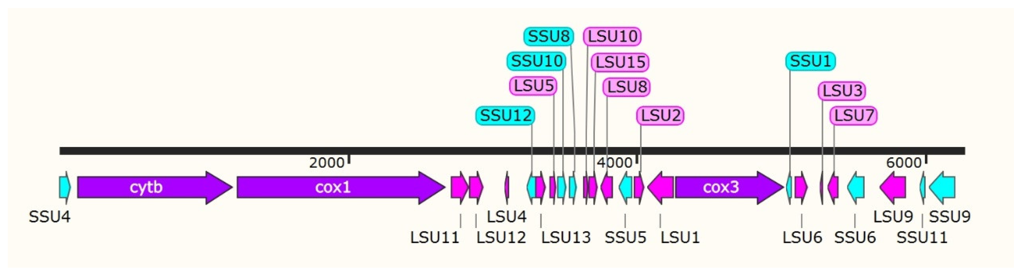
3.1. Analyzing the mtDNA Composition of E. kongi

In the existing literature, the mitochondrial genomes of apicomplexan protozoa, which include species such as Toxoplasma, Cyclospora, Isospora, and Eimeria, are relatively compact, averaging around 6 kilobases (kb) in size [23,24,25,26,27,28]. These organisms not only share a similar gene composition but they also exhibit a conserved gene arrangement. Specifically, they contain three cytochrome genes (cox1, cox3, cytb), as well as genes for the large- and small-subunit ribosomal RNAs (LSU and SSU rRNAs, respectively). Building upon this foundation, the present study aimed to annotate the mitochondrial genome of E. kongi by examining the complete mtDNA sequence. This was achieved through a comparative analysis of the gene content and genome architecture with 18 strains of Eimeria [29,30]. The complete mtDNA sequence of E. kongi spans 6258 bp and includes 3 protein-coding genes (cytb, cox1, and cox3), 14 fragments of the LSU rRNA gene, and 9 fragments of the SSU rRNA gene. Notably, the sequence lacks any transfer RNA (tRNA) genes, aligning with the characteristics observed in Eimeria species isolated from rabbits (Figure 1).
The cytb in E. kongi mtDNA spans a length of 1080 bp, located between nucleotide positions 128 and 1207, and initiates with the start codon ATG. The cytochrome c oxidase subunit I gene (cox1) extends over 1443 bp, ranging from nucleotide 1240 to 2682, also beginning with the ATG start codon. The cox3 is 756 bp in length, positioned between nucleotides 4279 and 5034, and it begins with the less common start codon TTA. Notably, all three protein-coding genes share the TAA stop codon (Table 2). Furthermore, the inferred translational direction for these genes—cytb, cox1, and cox3—conforms to the orientation observed in Eimeria species isolated from domestic rabbits (Figure 1).
The mitochondrial genome of E. kongi exhibits a pronounced adenine and thymine (A + T) content of 65.42%, with a corresponding guanine and cytosine (G + C) content of 34.58%. This can be further broken down into individual nucleotide frequencies: 29.80% adenine (A), 35.62% thymine (T), 16.95% guanine (G), and 17.63% cytosine (C). The sequence of E. kongi’s mtDNA offers novel genetic markers that are valuable for the study of molecular epidemiology and population genetics within the Eimeria genus. Moreover, it holds potential for advancing the molecular diagnosis and management strategies for rabbit coccidiosis. Among the 19 recognized species of Eimeria, the mtDNA lengths vary, with E. mitis featuring the longest mtDNA at 6408 bp, while E. brunetti has the shortest at 6156 bp. A notable characteristic across all Eimeria species is their high A + T content, which exceeds 60%. When comparing the nucleotide compositions, the six species of rabbit coccidia, particularly E. irresidua, show the closest similarity to E. kongi (Table 3).

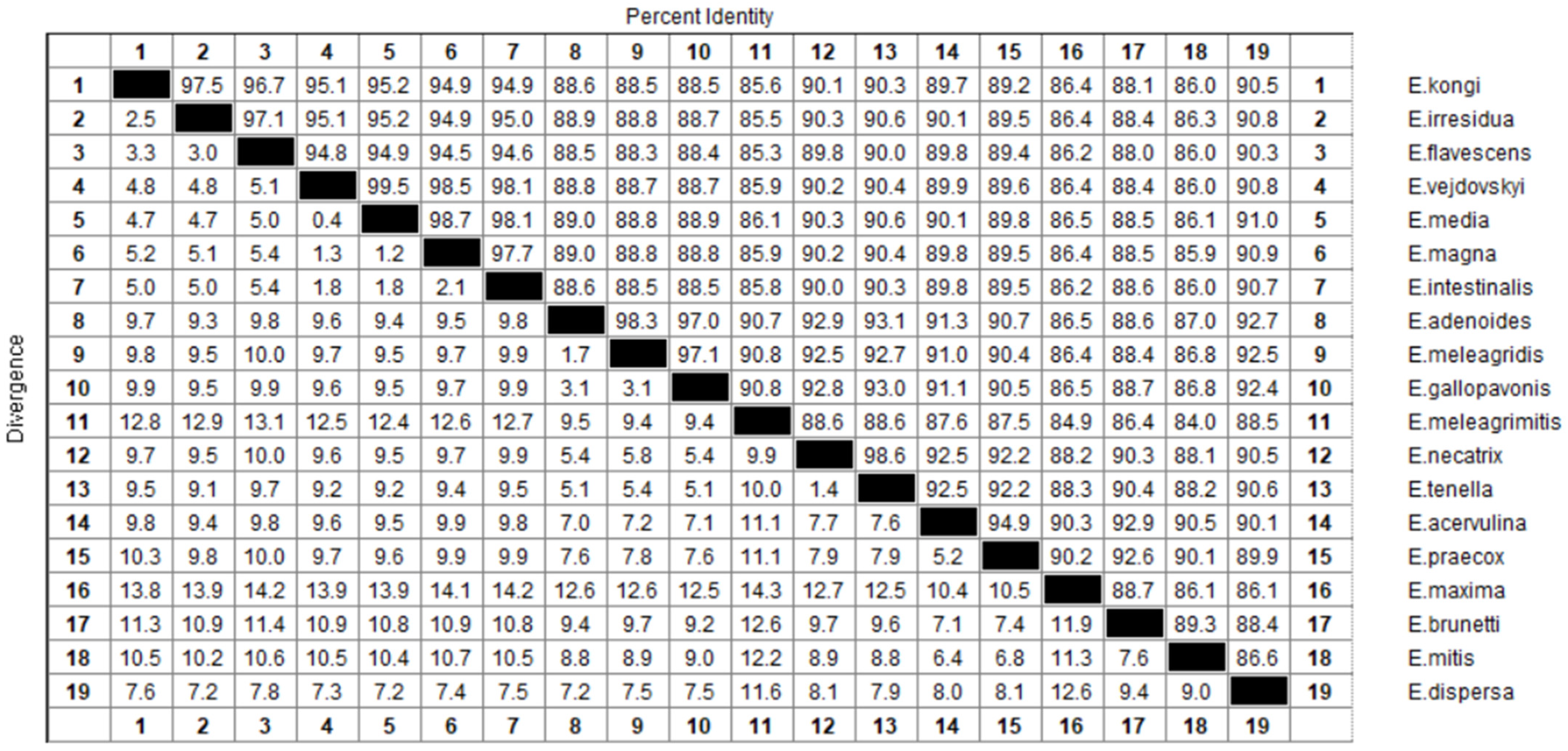
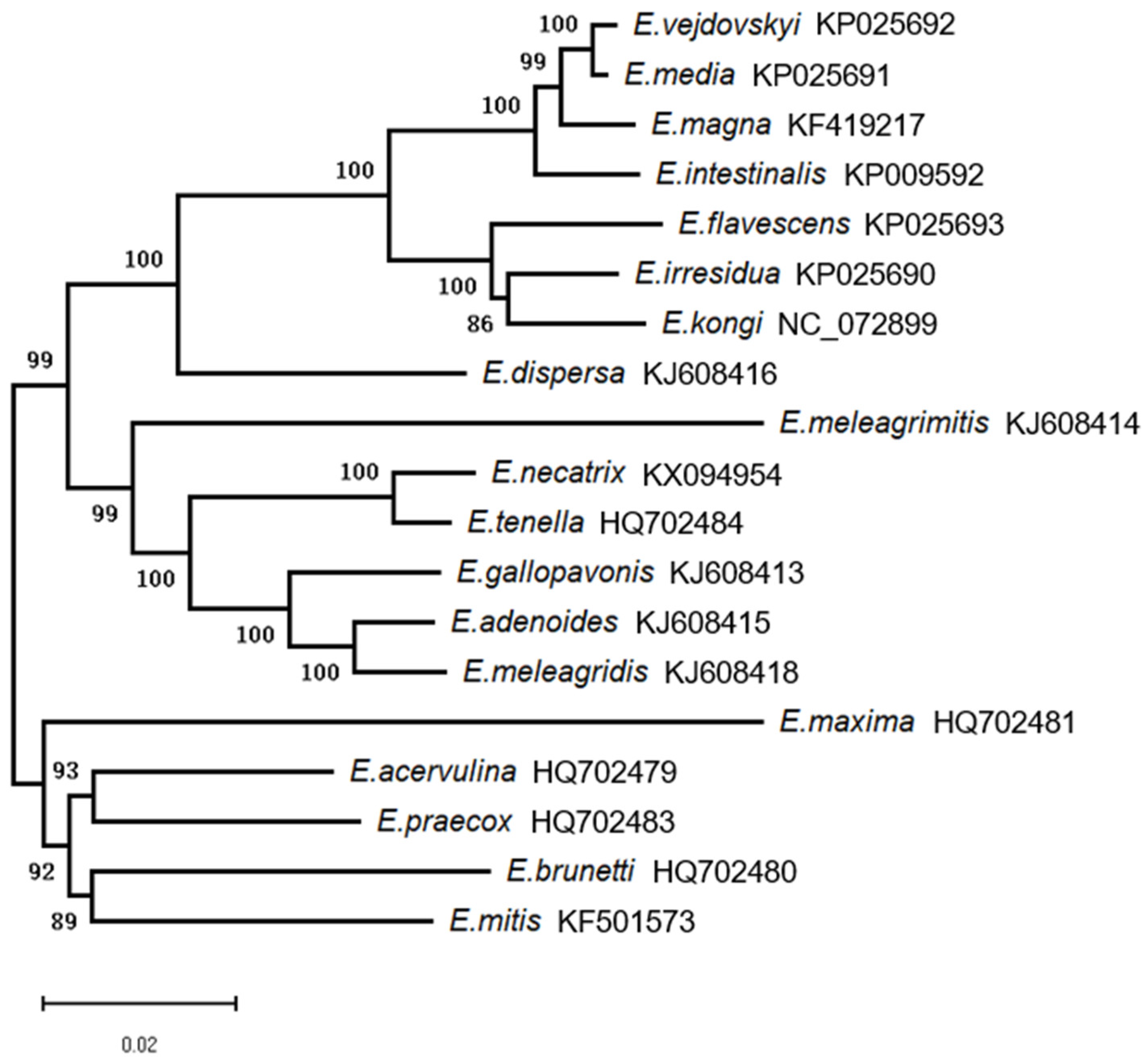
3.2. Clustal Alignment and Phylogenetic Analysis

The complete mitochondrial genome sequence of E. kongi was compared with those of 18 other Eimeria species, as shown in Figure 2. The sequence identity between E. kongi and rabbit coccidia species ranged from 94.9% to 97.5%, with E. irresidua and E. flavescens showing the highest identities at 97.5% and 96.7%, respectively. In contrast, other chicken and turkey coccidia species exhibited less than 90.5% sequence identity with E. kongi. Among these, E. meleagrimitis had the lowest identity at 85.6%, while E. dispersa had the highest at 90.5%.
As depicted in Figure 3, phylogenetic analysis revealed a pattern of clustering among certain Eimeria species that is congruent with their host organisms. Specifically, a monophyletic group encompasses seven species of rabbit coccidia, including E. kongi. However, this host-based clustering cannot be observed uniformly across all Eimeria species; for instance, chicken and turkey coccidia do not form such distinct host-related clusters. Among the chicken coccidia, E. necatrix and E. tenella, along with the turkey coccidia E. meleagrimitis, E. adenoides, E. gallopavonis, and E. meleagridis, form one monophyletic group. Conversely, the remaining five species of chicken coccidia—E. maxima, E. acervulina, E. praecox, E. brunetti, and E. mitis—constitute another monophyletic group. Interestingly, E. dispersa is not categorized within the previously mentioned monophyletic groups; instead, it is found to be closely related to the rabbit coccidia, belonging to the same monophyletic group. Furthermore, the monophyletic group of rabbit coccidia can be differentiated into two distinct sister lineages based on the presence or absence of oocyst residuum. E. kongi, E. irresidua, and E. flavescens are grouped within the lineage characterized by the absence of oocyst residuum. In contrast, E. magna, E. intestinalis, E. media, and E. vejdovskyi are classified within the lineage that exhibits oocyst residuum.
In summary, the phylogenetic tree derived from mtDNA sequences was found to be congruent with the trees constructed using 18S rRNA and ITS-1 sequences, as reported by Ping Cui and colleagues [2]. This concordance substantiates the classification of E. kongi within a monophyletic group alongside other species responsible for rabbit coccidiosis. Furthermore, we surmise that the oocyst residuum, a key morphological characteristic traditionally used in the taxonomy of rabbit coccidia, represents a phenotypic trait that arises from the gene expression of these organisms. Its significance in classification is as reliable as molecular markers. E. kongi is grouped with species that lack an oocyst residuum within the rabbit coccidia monophyletic group, a classification that aligns with its morphological features. The phylogenetic analysis based on multiple molecular sequences for E. kongi is consistent, and this genetic evidence, in conjunction with the organism’s morphological traits, strongly supports the designation of E. kongi as a distinct species within the rabbit coccidia, rather than a mere counterpart of existing species.

4. Conclusions

This study has elucidated that the mitochondrial genome of E. kongi is a linear DNA molecule, with a total length of 6258 bp. The nucleotide composition, as well as the directionality of protein translation and the utilization of start and stop codons within the E. kongi mitochondrial genome, closely resemble those observed in other Eimeria species’ mitochondrial genomes. Moreover, the phylogenetic analysis conducted using the complete mitochondrial genome sequence identified E. kongi as a novel species of rabbit coccidia endemic in China. It is positioned within a distinct sister lineage characterized by the absence of oocyst residuum, within the monophyletic group of rabbit coccidia. Ultimately, the mtDNA sequence of E. kongi offers novel DNA markers that will be instrumental for the investigation of molecular epidemiology and population genetics specific to this species. Furthermore, the mtDNA sequence aids in the molecular diagnosis of rabbit coccidiosis. Overall, the comprehensive mtDNA dataset serves as a valuable resource, providing essential information for phylogenetic analysis and population genetics research across various parasite populations.

Author Contributions

Conceptualization, P.C.; methodology, S.F. and F.D.; formal analysis, X.G. and Y.S.; investigation, Y.S. and C.H.; resources, P.C.; writing—original draft preparation, Y.S.; writing—review and editing, X.T.; project administration, S.F.; funding acquisition, P.C. All authors have read and agreed to the published version of the manuscript.

Funding

This study was supported by the Natural Science Foundation of Hebei Province and the Key Basic Research Project (grant number C2019405052), the Key R&D Program of Hebei Province (grant number 19226610D), and the Graduate Professional Degree Teaching Case (Library) Construction Project of Hebei Province (grant number KCJSZ2024101).

Data Availability Statement

The genome sequences are available in the NCBI, and Table 1 has listed all strains’ accession numbers.

Conflicts of Interest

The authors declare no conflicts of interest.

References

  1. Eckert, J.; Taylor, M.; Catchpole, J. Guidelines on Techniques in Coccidiosis Research; Directorate-General XII, Science, Research and Development Environment Research Programme; European Commission: Brussels, Belgium, 1995; pp. 103–119. [Google Scholar]
  2. Cui, P.; Liu, H.; Fang, S.; Gu, X.; Wang, P.; Liu, C.; Tao, G.; Liu, X.; Suo, X. A new species of Eimeria (Apicomplexa: Eimeriidae) from Californian rabbits in Hebei Province, China. Parasitol. Int. 2017, 66, 677–680. [Google Scholar] [CrossRef] [PubMed]
  3. Evans, J.D.; Lopez, D.L. Complete mitochondrial DNA sequence of the important honey bee pest, Varroa destructor (Acari: Varroidae). Exp. Appl. Acarol. 2002, 27, 69–78. [Google Scholar] [CrossRef] [PubMed]
  4. Virji, A.Z.; Thekkiniath, J.; Ma, W.; Lawres, L.; Knight, J.; Swei, A.; Roch, K.L.; Mamoun, C. Insights into the evolution and drug susceptibility of Babesia duncani from the sequence of its mitochondrial and apicoplast genomes. Int. J. Parasitol. 2019, 49, 105–113. [Google Scholar] [CrossRef] [PubMed]
  5. Vieira, L.M.D.C.; Pereira, P.H.O.; Vilela, D.A.D.R.; Landau, I.; Andreína Pacheco, M.; Escalante, A.A.; Ferreira, F.C.; Braga, É.M. Leucocytozoon cariamae n. sp. and Haemoproteus pulcher coinfection in Cariama cristata (Aves: Cariamiformes): First mitochondrial genome analysis and morphological description of a leucocytozoid in Brazil. Parasitology 2023, 150, 1296–1306. [Google Scholar] [CrossRef] [PubMed]
  6. Cao, J.; Guo, X.; Guo, C.; Wang, X.; Wang, Y.; Yan, F. Complete mitochondrial genome of Malenka flexura (Plecoptera: Nemouridae) and phylogenetic analysis. Genes 2022, 13, 911. [Google Scholar] [CrossRef] [PubMed]
  7. Aurongzeb, M.; Rashid, Y.; Habib Ahmed Naqvi, S.; Muhammad Talha Malik, H.; Kamran Azim, M.; Hassan, S.S.; Yasir, M.; Karim, A. Insights into genome evolution, pan-genome, and phylogenetic implication through mitochondrial genome sequence of Naegleria fowleri species. Sci. Rep. 2022, 12, 13152. [Google Scholar] [CrossRef] [PubMed]
  8. Song, Y.; Du, X.; Li, A.; Fan, A.; He, L.; Sun, Z.; Niu, Y.; Qiao, Y. Assembly and analysis of the complete mitochondrial genome of Forsythia suspensa (Thunb.) Vahl. BMC Genom. 2023, 24, 708. [Google Scholar] [CrossRef]
  9. Kim, N.Y.; Ahn, S.J.; Seo, J.S.; Jeon, E.J.; Cho, M.Y.; Choi, H.S. Characterization of the complete mitochondrial genome of Miamiensis avidus causing flatfish scuticociliatosis. Genetica 2022, 150, 407–420. [Google Scholar] [CrossRef] [PubMed]
  10. Ogedengbe, M.E.; El-Sherry, S.; Whale, J.; Barta, J.R. Complete mitochondrial genome sequences from five Eimeria species (Apicomplexa; Coccidia; Eimeriidae) infecting domestic turkeys. Parasit. Vectors 2014, 7, 335. [Google Scholar] [CrossRef]
  11. Gray, M.W.; Lang, B.F.; Cedergren, R.; Golding, G.B.; Lemieux, C.; Sankoff, D.; Turmel, M.; Brossard, N.; Delage, E.; Littlejohn, T.G.; et al. Genome structure and gene content in protist mitochondrial DNAs. Nucleic. Acids. Res. 1998, 26, 865–878. [Google Scholar] [CrossRef]
  12. Osman, S.A.M.; Nishibori, M.; Yonezawa, T. Complete mitochondrial genome sequence of Tosa-Jidori sheds light on the origin and evolution of Japanese native chickens. Anim. Biosci. 2021, 34, 941–948. [Google Scholar] [CrossRef] [PubMed]
  13. Hikosaka, K.; Nakai, Y.; Watanabe, Y.; Tachibana, S.I.; Arisue, N.; Palacpac, N.M.Q.; Toyama, T.; Honma, H.; Horii, T.; Kita, K.; et al. Concatenated mitochondrial DNA of the coccidian parasite Eimeria tenella. Mitochondrion 2011, 11, 273–278. [Google Scholar] [CrossRef] [PubMed]
  14. Jensen, M.R.; Sigsgaard, E.E.; Liu, S.; Manica, A.; Bach, S.S.; Hansen, M.M.; Møller, P.R.; Thomsen, P.F. Genome-scale target capture of mitochondrial and nuclear environmental DNA from water samples. Mol. Ecol. Resour. 2021, 21, 690–702. [Google Scholar] [CrossRef] [PubMed]
  15. Mathur, V.; Wakeman, K.C.; Keeling, P.J. Parallel functional reduction in the mitochondria of apicomplexan parasites. Curr. Biol. 2021, 31, 2920–2928. [Google Scholar] [CrossRef] [PubMed]
  16. Conrad, I.; Craft, A.; Thurman, C.L.; Baeza, J.A. The complete mitochondrial genome of the red-jointed brackish-water fiddler crab Minuca minax (LeConte 1855) (Brachyura: Ocypodidae): New family gene order, and purifying selection and phylogenetic informativeness of protein coding genes. Genomics 2021, 113, 565–572. [Google Scholar] [CrossRef] [PubMed]
  17. Fang, S.; Shi, Y.; Wang, P.; Guan, C.; Gu, X.; Guan, L.; Cui, P.; Suo, X. Study on the pathogenicity, immunogenicity, endogenous development and drug sensitivity of Eimeria kongi. Front. Vet. Sci. 2023, 10, 1134193. [Google Scholar] [CrossRef] [PubMed]
  18. Tenter, A.M.; Barta, J.R.; Beveridge, I.; Duszynski, D.W.; Mehlhorn, H.; Morrison, D.A.; Thompson, R.C.A.; Conrad, P.A. The conceptual basis for a new classification of the coccidia. Int. J. Parasitol. 2002, 32, 595–616. [Google Scholar] [CrossRef] [PubMed]
  19. Schmidt, V.; Dyachenko, V.; Aupperle, H.; Pees, M.; Krautwald-Junghanns, M.E.; Daugschies, A. Case report of systemic coccidiosis in a radiated tortoise (Geochelone radiata). Parasitol. Res. 2008, 102, 431–436. [Google Scholar] [CrossRef] [PubMed]
  20. Qin, Z.; Zhang, J.; Zhang, K.; Lang, J.; Wang, N.; Li, J.; Zhang, L. Morphological and molecular characteristics of a single oocyst for the identification of Eimeria species in domestic rabbits (Oryctolagus cuniculus f. domesticus). Vet. Parasitol. 2023, 321, 109986. [Google Scholar] [CrossRef]
  21. Šlapeta, J.R.; Modrý, D.; Votýpka, J.; Jirků, M.; Oborník, M.; Lukeš, J.; Koudela, B. Eimeria telekii n.sp. (Apicomplexa: Coccidia) from Lemniscomys striatus (Rodentia: Muridae): Morphology, pathology and phylogeny. Parasitology 2001, 122, 133–143. [Google Scholar] [CrossRef]
  22. Modrý, D.; Slapeta, J.R.; Jirků, M.; Oborník, M.; Lukeš, J.; Koudela, B. Phylogenetic position of a renal coccidium of the European green frogs, ‘Isospora’ lieberkuehni Labbé, 1894 (Apicomplexa: Sarcocystidae) and its taxonomic implications. Int. J. Syst. Evol. Microbiol. 2001, 53, 767–772. [Google Scholar] [CrossRef] [PubMed]
  23. González-Gómez, J.P.; Lozano-Aguirre, L.F.; Medrano-Félix, J.A.; Chaidez, C.; Gerba, C.P.; Betancourt, W.Q.; Castro-del Campo, N. Evaluation of nuclear and mitochondrial phylogenetics for the subtyping of Cyclospora cayetanensis. Parasitol. Res. 2023, 122, 2641–2650. [Google Scholar] [CrossRef] [PubMed]
  24. Kay, C.; Williams, T.A.; Gibson, W. Mitochondrial DNAs provide insight into trypanosome phylogeny and molecular evolution. BMC Evol. Biol. 2020, 20, 161. [Google Scholar] [CrossRef]
  25. Zhou, X.; Wang, Z.; Zhu, P.; Gu, X.; He, R.; Xu, J.; Jing, B.; Wang, L.; Chen, S.; Xie, Y. Eimeria zuernii (Eimeriidae: Coccidia): Mitochondrial genome and genetic diversity in the Chinese yak. Parasit. Vectors 2023, 16, 312. [Google Scholar] [CrossRef] [PubMed]
  26. Zhou, X.; Wang, L.; Zhu, P.; Yang, Z.; Wang, Z.; Chen, Y.; Gu, X.; He, R.; Xu, J.; Jing, B.; et al. Comprehensive molecular characterization of complete mitogenome assemblies of 33 Eimeria isolates infecting domestic chickens. Parasit. Vectors 2023, 16, 109. [Google Scholar] [CrossRef] [PubMed]
  27. Namasivayam, S.; Sun, C.; Bah, A.B.; Oberstaller, J.; Pierre-Louis, E.; Etheridge, R.D.; Feschotte, C.; Pritham, E.J.; Kissinger, J.C. Massive invasion of organellar DNA drives nuclear genome evolution in Toxoplasma. Proc. Natl. Acad. Sci. USA 2023, 120, e2308569120. [Google Scholar] [CrossRef] [PubMed]
  28. Yang, R.; Brice, B.; Oskam, C.; Zhang, Y.; Brigg, F.; Berryman, D.; Ryan, U. Characterization of two complete Isospora mitochondrial genomes from passerine birds: Isospora serinuse in a domestic canary and Isospora manorinae in a yellow-throated miner. Vet. Parasitol. 2017, 237, 137–142. [Google Scholar] [CrossRef] [PubMed]
  29. Tian, S.Q.; Cui, P.; Fang, S.F.; Liu, G.H.; Wang, C.R.; Zhu, X.Q. The complete mitochondrial genome sequence of Eimeria magna (Apicomplexa: Coccidia). Mitochondrial DNA 2015, 26, 714–715. [Google Scholar] [CrossRef]
  30. Liu, G.H.; Tian, S.Q.; Cui, P.; Fang, S.F.; Wang, C.R.; Zhu, X.Q. The complete mitochondrial genomes of five Eimeria species infecting domestic rabbits. Exp. Parasitol. 2015, 159, 67–71. [Google Scholar] [CrossRef]
Figure 1. Structure of the mtDNA of E. kongi. The spatial arrangement, length, and transcriptional orientation of genes within the mtDNA of E. kongi were elucidated through comparative analysis with the mitochondrial genomes of six rabbit coccidian species using Clustal v2.1. This comparative approach facilitated the prediction of gene locations within the E. kongi mitochondrial genome. In the resulting schematic representation, protein-coding genes are denoted in purple, fragments of LSU rRNA are highlighted in pink, and fragments of SSU rRNA are depicted in blue. The transcriptional direction of each gene is graphically represented by arrows at the respective gene termini.
Life 14 00699 g001
Figure 2. The nucleotides sequences of the complete mtDNA among the 18 Eimeria species and E. kongi.
Life 14 00699 g002
Figure 3. Phylogenetic analysis of E. kongi was conducted utilizing mtDNA sequences. The analysis employed the Neighbor-Joining (NJ) method, and nodes with percentage support exceeding 50% from 1000 pseudoreplicates are marked on the left side of the corresponding nodes.
Life 14 00699 g003
Table 1. GenBank accession numbers for the 19 complete mtDNA of Eimeria species in this study.
SpeciesAccession NumberSpeciesAccession Number
E. kongiNC_072899E. gallopavonisKJ608413
E. vejdovskyiKP025692E. meleagrimitisKJ608414
E. mediaKP025691E. necatrixKX094954
E. magnaKF419217E. tenellaHQ702484
E. intestinalisKP009592E. acervulinaHQ702479
E. flavescensKP025693E. praecoxHQ702483
E. irresiduaKP025690E. maximaHQ702481
E. dispersaKJ608416E. brunettiHQ702480
E. adenoidesKJ608415E. mitisKF501573
E. meleagridisKJ608418
Table 2. Complete mitochondrial genome of E. kongi.
GenesNucleotide No.Size (bp)Start CodonStop Codon
SSU41–8282
cytb128–12071080ATGTAA
cox11240–26821443ATGTAA
LSU112722–2849128
LSU122851–294999
LSU43090–311424
SSU123244–330461
LSU133305–337975
LSU53403–345149
SSU103460–352465
SSU83538–359659
LSU103637–366933
LSU153678–373356
LSU83757–383781
SSU53883–397492
LSU23992–405766
LSU14083–4261179
cox34279–5034756TTATAA
SSU15044–507330
LSU65102–519594
LSU35272–529524
LSU75331–540373
SSU65467–5582116
LSU95684–5871188
SSU115970–600637
SSU96032–6212181
Table 3. Nucleotide composition of the mitochondrial genome of the 19 Eimeria species.
SpeciesA (%)T (%)G (%)C (%)Total
E. mitis30.936.516.316.36408
E. irresidua29.835.61717.66259
E. kongi29.835.61717.66258
E. flavescens3035.516.917.66258
E. magna29.735.417.117.76249
E. intestinalis29.735.517.117.76247
E. media29.635.517.117.76245
E. vejdovskyi29.635.517.217.76240
E. dispersa29.935.616.817.66238
E. gallopavonis30.134.816.918.36215
E. tenella29.835.216.918.16213
E. meleagridis29.834.91718.36212
E. necatrix29.835.216.918.26212
E. adenoides29.834.81718.46211
E. acervulina30.135.31717.66179
E. praecox30.135.517.117.36172
E. maxima3034.517.517.96167
E. meleagrimitis3033.517.319.26165
E. brunetti29.835.817.117.46156
Disclaimer/Publisher’s Note: The statements, opinions and data contained in all publications are solely those of the individual author(s) and contributor(s) and not of MDPI and/or the editor(s). MDPI and/or the editor(s) disclaim responsibility for any injury to people or property resulting from any ideas, methods, instructions or products referred to in the content.

