Evaluation of 3D Footprint Morphology of Knee-Related Muscle Attachments Based on CT Data Reconstruction: A Feasibility Study
Abstract
:1. Introduction
2. Materials and Methods
2.1. Specimen Preparation
2.2. Bone Reconstruction
2.3. Generation of Coherent Models
2.4. Projection of Attachments on Bone Surfaces
2.5. Muscle Data Collection
2.6. Data Analysis
3. Results
3.1. Specimen Characterization
3.2. Segmentation Deviation Analysis
3.3. Morphological Data
4. Discussion
Variations of Muscle Attachments
5. Conclusions
Supplementary Materials
Author Contributions
Funding
Institutional Review Board Statement
Informed Consent Statement
Data Availability Statement
Acknowledgments
Conflicts of Interest
References
- D’Lima, D.D.; Fregly, B.J.; Patil, S.; Steklov, N.; Colwell, C.W. Knee Joint Forces: Prediction, Measurement, and Significance. Proc. Inst. Mech. Eng. H 2012, 226, 95–102. [Google Scholar] [CrossRef]
- Chan, C.W.; Rudins, A. Foot Biomechanics during Walking and Running. Mayo Clin. Proc. 1994, 69, 448–461. [Google Scholar] [CrossRef] [PubMed]
- Song, K.; Scattone Silva, R.; Hullfish, T.J.; Silbernagel, K.G.; Baxter, J.R. Patellofemoral Joint Loading Progression Across 35 Weightbearing Rehabilitation Exercises and Activities of Daily Living. Am. J. Sports Med. 2023, 51, 2110–2119. [Google Scholar] [CrossRef]
- Hiemstra, L.A.; Kerslake, S.; Irving, C. Anterior Knee Pain in the Athlete. Clin. Sports Med. 2014, 33, 437–459. [Google Scholar] [CrossRef] [PubMed]
- Harris, M.; Edwards, S.; Rio, E.; Cook, J.; Cencini, S.; Hannington, M.C.; Bonello, C.; Docking, S. Nearly 40% of Adolescent Athletes Report Anterior Knee Pain Regardless of Maturation Status, Age, Sex or Sport Played. Phys. Ther. Sport 2021, 51, 29–35. [Google Scholar] [CrossRef] [PubMed]
- DeHaven, K.E.; Lintner, D.M. Athletic Injuries: Comparison by Age, Sport, and Gender. Am. J. Sports Med. 1986, 14, 218–224. [Google Scholar] [CrossRef] [PubMed]
- Carbone, V.; Fluit, R.; Pellikaan, P.; van der Krogt, M.M.; Janssen, D.; Damsgaard, M.; Vigneron, L.; Feilkas, T.; Koopman, H.F.J.M.; Verdonschot, N. TLEM 2.0—A Comprehensive Musculoskeletal Geometry Dataset for Subject-Specific Modeling of Lower Extremity. J. Biomech. 2015, 48, 734–741. [Google Scholar] [CrossRef]
- Gerus, P.; Sartori, M.; Besier, T.F.; Fregly, B.J.; Delp, S.L.; Banks, S.A.; Pandy, M.G.; D’Lima, D.D.; Lloyd, D.G. Subject-Specific Knee Joint Geometry Improves Predictions of Medial Tibiofemoral Contact Forces. J. Biomech. 2013, 46, 2778–2786. [Google Scholar] [CrossRef]
- Davico, G.; Lloyd, D.G.; Carty, C.P.; Killen, B.A.; Devaprakash, D.; Pizzolato, C. Multi-Level Personalization of Neuromusculoskeletal Models to Estimate Physiologically Plausible Knee Joint Contact Forces in Children. Biomech. Model. Mechanobiol. 2022, 21, 1873–1886. [Google Scholar] [CrossRef]
- Putame, G.; Terzini, M.; Rivera, F.; Kebbach, M.; Bader, R.; Bignardi, C. Kinematics and Kinetics Comparison of Ultra-Congruent versus Medial-Pivot Designs for Total Knee Arthroplasty by Multibody Analysis. Sci. Rep. 2022, 12, 3052. [Google Scholar] [CrossRef]
- Ballal, M.S.; Walker, C.R.; Molloy, A.P. The Anatomical Footprint of the Achilles Tendon: A Cadaveric Study. Bone Jt. J. 2014, 96-B, 1344–1348. [Google Scholar] [CrossRef]
- Benninger, B.; Delamarter, T. Distal Semimembranosus Muscle-Tendon-Unit Review: Morphology, Accurate Terminology, and Clinical Relevance. Folia Morphol. 2013, 72, 1–9. [Google Scholar] [CrossRef]
- Dziedzic, D.; Bogacka, U.; Ciszek, B. Anatomy of Sartorius Muscle. Folia Morphol. 2014, 73, 359–362. [Google Scholar] [CrossRef]
- De Maeseneer, M.; Shahabpour, M.; Lenchik, L.; Milants, A.; De Ridder, F.; De Mey, J.; Cattrysse, E. Distal Insertions of the Semimembranosus Tendon: MR Imaging with Anatomic Correlation. Skelet. Radiol. 2014, 43, 781–791. [Google Scholar] [CrossRef]
- Lin, J.; Zhang, S.; Xin, E.; Liang, M.; Yang, L.; Chen, J. Anterior Cruciate Ligament Femoral Footprint Is Oblong-Ovate, Triangular, or Two-Tears Shaped in Healthy Young Adults: Three-Dimensional MRI Analysis. Knee Surg. Sports Traumatol. Arthrosc. 2023, 31, 5514–5523. [Google Scholar] [CrossRef]
- Westermann, R.W.; Sybrowsky, C.; Ramme, A.J.; Amendola, A.; Wolf, B.R. Three-Dimensional Characterization of the Femoral Footprint of the Posterior Cruciate Ligament. Arthroscopy 2013, 29, 1811–1816. [Google Scholar] [CrossRef]
- Karakostis, F.A. Statistical Protocol for Analyzing 3D Muscle Attachment Sites Based on the “Validated Entheses-based Reconstruction of Activity” (VERA) Approach. Int. J. Osteoarchaeol. 2023, 33, 461–474. [Google Scholar] [CrossRef]
- Karakostis, F.A.; Lorenzo, C. Morphometric Patterns among the 3D Surface Areas of Human Hand Entheses. Am. J. Phys. Anthropol. 2016, 160, 694–707. [Google Scholar] [CrossRef]
- Salhi, A.; Burdin, V.; Mutsvangwa, T.; Sivarasu, S.; Brochard, S.; Borotikar, B. Subject-Specific Shoulder Muscle Attachment Region Prediction Using Statistical Shape Models: A Validity Study. In Proceedings of the 2017 39th Annual International Conference of the IEEE Engineering in Medicine and Biology Society (EMBC), Jeju, Republic of Korea, 11–15 July 2017; pp. 1640–1643. [Google Scholar]
- Andreassen, T.E.; Hume, D.R.; Hamilton, L.D.; Walker, K.E.; Higinbotham, S.E.; Shelburne, K.B. Three Dimensional Lower Extremity Musculoskeletal Geometry of the Visible Human Female and Male. Sci. Data 2023, 10, 34. [Google Scholar] [CrossRef]
- Fukuda, N.; Otake, Y.; Takao, M.; Yokota, F.; Ogawa, T.; Uemura, K.; Nakaya, R.; Tamura, K.; Grupp, R.B.; Farvardin, A.; et al. Estimation of Attachment Regions of Hip Muscles in CT Image Using Muscle Attachment Probabilistic Atlas Constructed from Measurements in Eight Cadavers. Int. J. CARS 2017, 12, 733–742. [Google Scholar] [CrossRef]
- Herteleer, M.; Vancleef, S.; Herijgers, P.; Duflou, J.; Jonkers, I.; Vander Sloten, J.; Nijs, S. Variation of the Clavicle’s Muscle Insertion Footprints—A Cadaveric Study. Sci. Rep. 2019, 9, 16293. [Google Scholar] [CrossRef]
- Kamineni, S.; Bachoura, A.; Behrens, W.; Kamineni, E.; Deane, A. Distal Insertional Footprint of the Brachialis Muscle: 3D Morphometric Study. Anat. Res. Int. 2015, 2015, 786508. [Google Scholar] [CrossRef]
- Walton, C.; Li, Z.; Pennings, A.; Agur, A.; Elmaraghy, A. A 3-Dimensional Anatomic Study of the Distal Biceps Tendon: Implications for Surgical Repair and Reconstruction. Orthop. J. Sports Med. 2015, 3, 2325967115585113. [Google Scholar] [CrossRef]
- Philippon, M.J.; Ferro, F.P.; Campbell, K.J.; Michalski, M.P.; Goldsmith, M.T.; Devitt, B.M.; Wijdicks, C.A.; LaPrade, R.F. A Qualitative and Quantitative Analysis of the Attachment Sites of the Proximal Hamstrings. Knee Surg. Sports Traumatol. Arthrosc. 2015, 23, 2554–2561. [Google Scholar] [CrossRef]
- Bonnin, M.P.; de Kok, A.; Verstraete, M.; Van Hoof, T.; Van Der Straten, C.; Saffarini, M.; Victor, J. Popliteus Impingement after TKA May Occur with Well-Sized Prostheses. Knee Surg. Sports Traumatol. Arthrosc. 2017, 25, 1720–1730. [Google Scholar] [CrossRef]
- Van Hoof, T.; Cromheecke, M.; Tampere, T.; D’herde, K.; Victor, J.; Verdonk, P.C.M. The Posterior Cruciate Ligament: A Study on Its Bony and Soft Tissue Anatomy Using Novel 3D CT Technology. Knee Surg. Sports Traumatol. Arthrosc. 2013, 21, 1005–1010. [Google Scholar] [CrossRef]
- Neuschwander, T.B.; Benke, M.T.; Gerhardt, M.B. Anatomic Description of the Origin of the Proximal Hamstring. Arthroscopy 2015, 31, 1518–1521. [Google Scholar] [CrossRef]
- Klein Horsman, M.D.; Koopman, H.F.J.M.; van der Helm, F.C.T.; Prosé, L.P.; Veeger, H.E.J. Morphological Muscle and Joint Parameters for Musculoskeletal Modelling of the Lower Extremity. Clin. Biomech. 2007, 22, 239–247. [Google Scholar] [CrossRef]
- Arnold, E.M.; Ward, S.R.; Lieber, R.L.; Delp, S.L. A Model of the Lower Limb for Analysis of Human Movement. Ann. Biomed. Eng. 2010, 38, 269–279. [Google Scholar] [CrossRef]
- Kaya Keles, C.S.; Ates, F. How Mechanics of Individual Muscle-Tendon Units Define Knee and Ankle Joint Function in Health and Cerebral Palsy—A Narrative Review. Front. Bioeng. Biotechnol. 2023, 11, 1287385. [Google Scholar] [CrossRef]
- Scheys, L.; Desloovere, K.; Suetens, P.; Jonkers, I. Level of Subject-Specific Detail in Musculoskeletal Models Affects Hip Moment Arm Length Calculation during Gait in Pediatric Subjects with Increased Femoral Anteversion. J. Biomech. 2011, 44, 1346–1353. [Google Scholar] [CrossRef]
- Barzan, M.; Modenese, L.; Carty, C.P.; Maine, S.; Stockton, C.A.; Sancisi, N.; Lewis, A.; Grant, J.; Lloyd, D.G.; Brito Da Luz, S. Development and Validation of Subject-Specific Pediatric Multibody Knee Kinematic Models with Ligamentous Constraints. J. Biomech. 2019, 93, 194–203. [Google Scholar] [CrossRef]
- Soodmand, E.; Kluess, D.; Varady, P.A.; Cichon, R.; Schwarze, M.; Gehweiler, D.; Niemeyer, F.; Pahr, D.; Woiczinski, M. Interlaboratory Comparison of Femur Surface Reconstruction from CT Data Compared to Reference Optical 3D Scan. Biomed. Eng. Online 2018, 17, 29. [Google Scholar] [CrossRef]
- Wu, G.; Siegler, S.; Allard, P.; Kirtley, C.; Leardini, A.; Rosenbaum, D.; Whittle, M.; D’Lima, D.D.; Cristofolini, L.; Witte, H.; et al. ISB Recommendation on Definitions of Joint Coordinate System of Various Joints for the Reporting of Human Joint Motion--Part I: Ankle, Hip, and Spine. International Society of Biomechanics. J. Biomech. 2002, 35, 543–548. [Google Scholar] [CrossRef]
- Valente, G.; Pitto, L.; Testi, D.; Seth, A.; Delp, S.L.; Stagni, R.; Viceconti, M.; Taddei, F. Are Subject-Specific Musculoskeletal Models Robust to the Uncertainties in Parameter Identification? PLoS ONE 2014, 9, e112625. [Google Scholar] [CrossRef]
- Turcotte, C.M.; Rabey, K.N.; Green, D.J.; McFarlin, S.C. Muscle Attachment Sites and Behavioral Reconstruction: An Experimental Test of Muscle-bone Structural Response to Habitual Activity. Am. J. Biol. Anthropol. 2022, 177, 63–82. [Google Scholar] [CrossRef]
- Andriacchi, T.P.; Mündermann, A.; Smith, R.L.; Alexander, E.J.; Dyrby, C.O.; Koo, S. A Framework for the in Vivo Pathomechanics of Osteoarthritis at the Knee. Ann. Biomed. Eng. 2004, 32, 447–457. [Google Scholar] [CrossRef]
- Branch, E.A.; Anz, A.W. Distal Insertions of the Biceps Femoris: A Quantitative Analysis. Orthop. J. Sports Med. 2015, 3, 2325967115602255. [Google Scholar] [CrossRef]
- Lohrer, H.; Arentz, S.; Nauck, T.; Dorn-Lange, N.V.; Konerding, M.A. The Achilles Tendon Insertion Is Crescent-Shaped: An in Vitro Anatomic Investigation. Clin. Orthop. Relat. Res. 2008, 466, 2230–2237. [Google Scholar] [CrossRef]
- Lee, J.-H.; Kim, K.-J.; Jeong, Y.-G.; Lee, N.S.; Han, S.Y.; Lee, C.G.; Kim, K.-Y.; Han, S.-H. Pes Anserinus and Anserine Bursa: Anatomical Study. Anat. Cell Biol. 2014, 47, 127–131. [Google Scholar] [CrossRef]
- Olewnik, Ł.; Gonera, B.; Podgórski, M.; Polguj, M.; Jezierski, H.; Topol, M. A Proposal for a New Classification of Pes Anserinus Morphology. Knee Surg. Sports Traumatol. Arthrosc. 2019, 27, 2984–2993. [Google Scholar] [CrossRef] [PubMed]
- Tenforde, A.S.; Yin, A.; Hunt, K.J. Foot and Ankle Injuries in Runners. Phys. Med. Rehabil. Clin. N. Am. 2016, 27, 121–137. [Google Scholar] [CrossRef] [PubMed]
- Ryan, J.M.; Harris, J.D.; Graham, W.C.; Virk, S.S.; Ellis, T.J. Origin of the Direct and Reflected Head of the Rectus Femoris: An Anatomic Study. Arthroscopy 2014, 30, 796–802. [Google Scholar] [CrossRef] [PubMed]
- Sedaghat, S.; Langguth, P.; Larsen, N.; Campbell, G.; Both, M.; Jansen, O. Diagnostic Accuracy of Dual-Layer Spectral CT Using Electron Density Images to Detect Post-Traumatic Prevertebral Hematoma of the Cervical Spine. Rofo 2021, 193, 1445–1450. [Google Scholar] [CrossRef] [PubMed]


| Specimen | Femur CT2 | Femur CT3 | Femur CT4 | Pelvis CT2 | Pelvis CT3 | Tibia CT2 | Tibia CT3 | Tibia CT4 | Patella CT4 |
|---|---|---|---|---|---|---|---|---|---|
| 1 | 0.38 | 0.38 | 0.61 | 0.52 | 0.60 | 0.63 | 0.34 | 0.68 | 0.60 |
| 2 | 0.40 | 0.45 | 0.71 | 0.40 | 0.39 | 0.47 | 0.45 | 0.49 | 0.92 |
| 3 | 0.37 | 0.34 | 0.66 | 0.31 | 0.32 | 0.69 | 0.71 | 0.74 | 0.74 |
| 4 | 0.34 | 0.39 | 0.64 | 0.32 | 0.34 | 0.35 | 0.30 | 0.65 | 0.54 |
| Mean | 0.37 | 0.39 | 0.66 | 0.39 | 0.41 | 0.53 | 0.45 | 0.64 | 0.70 |
| STD | 0.02 | 0.04 | 0.04 | 0.08 | 0.11 | 0.13 | 0.16 | 0.09 | 0.15 |
| Bones | Volumes [mm3] | Surface Area [mm2] | Volume’s Center of Gravity [mm] | ||||
|---|---|---|---|---|---|---|---|
| Mean ± STD | CV [%] | Mean ± STD | CV [%] | Mean x ± STD | Mean y ± STD | Mean z ± STD | |
| Pelvis | 3.93 × 10⁵ ± 5.32 × 10⁴ | 13.54 | 7.11 × 10⁴ ± 6.21 × 10³ | 8.74 | 16.92 ± 6.27 | 454.79 ± 40.49 | 7.12 ± 4.32 |
| Femur | 6.45 × 10⁵ ± 1.11 × 10⁵ | 17.23 | 7.20 × 10⁴ ± 8.77 × 10³ | 12.17 | 5.75 ± 3.10 | 198.85 ± 24.32 | 16.72 ± 2.24 |
| Patella | 2.41 × 10⁴ ± 4.18 × 10³ | 17.38 | 4.76 × 10³ ± 5.64 × 10² | 11.85 | 49.73 ± 2.12 | 6.49 ± 3.72 | 4.23 ± 0.52 |
| Tibia/Fibula | 3.58 × 10⁵ ± 6.21 × 10⁴ | 17.33 | 6.71 × 10⁴ ± 8.99 × 10³ | 13.40 | 13.31 ± 9.16 | 167.38 ± 12.12 | 8.77 ± 5.64 |
| Foot | 2.45 × 10⁵ ± 3.49 × 10⁴ | 14.24 | 6.03 × 10⁴ ± 6.58 × 10³ | 10.92 | 44.91 ± 34.53 | 454.51 ± 42.08 | 28.18 ± 21.02 |
| Specimen | Femoral Morphological Parameter [mm] | |||
|---|---|---|---|---|
| MA | TEA | FHD | SD | |
| 1 | 430.40 | 97.80 | 50.60 | 36.60 |
| 2 | 403.70 | 95.00 | 54.70 | 40.40 |
| 3 | 442.10 | 88.50 | 51.30 | 34.90 |
| 4 | 345.30 | 85.30 | 47.10 | 30.90 |
| Mean | 405.38 | 91.65 | 50.93 | 35.70 |
| STD | 37.37 | 4.98 | 2.70 | 3.41 |
| CV [%] | 9.22 | 5.44 | 5.30 | 9.56 |
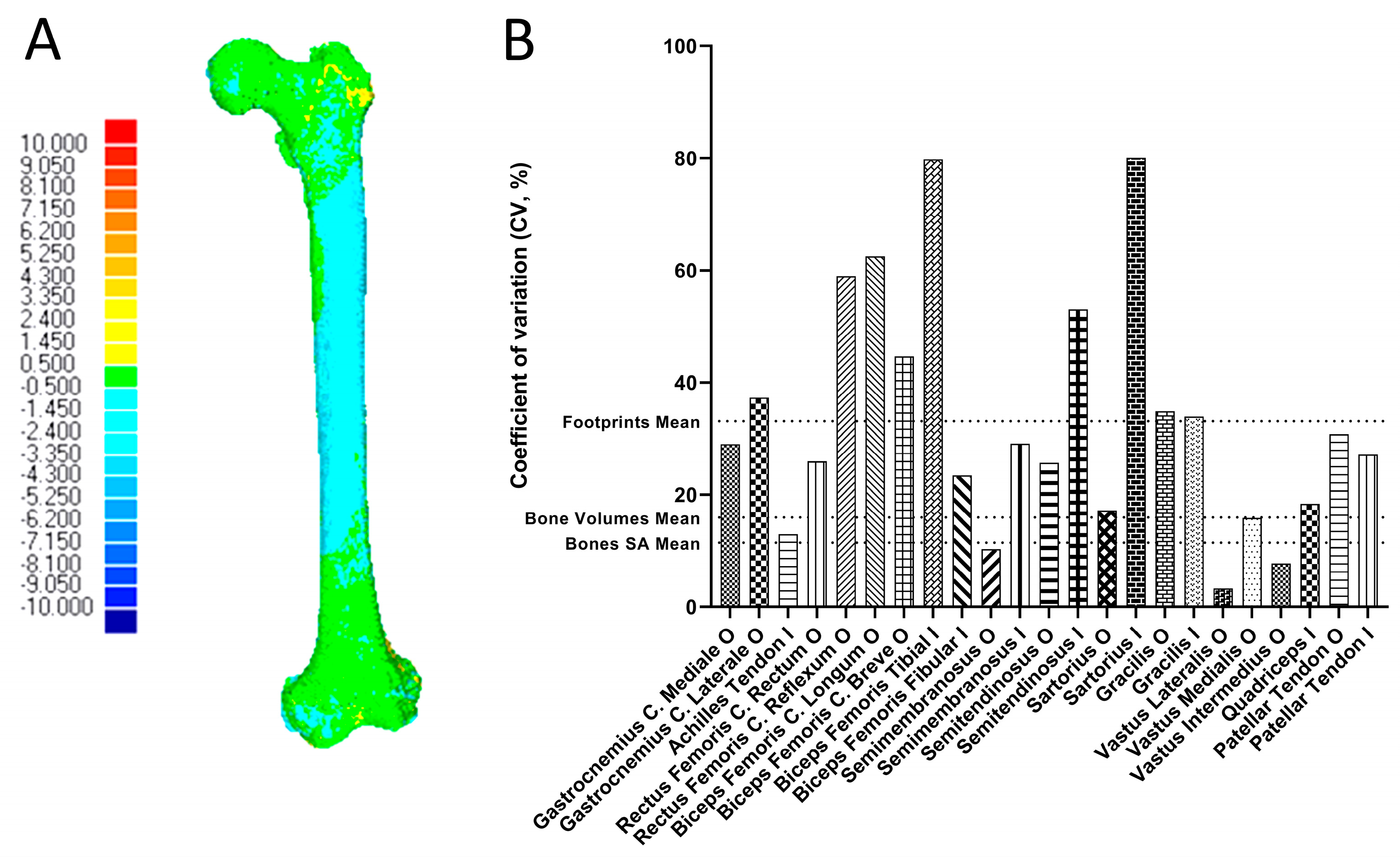
| Muscle Attachment | Surface Area [mm2] | CV [%] | Centroid [mm] | ||
|---|---|---|---|---|---|
| Mean ± STD | Mean x ± STD | Mean y ± STD | Mean z ± STD | ||
| Gastrocnemius Caput Mediale O | 630.12 ± 182.71 | 29.00 | 5.41 ± 1.72 | 24.21 ± 3.96 | 19.97 ± 3.51 |
| Gastrocnemius Caput Laterale O | 788.24 ± 293.77 | 37.27 | 2.89 ± 2.14 | 19.10 ± 8.41 | 35.02 ± 3.09 |
| Achilles Tendon I | 1000.21 ± 129.47 | 12.94 | 114.80 ± 41.42 | 417.36 ± 41.47 | 27.23 ± 17.40 |
| Rectus Femoris Caput Rectum O | 282.42 ± 73.21 | 25.92 | 40.90 ± 4.76 | 441.16 ± 39.09 | 4.05 ± 2.94 |
| Rectus Femoris Caput Reflexum O | 366.77 ± 216.17 | 58.94 | 4.90 ± 2.71 | 441.88 ± 40.23 | 18.03 ± 1.32 |
| Biceps Femoris Caput Longum O | 260.42 ± 162.77 | 62.50 | 62.22 ± 8.87 | 381.46 ± 41.80 | 7.38 ± 7.11 |
| Biceps Femoris Caput Breve O | 582.20 ± 259.99 | 44.66 | 2.71 ± 2.26 | 144.29 ± 17.82 | 19.04 ± 0.56 |
| Biceps Femoris Tibial I | 103.86 ± 82.87 | 79.79 | 8.13 ± 5.24 | 46.70 ± 4.90 | 43.34 ± 3.49 |
| Biceps Femoris Fibular I | 401.88 ± 94.16 | 23.43 | 22.57 ± 6.19 | 52.71 ± 6.37 | 48.95 ± 4.33 |
| Semimembranosus O | 471.78 ± 48.69 | 10.32 | 49.83 ± 9.45 | 370.54 ± 41.35 | 5.23 ± 4.93 |
| Semimembranosus I | 1048.10 ± 304.77 | 29.08 | 16.88 ± 3.21 | 55.22 ± 1.86 | 21.42 ± 0.73 |
| Semitendinosus O | 604.32 ± 155.25 | 25.69 | 64.19 ± 10.99 | 360.41 ± 41.71 | 10.73 ± 10.21 |
| Semitendinosus I | 226.50 ± 120.11 | 53.03 | 11.50 ± 5.61 | 97.74 ± 10.52 | 7.08 ± 4.02 |
| Sartorius O | 115.94 ± 19.92 | 17.18 | 62.48 ± 5.84 | 472.53 ± 39.85 | 9.45 ± 8.04 |
| Sartorius I | 313.18 ± 250.69 | 80.05 | 14.28 ± 3.34 | 86.16 ± 18.82 | 5.06 ± 4.82 |
| Gracilis O | 132.23 ± 46.13 | 34.88 | 16.69 ± 12.57 | 360.09 ± 34.53 | 79.06 ± 4.38 |
| Gracilis I | 67.18 ± 22.78 | 33.90 | 14.80 ± 6.41 | 85.25 ± 5.92 | 4.76 ± 3.73 |
| Vastus Lateralis O | 2493.56 ± 82.11 | 3.29 | 5.53 ± 3.11 | 238.36 ± 54.33 | 19.55 ± 8.51 |
| Vastus Medialis O | 3410.55 ± 541.07 | 15.86 | 1.54 ± 1.36 | 301.81 ± 35.94 | 50.51 ± 4.47 |
| Vastus Intermedius O | 10,968.48 ± 849.01 | 7.74 | 24.07 ± 4.15 | 224.31 ± 22.81 | 31.96 ± 2.28 |
| Quadriceps Tendon I | 1480.95 ± 271.94 | 18.36 | 56.66 ± 2.42 | 10.52 ± 6.66 | 4.08 ± 0.65 |
| Patellar Tendon O | 992.16 ± 305.28 | 30.77 | 50.81 ± 1.95 | 15.20 ± 6.47 | 5.20 ± 1.82 |
| Patellar Tendon I | 733.14 ± 198.92 | 27.13 | 24.11 ± 5.86 | 73.44 ± 8.94 | 16.76 ± 6.60 |
Disclaimer/Publisher’s Note: The statements, opinions and data contained in all publications are solely those of the individual author(s) and contributor(s) and not of MDPI and/or the editor(s). MDPI and/or the editor(s) disclaim responsibility for any injury to people or property resulting from any ideas, methods, instructions or products referred to in the content. |
© 2024 by the authors. Licensee MDPI, Basel, Switzerland. This article is an open access article distributed under the terms and conditions of the Creative Commons Attribution (CC BY) license (https://creativecommons.org/licenses/by/4.0/).
Share and Cite
Neumann, A.-M.; Kebbach, M.; Bader, R.; Hildebrandt, G.; Wree, A. Evaluation of 3D Footprint Morphology of Knee-Related Muscle Attachments Based on CT Data Reconstruction: A Feasibility Study. Life 2024, 14, 778. https://doi.org/10.3390/life14060778
Neumann A-M, Kebbach M, Bader R, Hildebrandt G, Wree A. Evaluation of 3D Footprint Morphology of Knee-Related Muscle Attachments Based on CT Data Reconstruction: A Feasibility Study. Life. 2024; 14(6):778. https://doi.org/10.3390/life14060778
Chicago/Turabian StyleNeumann, Anne-Marie, Maeruan Kebbach, Rainer Bader, Guido Hildebrandt, and Andreas Wree. 2024. "Evaluation of 3D Footprint Morphology of Knee-Related Muscle Attachments Based on CT Data Reconstruction: A Feasibility Study" Life 14, no. 6: 778. https://doi.org/10.3390/life14060778
APA StyleNeumann, A.-M., Kebbach, M., Bader, R., Hildebrandt, G., & Wree, A. (2024). Evaluation of 3D Footprint Morphology of Knee-Related Muscle Attachments Based on CT Data Reconstruction: A Feasibility Study. Life, 14(6), 778. https://doi.org/10.3390/life14060778


