Article

The Etiological and Antimicrobial Susceptibility Profiles of the Bacteria Obtained from Ovine Caseous Lymphadenitis Cases in the Çankırı Region, Türkiye

by
Serdal Tarhane
1,* and
Fatih Büyük
2
1
Veterinary Department, Eldivan Vocational School of Health Services, Çankırı Karatekin University, Çankırı 18200, Türkiye
2
Microbiology Department, Faculty of Veterinary Medicine, Kafkas University, Kars 36300, Türkiye
*
Author to whom correspondence should be addressed.
Life 2024, 14(9), 1078; https://doi.org/10.3390/life14091078
Submission received: 27 July 2024 / Revised: 15 August 2024 / Accepted: 24 August 2024 / Published: 28 August 2024
(This article belongs to the Section Microbiology)

Abstract

:
Sheep caseous lymphadenitis (CLA) causes significant economic losses in the livestock sector by causing a loss in the quantity and quality of animal products and a loss in the breeding value of animals. Although the primary agent in CLA’s etiology is Corynebacterium pseudotuberculosis, some other opportunistic microorganisms also play a role. Therefore, the control and treatment of CLA necessitates the identification of the relevant etiological agents. This study aimed to conduct an in vitro culture and molecular characterization (PCR analysis and 16S rRNA sequencing) of the bacteria involved in sheep CLA cases reported in the Çankırı province of Türkiye and determine the antibiotic susceptibility of the case isolates. In total, 82 (16.4%) of 500 sheep in five farms were diagnosed with CLA. Following the culture of the superficial abscesses samples, C. pseudotuberculosis was identified in 30 (36.59%) as a result of PCR, Pseudomonas spp. in 8 (9.76%), and Enterobacter cancerogenus in 1 (1.22%), as a result of 16S rRNA sequencing. These data revealed extensive heterogeneity among the Pseudomonas isolates, with hints of derivation from a common ancestry for some and phylogenetic similarity to isolates from Germany, Malaysia, and India. In contrast to the high susceptibility to cefoperazone and lincomycin, the high resistances of C. pseudotuberculosis and Pseudomonas spp. isolates to cephalothin, ceftiofur, cloxacillin, amoxicillin, and bacitracin were remarkable. Based on these findings, it was concluded that for an effective treatment and control of ovine CLA cases, there is a need to consider the possible involvement of opportunistic bacteria other than the primary causative agent, C. pseudotuberculosis. It also contributed to increasing the country-specific sequence data and establishing new taxa from a universal perspective.

1. Introduction

Animal breeding has an important place in global agricultural production and a share of 25% of all agricultural activities in Türkiye. Sheep breeding has an important place in small ruminant production, and the global sheep population was estimated as 1.1 billion heads in 2007. Türkiye ranks ninth in the world for its sheep population of 25,400,000 heads [1,2,3].
Ovine and caprine cases of caseous lymphadenitis (CLA) cause major economic losses due to adverse effects on meat, skin, and wool production. Furthermore, ovine and caprine CLA are also known to cause carcass losses, the loss of breeder animals, and a reduced circulation of animal products, due to infected animals being slaughtered [4]. CLA is described as a disease that is hard to control, due to the etiological agents frequently not responding to treatment and the disease being difficult to detect in the event of a subclinical course [5].
CLA is of bacterial origin and is described as a very common, chronic, and sporadic disease of sheep and goats. The primary causative agent of the disease is Corynebacterium pseudotuberculosis, which is a Gram-positive, asporogenic, and facultative anaerobic bacterium [6]. Today, CLA is still highly prevalent in sheep and goats. Recent research has shown that ovine CLA cases are not only caused by C. pseudotuberculosis but also by other bacteria, including among others, Staphylococcus aureus subsp. anaerobius, Pseudomonas aeruginosa, Pseudomonas hydrophila, Treuperella pyogenes, Staphylococcus lentus, and Actinomyces hyovaginalis [6,7].
Today, CLA is observed in sheep flocks all around the world. The mean prevalence of CLA in adult sheep in West Australia was reported as 58% in 1973 and 53% in 1984. In a slaughterhouse study conducted in Australia, meat inspection demonstrated the presence of signs of infection in 54% of the adult sheep carcasses and 3.4% of the lamb carcasses [8]. In another slaughterhouse study, the prevalence of CLA was observed to rise up to 61% in the adult animals of individual flocks raised in Tasmania and West Australia [9]. Subsequent studies demonstrated a continuous decrease in the prevalence of CLA, which was attributed to the introduction of a prophylactic vaccine in 1983 that gained popularity in the farmer community. In a field study carried out on 412 sheep flocks in West Australia, the mean prevalence of the disease was reported as 45% [10]. Reports have indicated the sensitivity of C. pseudotuberculosis to several antibiotics under in vitro conditions. However, this in vitro antimicrobial activity generally proves to be ineffective in clinical practice due to the thick capsule and dense pus content of abscesses that form in CLA cases. Nonetheless, following the resection of the lesion by surgical intervention, parenteral antibiotic treatment is administered [11,12].
The control of CLA, which is characterized by multi-etiology, requires the identification of the causative agents and the determination of their phenotypic and genotypic behavior. In this context, the present study was aimed at the investigation of the in vitro culturable bacteria, isolated from caseous lymphadenitis cases diagnosed in sheep flocks raised in the Çankırı region of Türkiye, using conventional (bacterial culture and phenotypic tests) and molecular (PCR and gene sequence analyses) methods, and the determination of the antimicrobial sensitivity of the case isolates.

2. Material and Method

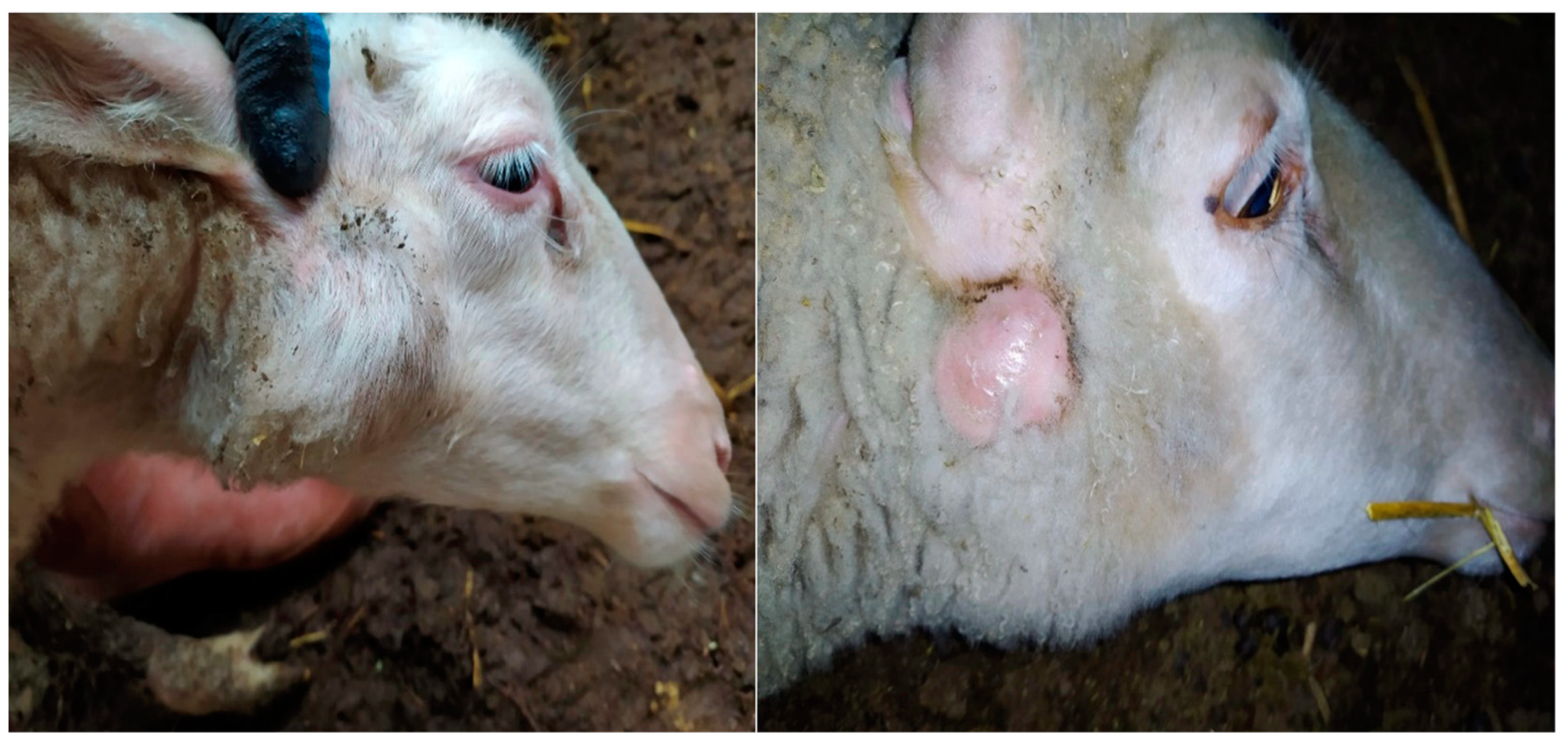
2.1. Acquisition of Samples

The study material comprised samples of abscess content from sheep diagnosed with caseous lymphadenitis in flocks raised in the Çankırı province and its vicinity in 2023. The samples were taken with the aid of sterile syringes from abscesses localized to the neck region of 82 animals, from 5 different flocks composed of a total of 500 sheep. The samples were stored at −80 °C until being used (maximum two weeks) for bacterial culture and molecular analyses.
All analyses of the biological samples at risk of contamination were carried out in BSL2 Plus laboratories equipped with the biosafety cabinets belonging to the Department of Microbiology, Faculty of Veterinary Medicine, Kafkas University.

2.2. Bacterial Culture

The abscess samples were inoculated onto 5% defibrinated sheep blood agar and incubated in an aerobic environment at 37 °C for 48 h. The bacteria, which formed the colonies that grew on the media, were assessed for their Gram-staining characteristics and microscopic morphology. The initial identification of Corynebacterium spp. was performed using phenotypic methods (colony morphology, hemolytic activity, and microscopic morphology) [13,14]. The other culture-positive isolates were identified by 16S rRNA gene sequence analysis [15].

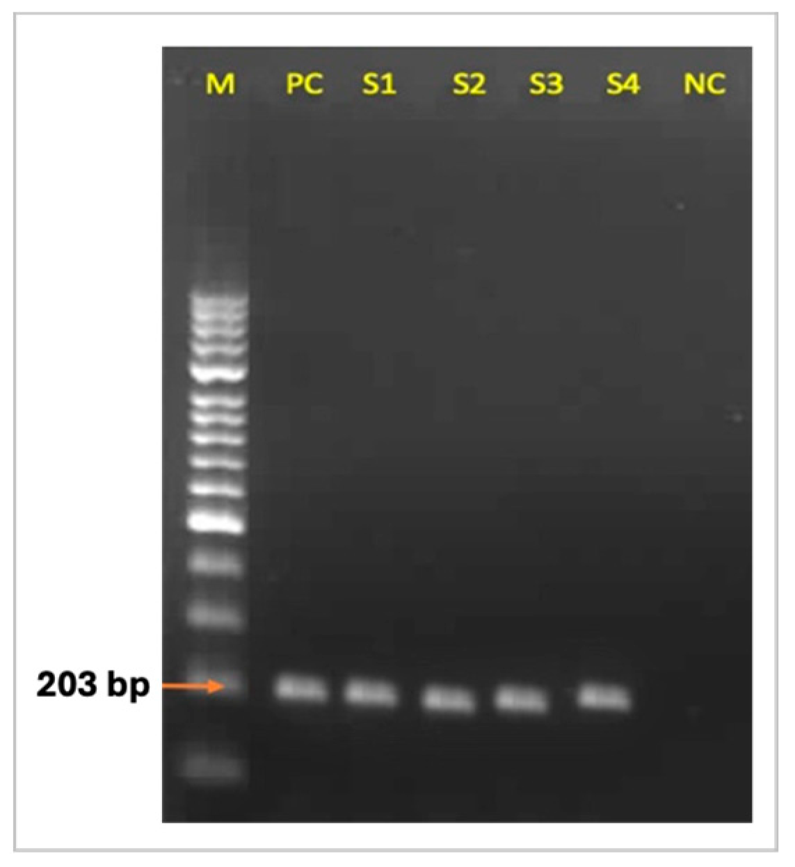
2.3. C. Pseudotuberculosis-Specific PCR

DNA extraction from pure bacterial colonies was performed using a commercial kit in accordance with the manufacturer’s instructions (DNeasy Blood & Tissue Kit®, Hilden, Germany). For the identification of C. pseudotuberculosis by PCR, primers PLDF, PLDR1, and PLDR2, targeting the pld gene, were used. The positive control strain was obtained from the Microbiology Department of Kafkas University, Faculty of Veterinary Medicine. DNA/RNA-free water was used as the negative control. The primers, PCR reaction mixture, and PCR thermal cycle are shown in Table 1. The PCR products were run on a 1.5% agarose gel, imaged with an UV trans-illuminator, and documented, and the 203 bp-bands were considered to be C. pseudotuberculosis [13].

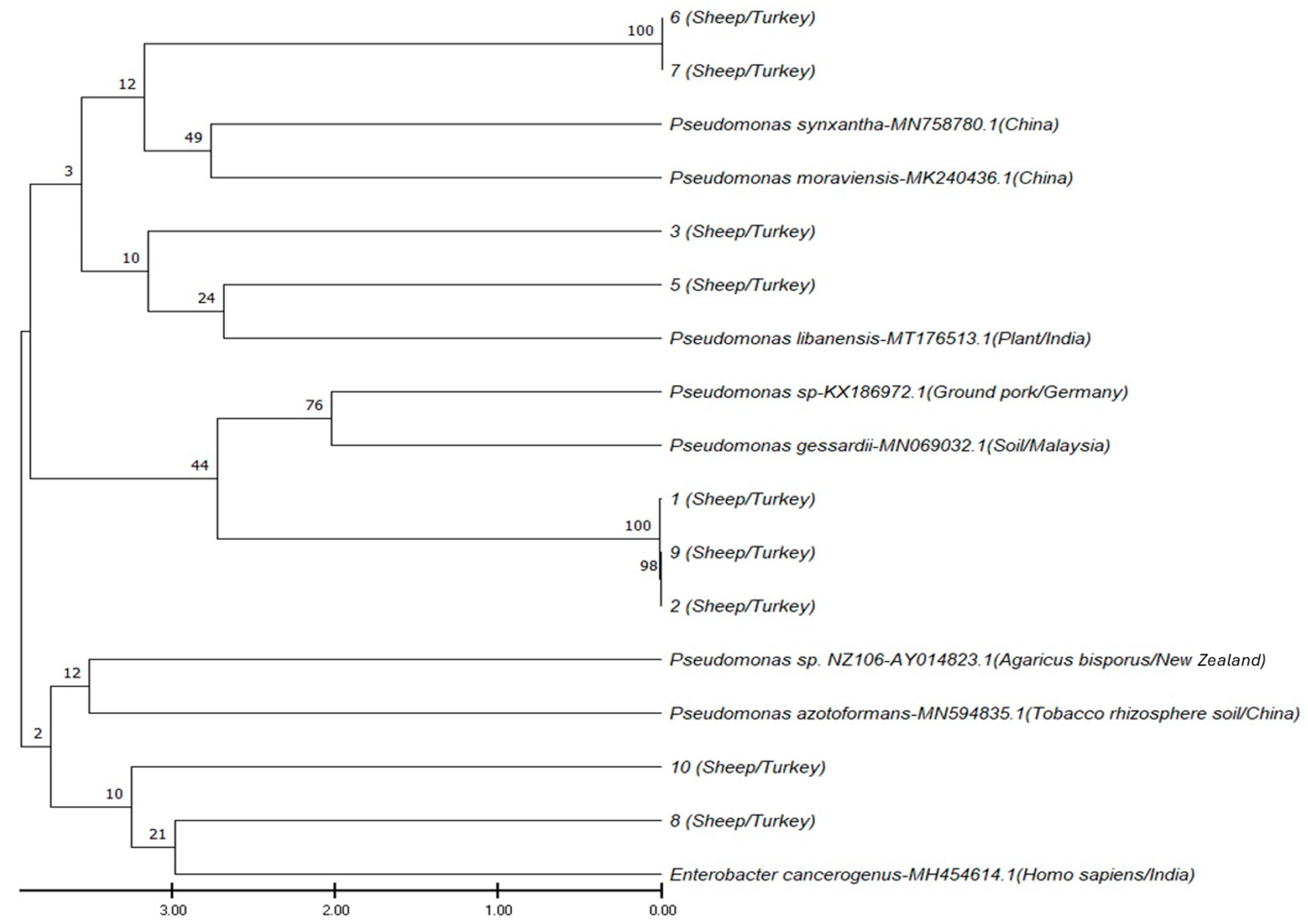
2.4. 16S rRNA Gene Sequencing and Phylogenetic Analysis

DNA extraction from pure bacterial colonies was performed using a commercial kit in accordance with the manufacturer’s instructions (DNeasy Blood & Tissue Kit®, Hilden, Germany). The Gram-negative bacteria were subjected to 16S rRNA-PCR and then 16S rRNA gene sequence analysis. For this purpose, the 27F and 1492R primers were used in the 16S rRNA-PCR amplification and sequencing. The primers, PCR reaction mixture, and PCR thermal cycle are shown in Table 1. The PCR products were run on a 1.5% agarose gel by electrophoresis, and the 1465 bp-products of the 16S rRNA gene were assessed. The PCR products were sequenced using the 27F and 1492R primers. For phylogenetic analysis, the resulting chromatograms were compared to the reference sequences available in the BLAST (Basic Local Alignment Search Tool) database of the National Center for Biotechnology Information (NCBI). The base identification of the chromatograms was performed by visual inspection using Finch TV (version V1.4). The evolutional analyses were performed using MEGA 11 software [16].

2.5. Antibiotic Sensitivity Test

The antibiotic sensitivity tests of the identified bacterial isolates were performed with the Kirby–Bauer disc diffusion test. For this purpose, firstly, fresh cultures of the bacteria were prepared on the appropriate solid growth media. Bacterial inocula adjusted to McFarland 0.5 were prepared with sterile physiological saline. Volumes of 100 µL of the bacterial inocula were streaked onto Mueller Hinton agar with a sterile loop. After the inoculum was absorbed, antibiotic discs containing 30 µg of cephalothin (KF), 30 µg of ceftiofur (EFT), 5 µg of cloxacillin (OBS), 10 µg of streptomycin (S), 75 µg of cefoperazone (CFP), 25 µg of amoxicillin (AML), 109 µg of lincomycin (LS), 40 µg of penicillin (PNV), and 10 units of bacitracin (BCDD) were placed onto the agar. The bacterial isolates inoculated onto Mueller Hinton agar plates containing these antibiotic discs were incubated at 37 °C for 24 h. The diameters of the inhibition zones were measured and assessed according to the guidelines of the Clinical and Laboratory Standards Institute [17].

3. Results

In this study, from five flocks composed of a total of 500 sheep, 82 animals (16.4%) with superficial neck abscesses were sampled for abscess content (Figure 1). It was determined that all of the sampled animals were female and breeder ewes.

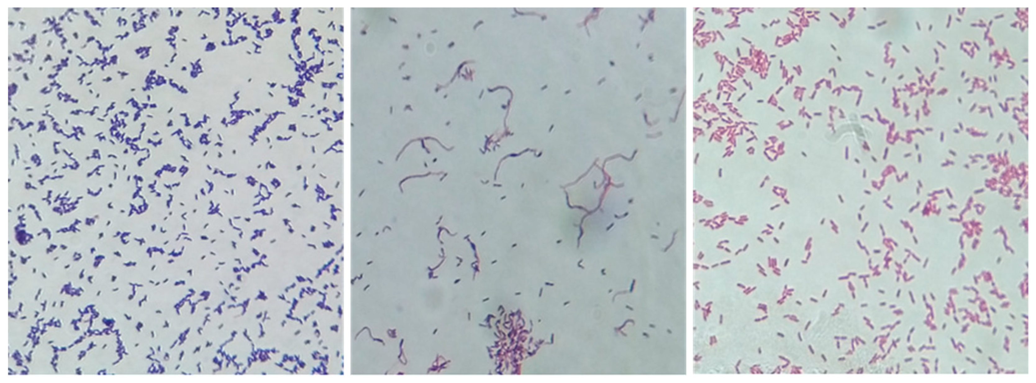
3.1. Bacterial Culture Findings

Out of the 82 samples, 39 (47.56%) were culture-positive. Of the derived isolates, 28 were Gram-positive bacilli (rods), 7 were Gram-negative bacilli and coccobacilli, and 2 displayed a mixed morphology (Figure 2). After the mixed isolates were purified, two were determined to be Gram-positive bacilli. The other two isolates were determined to be Gram-negative bacilli. Thus, 30 isolates were considered as Corynebacterium spp., based on their initial phenotypical characteristics.

3.2. C. Pseudotuberculosis-Specific PCR Findings

The species-specific PCR analysis yielded a 203 bp-band for each of the 30 isolates identified as Corynebacterium spp. by phenotypical tests, and thus, all of these isolates were confirmed to be C. pseudotuberculosis (Figure 3).

3.3. 16S rRNA Gene Sequencing and Phylogenetic Analysis Findings

As a result of the sequence analysis of the 16S rRNA-PCR products with 1465 bp, the Gram-negative isolates were identified as 8 Pseudomonas spp. and 1 Enterobacter cancerogenus (OR512110). The Pseudomonas isolates were identified as Pseudomonas helleri (OR512107), Pseudomonas synxantha (OR512109), Pseudomonas moraviensis (OR512111), Pseudomonas libanensis (OR512104), Pseudomonas fluorescens (OR512105), Pseudomonas fluorescens (OR512106), Pseudomonas azotoformans (OR512112), and Pseudomonas gessardii (OR512108) (Table 2).
The isolates obtained in the present study showed a highly heterogeneous distribution under two main clusters. The first cluster of the dendrogram included the isolates numbered 1, 2, 3, 5, 6, 7, and 9, which exhibited a paraphyletic distribution. Isolates 6 and 7 showed 100% similarity to each other, and likewise, isolates 1, 2, and 9 also showed 100% similarity among themselves and, thus, were characterized by a monophyletic distribution. Isolates 6 and 7 and isolates 1, 2, and 9 belonged to distant yet identical branches. The remaining isolates of the first cluster (isolates 3 and 5) presented with a paraphyletic distribution. The second cluster of the dendrogram included isolates 8 and 10, which also displayed a paraphyletic distribution (Figure 4).
Isolates 6 and 7, which were considered to have descended from a common ancestor, were included in the first cluster of the dendrogram. The isolates of this cluster, which showed a high level of similarity to each other (100%), were determined to belong to different taxa, based on comparison with GenBank sequences. Isolates 6 and 7 were determined to belong to taxa close to Pseudomonas synxantha strain JM20 (MN758780.1) and Pseudomonas moraviensis strain WTB8 (MK240436.1). The other three isolates of the first cluster, which showed 100% similarity to each other, namely isolates 1, 9, and 2, were determined to belong to a taxon close to Pseudomonas spp. strain TH21 (KX186972.1), which was isolated from minced pork in Germany in 2012, and Pseudomonas gessardii strain ST3SE (MN069032.1), isolated from soil in Malaysia. The paraphyletically distributed isolate 5 of the first cluster belonged to the same taxon with Pseudomonas libanensis strain DST45 (MT176513.1), which was isolated from plants in India in 2018. Isolate 3 belonged to a taxon close to that of isolate 5. As for the isolates included in the second cluster of the dendrogram, isolate 8 belonged to the same taxon as E. cancerogenus strain MB30 (MH454614.1), which was isolated from humans in India in 2018, and isolate 10 belonged to a taxon close to that of isolate 8 (Figure 4).

3.4. Antibiotic Sensitivity Test Findings

While 27 (90%) of the C. pseudotuberculosis isolates were found to be sensitive to cefoperazone and lincomycin, 3 (10%) were found to be resistant to these antibiotics. Furthermore, 18 (60%) of the C. pseudotuberculosis isolates showed sensitivity, and 12 (40%) showed resistance to penicillin. All (100%) of the C. pseudotuberculosis isolates were determined to be resistant to cephalothin, ceftiofur, cloxacillin, streptomycin, amoxicillin, and bacitracin (Table 3).
Out of the eight Pseudomanas spp. isolates, all (100%) were sensitive to cefoperazone, seven (87.5%) were sensitive to lincomycin, one (12.5%) was resistant to lincomycin, one (12.5%) was sensitive to streptomycin, and seven (87.5%) were resistant to streptomycin. Pseudomonas spp. were found to be resistant to all of the other tested antibiotics (cephalothin, ceftiofur, cloxacillin, amoxicillin, penicillin, and bacitracin) (Table 4).

4. Discussion

Caseous lymphadenitis (CLA), caused by C. pseudotuberculosis, still occurs and bears importance in regions, where sheep are raised [11]. The prevalence of CLA varies among countries. At the beginning of the nineties, an unavoidable incidence of CLA occurred in sheep and goat flocks raised in the United Kingdom, which caused great loss to the livestock sector [11,18]. While the primary causative agent involved in the etiology of CLA is known to be C. pseudotuberculosis, several reports have also indicated the involvement of secondary microorganisms. In a study carried out in Iraq, PCR results revealed C. pseudotuberculosis positivity for 22 (2.1%) out of 1090 abscesses detected in sheep and goat carcasses at a slaughterhouse [19]. In Egypt [20], researchers screened 1450 animals by molecular and cultural methods, detected CLA in 24 sheep, and reported the prevalence of C. pseudotuberculosis as 1.65%. In the same study, 10 of the ovine CLA cases were determined to have been caused by C. pseudotuberculosis alone, whilst in the other CLA cases, Staphylococcus aureus and Streptococcus pyogenes were isolated and identified together with C. pseudotuberculosis by bacterial culture. In a study on the prevalence of CLA in Kosovo, C. pseudotuberculosis was identified from 38 (84.2%) out of 284 ovine abscess samples that underwent cultural analysis and biochemical testing. In this study, the prevalence of CLA was determined as 11.3%, and the most prevalent agents after C. pseudotuberculosis were determined as Staphylococcus aureus (4/38, 10.5%) and Streptococcus pyogenes (2/38, 5.3%) [21]. In the present study, a total of 500 sheep were screened, and 82 were determined to have mandibular abscesses and were found to be positive for CLA. Our findings, indicating an overall CLA prevalence of 16.4%, are similar to those reported by Mazreku et al. [21] but differ from those reported by Ali et al. [20] and point out to a CLA positivity higher than those reported by these researchers. The results of the present study support the proposition of Baird and Fountain [11] that CLA prevalence varies among countries and regions and continues to be a major health problem for sheep and goats.
Previous studies carried out in sheep and goats have demonstrated that the causative agents of ovine and caprine CLA are not limited to C. pseudotuberculasis and S. aureus subsp. anaerobius and may include several other bacteria, namely P. aeruginosa, P. hydrophila, Treuperella pyogenes, Staphylococcus lentus, Actinomyces hyovaginalis, Streptococcus ovis, Staphylococcus simulans, Staphylococcus equorum, Staphylococcus caprae, and Staphylococcus warneri [7,22]. In a study conducted by Tiwari et al. [23] in India, the bacteria isolated and identified from 255 skin samples of various animal species, including cattle, buffaloes, dogs, goats, sheep, camels, and horses, among others, were Staphylococcus aureus (36.22%), E. coli (34.59%), Pseudomonas spp. (20.54%), Bacillus spp. (16.21%), Klebsiella spp. (12.43%), Micrococcus spp. (8.11%), Streptococcus pyogenes (7.56%), Proteus spp. (6.49%), Clostridium spp. (3.78%), a Gram-positive asporogenic rod (2.16%), and Fusobacterium spp. (0.54%). In another study conducted in Saudi Arabia, it was reported that bacteria were isolated from 97 (80.83%) out of 120 ovine abscess samples, and that the remaining 23 samples were sterile. In the same study, identification by the API-20 method revealed that the bacteria in 52 of the samples were C. pseudotuberculosis and S. aureus subsp. anaerobius and that these species constituted 54% of the isolates. The bacteria in the remaining 45 samples were identified as pyogenic bacteria, such as S. aureus, Streptococcus spp., Pseudomonas aeruginosa, and Actinomyces pyogenes, and these were ascertained to constitute approximately 46% of all the isolates. Based on these results, the low level of protection attained with the use of CLA vaccines was attributed to the abscesses having been caused by different bacterial species [5].
Although CLA has been shown to be contracted by the intranasal, intravenous, and intravaginal routes, the primary route of infection has been reported as the contamination of skin wounds. Such wounds may result from fights among animals, grooming, ear-tagging or the brushing of skin against dirty surfaces [24,25]. P. aeruginosa is an opportunistic environmental pathogen commonly found in soil, water, and barn flooring and may readily enter the body through skin lesions, eventually resulting in skin, ear, mammary gland, and lung infections in sheep [26]. Although known to be less virulent than P. aeruginosa, other Pseudomonas species, including P. fluorescens, P. luteola, P. putida, and P. stutzeri have been reported to be involved in various infections in immunocompromised hosts [27]. On the other hand, Enterobacter cancerogenus is a facultative aerobic bacterium belonging to the family Enterobacreiaceae and is commonly found in water, sewage, soil, and plants. Described as being an opportunistic pathogen, E. cancerogenus has been reported to may cause pneumonia, bacteremia, post-neurosurgical meningitis, skin and soft tissue infections, and urinary infections in both humans and animals [28,29]. In the present study, while most of the ovine CLA cases (36.59%) were determined to have been caused by C. pseudotuberculosis, the bacteria identified from 9.52% and 1.19% of the cases were Pseudomonas spp. and E. cancerogenus, respectively. This supports the opinions of Umer et al. [25] and Latif et al. [24] that factors, which disrupt the skin barrier, and thereby cause predisposition to microbial contamination and enable infection with opportunistic bacteria other than the primary causative agent of CLA, C. pseudotuberculosis, such as Pseudomanas spp. and E. cancerogenus. In fact, there are literature reports pointing out the isolation of these opportunistic pathogens from cases of CLA [5,7].
It has been reported that the administration of antibacterial protocols for the treatment of animals with caseous lymphadenitis may not result in acceptable levels of antimicrobial efficiency. Due to the abscesses being surrounded by a thick capsule and the causative microorganisms showing intracellular localization, it is indicated that some antibiotics may not reach effective concentrations within the abscesses [30]. In a study conducted in Egypt, 38 C. pseudotuberculosis isolates from 80 sheep and 36 goats were determined to be highly resistant to penicillin (96.2%) and erythromycin (92.3%) but highly sensitive to ciprofloxacin (96.2%), amikacin (90.4%), and neomycin (88.5%) [31]. Furthermore, out of 59 C. pseudotuberculosis isolates from 614 goats and 154 sheep destined for slaughter in Ethiopia, 35 (59.32%) were found to be sensitive to ampicillin and clindamycin, 43 (72.88%) were determined to be sensitive to doxycycline and tetracycline (72.88%), 41 (69.49%) were ascertained to be sensitive to vancomycin and kanamycin, and 46 (77.97%) were identified as being sensitive to norfloxacin [32]. Antibiotic sensitivity tests performed by El Damaty et al. [33] on 54 (24.5%) C. pseudotuberculosis isolates from CLA cases diagnosed in 150 sheep and 90 goats in the Sharkia state of Egypt demonstrated that, while all isolates (100%) were resistant to bacitracin and florphenicol, all isolates (100%) were sensitive to norfloxacin. The same study showed that while most of the isolates were highly resistant to penicillin and erythromycin (92.6% for each) as well as to cefradine (88.9%), 7.4% of the C. pseudotuberculosis isolates were resistant to vancomycin. Furthermore, out of 16 C. pseudotuberculosis isolates derived from 67 sheep raised in the Balıkesir province of Türkiye, sensitivity was determined in 14 (87.5%) to enrofloxacin, 13 (81.2%) to neomycin, bacitracin, and tetracycline, 12 (75.0%) to penicillin and novobiocin, 11 (68.7%) to oxytetracycline, 11 (68.7%) to amoxicillin and clavulanic acid, 10 (62.5%) to cloxacillin, 9 (56.2%) to tetracycline, 5 (31.2%) to ampicillin and sulbactam, and 3 (18.7%) to trimethoprim and sulfamethoxazole. In the present study, C. pseudotuberculosis isolates were tested for sensitivity to ten different antibiotics, such that 27 (90%) were found to be sensitive to cefoperazone and lincomycin, and 18 (60%) were determined to be sensitive to penicillin. All of the isolates were found to be resistant to cephalothin, ceftiofur, cloxacillin, streptomycin, amoxicillin, and bacitracin. The differences in the findings obtained in the present study and in previous research was attributed to the different study regions and the different antibiotics used for treatment, as suggested by Ilhan [34].
16S rRNA gene sequencing is described as a rather powerful tool for the identification of bacteria isolated from various sources [15]. While the use of physiological and biochemical characteristics has been proven to be of use for the classification of the genus Pseudomonas, it has been reported to fail in differentiating between Pseudomonas species. Nonetheless, 16S rRNA gene sequencing has also been reported to be an effective method for the identification of Pseudomonas-like microorganisms [35]. In the present study, excluding C. pseudotuberculosis, all of the other bacterial isolates were identified at the species level by means of 16S rRNA gene sequencing.
16S rRNA gene sequencing is frequently used in epidemiological and phylogenetic analyses [15]. It can reveal possible common ancestor epidemics and strain similarities from different geographies due to the border between countries. However, since there is no uniformity for the division of Pseudomonas isolates into various clusters after 16S rRNA sequencing, bacterial isolates obtained from different locations generally do not fully overlap each other and are take place in different taxa. In the present study, in addition to the widespread heterogeneity among the Pseudomonas isolates and the clues that some isolates came from a common ancestor (6 and 7 or 1, 2, and 9), it was determined that the isolates were phylogenetically related to isolates from Germany, Malaysia, and India, which are geographically distant from the country under study. The sequence data of the isolates obtained from CLA in our country are quite limited, and until the increase in sequence data and the creation and reinterpretation of new taxa, it is open to discussion that they are in the same taxon with isolates from countries that are not geographically close to this study’s country, unless there is a possible international animal movement.
The genus Pseudomonas includes more than 200 species, which are Gram-negative bacteria commonly found in animals, soil, and plants [36]. Pseudomonas spp. belong to the subclass of Gammaproteobacteria and show various life cycles in a wide environmental array from soil, water, and plant surfaces to animals. These bacteria are known for their prevalence in nature, the capability of using various organic compounds as energy sources, resistance to many medicinal and agricultural antimicrobial substances, and the ability to generate high levels of secondary metabolites [37]. Among all antibiotic-resistant pathogens, P. aeruginosa is especially highlighted for its continuously increasing resistance to antimicrobial agents, resulting in treatment complications and failures [38]. Several literature reports have been published on the occurrence of multiple antibiotic resistance in P. aeruginosa isolates from ovine CLA cases and other clinical cases of infection [39,40,41]; yet, there are only limited data available on the AST profiles of other Pseudomonas species and environmental isolates [42]. In a study carried out in Egypt on a total of 260 sheep and goats with respiratory infection, diarrhea, and abscessed skin lesions, 13 isolates were identified as P. aeruginosa by biochemical tests, and each of these isolates was found to be resistant to ampicillin, bacitracin, erythromycin, streptomycin, tetracycline, trimethoprim–sulfamethoxazole, and tobramycin and sensitive to ceftriaxone and norfloxacin. While all of the nine Pseudomonas species (100%) identified in the present study were found to be sensitive to cefoperazone, eight (89%) were determined to be sensitive to lincomycin, and one (11%) was ascertained to be sensitive to streptomycin. On the other hand, all of the Pseudomonas species identified in the present study were found to be resistant to cephalothin, ceftiofur, cloxacillin, cefoperazone, penicillin, and bacitracin. These results are attributed to the development of multiple antibiotic resistance in Pseudomonas species due to the nonselective use of antibiotics, as suggested by Dapgh et al. [39].

5. Conclusions

In conclusion, similar to the case in other countries, ovine CLA remains important in terms of animal health in Türkiye. Although the primary causative agent of CLA is known to be C. pseudotuberculosis, the possible involvement of other opportunistic bacteria (Pseudomonas spp., E. concerogenus) in the etiology of CLA should not be overlooked. We highlight the importance of considering the potential involvement of these secondary agents in CLA for achieving success in protection from the disease, as well as in the control and treatment of CLA. Furthermore, phylogenetic clustering of the strains can contribute to increasing country-specific sequence data and creating new taxa from a universal perspective.

Author Contributions

All authors contributed to the study conception and design. The first draft of the manuscript was written by S.T., and all authors commented on previous versions of the manuscript. S.T. Writing—Original Draft, Writing—Review and Editing, Project administration; S.T.: Writing—Review and Editing, Conceptualization, Project administration, Methodology; F.B.: Writing—Review and Editing, Visualization, Methodology; FB: Writing—Review and Editing, Visualization, Supervision. All authors have read and agreed to the published version of the manuscript.

Funding

This research received no external funding.

Institutional Review Board Statement

This study was conducted pursuant to the 2022/121 numbered approval of the Local Ethics Committee for Animal Experiments (HADYEK) of Kafkas University in May 2022 and the E-90434910-325.99(325.99)-5828556 numbered written permission of the Provincial Agriculture and Forestry Directorate of the Çankırı province, Türkiye.

Informed Consent Statement

Not applicable.

Data Availability Statement

Data are contained within the article.

Conflicts of Interest

The authors declare no conflicts of interest.

References

  1. Günaydın, G. Koyun yetiştiriciliğinin ekonomi politiği. Uludag Üniv. Ziraat Fak. Derg. 2009, 23, 15–32. Available online: https://dergipark.org.tr/tr/download/article-file/154093 (accessed on 7 July 2024). (In Turkish).
  2. De Pinho, R.B.; De Oliveira Silva, M.T.; Bezerra, F.S.B.; Borsuk, S. Vaccines for caseous lymphadenitis: Up-to-date and forward-looking strategies. Appl. Microbiol. Biotechnol. 2021, 105, 2287–2296. [Google Scholar] [CrossRef]
  3. Khanamir, R.A.; Issa, N.A.; Abdulrahman, R.F. First study on molecular epidemiology of caseous lymphadenitis in slaughtered sheep and goats in Duhok Province, Iraq. Open Vet. J. 2023, 13, 588–598. [Google Scholar] [CrossRef]
  4. Zidan, K.H.; Mazloum, K.; Saran, M.A.; Hatem, M.E. Abscesses in dromedary camels, sheep and goats etiology and pathology. In Proceedings of the 1st International Scientific of Pathology Conference, Faculty of Veterinary Medicine-Cairo University, Sheraton Dreamland Hotel Conference Center, Giza, Egypt, 25–27 April 2013; Available online: https://www.researchgate.net/publication/272090416_ABSCESSES_IN_DROMEDARY_CAMELS_SHEEP_AND_GOATS_ETIOLOGY_AND_PATHOLOGY (accessed on 7 July 2024).
  5. Al-Harbi, K.B. Prevalence and etiology of abscess disease of sheep and goats at Qassim region, Saudi Arabia. Vet. World 2011, 4, 495–499. [Google Scholar] [CrossRef]
  6. Mohamed, M.M.; Fouad, S.A.; Elshoky, H.A.; Mohammed, G.M.; Salaheldin, T.A. Antibacterial effect of gold nanoparticles against Corynebacterium pseudotuberculosis. Int. J. Vet. Sci. Med. 2017, 5, 23–29. [Google Scholar] [CrossRef]
  7. De La Fuente, R.; De Las Heras, M.; Torrijos, C.; De Tejada, P.D.; Pérez-Sancho, M.; Carrión, F.J.; Dominguez-Bernal, G. Isolation frequency of bacteria causing lymphadenitis and abscesses in small ruminants in central Spain. Small Rumin. Res. 2017, 154, 5–8. [Google Scholar] [CrossRef]
  8. Batey, R.G. Pathogenesis of caseous lymphadenitis in sheep and goats. Aust. Vet. J. 1986, 63, 269–272. [Google Scholar] [CrossRef]
  9. Osman, A.Y.; Nordin, M.L.; Kadir, A.A.; Saharee, A.A. The epidemiology and pathophysiology of caseous lymphadenitis: A review. J. Vet. Med. Res. 2018, 5, 1129. [Google Scholar] [CrossRef]
  10. Pepin, M.; Cannella, D.; Fontaine, J.J.; Pittet, J.C.; Le Pape, A. Ovine mononuclear phagocytes in situ: Identification by monoclonal antibodies and involvement in experimental pyogranulomas. J. Leukoc. Biol. 1992, 51, 188–198. [Google Scholar] [CrossRef]
  11. Baird, G.J.; Fontaine, M.C. Corynebacterium pseudotuberculosis and its role in ovine caseous lymphadenitis. J. Comp. Pathol. 2007, 137, 179–210. [Google Scholar] [CrossRef]
  12. Torky, H.A.; Saad, H.M.; Khaliel, S.A.; Kassih, A.T.; Sabatier, J.M.; Batiha, G.E.S.; Hetta, H.F.; Elghazaly, E.M.; De Waard, M. Isolation and molecular characterization of Corynebacterium pseudotuberculosis: Association with proinflammatory cytokines in caseous lymphadenitis pyogranulomas. Animals 2023, 13, 296. [Google Scholar] [CrossRef]
  13. Ilhan, Z. Detection of Corynebacterium pseudotuberculosis from sheep lymph nodes by PCR. Rev. Med. Vet. 2013, 164, 60–66. [Google Scholar]
  14. Sakmanoğlu, A.; Hadimli, H.H.; Erganiş, O.; Pınarkara, Y.; Sayın, Z.; Kav, K. Identification and antimicrobial susceptibility of Corynebacterium pseudotuberculosis isolated from sheep. Eurasian J. Vet. Sci. 2015, 31, 116–121. [Google Scholar] [CrossRef]
  15. Janda, J.M.; Abbott, S.L. 16S rRNA gene sequencing for bacterial identification in the diagnostic laboratory: Pluses, perils, and pitfalls. J. Clin. Microbiol. 2007, 45, 2761–2764. [Google Scholar] [CrossRef]
  16. Akça, D.; Büyük, F.; Karakaya, E.; Coşkun, M.R.; Çelik, E.; Şahin, M.; Çelebi, Ö.; Otlu, S.; Gülmez Sağlam, A.; Eray, B.; et al. Molecular characterization of Bacillus anthracis isolates recovered from nomic andnonnomic hosts. Turk. J. Vet. Anim. Sci. 2022, 46, 44–51. [Google Scholar] [CrossRef]
  17. Wayne, P.A. Clinical and laboratory standards institute. Performance standards for antimicrobial susceptibility testing. Inform. Suppl. 2011, 31, 100–121. Available online: https://www.sid.ir/paper/655659/en (accessed on 19 July 2024).
  18. Connor, K.M.; Quirie, M.M.; Baird, G.; Donachie, W. Characterization of United Kingdom isolates of Corynebacterium pseudotuberculosis using pulsed-field gel electrophoresis. J. Clin. Microbiol. 2000, 38, 2633–2637. [Google Scholar] [CrossRef]
  19. Abdulrahman, R.F. Virulence potential, antimicrobial susceptibility and phylogenetic analysis of corynebacterium pseudotuberculosis isolated from caseous lymphadenitis in sheep and goats in duhok city, Iraq. Adv. Anim. Vet. Sci. 2021, 9, 919–925. [Google Scholar] [CrossRef]
  20. Ali, A.D.; Mahmoud, A.A.; Khadr, A.M.; Elshemey, T.M.; Abdelrahman, A.H. Corynebacterium pseudotuberculosis: Disease prevalence, lesion distribution, and diagnostic comparison through microbiological culture and molecular diagnosis. Alex. J. Vet. Sci. 2016, 51, 189–198. [Google Scholar] [CrossRef]
  21. Mazreku, N.; Sylejmani, D.; Hamidi, A.; Robaj, A. The effect of vitamin h in aseptic laminitis prevention and its impact on blood indicators in dairy cows. Bulg. J. Agric. Sci. 2017, 23, 315–318. [Google Scholar]
  22. Saleh, W.M.M.; Lafta, M.H.; Abdulrazaq, A.W.; Habib, H.N.; Naeem, L.A. Bacteriological and histopathological evaluation of infectious lymphadenitis caused by Pseudomonas aeruginosa in awasi sheep. Adv. Anim. Vet. Sci. 2019, 7, 378–382. [Google Scholar] [CrossRef]
  23. Ruchi Tiwari, R.; Yadav, S.K.; Singh, S.; Gangwar, N.K. Bacterial etiology of skin and wound infections along with antibiogram profiling in reference to emerging antibiotic resistance in animals. Adv. Anim. Vet. Sci. 2015, 3, 259–268. [Google Scholar] [CrossRef]
  24. Latif, N.A.A.; Abdullah, F.F.J.; Othman, A.M.; Rina, A.; Chung, E.L.T.; Zamri-Saad, M.; Saharee, A.A.; Haron, A.W.; Lila, M.A.M. Isolation and detection of Corynebacterium pseudotuberculosis in the reproductive organs and associated lymph nodes of non-pregnant does experimentally inoculated through intradermal route in chronic form. Vet. World 2015, 8, 924–927. [Google Scholar] [CrossRef]
  25. Umer, M.; Abba, Y.; Abdullah, F.F.J.; Saleh, W.M.M.; Haron, A.W.; Saharee, A.A.; Ariff, A.B.; Baiee, F.H.A.; Hambali, I.U.; Sharif, A. Caseous lymphadenitis in small ruminants: An overview on reproductive implications. Int. J. Vet. Sci. Anim. Husb. 2017, 2, 23–31. [Google Scholar]
  26. Benito-Peña, A.; Peris, B.; Aduriz, G.; Martinez, J.; Corpa, J.M. Purulent nasomaxillary and mandibular osteomyelitis in sheep caused by Pseudomonas aeruginosa. Vet. Rec. 2010, 166, 115–116. [Google Scholar] [CrossRef]
  27. D’Agata, E.; Araos, R. Pseudomonas aeruginosa and other Pseudomonas species. In Mandell, Douglas, and Bennett’s Principles and Practice of Infectious Diseases, 9th ed.; Bennett, J.E., Dolin, R., Blaser, M.J., Eds.; Elseiver: Saunders, WB, USA, 2019; Volume 2, pp. 2686–2699. [Google Scholar]
  28. Abo-Amer, A.E.; Shobrak, M.Y. Antibiotic resistance and molecular characterization of Enterobacter cancerogenus isolated from wild birds in Taif province, Saudi Arabia. Thai J. Vet. Med. 2015, 45, 101–111. [Google Scholar] [CrossRef]
  29. Salimiyan Rizi, K.; Ghazvini, K.; Farsiani, H. Clinical and pathogenesis overview of Enterobacter infections. Rev. Clin. Med. 2020, 6, 146–154. [Google Scholar] [CrossRef]
  30. Washburn, K.E.; Bissett, W.T.; Fajt, V.R.; Libal, M.C.; Fosgate, G.T.; Miga, J.A.; Rockey, K.M. Comparison of three treatment regimens for sheep and goats with caseous lymphadenitis. J. Am. Vet. Med. Assoc. 2009, 234, 1162–1166. [Google Scholar] [CrossRef]
  31. Algammal, A. Molecular characterization and antibiotic susceptibility of Corynebacterium pseudotuberculosis isolated from sheep and goats suffering from caseous lymphadenitis. Zagazig Vet. J. 2016, 44, 1–8. [Google Scholar] [CrossRef]
  32. Abebe, D.; Sisay Tessema, T. Determination of Corynebacterium pseudotuberculosis prevalence and antimicrobial susceptibility pattern of isolates from lymph nodes of sheep and goats at an organic export abattoir, Modjo, Ethiopia. Lett. Appl. Microbiol. 2015, 61, 469–476. [Google Scholar] [CrossRef]
  33. El Damaty, H.M.; El-Demerdash, A.S.; Abd El-Aziz, N.K.; Yousef, S.G.; Hefny, A.A.; Abo Remela, E.M.; Shaker, A.; Elsohaby, I. Molecular characterization and antimicrobial susceptibilities of Corynebacterium pseudotuberculosis isolated from caseous lymphadenitis of smallholder sheep and goats. Animals 2023, 13, 2337. [Google Scholar] [CrossRef] [PubMed]
  34. İlhan, Z. In vitro antimicrobial susceptibility of Corynebacterium pseudotuberculosis isolated from sheep with caseous lymphadenitis. Kocatepe Vet. J. 2020, 13, 267–271. [Google Scholar] [CrossRef]
  35. Hussein, Z.T.; Mukhlif, S.S.; Taha, M.; Ali, S.A. Identification and characterization of Pseudomonas aeruginosa 16S rRNA gene isolated from contaminated soil with oil residues. In IOP Conference Series: Earth and Environmental Science, Proceedings of the Ninth National Conference on the Environment and Natural Resources (NCENR-2023), Basrah, Iraq, 8–9 Agust 2023; IOP Publishing: Bristol, UK, 2023; Volume 1215, p. 012001. [Google Scholar] [CrossRef]
  36. Lupo, A.; Haenni, M.; Madec, J.Y. Antimicrobial resistance in Acinetobacter spp. and Pseudomonas spp. Microbiol. Spectr. 2018, 6. [Google Scholar] [CrossRef]
  37. Yang, J.; Zhao, H.L.; Ran, L.Y.; Li, C.Y.; Zhang, X.Y.; Su, H.N.; Shi, M.; Zhou, B.C.; Chen, X.L.; Zhang, Y.Z. Mechanistic insights into elastin degradation by pseudolysin, the major virulence factor of the opportunistic pathogen Pseudomonas aeruginosa. Sci. Rep. 2015, 5, 9936. [Google Scholar] [CrossRef] [PubMed]
  38. Cabassi, C.S.; Sala, A.; Santospirito, D.; Alborali, G.L.; Carretto, E.; Ghibaudo, G.; Taddei, S. Activity of AMP2041 against human and animal multidrug resistant Pseudomonas aeruginosa clinical isolates. Ann. Clin. Microbiol. Antimicrob. 2017, 16, 17. [Google Scholar] [CrossRef]
  39. Dapgh, A.N.; Hakim, A.S.; Abouelhag, H.A.; Abdou, A.M.; Elgabry, E.A. Detection of virulence and multidrug resistance operons in Pseudomonas aeruginosa isolated from Egyptian Baladi sheep and goat. Vet. World 2019, 12, 1524–1528. [Google Scholar] [CrossRef]
  40. Mahmood, A.N.; Aljobori, A.H. Isolation and identification of Pseudomonas aeruginosa from infected sheep and detection of phosolipase C (lecithinase). Iraqi J. Vet. Medicine 2015, 39, 28–32. [Google Scholar] [CrossRef]
  41. Saha, T.K.; Begum, F.; Kabir, S.L.; Islam, M.S.; Khan, M.S.R. Characterization of bacterial isolates from skin lesions of sheep, goat and cattle in different rearing condition. Asian J. Med. Biol. Res. 2019, 5, 117–125. [Google Scholar] [CrossRef]
  42. Vaz-Moreira, I.; Nunes, O.C.; Manaia, C.M. Diversity and antibiotic resistance in Pseudomonas spp. from drinking water. Sci. Total Environ. 2012, 426, 366–374. [Google Scholar] [CrossRef]
Figure 1. The sampled abscesses located in the neck region of the sheep.
Life 14 01078 g001
Figure 2. C. pseudotuberculosis and the microscopic appearance of the mixed culture and other Gram-negative isolates (magnification 1000×).
Life 14 01078 g002
Figure 3. The electrophoresis of the PCR products specific to the pld gene. M: DNA ladder (Gene ruler 100 bp plus DNA ladder SM0321, Thermo Fischer Sci., Waltham, MA, USA). PC: positive control. S1, S2, S2, S3, S4: C. pseudotuberculosis field isolates. NC: negative control.
Life 14 01078 g003
Figure 4. Neighbor-joining phylogenetic tree constructed with 16S rRNA gene sequences showing the phylogenetic position of the isolates among the related taxa. Bar, 0.005 substitutions per nucleotide position.
Life 14 01078 g004
Table 1. Species-specific and 16S rRNA-PCR conditions of the isolates.
SpeciesPrimer
Sequence (5′—3′)
Target GenePCR Component
(25 μL)
Thermal CycleAmplicon (bp)Reference
C. pseudotuberculosisPLDF- ATAAGCGTAAGCAGGGAGCA
PLDR1- ATCAGCGGTGATTGTCTTCC
PLDR2- ATCAGCGGTGATTGTCTTCCAGG
pld
  • 2.5 μL PCR buffer (×10)
  • 2.5 μL MgCl2 (25 mM)
  • 0.5 dNTP (10 mM)
  • 1 μL each primer (10 pmol)
  • 0.5 μL Taq DNA polymerase (5 U)
  • 2.5 μL template DNA (40–80 ng/μL)
  • 13.5/14.5 μL nuclease free water
One cycle:
  • Denaturation at 94 °C for 5 min
Thirty-five cycles:
  • Denaturation at 94 °C for 1 min
  • Annealing at 56 °C for 1 min
  • Elongation at 72 °C for 2 min
One cycle:
  • Final elongation at 72 °C for 5 min
203[13]
Unknown species specific27F- AGAGTTTGATC(AC)TGGCTCAG
1492R- ACGG(CT)TTACCTTGTTACGACTT
16S rRNAOne cycle:
  • Denaturation at 95 °C for 5 min
Thirty cycles:
  • Denaturation at 94 °C for 15 s
  • Annealing at 59 °C for 30 s
  • Elongation at 72 °C for 45 s
One cycle:
  • Final elongation at 72 °C for 5 min
1465[16]
Table 2. The Gram-negative bacteria identified by 16S rRNA gene sequencing.
Number of IsolateCultural and Phenotypic CharacteristicsSpecies NameNCBI Accession Number
1Gram-negative, bacilliP. helleriOR512107
2Gram-negative, bacilliP. synxanthaOR512109
3Gram-negative, bacilliP. moraviensisOR512111
5Gram-negative, bacilliP. libanensisOR512104
6Gram-negative, bacilliP. fluorescensOR512105
7Gram-negative, bacilliP. fluorescensOR512106
8Gram-negative, bacilliP. azotoformansOR512112
9Gram-negative, bacilliP. gessardiiOR512108
10Gram-negative, cocobacilliE. cancerogenusOR512110
Table 3. Antibiotic sensitivity test results of C. pseudotuberculosis.
Antibiotic (Concentration)SusceptibleIntermediateResistant
KF (30 µg)0 (0%)0 (0%)30 (100%)
EFT (30 µg)0 (0%)0 (0%)30 (100%)
OBS (5 µg)0 (0%)0 (0%)30 (100%)
CFP (75 µg)27 (90%)0 (0%)3 (10%)
AML (25 µg)0 (0%)0 (0%)30 (100%)
LS (109 µg)27 (90%)0 (0%)3 (10%)
PNV (40 µg)18 (60%)0 (0%)12 (40%)
S (10 U)0 (0%)0 (0%)30 (100%)
BCDD (10 U)0 (0%)0 (0%)30 (100%)
KF: cephalothin, EFT: ceftiofur, OBS: cloxacillin, S: streptomycin, CFP: cefoperazone, AML: amoxicillin, LS: lincomycin, PNV: penicillin, BCDD: bacitracin.
Table 4. Antibiotic sensitivity test results of Pseudomonas spp.
Antibiotic (Concentration)SusceptibleIntermediateResistant
KF (30 µg)0 (0%)0 (0%)8 (100%)
EFT (30 µg)0 (100%)0 (0%)8 (100%)
OBS (5 µg)0 (0%)0 (0%)8 (100%)
CFP (75 µg)8 (100%)0 (0%)0 (0%)
AML (25 µg)0 (100%)1 (11%)8 (89%)
LS (109 µg)7 (87.5%)0 (0%)1 (12.5%)
PNV (40 µg)0 (0%)0 (0%)8 (0%)
S (10 U)1 (12.5%)4 (50%)3 (37.5%)
BCDD (10 U)0 (0%)0 (0%)8 (100%)
KF: cephalothin, EFT: ceftiofur, OBS: cloxacillin, S: streptomycin, CFP: cefoperazone, AML: amoxicillin, LS: lincomycin, PNV: penicillin, BCDD: bacitracin.
Disclaimer/Publisher’s Note: The statements, opinions and data contained in all publications are solely those of the individual author(s) and contributor(s) and not of MDPI and/or the editor(s). MDPI and/or the editor(s) disclaim responsibility for any injury to people or property resulting from any ideas, methods, instructions or products referred to in the content.

