Review

Enhancing Cartilage Repair: Surgical Approaches, Orthobiologics, and the Promise of Exosomes

Linda and Mitch Hart Regenerative and Personalized Medicine, Steadman Philippon Research Institute, Vail, CO 81657, USA
*
Author to whom correspondence should be addressed.
Life 2024, 14(9), 1149; https://doi.org/10.3390/life14091149
Submission received: 28 July 2024 / Revised: 22 August 2024 / Accepted: 30 August 2024 / Published: 11 September 2024
(This article belongs to the Special Issue Research Advances in Bone and Cartilage Tissue Engineering)

Abstract

:
Treating cartilage damage is challenging as its ability for self-regeneration is limited. Left untreated, it can progress to osteoarthritis (OA), a joint disorder characterized by the deterioration of articular cartilage and other joint tissues. Surgical options, such as microfracture and cell/tissue transplantation, have shown promise as techniques to harness the body’s endogenous regenerative capabilities to promote cartilage repair. Nonetheless, these techniques have been scrutinized due to reported inconsistencies in long-term outcomes and the tendency for the defects to regenerate as fibrocartilage instead of the smooth hyaline cartilage native to joint surfaces. Orthobiologics are medical therapies that utilize biologically derived substances to augment musculoskeletal healing. These treatments are rising in popularity because of their potential to enhance surgical standards of care. More recent developments in orthobiologics have focused on the role of exosomes in articular cartilage repair. Exosomes are nano-sized extracellular vesicles containing cargo such as proteins, lipids, and nucleic acids, and are known to facilitate intercellular communication, though their regenerative potential still needs to be fully understood. This review aims to demonstrate the advancements in cartilage regeneration, highlight surgical and biological treatment options, and discuss the recent strides in understanding the precise mechanisms of action involved.

1. Introduction

Cartilage is a complex tissue characterized by its strong and flexible properties, playing a key role in protecting joints from wear and tear. During embryologic development, stem cells from the mesoderm progeny undergo chondrogenesis, forming chondrocytes that produce a high concentration of extracellular matrix (ECM) [1,2]. This highly specialized matrix consists of fibrous tissue and various combinations of proteoglycans and glycosaminoglycans. There are two main subsets of cartilage: hyaline and fibrocartilage. Hyaline cartilage is the most abundant type of cartilage in the human body and resides in specific joints such as articular cartilage (AC). Composed primarily of type II collagen, it can resist compressive forces and reduce friction [3]. By nature, AC is predominantly avascular and acellular; composed mainly of secreted ECM, this tissue is optimal for handling the mechanical stresses of the joint. However, these characteristics make repairing damaged cartilage difficult [4,5]. When AC is damaged, it has a limited ability to regenerate autonomously due to low numbers of dividing cells and inefficient recruitment of cellular repair mechanisms resulting from a lack of vascularization that limits what is possible in native repair [4]. If left untreated, cartilage damage can become widespread and lead to diseases like osteoarthritis (OA). As such, there is a collective effort in orthopedics to reconstruct or regenerate articular hyaline cartilage to treat acute injury and delay the development of osteoarthritis.
While current interventions have shown effectiveness in managing the symptoms of cartilage damage, such as pain medications, anti-inflammatory injections, and surgical procedures, they have their limitations. These methods have succeeded in improving function and halting further degeneration, but achieving complete restoration of functional AC remains challenging [6]. Instead, the formation of mechanically inferior fibrocartilage is an expected outcome, contrasting with the desired hyaline cartilage native to joints. This is where the field of regenerative medicine comes in, aiming to overcome these limitations by harnessing the body’s innate healing capabilities.
This review will delve into the most prevalent therapeutics for articular cartilage injury and highlight promising developments in regenerative medicine. It will specifically explore how orthobiologics and the emerging therapeutic potential of exosomes have advanced our current understanding of cartilage repair and its unique anatomy that is challenging to regenerate. Exosomes present a unique opportunity as a bioactive molecule to modulate chondrogenic cellular processes in cartilage damage to actively communicate with neighboring chondrogenic cells to heal in a controlled and specified manner. The following sections will cover the current research and findings in cartilage damage in orthopedic conditions and discuss the future directions in translating orthobiologics and exosome-based therapies from bench to bedside.

2. Cartilage Overview

This section will cover the structure, composition, and types of human cartilage essential for maintaining healthy joints, further elaborating on damage to cartilage and its potential to lead to osteoarthritis (OA).

2.1. Human Cartilage: An Overview

Cartilage is a strong, flexible, non-vascular connective tissue. There are three types of cartilage with unique roles throughout the body: (1) hyaline cartilage (HC)—the most abundant form, has a glassy appearance and is found in joints at the ends of bones, the trachea and parts of the skull; (2) fibrocartilage (FC)—a solid and fibrous tissue, appears opaquely white and is found predominantly in the intervertebral disks of the spine and at osteotendinous/osteoligamentous junctions; and (3) elastic cartilage (EC)—a yellowish, pliable, non-load-bearing tissue found in the epiglottis, the external ear, and the auditory tube of the middle ear [7]. Each cartilage type has a low density of cells in structured extracellular matrices, the unique architectural components of which are vital for proper cartilage growth and function [8]. Most diseases of cartilage, including OA, involve significant, detrimental changes in the organization of their cartilaginous ECM due to contributing factors such as joint injury [8,9,10].

2.2. Articular Cartilage: Lessons from Early Chondrogenesis

AC is composed of hyaline cartilage found on bone surfaces within synovial joints or diarthroses. Synovial joints are contained by fibrous joint capsules that are continuous with the periosteum of each articulating bone and filled with synovial fluid [11]. Cartilage cells (chondrocytes) found in AC experience constant shifts in the joint’s physicochemical environment, receiving various mechano-chemical signals. The ECM secreted by AC chondrocytes is a unique blend of primarily type II collagen molecules, proteoglycans, and glycoproteins [7,12]. However, like other cartilage types, AC has minimal self-restoring properties when compared with bone, and defects frequently persist without healing [12,13,14].
The inability of AC to spontaneously heal presents a significant challenge, not only to surgeons and clinicians attempting to help patients recover from cartilage injury/degradation, but also to scientists searching for a reliable approach to regenerate AC. The only example of spontaneous AC formation in vivo is during the early stages of embryonic development [15]. Despite the challenges, significant advances in our understanding of AC chondrogenesis have highlighted key genetic, cellular, and molecular contributors, some of which are targets for new or improved regenerative therapies.
The process of AC chondrogenesis begins when mesenchymal stromal/progenitor cells (MSCs) from the mesoderm start to condense at the ends of bones, eventually committing to the chondrogenic lineage [16]. These MSCs differentiate into chondroblasts and chondrocytes, which begin secreting cartilage matrix, a behavior initially shown to correlate with intracellular collagen gene expression [17,18]. During MSC aggregation and throughout the maturation of chondrocytes and the ECM, chondrogenesis is controlled by a complex orchestra of genetic, molecular, and cellular signals, including balanced stimulation from the transforming growth factor-beta (TGF-ß) and bone morphogenic protein (BMP) families (Table 1) [19]. The expression of Sox9, the master regulator of the chondrocyte lineage, can be directly induced by BMP signaling, as demonstrated in vivo using animals with null mutations in Sox9. Animals with homozygous null mutations in Sox9 did not form cartilage, and those with heterozygous mutations exhibited defects in all cartilage primordia [16].

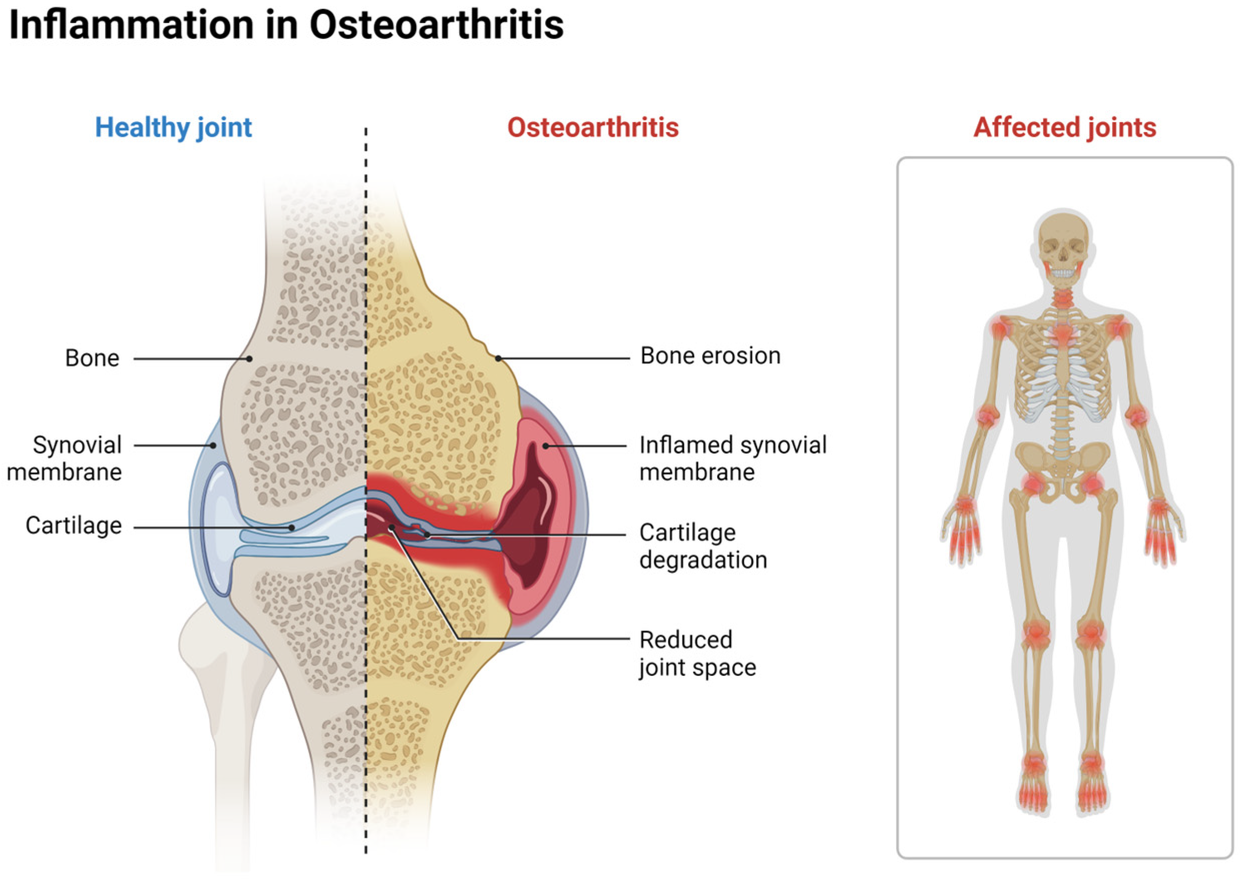
2.3. Cartilage Defects and Osteoarthritis

Focal cartilage injury is common in active patients. Typically, these injuries result in isolated damage and can be a result of high impact, impingement, fracture, surgical operation, or ligament tear. Focal injuries require treatment to reestablish quality of life and limit further systemic degradation to the joint [31]. OA correlates to previous joint trauma factors such as inflammation, joint abnormalities, avascular necrosis, and other conditions can lead to joint malformities, as well as cartilage degradation that commonly leads to the progression of OA [31].
OA is the most common joint disorder in the world and a significant burden on individuals and society. In 2019, The Lancet’s Global Burden of Disease study (GBD) reported 528 million people suffering from this condition, an increase of 113% since 1990 [32]. Caring for patients with OA is often costly to individuals, families, and healthcare systems. In 2017, total direct healthcare spending associated with OA reached USD 41.7 billion in the United States alone [33,34]. Despite high spending, there is still no known cure for OA, and many existing treatments seemingly target symptom management only. OA causes irreversible cartilage and subchondral bone degeneration, joint space narrowing, subchondral bone thickening (or sclerosis), bone spur (or osteophyte) formation, and painful loss of joint function over time. Due to the lack of a neural network in the joint space, cartilage damage can go unnoticed during the progression to OA; patients are usually asymptomatic until later stages, and diagnosis may be difficult until after irreparable joint damage has occurred [35]. Post-traumatic OA (PTOA) occurs when the onset of OA is triggered by joint trauma, which primarily affects the knee and ankle joints and accounts for about 12% of all OA cases [36,37]. Approximately 50% of all patients who suffer from traumatic joint injury (e.g., anterior cruciate ligament tears) will experience PTOA, though it may take 10–15 years to reach full development [36]. While the exact pathogenesis of OA is not understood, the current consensus suggests a complex interplay of dysfunctional cell behaviors, imbalanced metabolic homeostasis in the chondral/subchondral layers of bone ends, and significant inflammation and fibrosis in the surrounding joint (Figure 1) [38,39,40].
Current best practices for preventing and treating the cartilage degeneration seen in OA vary based on etiology and location, though generally, emphasis is placed foremost on maintaining joint health throughout life by engaging in regular exercise, avoiding joint overuse, making good nutrition choices, weight management, and early intervention when injuries occur. Few pharmacologic interventions for OA exist, however, anti-inflammatory medication and intra-articular injections are used for temporary pain relief [41]. For patients with highly active lifestyles—professional athletes, weekend warriors, or manual laborers who regularly experience intense joint strain—orthopedic and sports/occupational medicine specialists focus on optimizing joint biomechanics to restore adequate function. Non-surgical methods that cater to improving biomechanics may include targeted exercise programs or tailored physical therapy. Though some may find relief with these interventions, many seek out surgical treatment due to OA’s degrading nature. Various surgical techniques have differing levels of success, depending on the initial injury or condition and, in some cases, emerging biological approaches have been used [42]. These techniques include cartilage resurfacing, membrane-induced autologous chondrocyte implantation (MACI), microfracture, osteochondral allografting, and the osteoarticular transfer system (OATS) procedure. However, some surgical and regenerative techniques used to stimulate the biological process of cartilage formation have come under scrutiny due to mixed results from long-term follow-ups. Despite the brief pain relief experienced by patients under current OA management practices, many will eventually proceed with costly joint-replacement surgery (arthroplasty) to overcome progressive pain and disability.

2.4. Tissue Response to Cartilage Damage

Injury to AC and subchondral bone triggers several biological responses. Similar to bone cells, chondrocytes have mechanosensory receptors on their surface membrane, which allows them to react to shear stress, compression, or tension (Figure 2). The mechanic force on the membrane triggers intracellular signaling pathways, such as MAPK, Wnt/b-catenin, and Indian Hedgehog, that induce upregulation of genes involved in ECM production [43]. Furthermore, individually damaged chondrocytes may release damage-associated molecular patterns (DAMPs), such as fibronectin fragments, extracellular matrix metalloproteinase inducer (EMMPRIN), and high mobility group box 1 (HMGB1) [44]. These molecules interact with pattern recognition receptors (PRRs) like toll-like receptors (TLRs) on chondrocytes and other synovial cells to initiate repair pathways that trigger pro-inflammatory cytokine/chemokine release [45,46]. Undamaged, nearby chondrocytes may also augment their metabolic activity due to the influence of inflammatory cytokines such as IL-1b, TNF-α, monocyte chemoattractant protein-1 (MCP1), and Interleukin-6 (IL-6), which are often present in the synovial fluid following acute injury [47,48]. These mediators amplify the inflammatory response, stimulating chondrocytes to release matrix metalloproteinases (MMPs), like MMP3 and MMP13, which degrade the cartilage matrix into collagen or proteoglycan fragments that may also behave as pro-inflammatory agents (Figure 2) [49]. This degradation is typically tightly regulated by tissue inhibitors of metalloproteinases (TIMPs). Some ECM matrix fragments like collagen peptides and hyaluronan oligosaccharides can interact with cell surface receptors like CD44 and integrins, activating inflammatory signaling pathways such as NF-κB and leading to further production of catabolic cytokines and further enzymatic activity [50].
Despite the known healing benefits from acute inflammation in many injury states, synovial joints appear to lose the regenerative capacity for hyaline cartilage formation, generating fibrocartilage within defects [51]. Fibrocartilage, with a matrix rich in type I collagen, has been demonstrated to form more quickly than hyaline cartilage. The formation occurs because the type I collagen gene expression is less sensitive to the local environment than type II collagen expression, and readily forms even during acute inflammation [52]. Type II collagen, which is only upregulated in high enough concentrations for hyaline cartilage formation when the microenvironment is optimal for chondrogenesis, relies on specific growth factors and the absence of inflammatory cytokines [53]. In the setting of joint injury, the environment often does not adhere to the strict needs for chondrogenesis. The environment is particularly important when the zonal organization of the cartilage, critical for its normal function, is disrupted [54,55]. Hyaline cartilage exhibits a unique architecture from the superficial to the deep layers, with chondrocytes and the ECM organized to enable the tissue to withstand its mechanical demands [51,56,57]. This structural organization is difficult to replicate during the repair process, and the resultant fibrocartilage lacks the finely tuned mechanical properties of native hyaline cartilage [54,58,59].
Emerging regenerative medicine strategies aim to recreate the microenvironment necessary for hyaline cartilage formation. Techniques such as scaffold-based tissue engineering, autologous chondrocyte implantation (ACI), and the application of bioactive molecules encapsulated in biomaterials are developing to provide the cues necessary for MSCs to differentiate into chondrocytes capable of synthesizing a hyaline-like cartilage matrix [60,61]. These strategies mimic the embryonic development of articular cartilage, wherein a finely tuned cocktail of growth factors, mechanical cues, and cell–matrix interactions guide the formation of the functional zonal architecture seen in native hyaline cartilage.

3. Cartilage Repair Techniques

AC deterioration can be accelerated through sports injury, trauma, natural aging, and comorbidity. Current treatment options for AC damage only address pain and the consequences of deterioration since no procedure or treatment has yet to rejuvenate the joint and re-establish native function [62]. Existing treatments include corticosteroid injections, anti-inflammatory medications, and surgical interventions to debride painful osteophytes or replace the joint altogether [63,64].
The ideal treatment(s) for damaged cartilage would not only prevent or at least mitigate the metabolic changes that damaged chondrocytes undergo, but also recapitulate the balance of anabolic and catabolic events necessary for tissue homeostasis, and promote chondrogenic progenitor cells homing to the defect [65]. The potential of future research in this area is promising, as it could lead to innovative approaches that circumvent native repair shortcomings by sourcing the materials necessary for the body to complete healing of the defect, often delivering them locally. The remainder of this chapter will discuss the current standards of intervention and repair for AC damage, highlighting the pros and cons of various operative/non-operative regenerative techniques, and proposing these exciting future directions for research.

3.1. Surgical Approaches for Cartilage Repair

3.1.1. Microfracture or Bone Marrow Stimulation

Bone marrow stimulation through microfracture triggers the body’s natural healing response to promote cartilage defect repair [66]. Several standard arthroscopic procedures aim to trigger this response by establishing blood flow, allowing the recruitment of biologic materials and mechanisms to fill in the defect. The following standard methods have shown efficacy in symptom reduction and return to function, especially when the defect is minor.
Microfracture surgery, initially developed by Dr. Richard Steadman in the early 1980s, is a procedure to make small perforations in the subchondral bone perpendicular to the surface in full-thickness chondral defects ranging less than 2–3 cm in size [66,67]. A specialized awl creates perforations spaced 3–4 mm apart and reaches a specific 2–4 mm depth to release the crucial elements in bone marrow [68]. A microfracture is usually completed with another type of joint surgery to restore total joint functionality. Bone marrow contains mesenchymal stem cells, growth factors, and other healing factors such as platelets. Penetrating the subchondral bone allows for the promotion of a marrow clot to form at the base of the chondral lesion, providing an optimal environment for progenitor stem cells to differentiate into chondrocytes and undergo chondrogenesis. However, recent evidence has shown that bone marrow stimulation creates an environment that promotes the formation of fibrocartilage rather than the hyaline cartilage native to the synovial joints in which microfracture is commonly performed [69]. In some respects, stem cell differentiation bypasses distinct mechanisms important for chondrogenesis and the promotion of type II collagen; instead, it forms type I collagen that defies the natural niche of the synovial joint [70]. Due to this, creating an environment specific for chondrogenic production leading to hyaline cartilage is of utmost importance because the creation of cartilage not native to the site of injury could be a cause of failed surgical intervention, leading to revision cases and poor clinical outcomes. It is essential to replicate the native niche where chondrogenesis and aggrecans can perform to their full potential.

3.1.2. Cartilage Resurfacing

Resurfacing is often a necessary step in surgical procedures for significant defects [66,71]. The benefit of these procedures is that hyaline cartilage can be restored to the defect area, which is critical for more extensive defects where the inferior mechanics of fibrocartilage may play a more noticeable role. The downside to resurfacing surgeries is that they are often more involved and performed through open surgery rather than arthroscopically.
Matrix-induced autologous chondrocyte implantation (MACI) is a two-step procedure in which a small amount of healthy cartilage is removed from a non-weight-bearing area in the patient and sent to a lab where it is grown on a collagen matrix [4]. A second procedure is required to place the newly grown cartilage graft into the patient’s cartilage defect. MACI treatment is notably performed in chondral surface defects as it is not an ideal option in treating injuries involving the subchondral bone [4]. In addition, having to grow the new graft for one month ex vivo and then undergo additional surgery to implant the graft can lead to more complications.
Osteochondral Autograft Transplantation (OAT) is a procedure that treats articular cartilage repair when there is damage to the subchondral bone as well [72,73]. The method begins by harvesting a healthy cartilage graft from the patient’s knee from a non-weight-bearing area, then removing the damaged area of cartilage, creating a hole or plug for the insertion of the graft. The cylinder-shaped graft is then matched to the surface of the defect and pushed into place, leaving a smooth articular surface. However, when defects are too significant or widespread, it is challenging to treat autologously as there is only so much preserved cartilage to remove and transplant. Osteochondral allograft transplantation (OCA) is a procedure similar to the traditional OAT method. However, instead of using the patient’s tissue, the graft is taken from a cadaver tissue donor, which provides an option to treat more complex chondral defects [73]. Its limitation is that the tissue is not native to the patient, which can introduce a new environment to the knee joint, limiting its native potential.

3.2. Orthobiologics for Cartilage Repair

3.2.1. Current Orthobiologic Treatments

With the Food and Drug Administration (FDA) regulating the US utilization of more advanced/manipulated stem cell technologies, orthobiologics have emerged as therapeutic agents with the ability to promote tissue repair by recruiting pro- and anti-inflammatory responses, inducing faster healing times for soft tissue injuries [74,75]. Orthobiologics harness and enhance the patient’s natural healing properties and involve minimally manipulated and less invasive procedures, such as injections or the implantation of biological products. Orthobiologics have the potential to reduce pain and inflammation without surgical complications as well. Currently, available treatment options include platelet-rich plasma (PRP), bone marrow aspirate concentrate (BMAC), and micronized allogenic cartilage matrix (MCM) [76,77].
PRP is an autologous blood product where patients’ peripheral blood is drawn and processed to capture and concentrate the platelets, which are then resuspended in plasma [78,79]. Through centrifugation at a specific speed, usually 1500 g at 10 min, blood can be separated into three distinct layers: plasma (55% composition), the “buffy coat” (1% composition), and red blood cells (RBCs) (45% composition) [80]. Platelets reside in the “buffy coat”, a white-colored layer atop the RBCs containing platelets, mononuclear cells, and white blood cells (WBCs). This concentrate can then be prepared for injection into a specific site of injury that may promote pro- and anti-inflammatory reactions that induce innate healing by releasing essential growth factors, such as TGF-β, PDGF-α, and VEGF. The release of these biologically active molecules, platelets, and specific clotting factors promotes a cascade of events leading to a hemostatic plug formation at the injury site. This formation leads to a fibrin mesh, allowing tissue growth. Expanding upon the biologic affects from peripheral blood as a source for autologous orthobiologics, bone marrow consists of fat and immature blood-forming stem cells containing mesenchymal stromal cells (MSCs) that promote repair and regeneration [81]. Bone marrow aspirate concentrate (BMAC) involves a minimally invasive procedure where bone marrow aspirate (BMA) extraction occurs generally from the iliac crest (Figure 3) [82]. BMA is then concentrated, capturing the “buffy coat” containing the essential growth factors and stem cells circulating in the bone marrow [83]. Reducing the number of erythrocytes and leukocytes is important while increasing the concentration of the mononuclear region and hematopoietic stem cells [84,85]. BMAC is unique as it is an autologous product that can be retrieved through a minimally invasive procedure at a reasonable price, considering the market for stem cell therapy. Recent evidence shows that BMAC can augment cartilage repair and form more native articular cartilage [85,86]. However, BMAC only contains a small percentage of MSCs as they only make up 0.01–0.001% of the total nucleated cell population [87]. It is necessary to explore more optimal ways to retrieve BMAC and how lesser restrictions on cellular manipulation may increase the number and quality of MSCs captured in BMAC, which in return may lead to even better quality cartilage repair.
An additional modality of utilizing orthobiologics in the clinic space involves the use of a matrix to further mimic native environments for healing. Micronized Allogenic Cartilage Matrix (MCM) is dehydrated and decellularized cartilage that contains the native components of articular cartilage [88]. Recent evidence has shown that combining MCM with orthobiologics can synergistically lead to more native cartilage formation [89]. The scaffold-like template provides a support for the formation of hyaline cartilage and act as a delivery system for other therapies, such as PRP and stem cells [90,91]. Recent studies have shown the promise of using MCM as a scaffold for MSCs, promoting an environment suitable for MSCs to release bioactive factors that are essential for promoting cell migration and vascularization [92,93]. MCM provides MSCs with the appropriate structure for optimal cell viability, showing that rehydrating the MCM can lead to nearly 100% cell viability [88]. This platform has demonstrated the potential of maximizing the body’s incorporation of viable cartilage and loading systems for important biologic therapies like BMAC and PRP.

3.2.2. Limitations of Orthobiologics

Orthobiologics present a novel opportunity to use the body’s natural resources to initiate the innate healing response [33]. Since they are injected into the injury site to promote direct healing to the site needed for recovery, this targeted communication creates a fast-acting mechanism for cell-to-cell interaction. The efficacy of orthobiologics largely depends on what the therapy application is, as it is well known for its ability to help relieve inflammation and reduce pain. When combined with surgery, the success rate increases, however, it is still worth noting the quality of the orthobiologic products and the state of the native tissue after injury [94,95]. As an autologous product, the healing capacity of orthobiologics is limited to the patient’s age, extent of cartilage damage, and the specific type of orthobiologics used. Maximizing the concentration of MSCs in a reduced volume allows the therapeutic delivery of the cellular concentrate, secretome, and extracellular vesicles to a site of orthopedic injury or surgical repair. One limiting factor in BMAC is that the MSCs comprise only a tiny fraction of the total nucleated cell population. There is scientific debate about whether the small population of MSCs promotes the repair process or if the MSC derived extracellular vesicles that promote the healing process.

3.3. Microfracture Enhancement Therapy

Another emerging method to overcome current treatment disadvantages, specifically those seen in microfracture, is microfracture enhancement therapy. New research has shown that combining specific drugs with the procedure can lead to higher-quality cartilage, with higher concentrations of type II collagen representing hyaline cartilage (Table 2).
Losartan is a common drug used to treat high blood pressure and has recently shown evidence that, when given at a low dose, can block TGF-β1. The downregulation of TGF-β1 binding to fibroblast TGF-β receptors deactivates the pro-fibrotic response [99]. Recent evidence has shown that oral Losartan (10 mg/kg/day) improved microfracture-mediated cartilage repair and increased hyaline cartilage production in a rabbit osteochondral defect model [21]. Furthermore, intra-articular Losartan (1 mg/knee) has significantly improved microfracture-mediated cartilage repair [100,101]. These results have led to several clinical trials using Losartan to treat cartilage injury and disease [5,102,103].
Angiogenesis is also directly linked to inflammation and, when uncontrolled in the cartilage repair system, can lead to chondrocyte hypertrophy and endochondral ossification. These changes directly create an environment unsuitable for native tissue formation and homeostasis. Blocking angiogenesis has recently shown promise in treating cartilage defects through Avastin. This standard cancer treatment drug has shown promise in blocking angiogenesis by inhibiting catabolic reactions while stimulating anabolic functions [104,105]. Systemic administration of Avastin can improve microfracture-mediated cartilage repair [106]. More recently, Avastin has shown promise as an anti-VEGF therapeutic, directly inhibiting VEGF’s role in inhibiting the anabolic function of articular chondrocytes, leading to more production of chondrocytes, subsequently forming type II collagen. Intra-articular injection of FDA-approved Avastin (VEGF antibody) also enhanced microfracture-mediated cartilage repair by increasing hyaline cartilage [28,97,106,107]. Since cartilage repair depends on chondrogenic signaling to promote subchondral bone cells to differentiate into chondrocytes, the use of chondrogenic growth factor shows more significant differentiation potential. Delivery of BMP7 or BMP2 using different scaffolds shows hyaline cartilage regeneration [108,109]. However, it is important to note that further research is needed in this area to understand and fully optimize these methods.
Another issue that plagues individuals with cartilage repair is the accumulation of senescent cells at the injury site. Senescence is when cells reach a stable state of cell-cycle arrest while remaining metabolically active. During this state, senescent cells respond to molecular or cellular damage by initiating the release of various growth factors and cytokines, making up the senescence-associated secretory phenotype (SASP) [110]. Cellular senescence acts as a self-defense mechanism as it is known to arrest the proliferation of damaged cells. However, this mechanism creates an environment where the cells resist apoptosis while accumulating at the injury site, unable to be cleared out [111]. Cartilage damage has led to recent research indicating a chondrocyte SASP pathway that includes the upregulation of MMP-1 and MMP-13, known for its role in cartilage degradation and in the progression of OA [112,113,114]. Fisetin, a natural over-the-counter supplement, has shown remarkable promise as a senolytic drug that can promote apoptosis in senescent cells by turning off the pro-survival pathway, leading to the elimination of senescent cells [115,116]. As a polyphenol and flavonoid with antioxidants, fisetin has an anti-inflammatory and senolytic effect shown to improve microfracture-treated cartilage repair using osteochondral defect model animals [98,117]. Recent studies have shown the exciting potential of the combinatorial effect of fisetin and BMAC on cartilage repair, and the enhancement of microfracture. The synergy between the two created an environment to produce higher-quality hyaline cartilage with more robust mechanical and structural properties [117].

3.4. Exosomes, an Emerging Therapeutic for Cartilage Repair

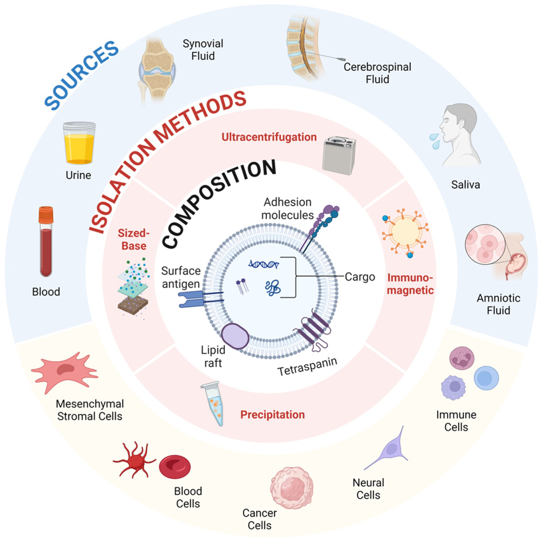
3.4.1. Introduction to Extracellular Vesicles

As discussed, the microenvironment of the joint space has a profound impact on the repair and regeneration of AC. Orthobiologics present a promising future for restoring AC damage, and exosomes are at the forefront due to their lack of immunoreactivity and simplicity as a result of their acellular nature [118,119,120]. Exosomes play a critical role in cellular communication, the transmission of materials, and cellular machinery [121,122]. Extracellular vesicles (EVs) are nanometer-sized lipid molecules that facilitate cell-to-cell communication and are present throughout the body [123,124,125]. They play a role in both stimulating and suppressing cellular responses through delivery of bioactive cargo derived from the signaling cell. EVs deliver their cargo systemically to cells through various targeting mechanisms. EVs present a unique opportunity to facilitate and enhance the intercommunication between impaired tissues and introduced therapeutic agents [126]. EVs’ ability to fuse with the plasma membrane through exocytosis and ectocytosis presents an opportunity to potentiate the reparative process and ensure vital cues are robustly transmitted and executed within biologic pathways (Figure 4). Tian et al. used rat pheochromocytoma (PC12) cell-derived exosomes to study the endocytosis pathway by live cell microscopy, revealing the PC12 cells were internalized and carried to the perinuclear region where they can directly fuse with lysosomes or the plasma membrane [127,128].

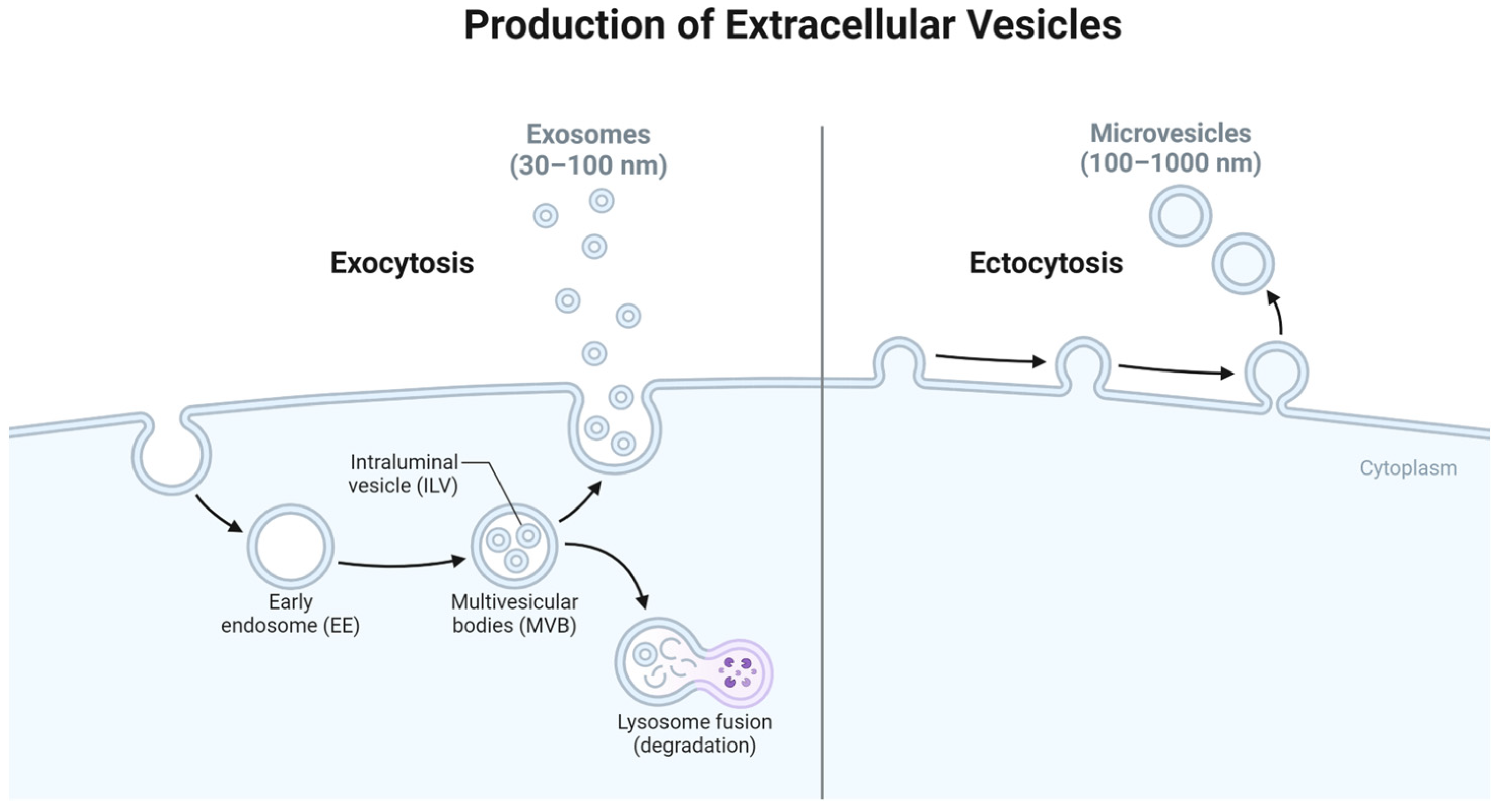
3.4.2. Origin of Exosomes
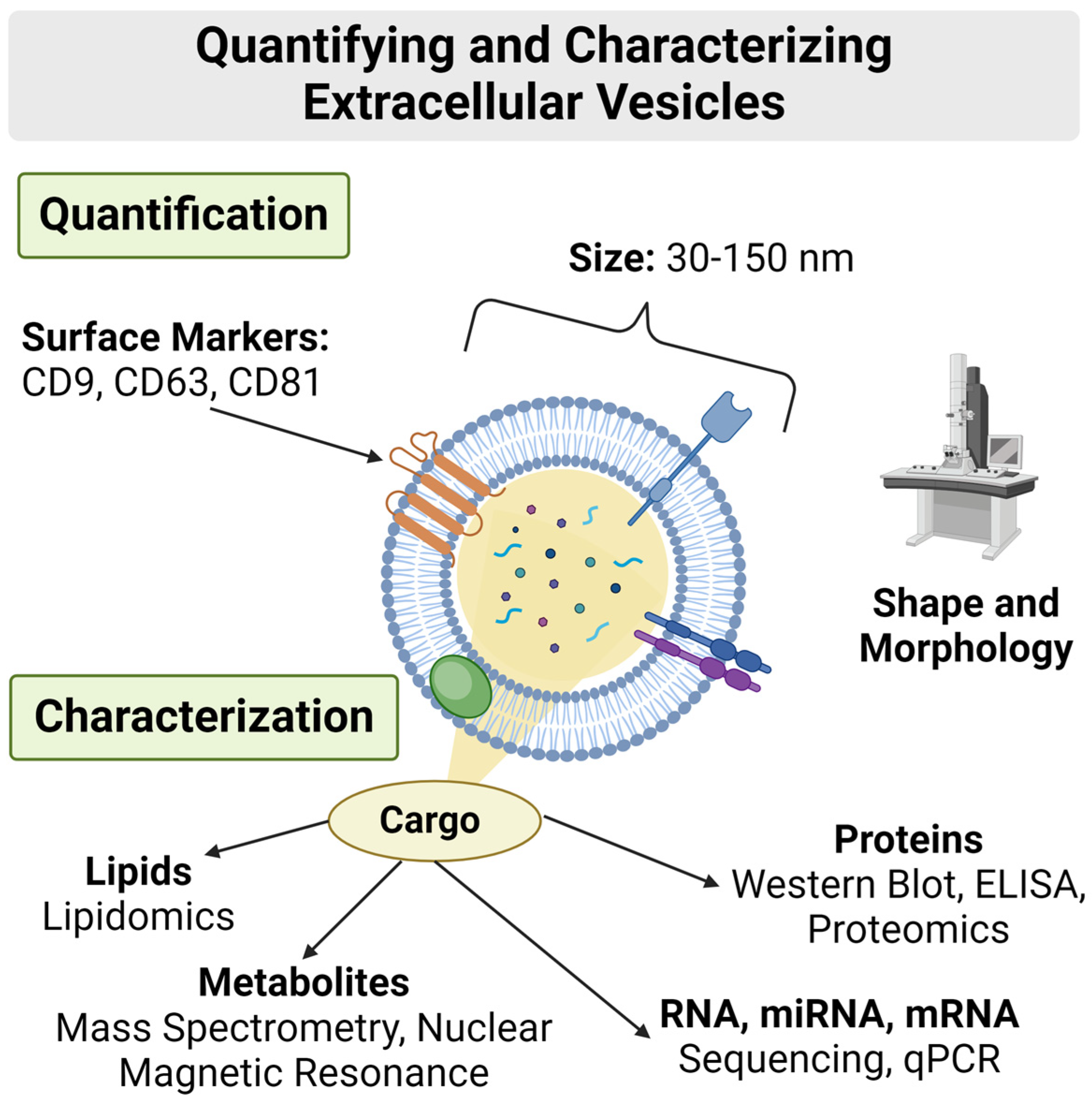
Exosomes are a type of extracellular vesicle that are characterized by their size of 30–150 nm in diameter and their endosomal origin (Figure 4) [129,130]. Exosomes originate from a variety of cells and include a variety of exosomal cargo including signaling proteins, microRNA (miRNA), and messenger RNA (mRNA) invaginated from cytoplasmic space [131]. Exosome production begins in endosomal specific organelles in which cytoplasmic cargo is sorted by inward budding of the membrane, forming intraluminal vesicles (ILVs) [132,133]. The formation of the ILVs are the precursors to multivesicular bodies (MVBs) that are either degraded by the lysosome or fused with the plasma membrane to release the sorted vesicles or exosomes [134].

3.4.3. Composition

Exosomes have a unique composition distinguishing them from other extracellular vesicles. Due to their biogenesis through the endosomal systems complex sorting, exosomes represent a more homogenous type of extracellular vesicle [135]. Comprised of a lipid bilayer different than that of the signaling cell, this distinct factor creates the optimal cargo transporter as it can easily pass through biologic barriers and be targeted for uptake by cells [136]. Distinctive surface markers integrated with the lipid bilayer unique to exosomes, such as CD9, CD63, and CD81, act as common biomarkers for identification and purification. Additionally, these surface markers, along with proteins specific for cellular signaling, act as binding receptors for exosomes to deliver their bioactive cargo to targeted cellular recipients. It has been found that, depending on their origin, exosomes can possess surface proteins that initiate immunomodulatory effects, showing promise in maintaining chronic inflammatory responses [137,138].
The cellular and metabolic state origin of an exosome has profound effects on its diverse bioactive cargo. Influenced primarily by external stimuli, the endosomal sorting and generation of MVBs pack exosomes with cargo intended to trigger a signaling cascade [135]. Toxicants can alter the folding of proteins and microRNA (miRNA), altering the microenvironment of recipient cells. If exosomal cargo are affected by stimuli that cause pathological processes to be more active, then finding ways to mediate these processes would be a key area of study. Bioactive cargo commonly found in exosomes include intracellular proteins, miRNA, mRNA, lipids, and metabolites. Exosomes hold the key to shaping the functions of recipient cells, exerting their influence through a plethora of mechanisms including RNA-mediated instructions, the generation of cellular machinery messenger RNA (mRNA), and the regulation of biochemical processes and cellular activation [125,139]. Recently, researchers have been utilizing the presence of known RNA subsets in exosomes—a diagnostic tool to help understand disease progression better [140]. Understanding these mechanisms is a crucial aspect of understanding the impact exosomes have in regulating and manipulating cellular environments (Figure 5). The ultimate objective is to gain precise control over exosome contents, paving the way for their optimization as versatile therapeutic or drug delivery devices, marking a pivotal chapter in biomedical innovation [141].

3.4.4. Targeting and Delivery

Exosomes are versatile cargo carriers and active participants in cellular communication, capable of interacting with cells throughout the body, triggering signaling and cellular events. Exosomes deliver their cargo with their target cells through membrane-bound receptors, direct membrane fusion, and endocytosis [142,143]. The structural characteristic of exosomes endows them with the ability to fuse directly with target cells, delivering their payload and ensuring the transfer of vital molecular information [136]. Surface proteins embellish exosomes and play a multifaceted role in cellular interactions. Some of these proteins engage in receptor–ligand interactions which can initiate a chain reaction, triggering intracellular signaling cascades that modulate various cellular responses. Endocytosis is a cellular process involving exosome engulfment by cellular membranes [131,144]. Once internalized, exosomes travel to lysosomes, where they face degradation, and their cargo is repurposed or discarded [90]. In some intriguing cases, exosomes may undergo modification within the endosomal pathway, acquiring additional cellular cargo before being re-secreted into the extracellular space (Figure 4). This process, known as exosome recycling, further underscores exosome-mediated communication’s dynamic and intricate nature [145,146].

3.4.5. Impact

Expanding pre-clinical research on exosomes has provided a template for developing therapeutics that utilize the unique characteristics of exosomes to enhance the regeneration of damaged tissues [147]. Due to exosomes’ ability to effectively shuttle stimuli-influenced cargo, the role of exosomes in AC damage may be either inducing or suppressive [148,149]. With the ability to utilize exosomes as potential diagnostic biomarkers, identifying the cargo-influenced mechanisms directing these contradictive effects is a key step in manipulating exosome cargo for targeted therapeutic approaches [143,150]. In addition, exosomes can act as drug carriers as they are non-immunogenic, avoid phagocytosis and engulfment by lysosomes, and expand their potential in pharmaceuticals [151,152,153]. Exosomes represent a promising therapeutic tool poised to enhance the effectiveness of existing orthobiologics while offering a safer and more targeted approach to delivering regenerative and immunomodulatory factors [85,154,155,156,157]. As previously stated, they can provide a more targeted and potent method of delivering regenerative and immunomodulatory factors, while reducing the potential risks associated with uncontrolled cellular responses. As versatile cargo, exosomes offer a unique opportunity to utilize therapeutic agents by encapsulating them within scaffolds or hydrogels as a new strategy for drug delivery in cartilage repair. These scaffolds can have a combined therapeutic effect by releasing specific drugs such as BMP with exosomes to enhance cartilage repair [158,159]. This approach allows for the controlled and sustained release of bioactive molecules directly to the site of injury [160]. Encapsulating exosomes in a localized manner can efficiently promote chondrocyte production while minimizing systemic side effects [161].

3.4.6. PRP-Derived Exosomes

Platelet activation is pivotal in the body’s natural response to tissue injury. Activated platelets release growth factors and signaling molecules, such as exosomes, that signal endogenous stem cells to migrate to the injury site, initiating the tissue repair process [155]. However, the traditional activation of platelets in PRP necessitates the addition of pro-clotting substances such as thrombin or calcium chloride (CaCl2) to induce fibrin formation to release platelet-derived growth factor (PDGF), which is important in cell growth and proliferation [29,162]. Recent research has shown that not all PRP products activate the platelets, limiting their potential [163]. PRP-derived exosomes provide an approach to produce more reliable and consistent results in PRP therapy. Liu, Xuchang, et al. compared PRP activation to PRP-derived exosomes, revealing that PRP-derived exosomes may have a better regenerative potential [155]. In addition, this study highlighted the potential for PRP-derived exosomes’ ability to decrease the apoptotic rate of OA chondrocytes, acting as a carrier for the specific growth factors that activate the Wnt/β-catenin signaling pathway [156]. PRP-derived exosomes provide a more reliable and consistent means of delivering regenerative signals to the injury site. This precision in molecular messaging ensures that the recruitment of endogenous stem cells is not dependent on platelet activation.

MSC vs. MSC-Derived Exosome Treatment

MSCs are multipotent precursor cells with adipogenic, osteogenic, and chondrogenic potential [164,165,166]. Through the secretion of various cytokines and growth factors, MSCs can promote paracrine anti-inflammatory and trophic effects that promote tissue repair [167]. Numerous studies have compared MSC treatment to treatment with MSC-derived exosomes [108]. It has recently become accepted that the role of MSCs in repair is not a mechanical response, but instead works as an environmental mediator [168]. MSCs may be responsible for stimulating native repair mechanisms and the recruitment of healing cells to the injury site [169]. This new research into MSC exosomes suggests that exosomes mediate the properties of MSCs. Specific exosomes, derived from chosen cell types, might be strategically utilized to modulate inflammatory responses, orchestrate stem cell differentiation, or advance tissue neovascularization. BM-MSCs-derived exosomes have shown promise in harnessing MSC potential by providing better instruction in promoting an environment for native cartilage repair [170,171,172].

BM-MSC Derived Exosomes Enhance Cartage Repair

The bone marrow microenvironment contains various cellular and structural components that mediate the health and function of resident MSCs [173]. Bone marrow’s cellular and acellular components regulate MSC proliferation, self-regeneration, and differentiations [84]. BMAC is known to promote a pro-regenerative MSC secretome and produce more chondrogenic cells, indicating the potential for cartilage repair [166]. Most studies have focused on the clinical improvement of orthobiologics rather than the regeneration potential. The few studies that have investigated the effect of bone marrow MSCs (BM-MSCs) for cartilage repair have revealed partial restoration of hyaline cartilage with issues of transient fibrocartilage formation and subchondral bone overgrowth [69,174]. These issues show the need to harness the potential of MSCs to communicate the modulating pathways to address these issues specifically.
Exosomes can potentially enhance the effects of BMAC and the crucial factors MSCs possess [86]. BM-derived exosomes have shown the potential to attenuate the proliferation and migration of chondrocytes. A recent study suggests that BM-MSC-derived exosomes are endocytosed into the chondrocytes, promoting direct communication to promote chondrogenesis into the injury site. Furthermore, BM-MSC-derived exosomes can reduce the effects of IL-1β, a proinflammatory cytokine that drives synovitis and is an inducer of cartilage degeneration, causing OA [175,176,177]. The BM-MSC-derived exosomes’ ability to inhibit IL-1β in any capacity can enhance the proliferation capacity of chondrocytes while increasing their migration to have a more sustained therapeutic effect. Treating cartilage damage with BM-MSC-derived exosomes can lead to the upregulation of COL2A1 protein, while downregulating MMP13 and Runx2, leading to native cartilage formation and ECM synthesis, and inhibiting apoptosis induction [178,179,180,181].

mRNA and miRNA Cargo in Exosomes

mRNA and miRNA play crucial roles in the complex functions of exosomes in cellular communication. mRNA acts as a blueprint for protein synthesis, while miRNA fine-tunes gene expression with precise control. MiRNAs are small non-coding RNA molecules, about 19–24 nucleotides long, serving as post-transcriptional gene regulators [182]. Their primary function is to inhibit the translation of target mRNA into proteins or degrade them, thereby modulating specific protein levels in the cell. MiRNA binds to complementary sequences in mRNA, silencing or optimizing gene expression in various cellular pathways [183]. This regulatory function is vital for maintaining cellular balance, ensuring genes are not overly expressed or suppressed. MiRNAs are involved in many diverse biological processes, from cell differentiation to immune responses.
Moreover, miRNAs carried by exosomes can travel between cells, enabling long-distance communication in the body. This property extends their impact beyond individual cells, allowing for orchestrating biological responses. MiRNA-mediated regulation, facilitated by exosomes, modulates cellular function, ensuring genes are expressed at the right time and in the right amounts to maintain health. This precision underscores miRNAs’ pivotal role in exosome-mediated cellular communication, suggesting their potential as therapeutic targets, especially in cartilage repair [184].
BM-MSC-derived exosome-specific miRNA plays a crucial role in promoting and enhancing chondrogenesis (Table 3). These exosomes navigate the complex processes by selectively modulating miRNA expression, targeting key factors in cartilage regeneration and inflammation, like IL-1β, while suppressing pro-inflammatory factors [185]. It is important to note that unregulated miRNA expression can lead to overexpression of pro-oncogene factors. Managing miRNA concentrations is critical in exosome-mediated therapies to balance the promotion of beneficial cellular responses and to avoid risks associated with unintended oncogene activation [186].

Exosomes as a Novel Orthobiologic Direction

Extracellular Vesicles, such as exosomes, present a novel opportunity to enhance orthobiologics in diagnosing, regenerating, and treating musculoskeletal injuries and diseases. While all cell-types secrete EVs, harnessing beneficial cargo that assists with the homeostasis of the cellular environment presents a strong potential for the targeted cell-sourcing of exosomes. Characterizing and quantifying key components of EV-mediated factors for their use in therapeutic applications has the potential to modulate the cellular environment to homeostasis, promoting healing through EVs promising regenerative and immunoregulatory properties. However, more research is needed to identify the optimal source, optimize isolation, and further characterize exosome-specific bioactive cargo before they are introduced into the clinical space (Figure 6).

4. Conclusions

Current therapeutics for cartilage repair have the potential to initiate cartilage regeneration and repair. However, regeneration is limited because of the unique characteristic of articular cartilage which is avascular in nature [14]. Surgical techniques utilize specific ways, such as microfracture, where stem cells in the subchondral bone are stimulated and released within the joint [10,199]. This stimulation process through microfracture leads primarily to the formation of fibrocartilage [21]. Orthobiologics promote the body’s healing response and yield better quality cartilage (hyaline like cartilage) that mimics the native environment [95]. However, orthobiologics such as PRP and BMAC present a similar issue to surgical techniques as there is no clear evidence for regenerating native AC within the joint [200].
Exosome therapy has emerged as an opportunity to direct specific communication within cells without the inherent risks of surgical intervention and the potential for uncontrolled responses to orthobiologics [132]. Exosomes being acellular minimizes the risk of an adverse immune response, and some of the dangers of MSC and stem cell use, such as immune-rejection, can also be minimized [134]. Finally, as exosome production and isolation research continue, approval of an allogeneic exosome product for the treatment of musculoskeletal conditions by the FDA may be more likely than for products with cellular components. Commercial production of an exosome product could lead to an opportunity to increase and control dosing that ensures the best environment for tissue regeneration in the joint.

5. Future Perspectives

Tissue engineering is an essential field in regenerative medicine that aims to create functional replacements of damaged or diseased tissue that resembles the native niche [201,202]. It is an emerging interdisciplinary field that uses biomedical techniques combined with current biologics to enhance and precisely regulate the local cellular environment, promoting a sustainable effect for proper tissue regeneration. Biomaterial scaffolds are a specific technique in tissue engineering that utilize a three-dimensional framework that supports the differentiation and proliferation of cells to repair and regenerate new tissue [203,204]. Scaffolds provide a porous structure that closely resembles the ECM found in tissues by utilizing biomaterials to influence biological processes to promote tissue regeneration [205,206]. New acellular scaffold therapies have become an interesting topic in cartilage repair as it offers an opportunity to aid, and one day replace, traditional surgeries such as bone marrow stimulation and ACI surgery.
To be successful in regenerating native tissue and maintaining structural compatibility, acellular scaffolds need to have a similar composition to AC [207]. They need to be constructed with mechanical strength and flexibility to withstand the force load that joints encounter while being biocompatible with the surrounding joint region to mimic and regenerate the existing ECM [208,209]. Supporting cell infiltration is critical for the proliferation and differentiation of chondrogenic and osteogenic cells; using natural and synthetic polymers while loading them with bioactive elements can enhance the cellular components needed for native tissue regeneration [210]. A multilayer and multipolymer approach has been shown to be the most effective acellular scaffold structure. A recent review article described two multilayered acellular scaffolds, MaioRegen and Agili-c, that have published data regarding osteochondral defects, revealing potential cartilage repair [211]. MaioRegen is a trilayered acellular scaffold that is composed of type 1 collagen and hydroxyapatite, while Agili-C is a crystalline aragonite bi-phasic scaffold that is combined with HA; the two unique properties can stimulate the cell infiltration process leading to better quality ECM production [211].
Acellular scaffolds can also be used as a delivery system for bioactive carriers such as MSCs and exosomes [209,212]. The fabrication of acellular scaffolds as a platform for the integration of exosomes has recently been studied, and this has shown that utilizing 3D culture, nanomaterials, and 3D biomaterials can enhance the exosome effect on the joint, leading to cartilage repair [207,213]. In one study, Liu et al. utilized a photoinduced imine crosslinking hydrogel glue as an exosome scaffold for cartilage defect regeneration [214]. This study showed that this “acellular tissue patch” could retain the bone marrow-derived exosomes and regulate chondrocyte production while integrating with native cartilage, resulting in cartilage repair [214]. In addition, an exciting class of biomaterials that are able help regenerate bone and cartilage regrowth is bioceramics, a biocompatible ceramic that has “apatite formation ability, allowing bonding to form between material and tissues” [215]. Bioactive bioceramics are commonly incorporated into scaffolds to provide structural support as they contain materials such as hydroxyapatite, tricalcium phosphate, and bioactive glass [215,216,217]. One group hypothesized that a scaffold based on gelatin/PRGF/lithium-doped bioactive glass seeded by endometrial stem cells forms a useful model for bone regeneration. Reza-Farmani et al. revealed that lithium ion increases cell survival through the activation of the Wnt/β-catenin pathway, and with the addition of PRGF it can increase it even further. In addition, lithium can “stimulate stem cells to secrete exosomes”, revealing the ability to elicit a paracrine effect [216]. The release of ions such as lithium is an essential part of the combinatorial effect that bioceramics can have with therapeutics, and exosomes would be a favorable target as this group showed the paracrine effect released by PRGF that increased cell viability [216,218].
These recent studies lay the groundwork for integrating biologics and biomaterials in order to stimulate healing within injured joints. Providing a dynamic scaffold to support the physical native structure while targeting the cellular environment indicates a novel approach towards regenerating AC. Combining targeted bioactive systems indicates the need for further research and preclinical applications to understand the exact mechanisms of action and their translational viability. Integrating cell signaling factors, such as exosomes, with acellular scaffolds is a new frontier in acellular therapies that have the potential to be translated from the bench to the bedside, filling the gap with current surgical and preventative techniques that avoid leaving patients at risk of worsening conditions, such as OA.

Funding

The authors would like to acknowledge funding from the National Institute of Health and the Department of Defense.

Data Availability Statement

No new data were created or analyzed in this study. Data sharing is not applicable to this article.

Conflicts of Interest

The authors declare no conflict of interest.

References

  1. Karamanos, N.K.; Theocharis, A.D.; Piperigkou, Z.; Manou, D.; Passi, A.; Skandalis, S.S.; Vynios, D.H.; Orian-Rousseau, V.; Ricard-Blum, S.; Schmelzer, C.E. A guide to the composition and functions of the extracellular matrix. FEBS J. 2021, 288, 6850–6912. [Google Scholar] [CrossRef] [PubMed]
  2. Akkiraju, H.; Nohe, A. Role of chondrocytes in cartilage formation, progression of osteoarthritis and cartilage regeneration. J. Dev. Biol. 2015, 3, 177–192. [Google Scholar] [CrossRef]
  3. Sophia Fox, A.J.; Bedi, A.; Rodeo, S.A. The basic science of articular cartilage: Structure, composition, and function. Sports Health 2009, 1, 461–468. [Google Scholar] [CrossRef]
  4. Medvedeva, E.V.; Grebenik, E.A.; Gornostaeva, S.N.; Telpuhov, V.I.; Lychagin, A.V.; Timashev, P.S.; Chagin, A.S. Repair of damaged articular cartilage: Current approaches and future directions. Int. J. Mol. Sci. 2018, 19, 2366. [Google Scholar] [CrossRef]
  5. Crane, J.L.; Cao, X. Bone marrow mesenchymal stem cells and TGF-β signaling in bone remodeling. J. Clin. Investig. 2014, 124, 466–472. [Google Scholar] [CrossRef]
  6. Guo, X.; Xi, L.; Yu, M.; Fan, Z.; Wang, W.; Ju, A.; Liang, Z.; Zhou, G.; Ren, W. Regeneration of articular cartilage defects: Therapeutic strategies and perspectives. J. Tissue Eng. 2023, 14, 1–27. [Google Scholar] [CrossRef]
  7. Wachsmuth, L.; Söder, S.; Fan, Z.; Finger, F.; Aigner, T. Immunolocalization of matrix proteins in different human cartilage subtypes. Histol. Histopathol. 2006, 21, 9. [Google Scholar]
  8. Stockwell, R. The cell density of human articular and costal cartilage. J. Anat. 1967, 101, 753. [Google Scholar]
  9. Mankin, H.J. The reaction of articular cartilage to injury and osteoarthritis. N. Engl. J. Med. 1974, 291, 1285–1292. [Google Scholar] [CrossRef] [PubMed]
  10. Krishnan, Y.; Grodzinsky, A.J. Cartilage diseases. Matrix Biol. 2018, 71–72, 51–69. [Google Scholar] [CrossRef] [PubMed]
  11. Pacifici, M.; Koyama, E.; Iwamoto, M. Mechanisms of synovial joint and articular cartilage formation: Recent advances, but many lingering mysteries. Birth Defects Res. Part C Embryo Today Rev. 2005, 75, 237–248. [Google Scholar] [CrossRef] [PubMed]
  12. Dudhia, J. Aggrecan, aging and assembly in articular cartilage. Cell. Mol. Life Sci. 2005, 62, 2241–2256. [Google Scholar] [CrossRef]
  13. Hall, A.C.; Horwitz, E.R.; Wilkins, R.J. The cellular physiology of articular cartilage. Exp. Physiol. Transl. Integr. 1996, 81, 535–545. [Google Scholar] [CrossRef]
  14. Newman, A.P. Articular cartilage repair. Am. J. Sports Med. 1998, 26, 309–324. [Google Scholar] [CrossRef]
  15. Decker, R.S. Articular cartilage and joint development from embryogenesis to adulthood. Semin. Cell Dev. Biol. 2017, 62, 50–56. [Google Scholar] [CrossRef]
  16. Shum, L.; Nuckolls, G. The life cycle of chondrocytes in the developing skeleton. Arthritis Res. Ther. 2001, 4, 94. [Google Scholar] [CrossRef]
  17. Godman, G.C.; Porter, K.R. Chondrogenesis, studied with the electron microscope. J. Cell Biol. 1960, 8, 719–760. [Google Scholar] [CrossRef]
  18. Kosher, R.A.; Kulyk, W.M.; Gay, S.W. Collagen gene expression during limb cartilage differentiation. J. Cell Biol. 1986, 102, 1151–1156. [Google Scholar] [CrossRef]
  19. van Beuningen, H.M.; Glansbeek, H.L.; van der Kraan, P.M.; van den Berg, W.B. Differential effects of local application of BMP-2 or TGF-β1 on both articular cartilage composition and osteophyte formation. Osteoarthr. Cartil. 1998, 6, 306–317. [Google Scholar] [CrossRef]
  20. Tekari, A.; Luginbuehl, R.; Hofstetter, W.; Egli, R.J. Transforming growth factor beta signaling is essential for the autonomous formation of cartilage-like tissue by expanded chondrocytes. PLoS ONE 2015, 10, e0120857. [Google Scholar] [CrossRef] [PubMed]
  21. Utsunomiya, H.; Gao, X.; Deng, Z.; Cheng, H.; Nakama, G.; Scibetta, A.C.; Ravuri, S.K.; Goldman, J.L.; Lowe, W.R.; Rodkey, W.G.; et al. Biologically regulated marrow stimulation by blocking TGF-β1 with losartan oral administration results in hyaline-like cartilage repair: A rabbit osteochondral defect model. Am. J. Sports Med. 2020, 48, 974–984. [Google Scholar] [CrossRef] [PubMed]
  22. Lópiz-Morales, Y.; Abarrategi, A.; Ramos, V.; Moreno-Vicente, C.; López-Durán, L.; López-Lacomba, J.L.; Marco, F. In vivo comparison of the effects of rhBMP-2 and rhBMP-4 in osteochondral tissue regeneration. Eur. Cells Mater. 2010, 20, e78. [Google Scholar] [CrossRef]
  23. Klein-Nulend, J.; Louwerse, R.; Heyligers, I.; Wuisman, P.; Semeins, C.; Goei, S.; Burger, E. Osteogenic protein (OP-1, BMP-7) stimulates cartilage differentiation of human and goat perichondrium tissue in vitro. J. Biomed. Mater. Res. 1998, 40, 614–620. [Google Scholar] [CrossRef]
  24. McQuillan, D.J.; Handley, C.J.; Campbell, M.A.; Bolis, S.; Milway, V.; Herington, A. Stimulation of proteoglycan biosynthesis by serum and insulin-like growth factor-I in cultured bovine articular cartilage. Biochem. J. 1986, 240, 423–430. [Google Scholar] [CrossRef]
  25. Sah, R.L.; Chen, A.C.; Grodzinsky, A.J.; Trippel, S. Differential effects of bFGF and IGF-I on matrix metabolism in calf and adult bovine cartilage explants. Arch. Biochem. Biophys. 1994, 308, 137–147. [Google Scholar] [CrossRef]
  26. Hung, G.; Galea-Lauri, J.; Mueller, G.; Georgescu, H.; Larkin, L.; Suchanek, M.; Tindal, M.; Robbins, P.; Evans, C. Suppression of intra-articular responses to interleukin-1 by transfer of the interleukin-1 receptor antagonist gene to synovium. Gene Ther. 1994, 1, 64–69. [Google Scholar] [PubMed]
  27. Gigout, A.; Guehring, H.; Froemel, D.; Meurer, A.; Ladel, C.; Reker, D.; Bay-Jensen, A.; Karsdal, M.; Lindemann, S. Sprifermin (rhFGF18) enables proliferation of chondrocytes producing a hyaline cartilage matrix. Osteoarthr. Cartil. 2017, 25, 1858–1867. [Google Scholar] [CrossRef]
  28. Hamilton, J.L.; Nagao, M.; Levine, B.R.; Chen, D.; Olsen, B.R.; Im, H.J. Targeting VEGF and its receptors for the treatment of osteoarthritis and associated pain. J. Bone Miner. Res. 2016, 31, 911–924. [Google Scholar] [PubMed]
  29. Schmidt, M.; Chen, E.; Lynch, S. A review of the effects of insulin-like growth factor and platelet derived growth factor on in vivo cartilage healing and repair. Osteoarthr. Cartil. 2006, 14, 403–412. [Google Scholar] [CrossRef]
  30. Xie, C.; Chen, Q. Adipokines: New therapeutic target for osteoarthritis? Curr. Rheumatol. Rep. 2019, 21, 71. [Google Scholar] [CrossRef]
  31. Dilley, J.E.; Bello, M.A.; Roman, N.; McKinley, T.; Sankar, U. Post-traumatic osteoarthritis: A review of pathogenic mechanisms and novel targets for mitigation. Bone Rep. 2023, 18, 101658. [Google Scholar] [CrossRef] [PubMed]
  32. GBD 2019 Diseases and Injuries Collaborators. Global burden of 369 diseases and injuries in 204 countries and territories, 1990–2019: A systematic analysis for the Global Burden of Disease Study 2019. Lancet 2020, 396, 1204–1222. [Google Scholar] [CrossRef] [PubMed]
  33. Dieleman, J.L.; Squires, E.; Bui, A.L.; Campbell, M.; Chapin, A.; Hamavid, H.; Horst, C.; Li, Z.; Matyasz, T.; Reynolds, A. Factors associated with increases in US health care spending, 1996–2013. JAMA 2017, 318, 1668–1678. [Google Scholar] [CrossRef]
  34. Dieleman, J.L.; Cao, J.; Chapin, A.; Chen, C.; Li, Z.; Liu, A.; Horst, C.; Kaldjian, A.; Matyasz, T.; Scott, K.W. US health care spending by payer and health condition, 1996–2016. JAMA 2020, 323, 863–884. [Google Scholar] [CrossRef]
  35. Bijlsma, J.W.; Berenbaum, F.; Lafeber, F.P. Osteoarthritis: An update with relevance for clinical practice. Lancet 2011, 377, 2115–2126. [Google Scholar] [CrossRef]
  36. Anderson, D.D.; Chubinskaya, S.; Guilak, F.; Martin, J.A.; Oegema, T.R.; Olson, S.A.; Buckwalter, J.A. Post-traumatic osteoarthritis: Improved understanding and opportunities for early intervention. J. Orthop. Res. 2011, 29, 802–809. [Google Scholar] [CrossRef] [PubMed]
  37. Brown, T.D.; Johnston, R.C.; Saltzman, C.L.; Marsh, J.L.; Buckwalter, J.A. Posttraumatic osteoarthritis: A first estimate of incidence, prevalence, and burden of disease. J. Orthop. Trauma 2006, 20, 739–744. [Google Scholar] [CrossRef]
  38. Fernandes, J.C.; Martel-Pelletier, J.; Pelletier, J.P. The role of cytokines in osteoarthritis pathophysiology. Biorheology 2002, 39, 237–246. [Google Scholar]
  39. Sanchez-Lopez, E.; Coras, R.; Torres, A.; Lane, N.E.; Guma, M. Synovial inflammation in osteoarthritis progression. Nat. Rev. Rheumatol. 2022, 18, 258–275. [Google Scholar]
  40. Poulet, B.; Staines, K.A. New developments in osteoarthritis and cartilage biology. Curr. Opin. Pharmacol. 2016, 28, 8–13. [Google Scholar] [CrossRef]
  41. Magni, A.; Agostoni, P.; Bonezzi, C.; Massazza, G.; Mene, P.; Savarino, V.; Fornasari, D. Management of Osteoarthritis: Expert Opinion on NSAIDs. Pain Ther. 2021, 10, 783–808. [Google Scholar] [CrossRef] [PubMed]
  42. Hunziker, E.B. Articular cartilage repair: Basic science and clinical progress. A review of the current status and prospects. Osteoarthr. Cartil. 2002, 10, 432–463. [Google Scholar] [CrossRef] [PubMed]
  43. Fang, Y.; Wu, D.; Birukov, K.G. Mechanosensing and mechanoregulation of endothelial cell functions. Compr. Physiol. 2019, 9, 873. [Google Scholar]
  44. Koenen, R.R. The prowess of platelets in immunity and inflammation. Thromb. Haemost. 2016, 116, 605–612. [Google Scholar] [CrossRef]
  45. Franz, S.; Rammelt, S.; Scharnweber, D.; Simon, J.C. Immune responses to implants–a review of the implications for the design of immunomodulatory biomaterials. Biomaterials 2011, 32, 6692–6709. [Google Scholar] [CrossRef]
  46. Sillat, T.; Barreto, G.; Clarijs, P.; Soininen, A.; Ainola, M.; Pajarinen, J.; Korhonen, M.; Konttinen, Y.T.; Sakalyte, R.; Hukkanen, M. Toll-like receptors in human chondrocytes and osteoarthritic cartilage. Acta Orthop. 2013, 84, 585–592. [Google Scholar] [CrossRef]
  47. Kim, H.A.; Cho, M.L.; Choi, H.Y.; Yoon, C.S.; Jhun, J.Y.; Oh, H.J.; Kim, H.Y. The catabolic pathway mediated by Toll-like receptors in human osteoarthritic chondrocytes. Arthritis Rheum. Off. J. Am. Coll. Rheumatol. 2006, 54, 2152–2163. [Google Scholar] [CrossRef]
  48. Kalaitzoglou, E.; Griffin, T.M.; Humphrey, M.B. Innate immune responses and osteoarthritis. Curr. Rheumatol. Rep. 2017, 19, 45. [Google Scholar] [CrossRef] [PubMed]
  49. Kandahari, A.M.; Yang, X.; Dighe, A.S.; Pan, D.; Cui, Q. Recognition of immune response for the early diagnosis and treatment of osteoarthritis. J. Immunol. Res. 2015, 2015, 192415. [Google Scholar] [CrossRef]
  50. Woodell-May, J.E.; Sommerfeld, S.D. Role of inflammation and the immune system in the progression of osteoarthritis. J. Orthop. Res. 2020, 38, 253–257. [Google Scholar] [CrossRef]
  51. Armiento, A.R.; Alini, M.; Stoddart, M.J. Articular fibrocartilage—Why does hyaline cartilage fail to repair? Adv. Drug Deliv. Rev. 2019, 146, 289–305. [Google Scholar] [CrossRef] [PubMed]
  52. Benjamin, M.; Ralphs, J. Biology of fibrocartilage cells. Int. Rev. Cytol. 2004, 233, 1–46. [Google Scholar]
  53. Yasui, N.; Nimni, M.E. Cartilage collagens. In Collagen; CRC Press: Boca Raton, FL, USA, 2018; pp. 225–242. [Google Scholar]
  54. Sgaglione, N.A. The future of cartilage restoration. J. Knee Surg. 2004, 17, 235–243. [Google Scholar] [CrossRef]
  55. Decker, R.S.; Koyama, E.; Pacifici, M. Articular cartilage: Structural and developmental intricacies and questions. Curr. Osteoporos. Rep. 2015, 13, 407–414. [Google Scholar] [CrossRef]
  56. Franke, O.; Durst, K.; Maier, V.; Göken, M.; Birkholz, T.; Schneider, H.; Hennig, F.; Gelse, K. Mechanical properties of hyaline and repair cartilage studied by nanoindentation. Acta Biomater. 2007, 3, 873–881. [Google Scholar] [CrossRef]
  57. Davis, S.; Roldo, M.; Blunn, G.; Tozzi, G.; Roncada, T. Influence of the mechanical environment on the regeneration of osteochondral defects. Front. Bioeng. Biotechnol. 2021, 9, 603408. [Google Scholar] [CrossRef] [PubMed]
  58. Coutts, R.D.; Healey, R.M.; Ostrander, R.; Sah, R.L.; Goomer, R.; Amiel, D. Matrices for cartilage repair. Clin. Orthop. Relat. Res. 2001, 391, S271–S279. [Google Scholar] [CrossRef]
  59. Wang, L.; Lazebnik, M.; Detamore, M. Hyaline cartilage cells outperform mandibular condylar cartilage cells in a TMJ fibrocartilage tissue engineering application. Osteoarthr. Cartil. 2009, 17, 346–353. [Google Scholar] [CrossRef]
  60. Wei, W.; Dai, H. Articular cartilage and osteochondral tissue engineering techniques: Recent advances and challenges. Bioact. Mater. 2021, 6, 4830–4855. [Google Scholar] [CrossRef]
  61. Buchanan, J.L. Types of fibrocartilage. Clin. Podiatr. Med. Surg. 2022, 39, 357–361. [Google Scholar] [CrossRef] [PubMed]
  62. Musumeci, G.; Loreto, C.; Castorina, S.; Imbesi, R.; Leonardi, R.; Castrogiovanni, P. Current concepts in the treatment of cartilage damage. A review. Ital. J. Anat. Embryol. 2013, 118, 189–203. [Google Scholar] [PubMed]
  63. Smith, G.; Knutsen, G.; Richardson, J. A clinical review of cartilage repair techniques. J. Bone Jt. Surg. Br. Vol. 2005, 87, 445–449. [Google Scholar] [CrossRef] [PubMed]
  64. Makris, E.A.; Gomoll, A.H.; Malizos, K.N.; Hu, J.C.; Athanasiou, K.A. Repair and tissue engineering techniques for articular cartilage. Nat. Rev. Rheumatol. 2015, 11, 21–34. [Google Scholar] [CrossRef] [PubMed]
  65. Goldring, M.B. Chondrogenesis, chondrocyte differentiation, and articular cartilage metabolism in health and osteoarthritis. Ther. Adv. Musculoskelet. Dis. 2012, 4, 269–285. [Google Scholar] [CrossRef]
  66. Steadman, J.R.; Rodkey, W.G.; Rodrigo, J.J. Microfracture: Surgical technique and rehabilitation to treat chondral defects. Clin. Orthop. Relat. Res. 2001, 391, S362–S369. [Google Scholar] [CrossRef]
  67. Steadman, J.R.; Rodkey, W.G.; Singleton, S.B.; Briggs, K.K. Microfracture technique forfull-thickness chondral defects: Technique and clinical results. Oper. Tech. Orthop. 1997, 7, 300–304. [Google Scholar] [CrossRef]
  68. Steadman, J.R.; Rodkey, W.G.; Briggs, K.K. Microfracture: Its history and experience of the developing surgeon. Cartilage 2010, 1, 78–86. [Google Scholar] [CrossRef]
  69. Hu, Y.; Chen, X.; Wang, S.; Jing, Y.; Su, J. Subchondral bone microenvironment in osteoarthritis and pain. Bone Res. 2021, 9, 20. [Google Scholar] [CrossRef]
  70. Goldring, M.B.; Tsuchimochi, K.; Ijiri, K. The control of chondrogenesis. J. Cell. Biochem. 2006, 97, 33–44. [Google Scholar] [CrossRef] [PubMed]
  71. Steadman, J.R.; Rodkey, W.G.; Briggs, K.K. Microfracture Chondroplasty: Indications, Techniques, and Outcomes. Sports Med. Arthrosc. Rev. 2003, 11, 236–244. [Google Scholar] [CrossRef]
  72. Bugbee, W.D.; Convery, F.R. Osteochondral allograft transplantation. Clin. Sports Med. 1999, 18, 67–75. [Google Scholar] [CrossRef] [PubMed]
  73. Redondo, M.L.; Beer, A.J.; Yanke, A.B. Cartilage restoration: Microfracture and osteochondral autograft transplantation. J. Knee Surg. 2018, 31, 231–238. [Google Scholar] [CrossRef] [PubMed]
  74. Jang, K.; Berrigan, W.A.; Mautner, K. Regulatory Considerations of Orthobiologic Procedures. Phys. Med. Rehabil. Clin. 2023, 34, 275–283. [Google Scholar] [CrossRef] [PubMed]
  75. Murray, I.R.; Chahla, J.; Wordie, S.J.; Shapiro, S.A.; Piuzzi, N.S.; Frank, R.M.; Halbrecht, J.; Okada, K.; Nakamura, N.; Mandelbaum, B. Regulatory and Ethical Aspects of Orthobiologic Therapies. Orthop. J. Sports Med. 2022, 10, 1–9. [Google Scholar] [CrossRef]
  76. Allickson, J. Emerging translation of regenerative therapies. Clin. Pharmacol. Ther. 2017, 101, 28–30. [Google Scholar] [CrossRef]
  77. Jessop, Z.M.; Al-Sabah, A.; Francis, W.R.; Whitaker, I.S. Transforming healthcare through regenerative medicine. BMC Med. 2016, 14, 115. [Google Scholar] [CrossRef]
  78. Boivin, J.; Tolsma, R.; Awad, P.; Kenter, K.; Li, Y. The biological use of platelet-rich plasma in skeletal muscle injury and repair. Am. J. Sports Med. 2023, 51, 1347–1355. [Google Scholar] [CrossRef]
  79. Fang, J.; Wang, X.; Jiang, W.; Zhu, Y.; Hu, Y.; Zhao, Y.; Song, X.; Zhao, J.; Zhang, W.; Peng, J. Platelet-rich plasma therapy in the treatment of diseases associated with orthopedic injuries. Tissue Eng. Part B Rev. 2020, 26, 571–585. [Google Scholar] [CrossRef]
  80. Andia, I.; Abate, M. Platelet-rich plasma in the treatment of skeletal muscle injuries. Expert Opin. Biol. Ther. 2015, 15, 987–999. [Google Scholar] [CrossRef]
  81. Gulati, G.L.; Ashton, J.K.; Hyun, B.H. Structure and function of the bone marrow and hematopoiesis. Hematol./Oncol. Clin. 1988, 2, 495–511. [Google Scholar] [CrossRef]
  82. Chahla, J.; Mannava, S.; Cinque, M.E.; Geeslin, A.G.; Codina, D.; LaPrade, R.F. Bone marrow aspirate concentrate harvesting and processing technique. Arthrosc. Tech. 2017, 6, e441–e445. [Google Scholar] [CrossRef] [PubMed]
  83. Cavallo, C.; Boffa, A.; Andriolo, L.; Silva, S.; Grigolo, B.; Zaffagnini, S.; Filardo, G. Bone marrow concentrate injections for the treatment of osteoarthritis: Evidence from preclinical findings to the clinical application. Int. Orthop. 2021, 45, 525–538. [Google Scholar] [CrossRef] [PubMed]
  84. Fortier, L.A.; Potter, H.G.; Rickey, E.J.; Schnabel, L.V.; Foo, L.F.; Chong, L.R.; Stokol, T.; Cheetham, J.; Nixon, A.J. Concentrated bone marrow aspirate improves full-thickness cartilage repair compared with microfracture in the equine model. JBJS 2010, 92, 1927–1937. [Google Scholar] [CrossRef] [PubMed]
  85. He, L.; He, T.; Xing, J.; Zhou, Q.; Fan, L.; Liu, C.; Chen, Y.; Wu, D.; Tian, Z.; Liu, B. Bone marrow mesenchymal stem cell-derived exosomes protect cartilage damage and relieve knee osteoarthritis pain in a rat model of osteoarthritis. Stem Cell Res. Ther. 2020, 11, 276. [Google Scholar] [CrossRef] [PubMed]
  86. Krych, A.J.; Nawabi, D.H.; Farshad-Amacker, N.A.; Jones, K.J.; Maak, T.G.; Potter, H.G.; Williams III, R.J. Bone marrow concentrate improves early cartilage phase maturation of a scaffold plug in the knee: A comparative magnetic resonance imaging analysis to platelet-rich plasma and control. Am. J. Sports Med. 2016, 44, 91–98. [Google Scholar] [CrossRef]
  87. Veronesi, F.; Giavaresi, G.; Tschon, M.; Borsari, V.; Nicoli Aldini, N.; Fini, M. Clinical use of bone marrow, bone marrow concentrate, and expanded bone marrow mesenchymal stem cells in cartilage disease. Stem Cells Dev. 2013, 22, 181–192. [Google Scholar] [CrossRef]
  88. Kreulen, C.; Giza, E.; Shieh, A.; Singh, S.; Nathe, C.; Lian, E.; Haudenschild, D. Effects of Micronized Cartilage Matrix on Cartilage Repair in Osteochondral Lesions of the Talus. Foot Ankle Orthop. 2017, 2, 1–2. [Google Scholar] [CrossRef]
  89. Bernhardt, A.; Lode, A.; Boxberger, S.; Pompe, W.; Gelinsky, M. Mineralised collagen—An artificial, extracellular bone matrix—Improves osteogenic differentiation of bone marrow stromal cells. J. Mater. Sci. Mater. Med. 2008, 19, 269–275. [Google Scholar] [CrossRef]
  90. Monibi, F.A.; Bozynski, C.C.; Kuroki, K.; Stoker, A.M.; Pfeiffer, F.M.; Sherman, S.L.; Cook, J.L. Development of a micronized meniscus extracellular matrix scaffold for potential augmentation of meniscal repair and regeneration. Tissue Eng. Part C Methods 2016, 22, 1059–1070. [Google Scholar] [CrossRef]
  91. Shin, J.J.; Mellano, C.; Cvetanovich, G.L.; Frank, R.M.; Cole, B.J. Treatment of glenoid chondral defect using micronized allogeneic cartilage matrix implantation. Arthrosc. Tech. 2014, 3, e519–e522. [Google Scholar] [CrossRef]
  92. Lai, W.; Li, Y.; Mak, S.; Ho, F.; Chow, S.; Chooi, W.; Chow, C.; Leung, A.; Chan, B. Reconstitution of bone-like matrix in osteogenically differentiated mesenchymal stem cell–collagen constructs: A three-dimensional in vitro model to study hematopoietic stem cell niche. J. Tissue Eng. 2013, 4, 1–14. [Google Scholar] [CrossRef] [PubMed]
  93. Bretschneider, H.; Quade, M.; Lode, A.; Gelinsky, M.; Rammelt, S.; Vater, C. Chemotactic and angiogenic potential of mineralized collagen scaffolds functionalized with naturally occurring bioactive factor mixtures to stimulate bone regeneration. Int. J. Mol. Sci. 2021, 22, 5836. [Google Scholar] [CrossRef] [PubMed]
  94. Huebner, K.; Frank, R.M.; Getgood, A. Ortho-biologics for osteoarthritis. Clin. Sports Med. 2019, 38, 123–141. [Google Scholar] [CrossRef] [PubMed]
  95. Mavrogenis, A.F.; Karampikas, V.; Zikopoulos, A.; Sioutis, S.; Mastrokalos, D.; Koulalis, D.; Scarlat, M.M.; Hernigou, P. Orthobiologics: A review. Int. Orthop. 2023, 47, 1645–1662. [Google Scholar] [CrossRef]
  96. Deng, Z.; Chen, F.; Liu, Y.; Wang, J.; Lu, W.; Jiang, W.; Zhu, W. Losartan protects against osteoarthritis by repressing the TGF-β1 signaling pathway via upregulation of PPARγ. J. Orthop. Transl. 2021, 29, 30–41. [Google Scholar] [CrossRef]
  97. Nagai, T.; Sato, M.; Kobayashi, M.; Yokoyama, M.; Tani, Y.; Mochida, J. Bevacizumab, an anti-vascular endothelial growth factor antibody, inhibits osteoarthritis. Arthritis Res. Ther. 2014, 16, 427. [Google Scholar] [CrossRef]
  98. Park, S.; Kim, B.-K.; Park, S.-K. Effects of fisetin, a plant-derived flavonoid, on response to oxidative stress, aging, and age-related diseases in Caenorhabditis elegans. Pharmaceuticals 2022, 15, 1528. [Google Scholar] [CrossRef]
  99. Border, W.A.; Noble, N.A. Transforming growth factor β in tissue fibrosis. N. Engl. J. Med. 1994, 331, 1286–1292. [Google Scholar]
  100. Logan, C.A.; Gao, X.; Utsunomiya, H.; Scibetta, A.C.; Talwar, M.; Ravuri, S.K.; Ruzbarsky, J.J.; Arner, J.W.; Zhu, D.; Lowe, W.R. The beneficial effect of an intra-articular injection of losartan on microfracture-mediated cartilage repair is dose dependent. Am. J. Sports Med. 2021, 49, 2509–2521. [Google Scholar] [CrossRef]
  101. Yamaura, K.; Nelson, A.; Nishimura, H.; Rutledge, J.; Ravuri, S.; Bahney, C.; Philippon, M.; Huard, J. The effects of losartan or angiotensin II receptor antagonists on cartilage: A systematic review. Osteoarthr. Cartil. 2023, 31, 435–446. [Google Scholar] [CrossRef]
  102. Wu, M.; Chen, G.; Li, Y.-P. TGF-β and BMP signaling in osteoblast, skeletal development, and bone formation, homeostasis and disease. Bone Res. 2016, 4, 16009. [Google Scholar] [CrossRef] [PubMed]
  103. Xu, X.; Zheng, L.; Yuan, Q.; Zhen, G.; Crane, J.L.; Zhou, X.; Cao, X. Transforming growth factor-β in stem cells and tissue homeostasis. Bone Res. 2018, 6, 2. [Google Scholar] [CrossRef] [PubMed]
  104. Ferrara, N.; Hillan, K.J.; Novotny, W. Bevacizumab (Avastin), a humanized anti-VEGF monoclonal antibody for cancer therapy. Biochem. Biophys. Res. Commun. 2005, 333, 328–335. [Google Scholar] [CrossRef] [PubMed]
  105. Lien, S.; Lowman, H. Therapeutic anti-VEGF antibodies. In Therapeutic Antibodies; Springer: Berlin/Heidelberg, Germany, 2008; pp. 131–150. [Google Scholar]
  106. Nagai, T.; Sato, M.; Kutsuna, T.; Kokubo, M.; Ebihara, G.; Ohta, N.; Mochida, J. Intravenous administration of anti-vascular endothelial growth factor humanized monoclonal antibody bevacizumab improves articular cartilage repair. Arthritis Res. Ther. 2010, 12, R178. [Google Scholar] [CrossRef] [PubMed]
  107. Utsunomiya, H.; Gao, X.; Cheng, H.; Deng, Z.; Nakama, G.; Mascarenhas, R.; Goldman, J.L.; Ravuri, S.K.; Arner, J.W.; Ruzbarsky, J.J. Intra-articular injection of bevacizumab enhances bone marrow stimulation–mediated cartilage repair in a rabbit osteochondral defect model. Am. J. Sports Med. 2021, 49, 1871–1882. [Google Scholar] [CrossRef] [PubMed]
  108. Kuo, A.; Rodrigo, J.; Reddi, A.; Curtiss, S.; Grotkopp, E.; Chiu, M. Microfracture and bone morphogenetic protein 7 (BMP-7) synergistically stimulate articular cartilage repair. Osteoarthr. Cartil. 2006, 14, 1126–1135. [Google Scholar] [CrossRef]
  109. Yang, H.S.; La, W.-G.; Bhang, S.H.; Kim, H.-J.; Im, G.-I.; Lee, H.; Park, J.-H.; Kim, B.-S. Hyaline cartilage regeneration by combined therapy of microfracture and long-term bone morphogenetic protein-2 delivery. Tissue Eng. Part A 2011, 17, 1809–1818. [Google Scholar] [CrossRef]
  110. Kumari, R.; Jat, P. Mechanisms of cellular senescence: Cell cycle arrest and senescence associated secretory phenotype. Front. Cell Dev. Biol. 2021, 9, 485. [Google Scholar] [CrossRef]
  111. Watanabe, S.; Kawamoto, S.; Ohtani, N.; Hara, E. Impact of senescence-associated secretory phenotype and its potential as a therapeutic target for senescence-associated diseases. Cancer Sci. 2017, 108, 563–569. [Google Scholar] [CrossRef]
  112. Coryell, P.R.; Diekman, B.O.; Loeser, R.F. Mechanisms and therapeutic implications of cellular senescence in osteoarthritis. Nat. Rev. Rheumatol. 2021, 17, 47–57. [Google Scholar] [CrossRef]
  113. Zheng, L.; Zhang, Z.; Sheng, P.; Mobasheri, A. The role of metabolism in chondrocyte dysfunction and the progression of osteoarthritis. Ageing Res. Rev. 2021, 66, 101249. [Google Scholar] [CrossRef] [PubMed]
  114. Loeser, R.F.; Collins, J.A.; Diekman, B.O. Ageing and the pathogenesis of osteoarthritis. Nat. Rev. Rheumatol. 2016, 12, 412–420. [Google Scholar] [CrossRef] [PubMed]
  115. Huard, C.A.; Gao, X.; Dey Hazra, M.E.; Dey Hazra, R.-O.; Lebsock, K.; Easley, J.T.; Millett, P.J.; Huard, J. Effects of Fisetin treatment on cellular senescence of various tissues and organs of old sheep. Antioxidants 2023, 12, 1646. [Google Scholar] [CrossRef]
  116. Zhu, Y.; Doornebal, E.J.; Pirtskhalava, T.; Giorgadze, N.; Wentworth, M.; Fuhrmann-Stroissnigg, H.; Niedernhofer, L.J.; Robbins, P.D.; Tchkonia, T.; Kirkland, J.L. New agents that target senescent cells: The flavone, fisetin, and the BCL-XL inhibitors, A1331852 and A1155463. Aging 2017, 9, 955. [Google Scholar] [CrossRef]
  117. Gao, X.; Hambright, S.; Whitney, K.; Huard, M.; Murata, Y.; Nolte, P.; Stake, I.; Huard, C.; Ravuri, S.; Philippon, M. Paper 40: Improved Cartilage Healing with Microfracture Augmented with Fisetin & Bone Marrow Aspirate Concentrate in Acute Osteochondral Defect. Orthop. J. Sports Med. 2022, 10, 1–6. [Google Scholar] [CrossRef]
  118. Chang, Y.-H.; Wu, K.-C.; Harn, H.-J.; Lin, S.-Z.; Ding, D.-C. Exosomes and stem cells in degenerative disease diagnosis and therapy. Cell Transplant. 2018, 27, 349–363. [Google Scholar] [CrossRef]
  119. Keshtkar, S.; Azarpira, N.; Ghahremani, M.H. Mesenchymal stem cell-derived extracellular vesicles: Novel frontiers in regenerative medicine. Stem Cell Res. Ther. 2018, 9, 63. [Google Scholar] [CrossRef]
  120. Harrell, C.R.; Markovic, B.S.; Fellabaum, C.; Arsenijevic, A.; Volarevic, V. Mesenchymal stem cell-based therapy of osteoarthritis: Current knowledge and future perspectives. Biomed. Pharmacother. 2019, 109, 2318–2326. [Google Scholar] [CrossRef]
  121. Bang, C.; Thum, T. Exosomes: New players in cell–cell communication. Int. J. Biochem. Cell Biol. 2012, 44, 2060–2064. [Google Scholar] [CrossRef]
  122. Alzahrani, F.A.; Saadeldin, I.M. Role of Exosomes in Biological Communication Systems; Springer: Berlin/Heidelberg, Germany, 2021. [Google Scholar]
  123. Chan, B.D.; Wong, W.Y.; Lee, M.M.L.; Cho, W.C.S.; Yee, B.K.; Kwan, Y.W.; Tai, W.C.S. Exosomes in inflammation and inflammatory disease. Proteomics 2019, 19, 1800149. [Google Scholar] [CrossRef]
  124. Console, L.; Scalise, M.; Indiveri, C. Exosomes in inflammation and role as biomarkers. Clin. Chim. Acta 2019, 488, 165–171. [Google Scholar] [CrossRef] [PubMed]
  125. Kalluri, R.; LeBleu, V.S. The biology, function, and biomedical applications of exosomes. Science 2020, 367, eaau6977. [Google Scholar] [CrossRef]
  126. Aheget, H.; Mazini, L.; Martin, F.; Belqat, B.; Marchal, J.A.; Benabdellah, K. Exosomes: Their role in pathogenesis, diagnosis and treatment of diseases. Cancers 2020, 13, 84. [Google Scholar] [CrossRef] [PubMed]
  127. Ghossoub, R.; Lembo, F.; Rubio, A.; Gaillard, C.B.; Bouchet, J.; Vitale, N.; Slavík, J.; Machala, M.; Zimmermann, P. Syntenin-ALIX exosome biogenesis and budding into multivesicular bodies are controlled by ARF6 and PLD2. Nat. Commun. 2014, 5, 3477. [Google Scholar] [CrossRef]
  128. Tian, T.; Wang, Y.; Wang, H.; Zhu, Z.; Xiao, Z. Visualizing of the cellular uptake and intracellular trafficking of exosomes by live-cell microscopy. J. Cell. Biochem. 2010, 111, 488–496. [Google Scholar] [CrossRef]
  129. Zhu, L.; Sun, H.-T.; Wang, S.; Huang, S.-L.; Zheng, Y.; Wang, C.-Q.; Hu, B.-Y.; Qin, W.; Zou, T.-T.; Fu, Y. Isolation and characterization of exosomes for cancer research. J. Hematol. Oncol. 2020, 13, 152. [Google Scholar] [CrossRef]
  130. Monguió-Tortajada, M.; Gálvez-Montón, C.; Bayes-Genis, A.; Roura, S.; Borràs, F.E. Extracellular vesicle isolation methods: Rising impact of size-exclusion chromatography. Cell. Mol. Life Sci. 2019, 76, 2369–2382. [Google Scholar] [CrossRef] [PubMed]
  131. Gonda, A.; Kabagwira, J.; Senthil, G.N.; Wall, N.R. Internalization of exosomes through receptor-mediated endocytosis. Mol. Cancer Res. 2019, 17, 337–347. [Google Scholar] [CrossRef]
  132. Mathivanan, S.; Ji, H.; Simpson, R.J. Exosomes: Extracellular organelles important in intercellular communication. J. Proteom. 2010, 73, 1907–1920. [Google Scholar] [CrossRef]
  133. Corrado, C.; Raimondo, S.; Chiesi, A.; Ciccia, F.; De Leo, G.; Alessandro, R. Exosomes as intercellular signaling organelles involved in health and disease: Basic science and clinical applications. Int. J. Mol. Sci. 2013, 14, 5338–5366. [Google Scholar] [CrossRef]
  134. Ludwig, A.-K.; Giebel, B. Exosomes: Small vesicles participating in intercellular communication. Int. J. Biochem. Cell Biol. 2012, 44, 11–15. [Google Scholar] [CrossRef] [PubMed]
  135. Lai, J.J.; Chau, Z.L.; Chen, S.Y.; Hill, J.J.; Korpany, K.V.; Liang, N.W.; Lin, L.H.; Lin, Y.H.; Liu, J.K.; Liu, Y.C. Exosome processing and characterization approaches for research and technology development. Adv. Sci. 2022, 9, 2103222. [Google Scholar] [CrossRef] [PubMed]
  136. Wiklander, O.P.; Brennan, M.Á.; Lötvall, J.; Breakefield, X.O.; El Andaloussi, S. Advances in therapeutic applications of extracellular vesicles. Sci. Transl. Med. 2019, 11, eaav8521. [Google Scholar] [CrossRef]
  137. Wu, S.-C.; Kuo, P.-J.; Rau, C.-S.; Wu, Y.-C.; Wu, C.-J.; Lu, T.-H.; Lin, C.-W.; Tsai, C.-W.; Hsieh, C.-H. Subpopulations of exosomes purified via different exosomal markers carry different microRNA contents. Int. J. Med. Sci. 2021, 18, 1058. [Google Scholar] [CrossRef] [PubMed]
  138. Zhou, Q.; Cai, Y.; Jiang, Y.; Lin, X. Exosomes in osteoarthritis and cartilage injury: Advanced development and potential therapeutic strategies. Int. J. Biol. Sci. 2020, 16, 1811. [Google Scholar] [CrossRef]
  139. Van Niel, G.; d’Angelo, G.; Raposo, G. Shedding light on the cell biology of extracellular vesicles. Nat. Rev. Mol. Cell Biol. 2018, 19, 213–228. [Google Scholar] [CrossRef]
  140. Kodam, S.P.; Ullah, M. Diagnostic and therapeutic potential of extracellular vesicles. Technol. Cancer Res. Treat. 2021, 20, 1–10. [Google Scholar] [CrossRef] [PubMed]
  141. Stoorvogel, W. Resolving sorting mechanisms into exosomes. Cell Res. 2015, 25, 531–532. [Google Scholar] [CrossRef] [PubMed]
  142. Li, Z.; Li, M.; Xu, P.; Ma, J.; Zhang, R. Compositional variation and functional mechanism of exosomes in the articular microenvironment in knee osteoarthritis. Cell Transplant. 2020, 29, 1–10. [Google Scholar] [CrossRef]
  143. Mosquera-Heredia, M.I.; Morales, L.C.; Vidal, O.M.; Barcelo, E.; Silvera-Redondo, C.; Vélez, J.I.; Garavito-Galofre, P. Exosomes: Potential disease biomarkers and new therapeutic targets. Biomedicines 2021, 9, 1061. [Google Scholar] [CrossRef]
  144. Morelli, A.E.; Larregina, A.T.; Shufesky, W.J.; Sullivan, M.L.; Stolz, D.B.; Papworth, G.D.; Zahorchak, A.F.; Logar, A.J.; Wang, Z.; Watkins, S.C. Endocytosis, intracellular sorting, and processing of exosomes by dendritic cells. Blood 2004, 104, 3257–3266. [Google Scholar] [CrossRef] [PubMed]
  145. de Gassart, A.; Géminard, C.; Hoekstra, D.; Vidal, M. Exosome secretion: The art of reutilizing nonrecycled proteins? Traffic 2004, 5, 896–903. [Google Scholar] [CrossRef]
  146. Jin, D.; Yang, F.; Zhang, Y.; Liu, L.; Zhou, Y.; Wang, F.; Zhang, G.-J. ExoAPP: Exosome-oriented, aptamer nanoprobe-enabled surface proteins profiling and detection. Anal. Chem. 2018, 90, 14402–14411. [Google Scholar] [CrossRef] [PubMed]
  147. Bunggulawa, E.J.; Wang, W.; Yin, T.; Wang, N.; Durkan, C.; Wang, Y.; Wang, G. Recent advancements in the use of exosomes as drug delivery systems. J. Nanobiotechnol. 2018, 16, 81. [Google Scholar] [CrossRef]
  148. Di Nicola, V. Degenerative osteoarthritis a reversible chronic disease. Regen. Ther. 2020, 15, 149–160. [Google Scholar] [CrossRef]
  149. Alcaraz, M.J.; Compañ, A.; Guillén, M.I. Extracellular vesicles from mesenchymal stem cells as novel treatments for musculoskeletal diseases. Cells 2019, 9, 98. [Google Scholar] [CrossRef] [PubMed]
  150. Fan, W.-J.; Liu, D.; Pan, L.-Y.; Wang, W.-Y.; Ding, Y.-L.; Zhang, Y.-Y.; Ye, R.-X.; Zhou, Y.; An, S.-B.; Xiao, W.-F. Exosomes in osteoarthritis: Updated insights on pathogenesis, diagnosis, and treatment. Front. Cell Dev. Biol. 2022, 10, 949690. [Google Scholar] [CrossRef]
  151. Butreddy, A.; Kommineni, N.; Dudhipala, N. Exosomes as naturally occurring vehicles for delivery of biopharmaceuticals: Insights from drug delivery to clinical perspectives. Nanomaterials 2021, 11, 1481. [Google Scholar] [CrossRef]
  152. Ha, D.; Yang, N.; Nadithe, V. Exosomes as therapeutic drug carriers and delivery vehicles across biological membranes: Current perspectives and future challenges. Acta Pharm. Sin. B 2016, 6, 287–296. [Google Scholar] [CrossRef]
  153. Kooijmans, S.A.; Vader, P.; van Dommelen, S.M.; van Solinge, W.W.; Schiffelers, R.M. Exosome mimetics: A novel class of drug delivery systems. Int. J. Nanomed. 2012, 7, 1525–1541. [Google Scholar]
  154. Rodeo, S.A. Exosomes: The New Kid on the Block in Orthobiologics; SAGE Publications: Los Angeles, CA, USA, 2023; Volume 51, pp. 3363–3366. [Google Scholar]
  155. Liu, X.; Wang, L.; Ma, C.; Wang, G.; Zhang, Y.; Sun, S. Exosomes derived from platelet-rich plasma present a novel potential in alleviating knee osteoarthritis by promoting proliferation and inhibiting apoptosis of chondrocyte via Wnt/β-catenin signaling pathway. J. Orthop. Surg. Res. 2019, 14, 470. [Google Scholar] [CrossRef] [PubMed]
  156. Tao, S.-C.; Yuan, T.; Rui, B.-Y.; Zhu, Z.-Z.; Guo, S.-C.; Zhang, C.-Q. Exosomes derived from human platelet-rich plasma prevent apoptosis induced by glucocorticoid-associated endoplasmic reticulum stress in rat osteonecrosis of the femoral head via the Akt/Bad/Bcl-2 signal pathway. Theranostics 2017, 7, 733. [Google Scholar] [CrossRef] [PubMed]
  157. Zhang, Y.; Wang, X.; Chen, J.; Qian, D.; Gao, P.; Qin, T.; Jiang, T.; Yi, J.; Xu, T.; Huang, Y. Exosomes derived from platelet-rich plasma administration in site mediate cartilage protection in subtalar osteoarthritis. J. Nanobiotechnol. 2022, 20, 56. [Google Scholar] [CrossRef]
  158. Khayambashi, P.; Iyer, J.; Pillai, S.; Upadhyay, A.; Zhang, Y.; Tran, S.D. Hydrogel encapsulation of mesenchymal stem cells and their derived exosomes for tissue engineering. Int. J. Mol. Sci. 2021, 22, 684. [Google Scholar] [CrossRef]
  159. Zhang, Y.; Xie, Y.; Hao, Z.; Zhou, P.; Wang, P.; Fang, S.; Li, L.; Xu, S.; Xia, Y. Umbilical mesenchymal stem cell-derived exosome-encapsulated hydrogels accelerate bone repair by enhancing angiogenesis. ACS Appl. Mater. Interfaces 2021, 13, 18472–18487. [Google Scholar] [CrossRef] [PubMed]
  160. Bei, H.P.; Hung, P.M.; Yeung, H.L.; Wang, S.; Zhao, X. Bone-a-petite: Engineering exosomes towards bone, osteochondral, and cartilage repair. Small 2021, 17, 2101741. [Google Scholar] [CrossRef]
  161. Huang, J.; Xiong, J.; Yang, L.; Zhang, J.; Sun, S.; Liang, Y. Cell-free exosome-laden scaffolds for tissue repair. Nanoscale 2021, 13, 8740–8750. [Google Scholar] [CrossRef]
  162. Okuda, K.; Kawase, T.; Momose, M.; Murata, M.; Saito, Y.; Suzuki, H.; Wolff, L.F.; Yoshie, H. Platelet-rich plasma contains high levels of platelet-derived growth factor and transforming growth factor-β and modulates the proliferation of periodontally related cells in vitro. J. Periodontol. 2003, 74, 849–857. [Google Scholar] [CrossRef]
  163. Arnoczky, S.P.; Shebani-Rad, S. The basic science of platelet-rich plasma (PRP): What clinicians need to know. Sports Med. Arthrosc. Rev. 2013, 21, 180–185. [Google Scholar] [CrossRef]
  164. Börger, V.; Bremer, M.; Ferrer-Tur, R.; Gockeln, L.; Stambouli, O.; Becic, A.; Giebel, B. Mesenchymal stem/stromal cell-derived extracellular vesicles and their potential as novel immunomodulatory therapeutic agents. Int. J. Mol. Sci. 2017, 18, 1450. [Google Scholar] [CrossRef]
  165. Ferreira, J.R.; Teixeira, G.Q.; Santos, S.G.; Barbosa, M.A.; Almeida-Porada, G.; Gonçalves, R.M. Mesenchymal stromal cell secretome: Influencing therapeutic potential by cellular pre-conditioning. Front. Immunol. 2018, 9, 2837. [Google Scholar] [CrossRef] [PubMed]
  166. Harrell, C.R.; Fellabaum, C.; Jovicic, N.; Djonov, V.; Arsenijevic, N.; Volarevic, V. Molecular mechanisms responsible for therapeutic potential of mesenchymal stem cell-derived secretome. Cells 2019, 8, 467. [Google Scholar] [CrossRef]
  167. Zhang, R.; Ma, J.; Han, J.; Zhang, W.; Ma, J. Mesenchymal stem cell related therapies for cartilage lesions and osteoarthritis. Am. J. Transl. Res. 2019, 11, 6275. [Google Scholar]
  168. Phinney, D.G.; Pittenger, M.F. Concise review: MSC-derived exosomes for cell-free therapy. Stem Cells 2017, 35, 851–858. [Google Scholar] [CrossRef]
  169. Pittenger, M.F.; Discher, D.E.; Péault, B.M.; Phinney, D.G.; Hare, J.M.; Caplan, A.I. Mesenchymal stem cell perspective: Cell biology to clinical progress. NPJ Regen. Med. 2019, 4, 22. [Google Scholar] [CrossRef] [PubMed]
  170. Ohishi, M.; Schipani, E. Bone marrow mesenchymal stem cells. J. Cell. Biochem. 2010, 109, 277–282. [Google Scholar] [CrossRef] [PubMed]
  171. Konala, V.B.R.; Mamidi, M.K.; Bhonde, R.; Das, A.K.; Pochampally, R.; Pal, R. The current landscape of the mesenchymal stromal cell secretome: A new paradigm for cell-free regeneration. Cytotherapy 2016, 18, 13–24. [Google Scholar] [CrossRef]
  172. Fan, X.-L.; Zhang, Y.; Li, X.; Fu, Q.-L. Mechanisms underlying the protective effects of mesenchymal stem cell-based therapy. Cell. Mol. Life Sci. 2020, 77, 2771–2794. [Google Scholar] [CrossRef]
  173. Jones, E.; McGonagle, D. Human bone marrow mesenchymal stem cells in vivo. Rheumatology 2008, 47, 126–131. [Google Scholar] [CrossRef]
  174. Charbord, P. Bone marrow mesenchymal stem cells: Historical overview and concepts. Hum. Gene Ther. 2010, 21, 1045–1056. [Google Scholar] [CrossRef]
  175. Harrell, C.R.; Jovicic, N.; Djonov, V.; Arsenijevic, N.; Volarevic, V. Mesenchymal stem cell-derived exosomes and other extracellular vesicles as new remedies in the therapy of inflammatory diseases. Cells 2019, 8, 1605. [Google Scholar] [CrossRef] [PubMed]
  176. Li, Z.-q.; Kong, L.; Liu, C.; Xu, H.-G. Human bone marrow mesenchymal stem cell-derived exosomes attenuate IL-1β-induced annulus fibrosus cell damage. Am. J. Med. Sci. 2020, 360, 693–700. [Google Scholar] [CrossRef] [PubMed]
  177. Cosenza, S.; Ruiz, M.; Toupet, K.; Jorgensen, C.; Noël, D. Mesenchymal stem cells derived exosomes and microparticles protect cartilage and bone from degradation in osteoarthritis. Sci. Rep. 2017, 7, 16214. [Google Scholar] [CrossRef] [PubMed]
  178. Yano, F.; Ohba, S.; Murahashi, Y.; Tanaka, S.; Saito, T.; Chung, U.-I. Runx1 contributes to articular cartilage maintenance by enhancement of cartilage matrix production and suppression of hypertrophic differentiation. Sci. Rep. 2019, 9, 7666. [Google Scholar] [CrossRef]
  179. Zhang, Y.; Zuo, T.; McVicar, A.; Yang, H.-L.; Li, Y.-P.; Chen, W. Runx1 is a key regulator of articular cartilage homeostasis by orchestrating YAP, TGFβ, and Wnt signaling in articular cartilage formation and osteoarthritis. Bone Res. 2022, 10, 63. [Google Scholar] [CrossRef]
  180. Jin, Y.; Xu, M.; Zhu, H.; Dong, C.; Ji, J.; Liu, Y.; Deng, A.; Gu, Z. Therapeutic effects of bone marrow mesenchymal stem cells-derived exosomes on osteoarthritis. J. Cell. Mol. Med. 2021, 25, 9281–9294. [Google Scholar] [CrossRef]
  181. Lu, K.; Li, H.-Y.; Yang, K.; Wu, J.-L.; Cai, X.-W.; Zhou, Y.; Li, C.-Q. Exosomes as potential alternatives to stem cell therapy for intervertebral disc degeneration: In-vitro study on exosomes in interaction of nucleus pulposus cells and bone marrow mesenchymal stem cells. Stem Cell Res. Ther. 2017, 8, 108. [Google Scholar] [CrossRef]
  182. Zhang, X.; Wang, W.; Zhu, W.; Dong, J.; Cheng, Y.; Yin, Z.; Shen, F. Mechanisms and functions of long non-coding RNAs at multiple regulatory levels. Int. J. Mol. Sci. 2019, 20, 5573. [Google Scholar] [CrossRef]
  183. Stefani, G.; Slack, F.J. Small non-coding RNAs in animal development. Nat. Rev. Mol. Cell Biol. 2008, 9, 219–230. [Google Scholar] [CrossRef]
  184. Velez, C. Unraveling the Mystery of Non-Coding Genomic Content: Evolution, Regulation, and Functional Significance. Master’s Thesis, SUNY Downstate Health Sciences University, Brooklyn, NY, USA, 2023. [Google Scholar]
  185. Baglio, S.R.; Rooijers, K.; Koppers-Lalic, D.; Verweij, F.J.; Pérez Lanzón, M.; Zini, N.; Naaijkens, B.; Perut, F.; Niessen, H.W.; Baldini, N. Human bone marrow-and adipose-mesenchymal stem cells secrete exosomes enriched in distinctive miRNA and tRNA species. Stem Cell Res. Ther. 2015, 6, 127. [Google Scholar] [CrossRef]
  186. Asgarpour, K.; Shojaei, Z.; Amiri, F.; Ai, J.; Mahjoubin-Tehran, M.; Ghasemi, F.; ArefNezhad, R.; Hamblin, M.R.; Mirzaei, H. Exosomal microRNAs derived from mesenchymal stem cells: Cell-to-cell messages. Cell Commun. Signal. 2020, 18, 149. [Google Scholar] [CrossRef] [PubMed]
  187. Li, P.; Wei, X.; Guan, Y.; Chen, Q.; Zhao, T.; Sun, C.; Wei, L. MicroRNA-1 regulates chondrocyte phenotype by repressing histone deacetylase 4 during growth plate development. FASEB J. 2014, 28, 3930. [Google Scholar] [CrossRef]
  188. Wang, K.; Li, F.; Yuan, Y.; Shan, L.; Cui, Y.; Qu, J.; Lian, F. Synovial mesenchymal stem cell-derived EV-packaged miR-31 downregulates histone demethylase KDM2A to prevent knee osteoarthritis. Mol. Ther.-Nucleic Acids 2020, 22, 1078–1091. [Google Scholar] [CrossRef]
  189. Hou, C.; Zhang, Z.; Zhang, Z.; Wu, P.; Zhao, X.; Fu, M.; Sheng, P.; Kang, Y.; Liao, W. Presence and function of microRNA-92a in chondrogenic ATDC5 and adipose-derived mesenchymal stem cells. Mol. Med. Rep. 2015, 12, 4877–4886. [Google Scholar] [CrossRef]
  190. Mao, G.; Zhang, Z.; Hu, S.; Zhang, Z.; Chang, Z.; Huang, Z.; Liao, W.; Kang, Y. Exosomes derived from miR-92a-3p-overexpressing human mesenchymal stem cells enhance chondrogenesis and suppress cartilage degradation via targeting WNT5A. Stem Cell Res. Ther. 2018, 9, 247. [Google Scholar] [CrossRef]
  191. Mao, G.; Hu, S.; Zhang, Z.; Wu, P.; Zhao, X.; Lin, R.; Liao, W.; Kang, Y. Exosomal miR-95-5p regulates chondrogenesis and cartilage degradation via histone deacetylase 2/8. J. Cell. Mol. Med. 2018, 22, 5354–5366. [Google Scholar] [CrossRef] [PubMed]
  192. Wu, J.; Kuang, L.; Chen, C.; Yang, J.; Zeng, W.-N.; Li, T.; Chen, H.; Huang, S.; Fu, Z.; Li, J. miR-100-5p-abundant exosomes derived from infrapatellar fat pad MSCs protect articular cartilage and ameliorate gait abnormalities via inhibition of mTOR in osteoarthritis. Biomaterials 2019, 206, 87–100. [Google Scholar] [CrossRef] [PubMed]
  193. Wang, R.; Xu, B.; Xu, H. TGF-β1 promoted chondrocyte proliferation by regulating Sp1 through MSC-exosomes derived miR-135b. Cell Cycle 2018, 17, 2756–2765. [Google Scholar] [CrossRef] [PubMed]
  194. Tao, S.-C.; Yuan, T.; Zhang, Y.-L.; Yin, W.-J.; Guo, S.-C.; Zhang, C.-Q. Exosomes derived from miR-140-5p-overexpressing human synovial mesenchymal stem cells enhance cartilage tissue regeneration and prevent osteoarthritis of the knee in a rat model. Theranostics 2017, 7, 180. [Google Scholar] [CrossRef]
  195. Zhao, C.; Chen, J.Y.; Peng, W.M.; Yuan, B.; Bi, Q.; Xu, Y.J. Exosomes from adipose-derived stem cells promote chondrogenesis and suppress inflammation by upregulating miR-145 and miR-221. Mol. Med. Rep. 2020, 21, 1881–1889. [Google Scholar] [CrossRef]
  196. Wang, Z.; Yan, K.; Ge, G.; Zhang, D.; Bai, J.; Guo, X.; Zhou, J.; Xu, T.; Xu, M.; Long, X. Exosomes derived from miR-155-5p–overexpressing synovial mesenchymal stem cells prevent osteoarthritis via enhancing proliferation and migration, attenuating apoptosis, and modulating extracellular matrix secretion in chondrocytes. Cell Biol. Toxicol. 2021, 37, 85–96. [Google Scholar] [CrossRef] [PubMed]
  197. Meng, F.; Zhang, Z.; Chen, W.; Huang, G.; He, A.; Hou, C.; Long, Y.; Yang, Z.; Liao, W. MicroRNA-320 regulates matrix metalloproteinase-13 expression in chondrogenesis and interleukin-1β-induced chondrocyte responses. Osteoarthr. Cartil. 2016, 24, 932–941. [Google Scholar] [CrossRef] [PubMed]
  198. Shi, J.; Guo, K.; Su, S.; Li, J.; Li, C. miR-486-5p is upregulated in osteoarthritis and inhibits chondrocyte proliferation and migration by suppressing SMAD2. Mol. Med. Rep. 2018, 18, 502–508. [Google Scholar] [CrossRef] [PubMed]
  199. Song, S.J.; Park, C.H. Microfracture for cartilage repair in the knee: Current concepts and limitations of systematic reviews. Ann. Transl. Med. 2019, 7, S108. [Google Scholar] [CrossRef]
  200. Budhiparama, N.C.; Putramega, D.; Lumban-Gaol, I. Orthobiologics in knee osteoarthritis, dream or reality? Arch. Orthop. Trauma Surg. 2024. [Google Scholar] [CrossRef]
  201. Tabata, Y. Biomaterial technology for tissue engineering applications. J. R. Soc. Interface 2009, 6, S311–S324. [Google Scholar] [CrossRef]
  202. O’brien, F.J. Biomaterials & scaffolds for tissue engineering. Mater. Today 2011, 14, 88–95. [Google Scholar]
  203. Kalkan, R.; Nwekwo, C.W.; Adali, T. The use of scaffolds in cartilage regeneration. Crit. Rev.™ Eukaryot. Gene Expr. 2018, 28, 343–348. [Google Scholar] [CrossRef] [PubMed]
  204. Dhandayuthapani, B.; Yoshida, Y.; Maekawa, T.; Kumar, D.S. Polymeric scaffolds in tissue engineering application: A review. Int. J. Polym. Sci. 2011, 2011, 290602. [Google Scholar] [CrossRef]
  205. Woodfield, T.; Bezemer, J.; Pieper, J.; Van Blitterswijk, C.; Riesle, J. Scaffolds for tissue engineering of cartilage. Crit. Rev.™ Eukaryot. Gene Expr. 2002, 12, 28p. [Google Scholar] [CrossRef]
  206. Hutmacher, D.W.; Sittinger, M.; Risbud, M.V. Scaffold-based tissue engineering: Rationale for computer-aided design and solid free-form fabrication systems. Trends Biotechnol. 2004, 22, 354–362. [Google Scholar] [CrossRef] [PubMed]
  207. Yang, Z.; Shi, Y.; Wei, X.; He, J.; Yang, S.; Dickson, G.; Tang, J.; Xiang, J.; Song, C.; Li, G. Fabrication and repair of cartilage defects with a novel acellular cartilage matrix scaffold. Tissue Eng. Part C Methods 2010, 16, 865–876. [Google Scholar] [CrossRef] [PubMed]
  208. Jia, L.; Zhang, P.; Ci, Z.; Hao, X.; Bai, B.; Zhang, W.; Jiang, H.; Zhou, G. Acellular cartilage matrix biomimetic scaffold with immediate enrichment of autologous bone marrow mononuclear cells to repair articular cartilage defects. Mater. Today Bio 2022, 15, 100310. [Google Scholar] [CrossRef] [PubMed]
  209. Wang, Y.; Xu, Y.; Zhou, G.; Liu, Y.; Cao, Y. Biological evaluation of acellular cartilaginous and dermal matrixes as tissue engineering scaffolds for cartilage regeneration. Front. Cell Dev. Biol. 2021, 8, 624337. [Google Scholar] [CrossRef]
  210. Saurav, S.; Sharma, P.; Kumar, A.; Tabassum, Z.; Girdhar, M.; Mamidi, N.; Mohan, A. Harnessing Natural Polymers for Nano-Scaffolds in Bone Tissue Engineering: A Comprehensive Overview of Bone Disease Treatment. Curr. Issues Mol. Biol. 2024, 46, 585–611. [Google Scholar] [CrossRef]
  211. Debieux, P.; Mameri, E.S.; Medina, G.; Wong, K.L.; Keleka, C.C. Acellular scaffolds, cellular therapy and next generation approaches for knee cartilage repair. J. Cartil. Jt. Preserv. 2024, 4, 100180. [Google Scholar] [CrossRef]
  212. Demmer, W.; Schinacher, J.; Wiggenhauser, P.S.; Giunta, R.E. Use of Acellular Matrices as Scaffolds in Cartilage Regeneration: A Systematic Review. Adv. Wound Care 2024. [Google Scholar] [CrossRef] [PubMed]
  213. Barrere, F.; Mahmood, T.; De Groot, K.; Van Blitterswijk, C. Advanced biomaterials for skeletal tissue regeneration: Instructive and smart functions. Mater. Sci. Eng. R Rep. 2008, 59, 38–71. [Google Scholar] [CrossRef]
  214. Liu, Y.; Ma, Y.; Zhang, J.; Yuan, Y.; Wang, J. Exosomes: A novel therapeutic agent for cartilage and bone tissue regeneration. Dose-Response 2019, 17, 1–11. [Google Scholar] [CrossRef]
  215. Benrashed, M.A.; Alyousef, N.I.; AlQahtani, N.H.; AlMaimouni, Y.K.; Khan, M.; Khan, A.S. Conventional to advanced endodontics: Use of bioactive materials. In Biomaterials in Endodontics; Elsevier: Amsterdam, The Netherlands, 2022; pp. 169–194. [Google Scholar]
  216. Farmani, A.R.; Nekoofar, M.H.; Ebrahimi-Barough, S.; Azami, M.; Najafipour, S.; Moradpanah, S.; Ai, J. Preparation and in vitro osteogenic evaluation of biomimetic hybrid nanocomposite scaffolds based on gelatin/plasma rich in growth factors (PRGF) and lithium-doped 45s5 bioactive glass nanoparticles. J. Polym. Environ. 2023, 31, 870–885. [Google Scholar] [CrossRef]
  217. Thamaraiselvi, T.; Rajeswari, S. Biological evaluation of bioceramic materials—A review. Carbon 2004, 24, 172. [Google Scholar]
  218. Zhang, X.; Tang, Y.; Liu, S.; Zhang, Y. Influence of lithium ion doping and mitoxantrone hydrochloride loading on the structure and in vitro biological properties of mesoporous bioactive glass microspheres in the treatment of multiple myeloma. Colloids Surf. A Physicochem. Eng. Asp. 2024, 695, 134168. [Google Scholar] [CrossRef]
Figure 1. Osteoarthritis is the most common joint disorder that is associated with reduced joint space, inflammation, and cartilage degradation. Created with Biorender.
Life 14 01149 g001
Figure 2. A mechanical stress injury or infection to the joint triggers the osteoclast and damaged chondrocytes to release alarmins into the synovia (DAMPs, EMMPRIN, HMGB1). Alarmins then bind to pattern-recognition receptors (PRR) on synovial cells, which then release pro-inflammatory mediators (TNFa, MCPI, IL-6). Amplification of inflammation within the synovia causes chondrocytes to release degrading factors (MMP3, MMP13) that can lead to further cartilage degradation, joint inflammation, and bone remodeling. Created with Biorender.
Life 14 01149 g002
Figure 3. Depiction of the extraction of BMA from the iliac crest. BMA is centrifuged to separate the specific blood marrow components and concentrated to BMA. Created with Biorender.
Life 14 01149 g003
Figure 4. The biogenesis of extracellular vesicles (EVs)—exosomes are the smallest EVs, originating from the endosomal pathway of the signaling cell and released through exocytosis. Larger microvesicles’ origins differ from those of their smaller counterparts as their cargo comes directly from the cytoplasm of the signaling cell and is released through ectocytosis. Created with Biorender.
Life 14 01149 g004
Figure 5. Quantifying and characterizing EVs is an important aspect in identifying their origin and therapeutic potential. Quantification includes analyzing surface markers, particle sizes, particle shapes, and morphology, which is pertinent to differentiating exosomes from their microvesicle counterparts. Characterization of EV cargo furthers our understanding of (1) the environmental stimuli that influences the bioactive cargo from the signaling cell and (2) the signaling expression or suppression in the target cell influenced by the EVs. Created with Biorender.
Life 14 01149 g005
Figure 6. Extracellular overview—EVs are secreted by nearly all cell-types and are found in all bodily fluids and tissues. Common methods of isolating EVs are (1) sized-based filtration, (2) precipitation, (3) immunomagnetic, and (4) ultracentrifugation. Analysis of EV composition provides insight into optimizing recovery efficiency, purity, intact vesicles, and reproducibility. Created with Biorender.
Life 14 01149 g006
Table 1. Pertinent growth factors for cartilage repair and maintenance.
Growth FactorImportanceSources
Transforming Growth Factor-β (TGF-β)TGF-β is essential for autonomous cartilage formation. In the absence of TGF-β, cartilage is not formed. However, it has been demonstrated that downregulation of TGF-β can lead to less fibrotic cartilage repair, suggesting that there is an optimum concentration level.[20,21]
Bone Morphogenic Protein
(BMP)
BMPs are prominent growth factors for regenerating osteochondral tissue. Often expressed throughout the whole chondrogenic process, BMP2 has been shown to improve subchondral bone, while BMP4 has been shown to be superior in hyaline cartilage formation. BMP7 has demonstrated efficacy in promoting cartilage differentiation, proliferation, and retention of ECM. Many BMPs have been studied, all sharing similar roles in cartilage formation and maintenance.[22,23]
Insulin-Like Growth Factor (IGF) IGF-1 has been linked to increases in proteoglycan and collagen synthesis, as well as to the reduction of ECM degradation. [24,25]
Interleukin-1 (IL-1)IL-1 inflammatory cytokine has been shown to lead to cartilage degradation. IL-1 receptor antagonist reduces proteoglycan breakdown. [26]
Fibroblast Growth Factor (FGF) Recombinant human FGF-18 has been shown to stimulate chondrocyte proliferation and SOX-9 expression, as well as a marked decrease in type I collagen expression. [27]
Vascular Endothelial Growth Factor (VEGF)Increased levels of VEGF have been correlated to OA progression. Specifically, VEGF appears to be involved in endochondral ossification, osteocyte formation, synovitis, and pain. Anti-VEGF treatments show promise for protecting cartilage from degradation and reducing the progression of OA. [28]
Platelet-Derived Growth Factor (PDGF) PDGF is highly expressed in the early stages of wound healing and prevalent in platelets and Platelet Rich Plasma (PRP). PDGF plays a role in chondrocyte proliferation and inhibits the endochondral maturation process.[29]
Tumor Necrosis Factor-alpha
(TNF-α)
Another inflammatory cytokine, TNF-α, plays a role in OA progression. TNF-α inhibition has shown efficacy in reducing the progression of OA. [30]
Table 2. Overview of interventional drugs under investigation for microfracture enhancement therapy.
Interventional DrugCommon UseKnown BenefitsTargeted UseSources
LosartanHigh blood pressure
-
Block TGF-β1
-
Decrease Fibrosis
-
Increase Hyaline cartilage production
-
Production of type II collagen and hyaline cartilage
[96]
AvastinGeneric cancer treatment
-
Block angiogenesis
-
Inhibiting VEGF
-
Increase Hyaline cartilage production
-
Production of chondrocytes
[97]
FisetinSupplement
-
Promote apoptosis in senescent cells
-
Elimination of senescent cells
[98]
Table 3. Common miRNAs pertinent to orthopedics found in exosomes and their function.
miRNAOrthopedic FunctionSources
miRNA-1Promote the growth of cartilage through HDAC4.[187]
miRNA-31 Factor important for growth and proliferation of chondrocytes, and found to be chondroprotective in OA models. [188]
miRNA-92a Promote proliferation of cartilage progenitor cells through PI3K. [189]
miRNA-92a-3p Regulates cartilage development and homeostasis through Wnt5a. [190]
miRNA-95-5p Regulates cartilage development and homeostasis through HDAC2. [191]
miRNA-100-5p Maintains cartilage homeostasis through mTOR. [192]
miRNA-135b Promotes chondrocyte proliferation and cartilage repair through SP1. [193]
miRNA-140-5p Enhances proliferation and migration of chondrocytes through RALA. [194]
miRNA-145 Promotes chondrogenesis in periosteal cells. [195]
miRNA-155-5p Shown to play a role in cell proliferation and apoptosis. Downregulated in OA. [196]
miRNA-221 Suppresses pro-inflammatory cytokines. [195]
miRNA-320 Promote the growth of cartilage through mmp13. [197]
miRNA-486-5p Upregulated in OA. Linked to cartilage degradation. [198]
Disclaimer/Publisher’s Note: The statements, opinions and data contained in all publications are solely those of the individual author(s) and contributor(s) and not of MDPI and/or the editor(s). MDPI and/or the editor(s) disclaim responsibility for any injury to people or property resulting from any ideas, methods, instructions or products referred to in the content.

