Article

High Frequency of PIK3CA Mutations in Low-Grade Serous Ovarian Carcinomas of Japanese Patients

1
Department of Obstetrics and Gynecology, Shimane University School of Medicine, Izumo 6938501, Japan
2
Department of Obstetrics and Gynecology, Shimane Prefectural Central Hospital, Izumo 6938555, Japan
3
Department of Obstetrics and Gynecology, Seirei Hamamatsu Hospital, Hamamatsu 4308558, Japan
4
Department of Organ Pathology, Seirei Hamamatsu Hospital, Hamamatsu 4308558, Japan
5
Department of Organ Pathology, Shimane University School of Medicine, Izumo 6938501, Japan
*
Author to whom correspondence should be addressed.
Diagnostics 2020, 10(1), 13; https://doi.org/10.3390/diagnostics10010013
Submission received: 26 November 2019 / Revised: 24 December 2019 / Accepted: 25 December 2019 / Published: 27 December 2019
(This article belongs to the Section Pathology and Molecular Diagnostics)

Abstract

:
The frequency of KRAS/BRAF mutations associated with low-grade serous ovarian carcinoma (LGSC)/serous borderline tumors (SBTs) in Japan is unknown. We aimed to identify genetic variations in KRAS, BRAF, PIK3CA, and ERBB2 in LGSC/SBT/serous cystadenomas (SCAs) in a Japanese population. We performed a mutation analysis (by Sanger sequencing) of 33 cases of LGSC/SBT/SCA and 4 cases of LGSC with synchronous SBTs using microdissected paraffin-embedded sections. Immunohistochemistry of p53 and ARID1A was also performed. The frequency of oncogenic mutations in PIK3CA was 60.0% (6/10) in LGSCs, 63.6% (7/11) in SBTs, and 8.3% (1/12) in SCAs. All cases harbored wild-type KRAS. The frequency of BRAF mutations was 20.0% (2/10) in LGSCs, whereas all SBTs and SCAs harbored the wild-type allele. The frequency of ERBB2 mutations was 30.0% (3/10) in LGSCs, 0.0% (0/11) in SBTs, and 16.7% (2/12) in SCAs. ARID1A staining was positive in all cases. p53 staining was positive in 0% (0/10) LGSCs, 9.1% (1/11) SBTs, and 0.0% (0/12) SCAs. One LGSC case had two PIK3CA mutations (G1633A and G3149A) in both LGSC and SBT lesions, but a BRAF mutation was detected only in an LGSC lesion. These results suggest that, compared with the values in Western populations (16–54%), the KRAS mutation frequency in LGSCs/SBTs is lower and that of PIK3CA mutations in LGSCs/SBTs is much higher in Japanese populations. Therefore, the main carcinogenesis signaling pathways may be different between Japanese and Western LGSCs. Molecular therapies targeting the PIK3CA/AKT pathway may be effective in LGSCs in Japan.

Graphical Abstract

1. Introduction

Ovarian cancer is the leading cause of death owing to gynecologic malignancies in the world [1]. Recently, ovarian cancer was subdivided into two categories, Type I and Type II [2]. Type II tumors mainly include high-grade serous carcinomas (HGSCs) with TP53 mutations and show an aggressive clinical course. In contrast, Type I tumors include low-grade serous carcinomas (LGSCs), mucinous carcinomas, and clear cell carcinomas. LGSCs are more common in younger patients and associated with chemoresistance than HGSCs. Previous reports from Western countries have indicated that LGSCs have a higher frequency of KRAS (16–54%) or BRAF (2–33%) mutations [3,4,5]. Therefore, KRAS/BRAF/ERK signaling pathways are thought to be essential in the carcinogenesis of LGSC in Europe.
However, molecular profiles of LGSC in Japanese patients have not been determined. Recently, we identified a case of LGSC with synchronous pathological precursor tissues but without either KRAS or BRAF mutations in any lesions [6]. Therefore, we speculated that LGSCs in Japanese patients might have a low frequency of KRAS and BRAF mutations, but could be associated with other oncogenic mutations. In the current study, we evaluated the prevalence of KRAS, BRAF, PIK3CA, and ERBB2 mutations in Japanese LGSCs, not only clarifying the genetic drivers of these mutations but also the difference in mechanisms of carcinogenesis between Japanese and European LGSCs. Furthermore, immunohistochemistry of p53 and ARID1A was performed as a surrogate for identifying inactivating mutations in these genes.

2. Materials and Methods

2.1. Tumor Aamples

Formalin-fixed paraffin-embedded tissue samples from 10 LGSC, 17 SBT, and 12 SCA patients were analyzed in this study. The samples were retrieved from the Department of Obstetrics and Gynecology, Shimane University Hospital (Izumo, Japan), Seirei Hamamatsu General Hospital, and Shimane Prefectural Central Hospital from 2007 to 2017. Pathological diagnoses were determined by histopathologic examination of hematoxylin and eosin-stained sections. The tumors were categorized according to the World Health Organization subtype criteria, and staged according to the International Federation of Gynecology and Obstetrics classification system. All patients were treated with primary debulking surgery (i.e., total abdominal hysterectomy, bilateral salpingo-oophorectomy, and omentectomy) with or without pelvic and para-aortic lymph node dissection and adjuvant taxane and platinum combination chemotherapy. The surgical specimens from each case were reviewed by a gynecological pathologist (N.I.). This human subjects research was approved by the Ethics Committee of the Shimane University Hospital (approval no. 2004-0381), and written informed consent was obtained from all patients. The study was conducted in accordance with the tenets of the Declaration of Helsinki and Title 45 (United States Code of Federal Regulations), Part 46 (Protection of Human Subjects), effective 13 December 2001.

2.2. Microdissection and DNA Extraction

Ten LGSC, 11 SBT, and 12 SCA cases had sufficient tumor tissue for DNA extraction and sequence analysis. Tissue sections reviewed and marked with lines by a skilled gynecological pathologist were placed on membrane slides and counterstained with hematoxylin. Selected tumor tissues dissected in 10-mm sections under a microscope using a 24-gauge needle to obtain a high percentage of tumor cells. After 48 h of digestion with proteinase K, DNA was extracted from the microdissected samples using a QIAmp DNA Micro Kit (Qiagen, Valencia, CA, USA) according to the manufacturer’s instructions.

2.3. Direct Sequence Analysis

Sanger sequencing was performed on polymerase chain reaction (PCR)-amplified KRAS, BRAF, PIK3CA, and ERBB2 using genomic DNA obtained from microdissected formalin-fixed paraffin-embedded tissue. We focused on analyzing exons that were reported to harbor the majority of mutations in each of the genes. The primer sequences and PCR protocol used in this study were described previously [7]. Supplementary Table S1 shows sequencing primers for all exons that were sequenced in the current study. We confirmed the pathogenicity associated with each mutation using the Catalogue of Somatic Mutations in Cancer (COSMIC) [8].

2.4. Immunostaining of p53 and ARID1A
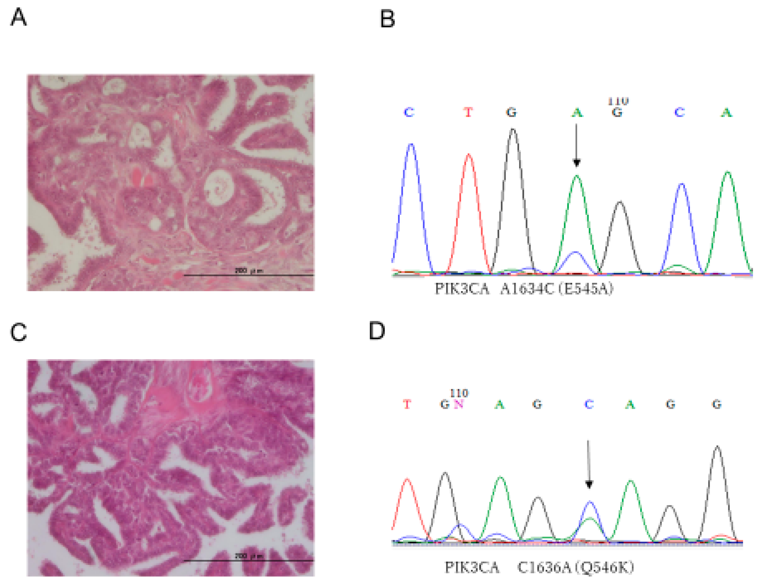
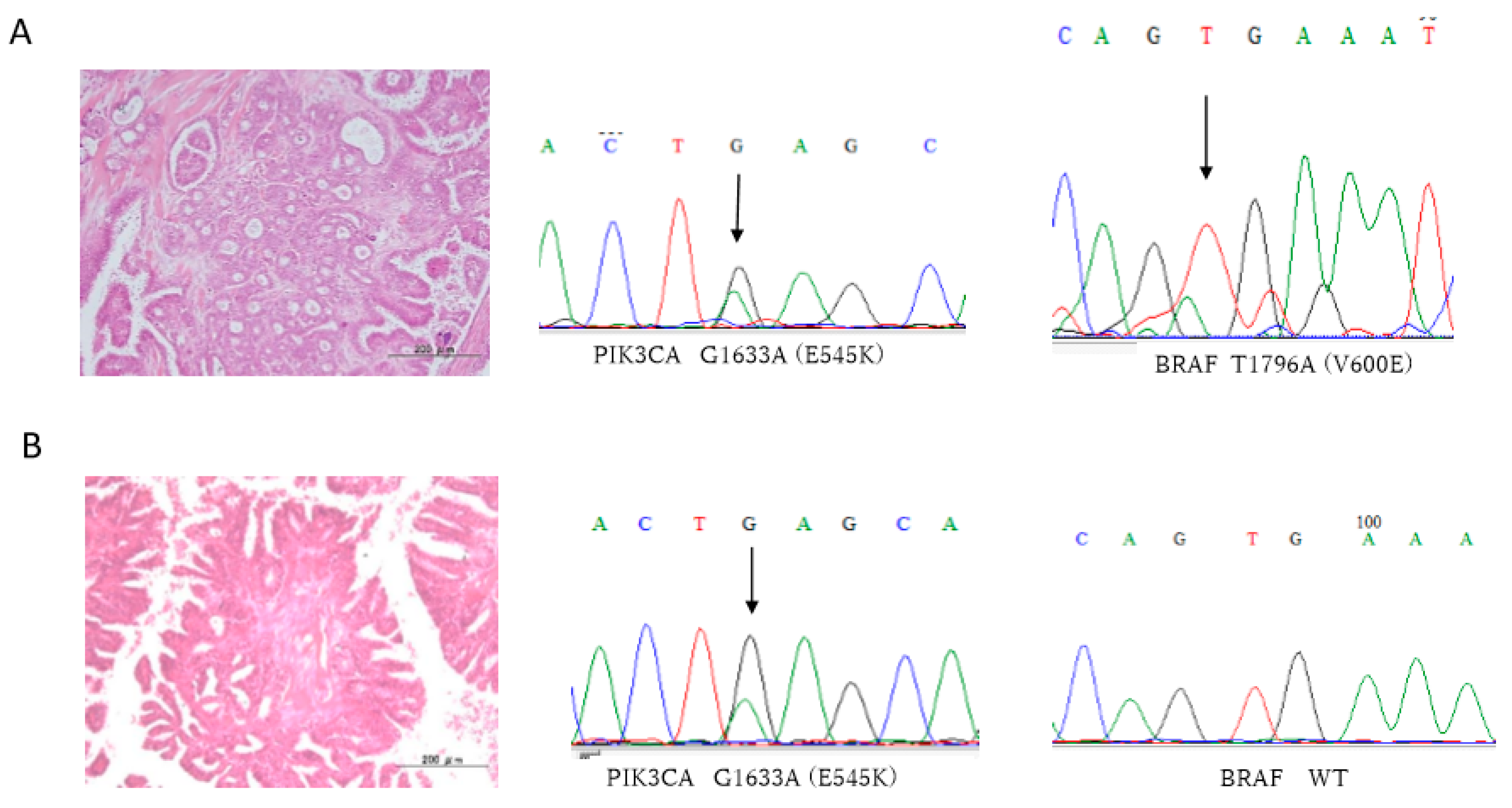
Loss of ARID1A expression in tumor cell nuclei was used as a surrogate for the presence of ARID1A loss-of-function mutations [9]. Similarly, p53 immunoreactivity was used as a surrogate for the presence of p53 loss-of-function mutations. The antibodies used in this study were a mouse monoclonal antibody against ARID1A (BAF250a) (Santa Cruz Biotechnology Santa Cruz, CA, USA) and mouse monoclonal antibody against p53 (clone DO-7, DAKO, Carpinteria, CA, USA). Immunohistochemistry for ARID1A and p53 was performed on tissue specimens at a dilution of 1:50 or 1:100, followed by detection using an EnVision+ System with the peroxidase method (DAKO, Carpinteria, CA, USA). The detail protocols for immunostaining and evaluation of ARID1A and P53 have been described in previous reports [9,10].

3. Results

All 33 ovarian serous tumors were assessed for mutations in KRAS, BRAF, PIK3CA, and ERBB2. Interestingly, all LGSC, SBT, and SCA cases showed wild-type KRAS variant (Table 1, Table 2 and Table 3). The prevalence of oncogenic mutation of PIK3CA was 60.0% (6/10) in LGSCs, 63.6% (7/11) in SBTs, and 8.3% (1/12) in SCAs. Representative histological images and nucleotide sequences in PIK3CA are shown in Figure 1. The prevalence of BRAF mutations was 20.0% (2/10) in LGSCs, whereas BRAF in both SBTs and SCAs were all wild-type. The prevalence of ERBB2 mutations was 30.0% (3/10) in LGSCs, 0.0% (0/10) in SBTs, and 16.7% (2/12) in SCAs. Details of PIK3CA, BRAF, and ERBB2 mutation types are shown in Table 1, Table 2 and Table 3. ARID1A staining was observed in all cases. Staining of p53 was found in 0.0% (0/10) of LGSCs, 9.1% (1/11) of SBTs, and 0.0% (0/12) of SCAs. Representative images of p53 and ARID1A staining are shown in Figure 2. We also analyzed the mutation status of KRAS, PIK3CA, BRAF, and ERBB2 in LGSCs with synchronous SBTs. Representative histological images and nucleotide sequences in PIK3CA and BRAF in a case with both LGSC and SBT are shown in Figure 3 and Table 4. One LGSC case had PIK3CA, BRAF, and ERBB2 mutations in the LGSC lesion but not in the SBT lesion. Another LGSC case had PIK3CA mutations (G1633A) in both LGSC and SBT lesions and a BRAF mutation only in the LGSC lesion. One other case had an ERBB2 mutation in both LGSC and SBT lesions, and another had no mutations in either lesion (Table 4).

4. Discussion

In the current study, wild-type KRAS was found in all Japanese LGSC, SBT, and SCA cases. In contrast, BRAF mutations were detected in 20% (2/10) of LGSCs. These findings are consistent with a recent report of a low frequency of KRAS and BRAF mutations in Chinese patients [11], suggesting that genes driving LGSC may be different in Asian and Western populations. Furthermore, in the current study, 3 out of 10 (30%) LGSC cases showed ERBB2 mutations. Previously, we identified ERBB2 mutations (9.5%) in Western patients [7]. The prevalence of ERBB2 mutations in the current study was much higher than that indicated in previous reports [7,12]. Interestingly, 16.7% of ERBB2 mutations were detected in SCAs, suggesting that ERBB2 mutation may be an early event in LGSC carcinogenesis. Taken together, the results of the current and previous reports also suggest that carcinogenesis of Japanese LGSCs may be different from that of Western LGSCs. Furthermore, the prevalence of oncogenic mutations in PIK3CA in both LGSCs and SBTs was much higher in Japanese patients than in Western patients [4,7,12,13]. Supplementary Table S2 shows the prevalence of KRAS, BRAF, PIK3CA, and ERBB2 mutations in European LGSCs. This high prevalence of oncogenic PIK3CA mutations in both SBTs and LGSCs suggests that these mutation events occur early in LGSC carcinogenesis. How does this discrepancy in PIK3CA mutation occur in patients with LGSC among different studies? One possibility is that the molecular mechanism of LGSC carcinogenesis differs between Japanese and other ethnicities. Another possibility is that the sample size in the current study was small and not representative of the Japanese population as a whole. The incidence of LGSC is quite low in Japan; therefore, a large multi-institutional cohort study is needed to confirm the current findings.
To determine the significance of PIK3CA, BRAF, and ERBB2 mutations in carcinogenesis of LGSCs, we analyzed these mutations in LGSCs with synchronous SBTs. One LGSC case had PIK3CA, BRAF, and ERBB2 mutations only in LGSC lesions but not in SBT lesions, whereas another LGSC case had PIK3CA mutations in both LGSC and SBT lesions. In contrast, BRAF mutations were detected only in LGSC lesions, suggesting that BRAF mutation is an important event in the SBT to LGSC transition. These findings suggest that both PIK3CA and ERBB2 mutations are important carcinogenesis events, leading from SCA to SBT and SBT to LGSC. In contrast, BRAF mutation may be a late event in LGSC carcinogenesis.
According to results of the current immunohistochemical study, loss of function mutations in ARID1A and TP53 are not critical events in LGSC carcinogenesis in Japanese patients. However, tumor cells from one SBT case stained positive for p53 (an indication of mutant TP53), suggesting that this SBT is a precursor of HGSC, and that there may be a pathway from Type I to Type II ovarian cancer [14].
Based on the current findings, we hypothesize that the main oncogenic signaling pathway in Japanese LGSCs is PIK3CA/AKT, whereas that in Western LGSCs is KRAS/BRAF/ERK (Figure 4). Recently, gene panel sequencing was introduced in clinical settings in Japan [15]. Current findings may contribute to genotyped matched therapies against LGSC in Japanese patients.
Oncology research teams from Johns Hopkins University and MD Anderson Cancer Center recently reported two lines of evidence supporting an LGSC carcinogenesis pathway. Peritoneal implants of SBTs and atypical proliferative serous tumors, which are considered precancerous lesions that will lead to LGSCs, had the same KRAS/BRAF mutation patterns as those of primary lesions, suggesting that these are metastatic lesions arising from primary lesions [16]. In addition, KRAS mutations, but not BRAF mutations, are involved in the progression of SBT to LGSC, and KRAS, a G12V mutation, indicates a poor prognosis [17]. Recently, Emmanuel et al. reported NRAS mutations in 9% of LGSCs with adjacent SBTs, suggesting NRAS is an oncogenic driver of LGSCs [18]. Furthermore, Gershenson et al. reported that patients with KRAS or BRAF mutations had significantly better overall survival times that those with wild-type KRAS or BRAF [19]. The Gynecologic Oncology Group recently performed a phase II study in the USA of selumentinib (AZD6244, ARRY142866), a MEK1/2 inhibitor in patients with recurrent LGSC [20]. According to their report, stable disease was observed in 65% of patients, with an overall disease control rate of 81%. Furthermore, there was no correlation between KRAS/BRAF mutations and response rates, so the predictive value of identifying KRAS/BRAF mutations against MEK inhibitor as biomarkers is still unclear. However, very recently, successful genotype-matched therapies against BRAF, KRAS and NRAS in patients with LGSC have been reported [21,22,23]. Unfortunately, these studies were based only on a Western, not Japanese or even Asian, population. Therefore, a large multi-institutional cohort study with genetic and epigenetic analyses is needed to elucidate the carcinogenic mechanism underlying LGSC in Japanese patients.
The current study has several limitations. First, the number of samples in this study was small. A follow-up study with an increased number of subjects is ongoing. This will enable us to determine, statistically, the relationship between the mutations identified in the present study and patient outcomes. Second, we identified genetic mutations via Sanger sequencing; therefore, the kinds of gene mutations assessed were limited. Further analyses using next-generation sequencing will also be needed to determine the molecular mechanism that underlies progression to LGSC in Japanese patients.
In summary, the current findings suggest that the mutation frequency of KRAS in LGSCs/SBTs in Japan is lower than that in Western countries. In addition, the mutation frequency of PIK3CA in LGSCs/SBTs appears to be very high in a Japanese population compared to Western populations. PIK3CA mutation may be a main driver and BRAF or ERBB2 mutation may be a sub-driver event in Japanese LGSCs. Therefore, molecular therapies targeting the PIK3CA/AKT pathway may be effective in LGSCs in Japan.

Supplementary Materials

The following are available online at https://www.mdpi.com/2075-4418/10/1/13/s1.

Author Contributions

T.I. and K.N. (Kentaro Nakayama) drafted the manuscript. T.I., M.I., K.N. (Kohei Nakamura), H.Y., K.I., N.I., P.D., and S.R. carried out the molecular genetic studies. K.N. (Kentaro Nakayama) participated in the design of the study. H.K., S.N., Y.O., and N.I. carried out the pathological diagnosis. S.K. conceived of the study, participated in its design and coordination, and helped in drafting the manuscript. All authors have read and agreed to the published version of the manuscript.

Funding

This work was supported by JSPS KAKENHI grant numbers 18K09229 and 18K09291.

Conflicts of Interest

The authors declare no conflicts of interest.

References

  1. Siegel, R.L.; Miller, K.D.; Jemal, A. Cancer statistics, 2019. CA Cancer J. Clin. 2019, 69, 7–34. [Google Scholar] [CrossRef] [Green Version]
  2. Kurman, R.J.; Shih, I.M. The dualistic model of ovarian carcinogenesis: Revisited, revised, and expanded. Am. J. Pathol. 2016, 186, 733–747. [Google Scholar] [CrossRef] [PubMed] [Green Version]
  3. Wong, K.K.; Tsang, Y.T.; Deavers, M.T.; Mok, S.C.; Zu, Z.; Sun, C.; Malpica, A.; Wolf, J.K.; Lu, K.H.; Gershenson, D.M. BRAF mutation is rare in advanced-stage low-grade ovarian serous carcinomas. Am. J. Pathol. 2010, 177, 1611–1617. [Google Scholar] [CrossRef] [PubMed]
  4. Jones, S.; Wang, T.L.; Kurman, R.J.; Nakayama, K.; Velculescu, V.E.; Vogelstein, B.; Kinzler, K.W.; Papadopoulos, N.; Shih, I.M. Low-grade serous carcinomas of the ovary contain very few point mutations. J. Pathol. 2012, 226, 413–420. [Google Scholar] [CrossRef]
  5. Singer, G.; Kurman, R.J.; Chang, H.W.; Cho, S.K.; Shih, I.M. Diverse tumorigenic pathways in ovarian serous carcinoma. Am. J. Pathol. 2002, 160, 1223–1228. [Google Scholar] [CrossRef] [Green Version]
  6. Nakamura, K.; Nakayama, K.; Ishibashi, T.; Ishikawa, N.; Ishikawa, M.; Katagiri, H.; Minamoto, T.; Sato, E.; Sanuki, K.; Yamashita, H.; et al. KRAS/BRAF analysis in ovarian low-grade serous carcinoma having synchronous all pathological precursor regions. Int. J. Mol. Sci. 2016, 17, 625. [Google Scholar] [CrossRef] [Green Version]
  7. Nakayama, K.; Nakayama, N.; Kurman, R.J.; Cope, L.; Pohl, G.; Samuels, Y.; Velculescu, V.E.; Wang, T.L.; Shih, I.M. Sequence mutations and amplification of PIK3CA and AKT2 genes in purified ovarian serous neoplasms. Cancer Biol. Ther. 2006, 5, 779–785. [Google Scholar] [CrossRef] [PubMed] [Green Version]
  8. Forbes, S.A.; Beare, D.; Boutselakis, H.; Bamford, S.; Bindal, N.; Tate, J.; Cole, C.G.; Ward, S.; Dawson, E.; Ponting, L.; et al. COSMIC: Somatic cancer genetics at high-resolution. Nucleic Acids Res. 2017, 45, D777–D783. [Google Scholar] [CrossRef] [PubMed]
  9. Katagiri, A.; Nakayama, K.; Rahman, M.T.; Rahman, M.; Katagiri, H.; Nakayama, N.; Ishikawa, M.; Ishibashi, T.; Iida, K.; Kobayashi, H.; et al. Loss of ARID1A expression is related to shorter progression-free survival and chemoresistance in ovarian clear cell carcinoma. Mod. Pathol. 2012, 25, 282–288. [Google Scholar] [CrossRef] [PubMed] [Green Version]
  10. Nakayama, K.; Takebayashi, Y.; Nakayama, S.; Hata, K.; Fujiwaki, R.; Fukumoto, M.; Miyazaki, K. Prognostic value of overexpression of p53 in human ovarian carcinoma patients receiving cisplatin. Cancer Lett. 2003, 192, 227–235. [Google Scholar] [CrossRef]
  11. Xu, Y.; Bi, R.; Xiao, Y.; Tu, X.; Li, M.; Li, A.; Shan, L.; Zhou, S.; Yang, W. Low frequency of BRAF and KRAS mutations in Chinese patients with low-grade serous carcinoma of the ovary. Diagn. Pathol. 2017, 12, 87. [Google Scholar] [CrossRef] [Green Version]
  12. Van Nieuwenhuysen, E.; Busschaert, P.; Laenen, A.; Moerman, P.; Han, S.N.; Neven, P.; Lambrechts, D.; Vergote, I. Loss of 1p36.33 frequent in low-grade serous ovarian cancer. Neoplasia 2019, 21, 582–590. [Google Scholar] [CrossRef]
  13. Hunter, S.M.; Anglesio, M.S.; Ryland, G.L.; Sharma, R.; Chiew, Y.E.; Rowley, S.M.; Doyle, M.A.; Li, J.; Gilks, C.B.; Moss, P.; et al. Molecular profiling of low grade serous ovarian tumours identifies novel candidate driver genes. Oncotarget 2015, 6, 37663–37677. [Google Scholar] [CrossRef] [Green Version]
  14. Kurman, R.J.; Shih, I.M. Pathogenesis of ovarian cancer: Lessons from morphology and molecular biology and their clinical implications. Int. J. Gynecol. Pathol. 2008, 27, 151–160. [Google Scholar] [CrossRef] [Green Version]
  15. Kohno, T. Implementation of “clinical sequencing” in cancer genome medicine in Japan. Cancer Sci. 2018, 109, 507–512. [Google Scholar] [CrossRef] [Green Version]
  16. Ardighieri, L.; Zeppernick, F.; Hannibal, C.G.; Vang, R.; Cope, L.; Junge, J.; Kjaer, S.K.; Kurman, R.J.; Shih, I.M. Mutational analysis of BRAF and KRAS in ovarian serous borderline (atypical proliferative) tumours and associated peritoneal implants. J. Pathol. 2014, 232, 16–22. [Google Scholar] [CrossRef] [Green Version]
  17. Tsang, Y.T.; Deavers, M.T.; Sun, C.C.; Kwan, S.Y.; Kuo, E.; Malpica, A.; Mok, S.C.; Gershenson, D.M.; Wong, K.K. KRAS (but not BRAF) mutations in ovarian serous borderline tumour are associated with recurrent low-grade serous carcinoma. J. Pathol. 2013, 231, 449–456. [Google Scholar] [CrossRef] [Green Version]
  18. Emmanuel, C.; Chiew, Y.E.; George, J.; Etemadmoghadam, D.; Anglesio, M.S.; Sharma, R.; Russell, P.; Kennedy, C.; Fereday, S.; Hung, J.; et al. Australian Ovarian Cancer Study (AOCS). Genomic classification of serous ovarian cancer with adjacent borderline differentiates RAS pathway and TP53-mutant tumors and identifies NRAS as an oncogenic driver. Clin. Cancer Res. 2014, 20, 6618–6630. [Google Scholar] [CrossRef] [Green Version]
  19. Gershenson, D.M.; Sun, C.C.; Wong, K.K. Impact of mutational status on survival in low-grade serous carcinoma of the ovary or peritoneum. Br. J. Cancer 2015, 113, 1254–1258. [Google Scholar] [CrossRef] [Green Version]
  20. Farley, J.; Brady, W.E.; Vathipadiekal, V.; Lankes, H.A.; Coleman, R.; Morgan, M.A.; Mannel, R.; Yamada, S.D.; Mutch, D.; Rodgers, W.H.; et al. Selumetinib in women with recurrent low-grade serous carcinoma of the ovary or peritoneum: An open-label, single-arm, phase 2 study. Lancet Oncol. 2013, 14, 134–140. [Google Scholar] [CrossRef] [Green Version]
  21. Mendivil, A.A.; Tung, P.K.; Bohart, R.; Bechtol, K.; Goldstein, B.H. Dramatic clinical response following dabrafenib and trametinib therapy in a heavily pretreated low grade serous ovarian carcinoma patient with a BRAF V600E mutation. Gynecol. Oncol. Rep. 2018, 26, 41–44. [Google Scholar] [CrossRef] [PubMed]
  22. Champer, M.; Miller, D.; Kuo, D.Y. Response to trametinib in recurrent low-grade serous ovarian cancer with NRAS mutation: A case report. Gynecol. Oncol. Rep. 2019, 28, 26–28. [Google Scholar] [CrossRef] [PubMed]
  23. Rodriguez-Freixinos, V.; Lheureux, S.; Mandilaras, V.; Clarke, B.; Dhani, N.C.; Mackay, H.; Butler, M.O.; Wang, L.; Siu, L.L.; Kamel-Reid, S.; et al. Impact of somatic molecular profiling on clinical trial outcomes in rare epithelial gynecologic cancer patients. Gynecol. Oncol. 2019, 153, 304–311. [Google Scholar] [CrossRef] [PubMed]
Figure 1. Histopathological images and nucleotide sequences of PIK3CA in representative LGSC and SBT cases. (A) Hematoxylin and eosin staining of LGSC sections. (B) Nucleotide sequence chromatogram showing a mutation, E545A (1634 A > C), in PIK3CA of an LGSC. (C) Hematoxylin and eosin staining of SBT sections. (D) Nucleotide sequence chromatogram showing a mutation, Q546K (1636 C > A), in PIK3CA of an SBT. Scale bar = 200 µm. C; cytosine, T; thymine, G; guanine, A; adenine.
Diagnostics 10 00013 g001
Figure 2. Representative positive staining of p53 (A) and ARID1A (B). Nuclear staining of p53 and ARID1A in an LGSC case.
Diagnostics 10 00013 g002
Figure 3. Synchronous LGSCs and SBTs with matched PIK3CA and BRAF sequences in an LGSC case. (A) LGSC showing mutations E545K (1633 G > A), in PIK3CA, and V600E (1796T > A), in BRAF. (B) SBT showing a mutation, E545K (1633 G > A), in PIK3CA, and wild-type BRAF. Scale bar = 200 µm. C; cytosine, T; thymine, G; guanine, A; adenine.
Diagnostics 10 00013 g003
Figure 4. Hypothesized differences in carcinogenesis between Japanese and European patients with LGSC. LGSC in Japanese patients may depend on alterations in the PIK3CA/AKT pathway, whereas in Europeans it may depend on alterations in the KRAS/BRAF/AKT pathway.
Diagnostics 10 00013 g004
Table 1. Mutation and imunohistochemical analysis of low grade serous ovarian carcinoma (LGSC). WT; Wild Type.
No.AgeFIGO StageKRASBRAFPI3KCA E9PI3KCA E20ERBB2P53ARID1A
137II cWTWTWTWTA2384G (Q795R)NormalNormal
261IV bWTWTG1633A (E545K)WTWTNormalNormal
383I aWTWTA1634C (E545A)WTWTNormalNormal
461I cWTWTWTWTA2384G (Q795R)NormalNormal
537III cWTWTG1633C (E545Q)WTWTNormalNormal
627I cWTT1796A (V600E)A1634C (E545A)WTA2384G (Q795R)NormalNormal
761III cWTWTWTWTWTNormalNormal
848I cWTWTWTWTWTNormalNormal
926III cWTWTG1633A (E545K)WTWTNormalNormal
1040I cWTT1796A (V600E)A1634C (E545A)WTWTNormalNormal
Table 2. Mutation and immunohistochemical analysis of serous borderline tumor (SBT). WT; Wild Type.
No.AgeFIGO StageKRASBRAFPI3KCA E9PI3KCA E20ERBB2P53ARID1A
1132I aWTWTWTWTWTNormalNormal
1239I cWTWTC1636A (Q546K)WTWTNormalNormal
1344III cWTWTWTWTWTNormalNormal
1445I cWTWTC1636A (Q546K)WTWTNormalNormal
1545III cWTWTG1633C (E545Q)WTWTNormalNormal
1638I aWTWTA1634C (E545A)WTWTPositiveNormal
1725I aWTWTWTWTWTNormalNormal
1848I aWTWTC1636A (Q546K)WTWTNormalNormal
1969I aWTWTC1636A (Q546K)WTWTNormalNormal
2057I aWTWTC1636A (Q546K)WTWTNormalNormal
2166I cWTWTWTWTWTNormalNormal
Table 3. Mutation and immunohistochemical analysis of serous cystadenoma (SCA). WT; Wild Type.
No.AgeKRASBRAFPI3KCA E9PI3KCA E20ERBB2P53ARID1A
2225WTWTWTWTWTNormalNormal
2373WTWTWTWTWTNormalNormal
2481WTWTWTWTWTNormalNormal
2547WTWTWTWTWTNormalNormal
2652WTWTWTWTA2384G (Q795R)NormalNormal
2771WTWTWTWTWTNormalNormal
2872WTWTWTWTWTNormalNormal
2975WTWTA1634C (E545A)WTWTNormalNormal
3055WTWTWTWTA2384G (Q795R)NormalNormal
3163WTWTWTWTWTNormalNormal
3263WTWTWTWTWTNormalNormal
3326WTWTWTWTWTNormalNormal
Table 4. Mutation and immunohihistochemical analysis in four cases of LSGC accompanied by SBT. WT; Wild Type.
No.AgeStageLGSC/SBTKRASBRAFPI3KCA E9PI3KCA E20ERBB2P53ARID1A
137III cLGSC
SBT
WT
WT
WT
WT
WT
WT
WT
WT
A2384G (Q795R)
A2384G (Q795R)
Normal
Normal
Normal
Normal
627I cLGSC
SBT
WT
WT
T1796A (V600E)
WT
A1634G (E545G)
WT
WT
WT
A2384G (Q795R)
WT
Normal
Normal
Normal
Normal
848I cLGSC
SBT
WT
WT
WT
WT
WT
WT
WT
WT
WT
WT
Normal
Normal
Normal
Normal
1040I cLGSC
SBT
WT
WT
T1796A (V600E)
WT
A1634C (E545A)
A1634C (E545A)
WT
WT
WT
WT
Normal
Normal
Normal
Normal

