Clinico-Pathological Presentations of Cystic and Classic Adenomatoid Odontogenic Tumors
Abstract
:1. Introduction
2. Materials and Method
3. Results
4. Discussion
Author Contributions
Funding
Conflicts of Interest
References
- Barnes, L.; Eveson, J.W.; Reichart, P.; Sidransky, D. World Health Organization Classification of Tumours, Pathology and Genetics of Head and Neck Tumours; Philipsen, H.P., Nikai, H., Eds.; IARC Press: Lyon, France, 2005; Chapter 6; pp. 304–305. [Google Scholar]
- Kramer, I.R.H.; Pindborg, J.J.; Shear, M. Histological typing of odontogenic tumours. In WHO International Histological Classification of Tumours, 2nd ed.; Springer: London, UK, 1992. [Google Scholar]
- Jivan, V.; Altini, M.; Meer, S. Secretory cells in adenomatoid odontogenic tumour: Tissue induction or metaplastic mineralization? Oral Dis. 2008, 14, 445–449. [Google Scholar] [CrossRef] [PubMed]
- El-Naggar, A.K.; Chan, J.K.C.; Grandis, J.R.; Takata, T.; Slootweg, P.J. World Health Organization Classification of Head and Neck Tumours; Wright, J.M., Kusama, K., Eds.; IARC Press: Lyon, France, 2017; Chapter 8; pp. 221–222. [Google Scholar]
- Gadewar, D.R.; Srikant, N. Adenomatoid odontogenic tumour: Tumour or a cyst, a histopathological support for the controversy. Int. J. Pediatric Otorhinolaryngol. 2010, 74, 333–337. [Google Scholar] [CrossRef] [PubMed]
- Grover, S.; Rahim, A.M.B.; Parakkat, N.K.; Kapoor, S.; Mittal, K.; Sharma, B.; Shivappa, A.B. Cystic Adenomatoid Odontogenic Tumour. Case Rep. Dent. 2015. [Google Scholar] [CrossRef] [PubMed] [Green Version]
- Ide, F.; Muramatsu, T.; Ito, Y.; Kikuchi, K.; Miyazaki, Y.; Saito, I.; Kusama, K. An expanded and revised early history of the adenomatoid odontogenic tumour. Oral Surg. Oral Med. Oral Pathol. Oral Radiol. 2013, 115, 646–651. [Google Scholar] [CrossRef] [PubMed]
- Philipsen, H.P.; Reichart, P.A.; Zhang, K.H.; Nikai, H.; Yu, X.U. Adenomatoid odontogenic tumour: Biological profile based on 499 cases. J. Oral Pathol. Med. 1991, 20, 149–158. [Google Scholar] [CrossRef] [PubMed]
- Philipsen, H.P.; Reichart, P.A. Adenomatoid odontogenic tumour: Facts and figures. Oral Oncol. 1999, 35, 125–131. [Google Scholar] [CrossRef]
- Philipsen, H.P.; Reichart, P.A.; Siar, C.H.; Ng, K.H.; Lau, S.H.; Zhang, X.; Jivan, V. An updated clinical and epidemiological profile of the adenomatoid odontogenic tumour: A collaborative retrospective study. J. Oral Pathol. Med. 2007, 36, 383–393. [Google Scholar] [CrossRef] [PubMed]
- Leon, J.E.; Mata, G.M.; Fregnani, E.R.; Carlos-Bregni, R.; de Almeida, O.P.; Mosqueda-Taylor, A.; Vargas, P.A. Clinicopathological and immunohistochemical study of 39 cases of Adenomatoid Odontogenic Tumour: A multicentric study. Oral Oncol. 2005, 41, 835–842. [Google Scholar] [CrossRef] [PubMed]
- De Matos, F.R.; Nonaka, C.F.; Pinto, L.P.; De Souza, L.B.; Freitas, R.D.A. Adenomatoid odontogenic tumour: Retrospective study of 15 cases with emphasis on histopathological features. Head Neck Pathol. 2012, 6, 430–437. [Google Scholar] [CrossRef] [PubMed] [Green Version]
- Muzio, L.L.; Mascitti, M.; Santarelli, A.; Rubini, C.; Bambini, F.; Procaccini, M.; Nocini, P.F. Cystic lesions of the jaws: A retrospective clinicopathological study of 2030 cases. Oral Surg. Oral Med. Oral Pathol. Oral Radiol. 2017, 124, 128–138. [Google Scholar] [CrossRef] [PubMed]
- Saxena, K.; Jose, M.; Chatra, L.K.; Sequiera, J. Adenoid ameloblastoma with dentinoid. J. Oral Maxillofac. Pathol. 2012, 16, 272–276. [Google Scholar] [CrossRef] [PubMed] [Green Version]
- Harnet, J.C.; Pedeutour, F.; Raybaud, H.; Ambrosetti, D.; Fabas, T.; Lombardi, T. Immunohistochemical features in adenomatoid odontogenic tumour: Review of the literature and first expression and mutational analysis of beta-Catenin in this unusual lesion of the jaws. J. Oral Maxillofac. Surg. 2013, 71, 706–713. [Google Scholar] [CrossRef] [PubMed] [Green Version]
- Chuan-Xiang, Z.; Yan, G. Adenomatoid odontogenic tumour: A report of a rare case with recurrence. J. Oral Pathol. Med. 2007, 36, 440–443. [Google Scholar] [CrossRef] [PubMed]
- Ide, F.; Mishima, K.; Saito, I.; Kusama, K. Diagnostically challenging epithelial odontogenic tumours: A selective review of 7 jawbone lesions. Head Neck Pathol. 2009, 3, 18–26. [Google Scholar] [CrossRef] [PubMed] [Green Version]
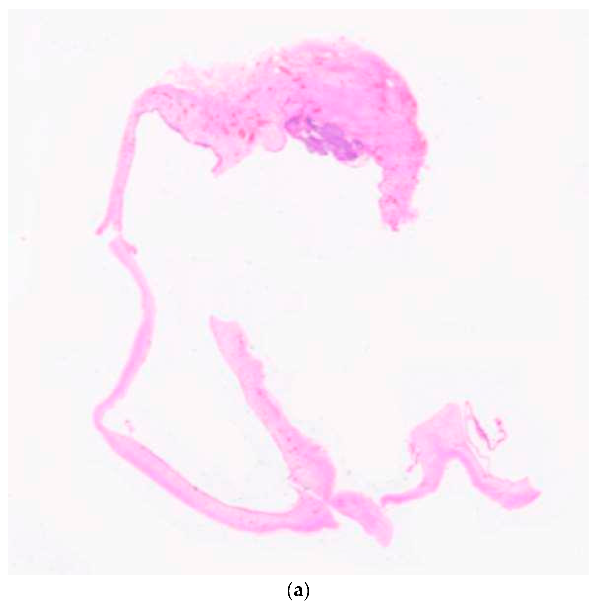
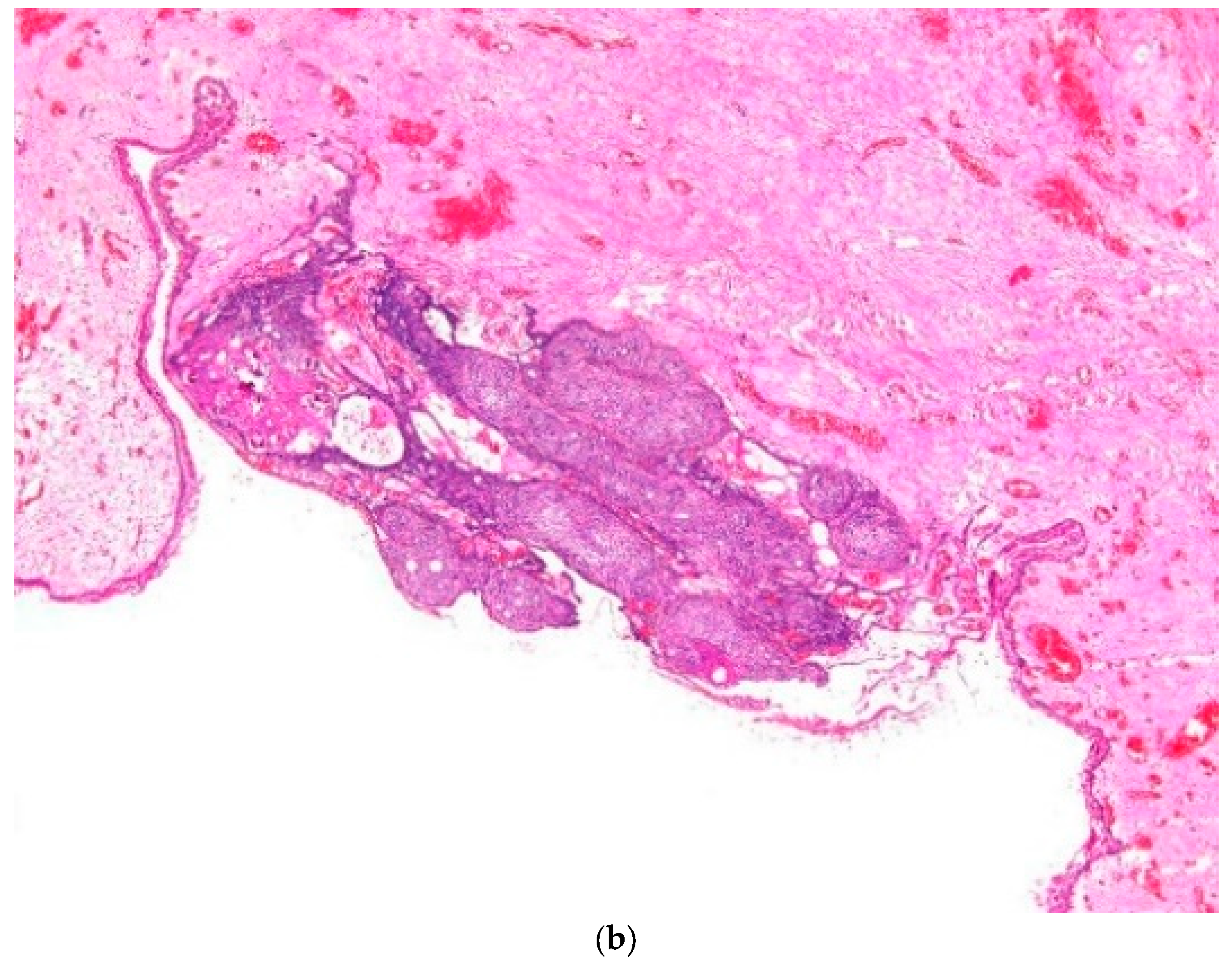
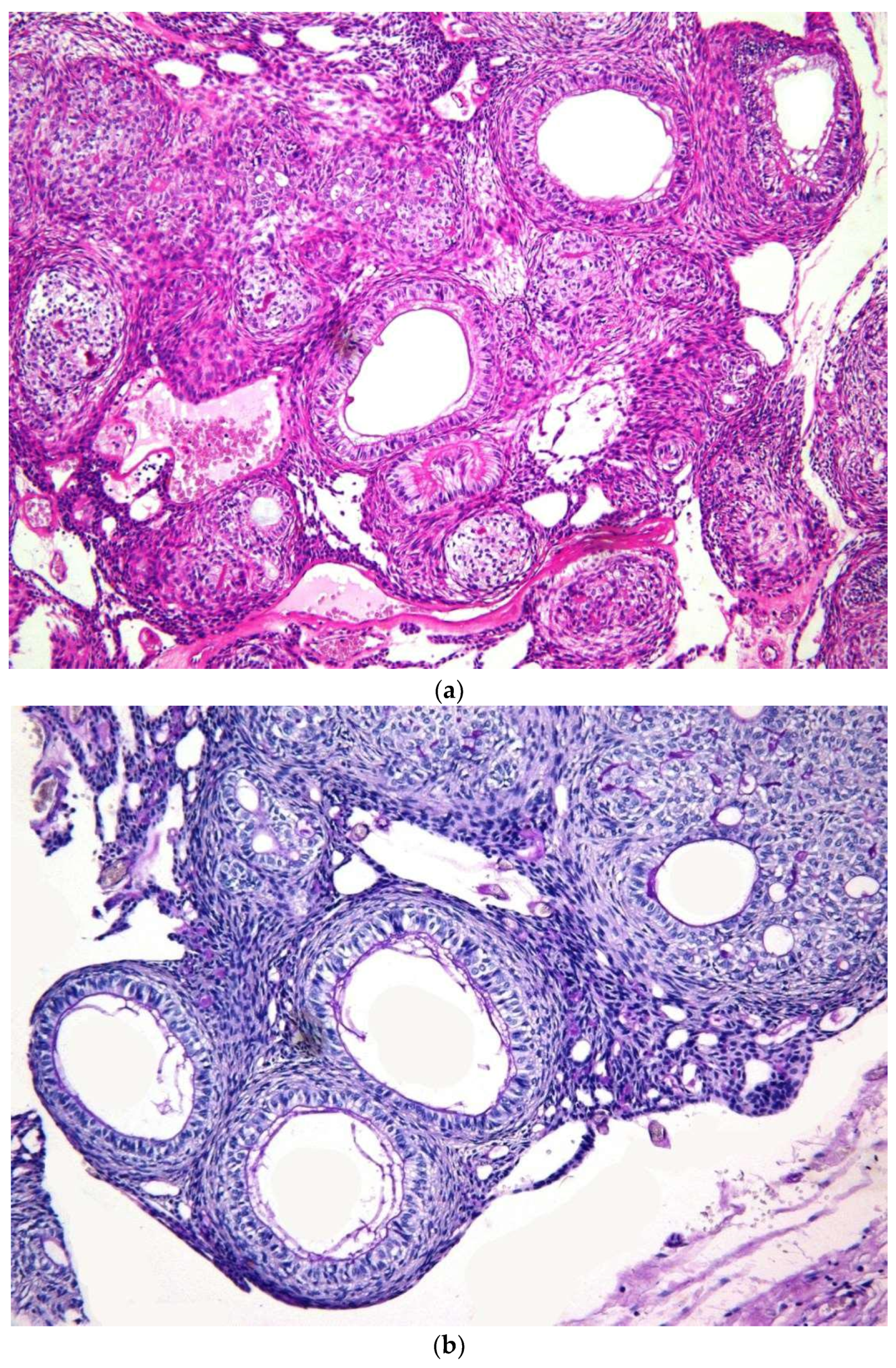
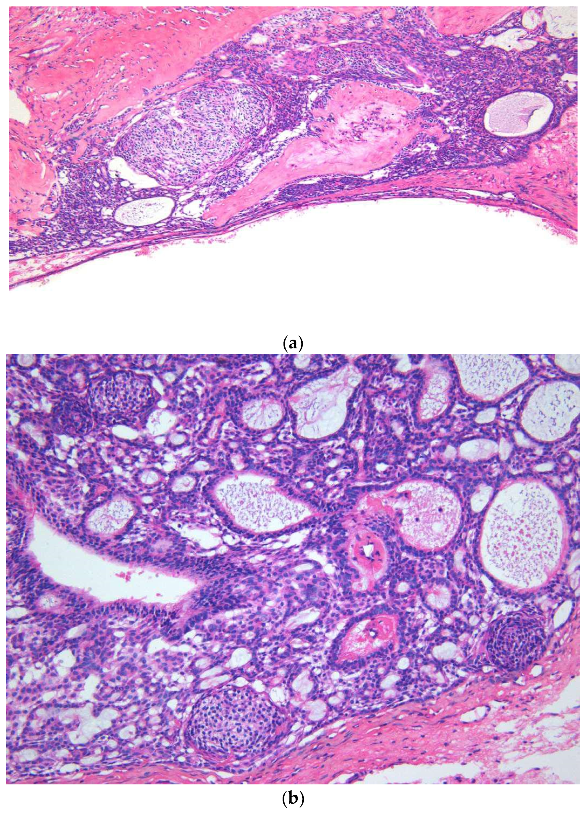
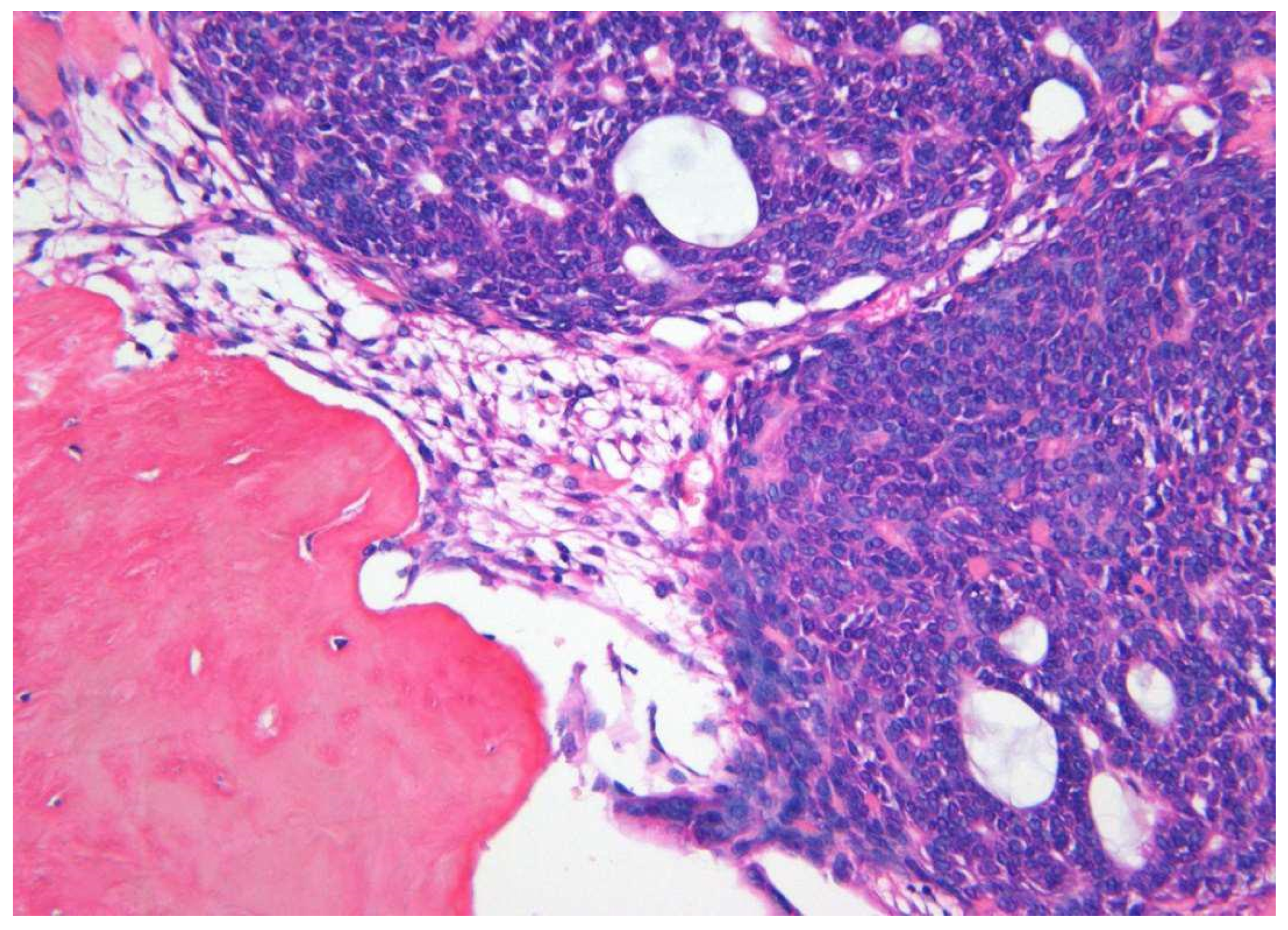


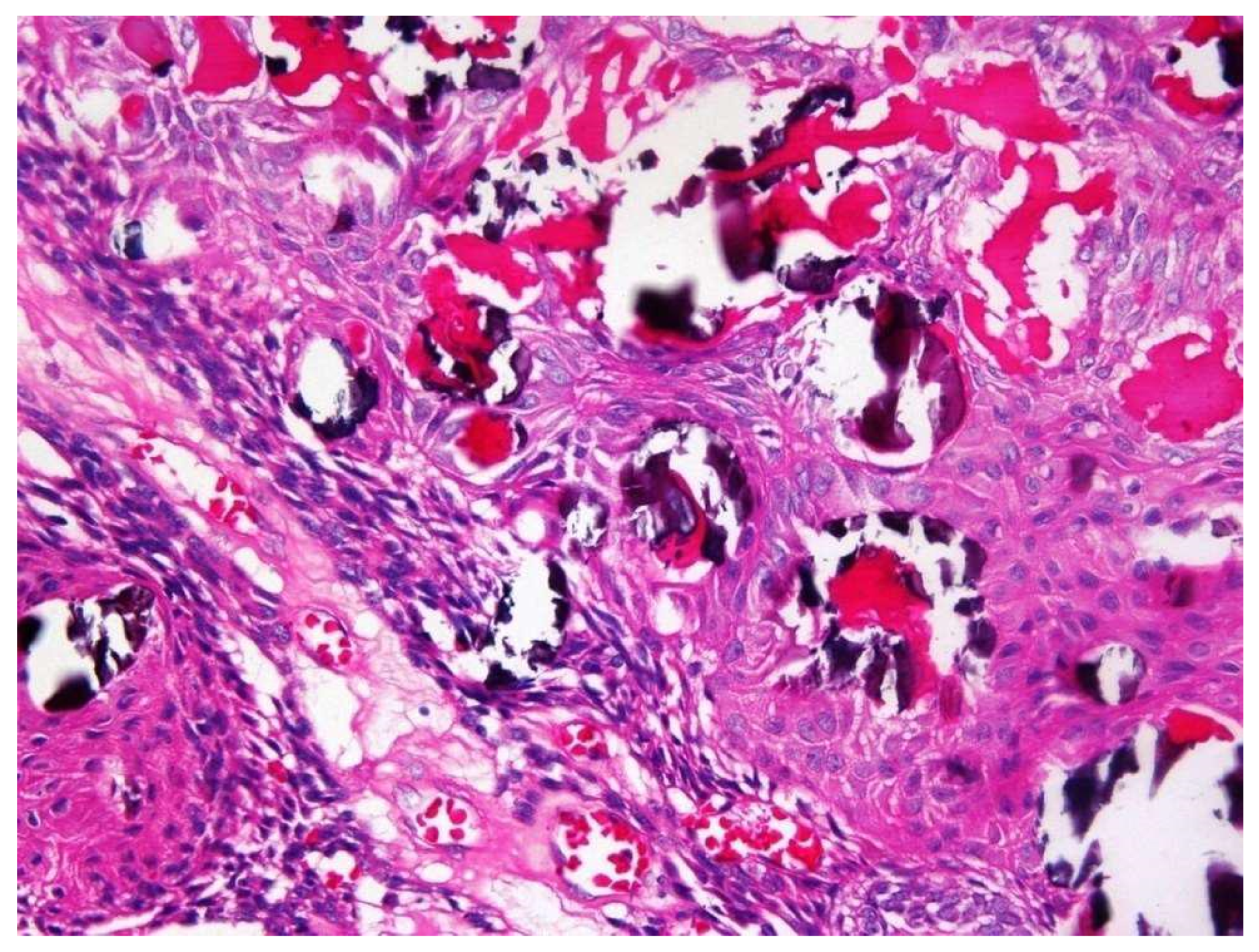
| Clinical Feature | Cystic AOT (%) n = 11 | Classic AOT (%) n = 30 | Total (%) n = 41 | p Value |
|---|---|---|---|---|
| Age | ||||
| 10–15 yrs | 04 (36.4) | 11 (36.7) | 15 | |
| 16–20 yrs | 04 (36.4) | 15 (50.0) | 19 | p = 0.4 |
| 21–25 yrs | 00 | 03 (10.0) | 03 | |
| >26 yrs | 03 (27.2) | 01 (03.3) | 04 | |
| Gender | ||||
| Female | 06 (54.5) | 22 (73.3) | 28 | p = 0.3 |
| Male | 05 (45.5) | 08 (26.7) | 13 | |
| Site | ||||
| Maxilla | 08 (72.8) | 19 (63.3) | 27 | |
| Mandible | 03 (27.2) | 10 (33.4) | 13 | p = 0.7 |
| Unknown | 00 | 01 (03.3) | 01 | |
| Size | ||||
| Less than 3 × 3 cm | 01 (09.1) | 12 (40.0) | 13 | |
| More than 3 × 3 cm | 07 (63.6) | 08 (26.7) | 15 | p = 0.2 |
| unknown | 03 (27.3) | 10 (33.3) | 13 | |
| Radiological presentation | ||||
| Follicular | 08 (72.8) | 14 (46.7) | 22 (53.7) | |
| Extra follicular | 03 (27.2) | 14 (46.7) | 17 (41.5) | p = 0.2 |
| Peripheral | 00 | 01 (03.3) | 01 (02.4) | |
| Unknown | 00 | 01 (03.3) | 01 (02.4) | |
| Histopathology | ||||
| 1. Capsule-present | 10 (90.9) | 24 (80.0) | 34 | |
| 2. Epithelial component | ||||
| 2a. Duct like structures | 09 (81.8) | 27 (90.0) | 36 | |
| 2b. Epithelial whorls | 11 (100) | 29 (96.6) | 40 | p = 0.7 |
| 2c. Rosettes | 03 (27.2) | 19 (63.3) | 22 | |
| 2d. Trabeculae | 08 (72.8) | 25 (83.3) | 33 | |
| 3. Stromal component | ||||
| 3a. Tumour droplets | 08 (72.8) | 20 (66.6) | 28 | |
| 3b. Calcifications | 11 (100) | 29 (96.6) | 40 | p = 0.8 |
| 3c. Osteo-dentine | 01 (09.1) | 01 (03.3) | 02 | |
| 3d. Melanin | 00 | 01 (03.3) | 01 | |
| Type of surgery | ||||
| Enucleation | 10 (90.9) | 28 (93.3) | 38 | p = 0.7 |
| Radical surgery | 1 (03.3) | 2 (06.7) | 3 |
| Clinical Features of Cystic AOT | Published Cases | Present Cases (n = 11) | ||
|---|---|---|---|---|
| All cystic AOT (n = 19) | AOT arising in dentigerous cysts (n = 12) | AOT arising in dentigerous cysts (n = 08) | AOT arising in unclassifiable odontogenic cysts (n = 03) | |
| Age | Range 0–40 yrs | Range 8–25 yrs | Range 13–27 yrs | Range 18–29yrs |
| Average 19.5 yrs | Average 15.5 yrs | Average 16.7yrs | Average 24.3 yrs | |
| Gender | 12 out of 19 occurred in males | 7 out of 12 occurred in males | 5 out of 8 occurred in males | One out of 3 occurred in a male |
| Male:female ratio 1.7:1 | Male: female ratio 1.4:1 | Male: female ratio 1.6:1 | Male:female ratio 0.5:1 | |
| Site | 11 out of 19 occurred in maxilla | 11 out of 12 occurred in the maxilla | 5 out of 8 cases occurred in the maxilla | All 3 cases occurred in the maxilla |
| Maxilla:mandible ratio 1.4:1 | Maxilla:mandible ratio 11:1 | Maxilla:mandible ratio 1.6:1 | ||
| Location according to teeth present | Canine n = 9, premolar/molar n = 6 | Canine n = 7, premolar = 3, molar = 2 | Incisor n = 1, canine n = 5, premolar n = 1, molar n = 1 | Incisor n = 1, premolar/molar n = 2 |
| Clinical Feature | Classic AOT (n = 30) | AOT + CEOT (n = 9) | Classic CEOT (n = 9) |
|---|---|---|---|
| Age | Range 13–33 yrs | Range 15–25 yrs | Range 26–58 yrs |
| Average 18 yrs | Average 17.8 yrs | Average 40 yrs | |
| Gender | 22 out of 30 occurred in females | 7 out of 9 occurred in females | 5 out of 9 occurred in females |
| Male: female ratio 1:2.75 | Male:female ratio 1: 3.5 | Male:female ratio 1: 1.25 | |
| Site | 19 out of 29 occurred in maxilla | 6 out of 9 occurred in maxilla | 1 out of 9 occurred in maxilla |
| Maxilla:mandible ratio 1.9:1 | Maxilla:mandible ratio 2:1 | Maxilla:mandible ratio 1:9 | |
| Location in the jaw bones | Anterior = 24 | Anterior = 7 | Anterior = 1 |
| Premolar/molar = 5 | Premolar/molar = 2 | Premolar/molar = 5 | |
| Angle of the mandible = 0 | Angle of the mandible = 0 | Angle of the mandible = 3 | |
| Recurrences | None | None | 2 out of 9 lesions presented with recurrences within 5 years after treatment |
© 2019 by the authors. Licensee MDPI, Basel, Switzerland. This article is an open access article distributed under the terms and conditions of the Creative Commons Attribution (CC BY) license (http://creativecommons.org/licenses/by/4.0/).
Share and Cite
Jayasooriya, P.R.; Rambukewella, I.K.; Tilakaratne, W.M.; Mendis, B.R.R.N.; Lombardi, T. Clinico-Pathological Presentations of Cystic and Classic Adenomatoid Odontogenic Tumors. Diagnostics 2020, 10, 3. https://doi.org/10.3390/diagnostics10010003
Jayasooriya PR, Rambukewella IK, Tilakaratne WM, Mendis BRRN, Lombardi T. Clinico-Pathological Presentations of Cystic and Classic Adenomatoid Odontogenic Tumors. Diagnostics. 2020; 10(1):3. https://doi.org/10.3390/diagnostics10010003
Chicago/Turabian StyleJayasooriya, Primali Rukmal, Inoka Krishanthi Rambukewella, Wanninayake Mudiyanselage Tilakaratne, Balapuwaduge Ranjit Rigobert Nihal Mendis, and Tommaso Lombardi. 2020. "Clinico-Pathological Presentations of Cystic and Classic Adenomatoid Odontogenic Tumors" Diagnostics 10, no. 1: 3. https://doi.org/10.3390/diagnostics10010003
APA StyleJayasooriya, P. R., Rambukewella, I. K., Tilakaratne, W. M., Mendis, B. R. R. N., & Lombardi, T. (2020). Clinico-Pathological Presentations of Cystic and Classic Adenomatoid Odontogenic Tumors. Diagnostics, 10(1), 3. https://doi.org/10.3390/diagnostics10010003


