Article

Non-Small-Cell Lung Cancer-Sensitive Detection of the p.Thr790Met EGFR Alteration by Preamplification before PNA-Mediated PCR Clamping and Pyrosequencing

by
Amandine Billaud
1,2,
Veronique Verriele
2,
Jonathan Dauvé
2,
Louise-Marie Chevalier
1,2 and
Alain Morel
1,2,*
1
Université d’Angers, Inserm, CRCINA, F-49000 Angers, France
2
Institut de Cancérologie de l’Ouest Nantes-Angers, 49000 Angers, France
*
Author to whom correspondence should be addressed.
Diagnostics 2020, 10(8), 527; https://doi.org/10.3390/diagnostics10080527
Submission received: 27 May 2020 / Revised: 19 July 2020 / Accepted: 27 July 2020 / Published: 29 July 2020
(This article belongs to the Section Pathology and Molecular Diagnostics)

Abstract

:
Targeted therapies and, more precisely, EGFR tyrosine kinase inhibitors (TKIs) have been a major improvement in the therapeutic management of EGFR-mutated non-small-cell lung cancers (NSCLCs). Earlier administration of these TKIs throughout tumor progression is imperative to improve patient outcomes. Consequently, studies have focused on refining the characterization of biomarkers, especially concerning the resistance mutation p.Thr790Met of EGFR. Herein, we developed peptide nucleic acid (PNA)-mediated PCR clamping followed by pyrosequencing, favoring enrichment of the mutated fraction. A preamplification step was first added to increase the amplifiable DNA fraction. Throughout the application of our method on DNA extracted from FFPE samples of 46 patients with NSCLC who had relapsed under first-generation EGFR TKI, we evaluated a sensitivity of 93.3% and a specificity of 100%. All 19 patients who were positive for the p.Thr790Met mutation with NGS were also found to be positive with our protocol. The only discordant case was a sample with no mutation detected with NGS, but which was positive with PNA. This protocol allows for the detection of the p.Thr790Met mutation with a sensitivity of 0.5% which will permit earlier detection and an improvement of therapeutic management.

1. Introduction

As a leading cause of cancer-related death worldwide (18.6%) [1], lung cancers are divided into two classes: small-cell lung cancer (SCLC) and non-small-cell lung cancer (NSCLC), which is the most common type (approximately 85%) [2]. Direct exposure to carcinogens through airways and important renewal of the pulmonary epithelium make NSCLC the second most heterogeneous cancer with a major diversity of oncogenic drivers [3]. Despite the predominance of KRAS mutations (32%) in those tumors, EGFR alterations represent 12% of cases [4,5], affecting mainly the tyrosine kinase domain of the receptor [6], which becomes constitutively active [7,8]. The p.Leu858Arg substitution (40%) and the deletion of exon 19 (45%) are, therefore, the most frequent alterations of EGFR [6]. These molecular characterizations led to the appearance of new targeted therapies. The first generation of EGFR tyrosine kinase inhibitors (TKIs), gefitinib and erlotinib, improved the 12 month progression-free survival rate, from 6.7% with chemotherapy to 24.9% with gefitinib and from 10% to 40% with erlotinib [9,10,11]. However, after 9 to 12 months of continuous treatment, acquired resistance appears mainly due to a second mutation in EGFR that changes the oncogenic addiction [12,13]. The diversity of these mechanisms of resistance is linked to the major tumor heterogeneity of lung cancers [14]. However, the p.Thr790Met alteration of EGFR is responsible for more than 50% of acquired resistance to first-generation TKIs, increasing the receptor affinity for ATP and competitively inhibiting TKIs [15]. Targeting this mutation specifically and covalently and bypassing the hemato-encephalic barrier, a third-generation TKI (osimertinib) improved progression-free survival from 4.4 months to 10.1 months compared to platinum therapy plus pemetrexed [16,17]. Consequently, the detection of this alteration has become crucial for its administration.
Following a relapse with first-generation EGFR TKIs, a second biopsy is not always possible. Moreover, this invasive technique represents only a snapshot of the disease, without ideas of tumor heterogeneity and evolution in space and time [14,18]. A constant follow up is thus a necessity to detect disease progression earlier and adapt treatment. Consequently, analysis procedures for circulating tumor DNA (ctDNA) and liquid biopsies are developing, resulting in findings of tumor heterogeneity, progression and eventually residual disease [19,20,21]. Concordance between ctDNA and tumor genotype has already been demonstrated, promoting ctDNA as an effective biomarker [22,23]. The quantity of ctDNA detected in blood is a function of tumor stage, localization and oncogene driver and is also associated with patient prognosis [24]. ctDNA can also be detected in non-blood body fluids such as urine, cerebrospinal fluid and pleural liquid [25]. Increasing the sensitivity of mutation detection is a challenge when faced with small concentrations of degraded tumoral DNA (mean of 166 bp) [26] and dilutions in wild-type DNA (mutant allele < 1% of DNA) [27]. Several methods have been used in recent years, including droplet digital PCR, real-time PCR, PNA-LNA PCR clamping and next-generation sequencing (NGS) [28,29,30].
Peptide nucleic acid (PNA) is an artificial DNA with a sugar phosphate backbone that is replaced by pseudopeptide (N-(2-aminoethyl) glycine units), allowing it to bind DNA with higher affinity [31] and to resist exonuclease activities. Included in PCR, PNA hybridizes with the wild-type sequence, blocking the action of the polymerase and, thereby, its amplification. Mutated DNA is preferentially amplified causing enrichment and increasing its detection sensitivity [32]. Herein, we started with a preamplification step increasing the amplifiable fraction before PNA-mediated PCR clamping followed by pyrosequencing. Because of sample scarcity, we proved the efficiency and sensitivity of our protocol by analyzing DNA from somatic tissue.

2. Materials and Methods

2.1. Patient Samples and DNA Extraction

Tumor specimens were obtained from 46 patients diagnosed with NSCLC between years 2015 and 2018 from the Institut de Cancérologie de l’Ouest (ICO). For all of them, the p.Thr790Met mutation in EGFR was assessed by NGS using a S5 apparatus (ThermoFisher) or pyrosequencing and 19 results were positive. The included patients were 23 men and 23 women from ages 50.8 to 87.8. This retrospective study was approved by the local ethics committee of Angers medical university on April 2020 under the reference number 2020-36. This study was conducted on formalin-fixed paraffin-embedded (FFPE) tissue samples (6 h for solid biopsies and 24 h for surgical samples). Hematoxylin and eosin (H&E)-stained slides were used to determine tumor cell content by area. All genomic DNA was extracted from 10 µm sections from FFPE blocks using the NucleoSpin DNA FFPE XS Kit (Macherey Nagel, Düren, Germany,) and stored at −20 °C.

2.2. Next-Generation Sequencing

NGS barcoded library preparations of all samples were carried out with 20 ng of DNA with the Ion AmpliSeq 2.0 Library Kit (Thermo Fisher Scientific, Waltham, MA, USA). A pool of primer pairs for entire EGFR tyrosin kinase domain and boundaries, among other oncogenic drivers, were used to generate the sequencing libraries. Each read has a mean size of 102 bases, with a total size of 5.2 kb for the whole panel. Clonal amplification of the libraries was carried out by emulsion PCR using an Ion OneTouch 2 (Thermo Fisher Scientific, Waltham, MA, USA) and the Ion PGM Hi-Q View OT2 Kit (Thermo Fisher Scientific, Waltham, MA, USA) according to the manufacturer’s instructions. The prepared libraries were sequenced on the Ion PGM (Thermo Fisher Scientific) using an Ion PGM Hi-Q View Sequencing Kit (Thermo Fisher Scientific, Waltham, MA, USA). Variants of interest were visualized in Integrative Genomics Viewer (IGV), a high performance visualization tool for interactive exploration of large integrated genomic dataset on standard desktop computers. The minimal cover established to analyze a sample was 300X for each position.

2.3. Analysis of the Sensitivity and Specificity of the Detection of the p.Thr790Met Alteration

To assess the specificity and sensitivity of our technique, a p.Thr790Met highly mutated sample (50%) was used as a reference standard (Horizon Discovery, Waterbeach, UK), and different dilutions were made from it to establish three ranges. Wild-type DNA was extracted from peripheral blood nuclear cells (PBNC) of three healthy donors, using the Maxwell16 Blood DNA Purification Kit (Promega, Madison, WI, USA). Dilutions were made to obtain the following percentages of p.Thr790Met mutation: 50, 10, 5, 1, 0.5 and 0.1%. The protocol used on patient DNA was also used on these three ranges, and they were pyrosequenced with or without PNA.

2.4. PNA-Mediated PCR Clamping on the Wild-Type EGFR Sequence

PNA sequence targeting the genomic region encompassing the p.Thr790Met alteration of EGFR (Table 1) was resuspended in DNase/RNase-free water (Gibco) at 100 mm and stored at −20 °C. This concentration was diluted to 10 mm before use. A first step of preamplification with 12 cycles was used to enhance the amplifiable fraction of the DNA. PCR cycling conditions of the preamplification included therefore a 10 min hold at 94 °C followed by 12 cycles of three temperature steps (30 s at 94 °C, 30 s at 57 °C and 1 min at 72 °C) and a 5 min final extension at 72 °C. Five nanograms of genomic DNA was mixed with Ampli Taq Gold DNA polymerase (5 U/µL, Thermo Fisher Scientific, Waltham, MA, USA), dNTP (10 mm, Invitrogen, Carlsbad, CA, USA), primers (4 µm, sequences in Table 1) and an MgCl2 concentration of 1.5 µm. Then, a second PCR amplification was performed using the same primers and mixes and 10 µL of the preamplification products were added followed by 0.2 µm of the PNA targeting the wild-type sequence. The same reactions were also conducted as control experiments without PNA. PCR cycling conditions included a 10 min hold at 94 °C followed by 35 cycles of three temperature steps (30 s at 94 °C, 30 s at 57 °C and 1 min at 72 °C) and a 5 min final extension at 72 °C. Amplifications and sizes were checked with Qiaxcel (Qiagen) before pyrosequencing.

2.5. Pyrosequencing

The genomic region encompassing the p.Thr790Met alteration of EGFR was characterized by pyrosequencing with PSQ-96MA (Qiagen). Briefly, 40 µL of the second PCR amplification with or without PNA was bound to streptavidin beads. Then, the beads were washed before being added to the sequencing primer (0.2 µm) (Table 1). The samples were incubated at 80 °C for two minutes. dNTPs were added following the dispensation order (Table 1), and the pyrophosphates resulting from the incorporation of dNTPs were measured. Pyrogram outputs were analyzed using Pyromark Q24 (Qiagen) software to determine the percentages of p.Thr790Met mutation over the wild-type sequence.

2.6. Statistics

A linear regression of the mutation frequencies was calculated to evaluate the precision of the dilutions before comparison with the results obtained with PNA. A detection limit was calculated with the formula: detection limit = mean (p.Thr790Met frequency) + 3 * SD (p.Thr790Met frequency), using DNA from three healthy donors. The frequency of p.Thr790Met alteration was searched with or without PNA, six times each, in those samples. Κ analysis was also performed to compare our results to those obtained with the technique of reference, NGS. Statistical analyses were performed with GraphPad Prism analysis Software.

3. Results

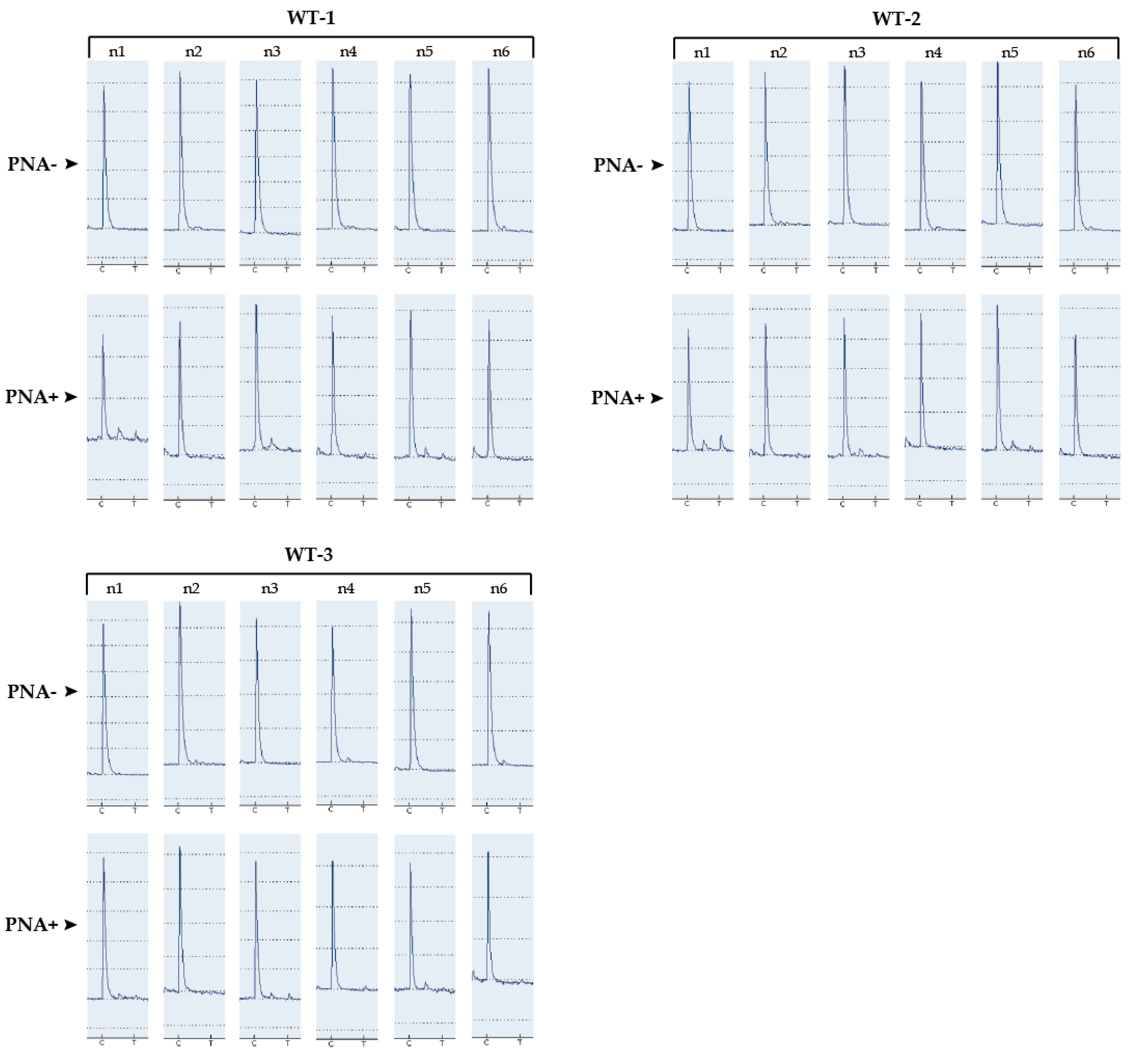
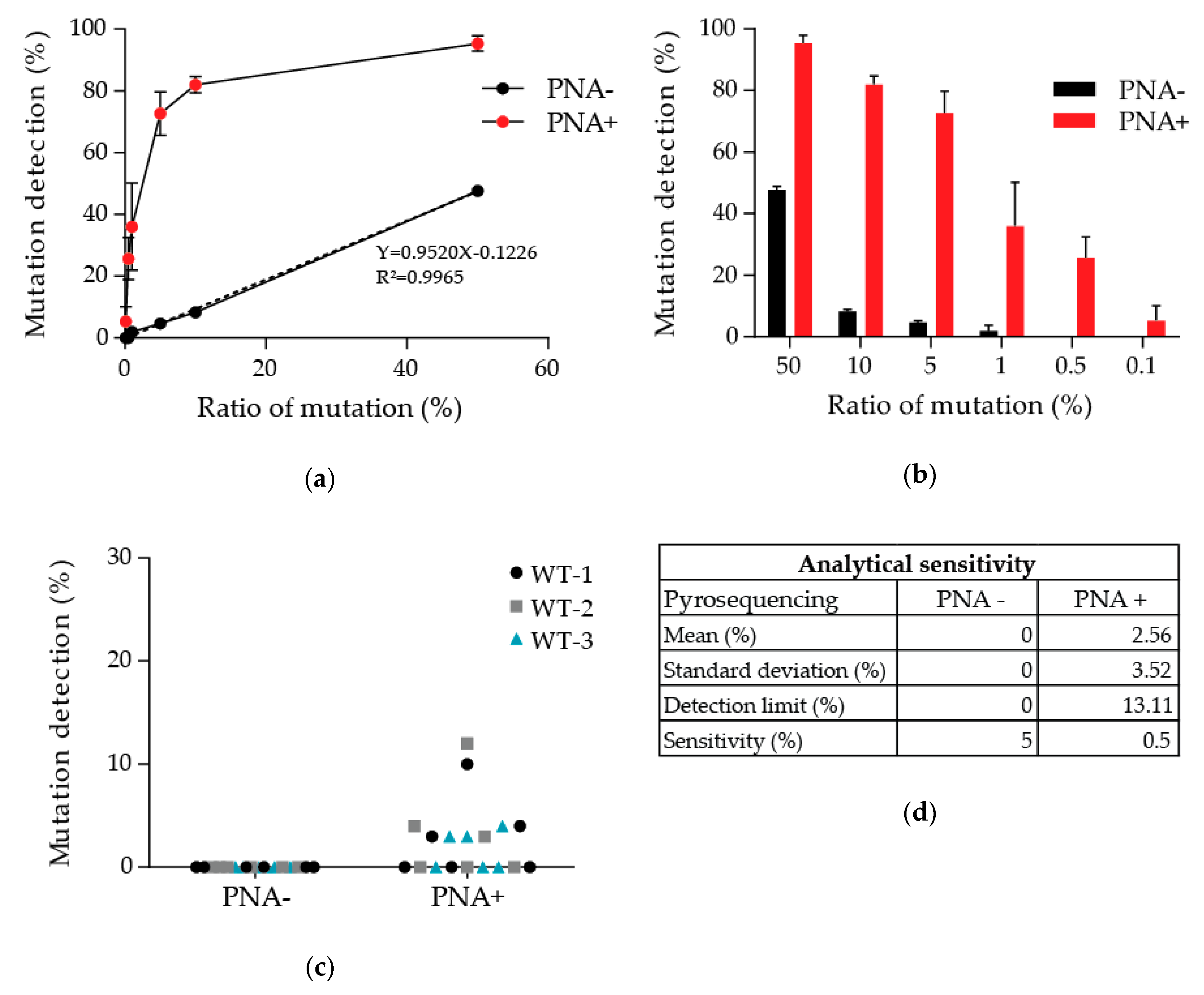
3.1. Determination of the Detection Limit of Our PNA-Mediated PCR Clamping

The sensitivity of our PNA-mediated PCR clamping method was assessed using a range with different percentages, from 50 to 0.1%, of the p.Thr790Met alteration of EGFR. Three ranges were realized by diluting an EGFR p.Thr790Met reference standard, 50% mutated, in wild-type DNA extracted from lymphocytes of healthy donors to avoid cancer cell aneuploidy, which would perturb the precision of our dilutions. The linearity of our ranges was first verified without PNA, and an R² of 0.9965 was obtained (Figure 1a). PNAs were added to the PCR mix in parallel, and the same protocol was followed to evaluate the p.Thr790Met frequencies at the different points of the ranges. Where PNAs were added, mutation frequencies tended to saturate where there were over five to 10% in the original sample (Figure 1a,b). The different points of the ranges with or without PNA were compared to validate the enrichment of the p.Thr790Met mutation in the samples. Without PNA, the frequencies of the p.Thr790Met alteration in mutated samples are similar whether or not a preamplification had been performed (Table 2). However, pyrogram signals are better after preamplification (Appendix A Figure A1). This is even clearer where PNA had been added. The preamplification step enhanced the fraction of amplifiable DNA allowing the detection of signals, which were otherwise drown in background noise.
The detection limit of our method was then evaluated. Wild-type DNA extracted from three healthy donors was tested several times with or without PNA (Figure 1c,d). Due to the prior preamplification step, background noise was present with PNAs (Appendix A Figure A2). A limit of detection of 13.11% was consequently calculated (limit = mean + 3*standard deviation) based on the frequencies of the p.Thr790Met alteration detected, which is mainly background noise (Appendix A Figure A2 WT-1 n1 and WT-2 n1 for the highest frequencies). The EGFR p.Thr790Met reference standard also allowed the determination of the analytical sensitivity of PNA-mediated PCR clamping followed by pyrosequencing. Contrary to classical pyrosequencing, the detection limit of which remains 5%, the addition of PNA in the PCR mix enabled the detection of smaller percentages up to 0.5%.

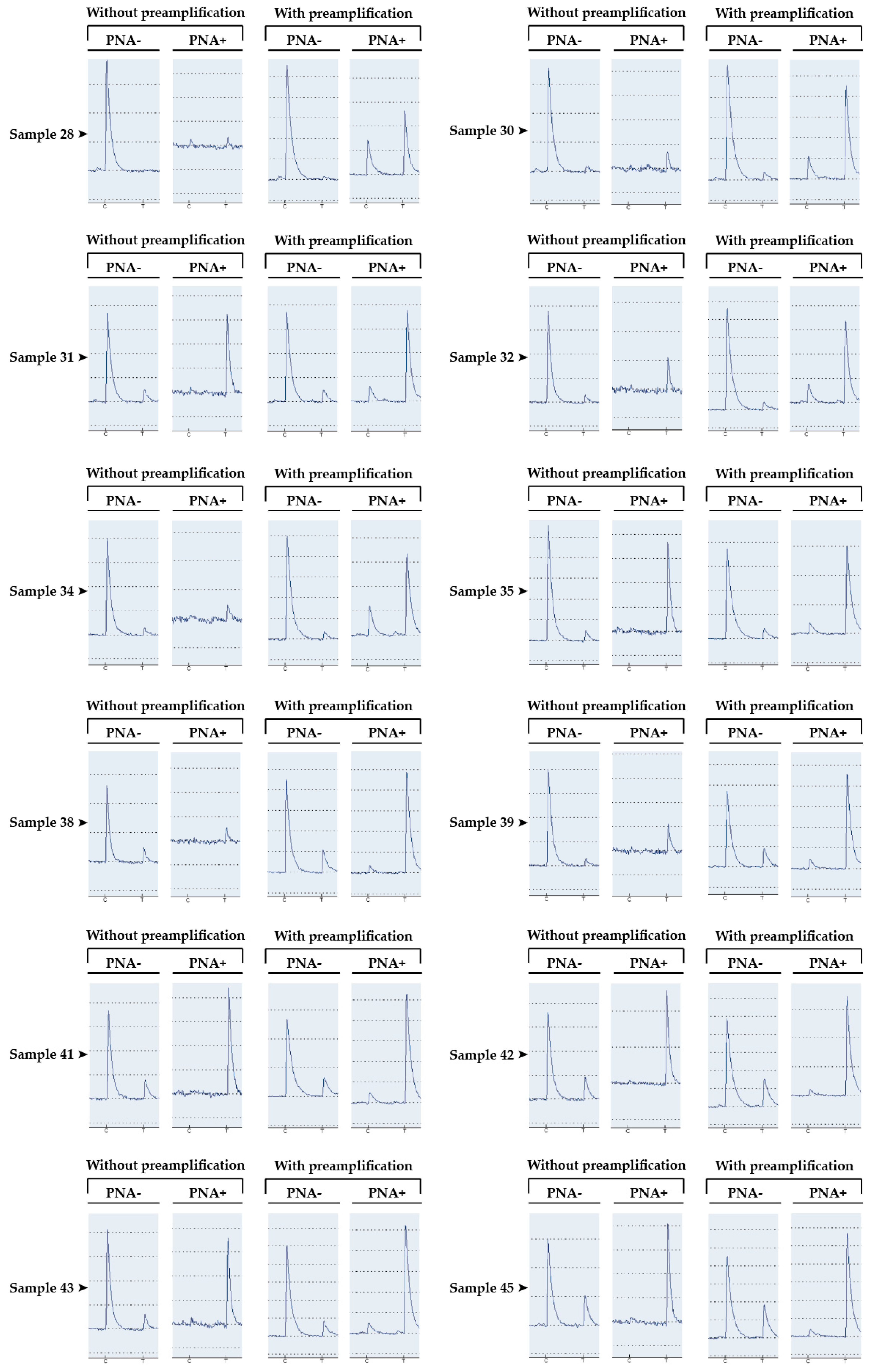
3.2. Detection of the p.Thr790Met Alteration of EGFR in Patients’ Somatic DNA

Because of ctDNA sample scarcity and poor DNA concentration and quality, we applied our method to the analysis of DNA extracted from solid tumors. Somatic DNA extracted from 46 patients, suffering from NSCLC was first tested using NGS analysis for the presence of the p.Thr790Met alteration. This resistance alteration was detected in 19 samples (Figure 2a). Preamplification followed by PCR with and without PNA and pyrosequencing was performed for each sample. Herein, the pyrograms obtained for mutated and wild-type samples with and without PNA are represented (Figure 2b). An enrichment of the mutation frequencies was noticed for all mutated samples, increasing from two to 26% with NGS to over 28% with PNA (Table 3). All the results were compared to those obtained with NGS (Table 3 and Figure 2a). Seven samples that were found wild type had a few percentages with PNA but were below the calculated limit of detection, so they were considered wild type. Only one patient had discordant results. He was wild type with NGS but was found positive with our PNA-mediated PCR method. Consequently, the comparative analysis of all results allowed us to determine a sensitivity of 96.3% and a specificity of 100% with our protocol with a κ value of 95.6% (Figure 2c). The positive predictive value (PPV) was 100% and the negative predictive (NPV) value was 95%.

3.3. Patient with Discordant Results

The somatic DNA from the discordant sample presented a tumor cellularity of 80% and a concentration of 44.2 ng/µL. No mutation was detected without PNA, but its addition allowed the appearance of an 18% peak at the p.Thr790Met alteration localization, so above the limit of detection calculated earlier (Figure 3a). This patient was a 51-year-old woman who had breast cancer in 2015 treated by surgery and hormonotherapy and was diagnosed with advanced pulmonary adenocarcinoma in 2017. NGS was performed and a BRAF mutation was detected with a frequency of 40.4% (Figure 3b). Following our analysis with PNA and pyrosequencing, we searched for the p.Thr790Met mutation of EGFR in the NGS results. Indeed, only two reads were detected, giving a frequency of 0.06% so far under the threshold of NGS.

4. Discussion

Over half of the acquired resistance to first-generation EGFR tyrosine kinase inhibitors in NSCLC is due to the appearance of the p.Thr790Met variant of EGFR. Currently, the acquisition of this mutation is no longer such a poor prognosis thanks to a third-generation EGFR TKI called osimertinib. Consequently, there is a need for a highly sensitive method of detection for this variant, enabling an earlier adaptation of treatment. Herein, we described an improvement of the PNA-mediated PCR clamping method preceding pyrosequencing. Indeed, a first step of preamplification, without PNA enhanced the fraction of amplifiable DNA (Table 2 and Appendix A Figure A1). We therefore were able to demonstrate an analytical sensitivity of 0.5% with a detection limit of 13.11% due to background noise (Figure 1c,d and Appendix A Figure A2). The second amplification with PNA allowed an enrichment of the mutated fraction of the sample and tended to saturate where there was more than 10% mutation in the original sample (Figure 1a,b). Hence, our method favors an easier and inexpensive detection of small percentages and is qualitative rather than quantitative.
To evaluate the sensitivity and specificity of our method, we analyzed 46 DNA samples extracted from solid tumors from patients who relapsed after the appearance of acquired resistance to first-generation EGFR TKIs. NGS, our method of reference, determined that 19 of them had this resistance alteration. They were all also positive after utilization of PNA (Figure 2a and Table 3). Only one discordant sample, which was wild type with NGS, was positive for the p.Thr790Met alteration with PNA (Table 3, Figure 2a and Figure 3a). These results allowed the calculation of a κ-value of 0.956 with a sensitivity of 96.3% and a specificity of 100%, resulting in very strong agreement [33] (Figure 2c). Moreover, the analytical sensitivity was greatly improved (over 0.5%) but to the detriment of the statistical sensitivity for such small percentages. However, when we closely analyzed the NGS results from the discordant case, which had a frequency of 18% of the p.Thr790Met variant with PNA (Figure 3a), two reads indeed carried this alteration, giving a frequency of 0.06%, below its threshold of 2% (Figure 3b). This discordant case could truly be mutated, thereby favoring a good statistical sensitivity of our method.
Currently, the third-generation EGFR TKI (osimertinib) is now accessible as a first-line treatment where an activating mutation of EGFR is characterized in a patient with NSCLC. A longer progression-free survival was shown compared to first-line EGFR TKI, and the p.Thr790Met alteration is no longer a factor of resistance to that treatment [34]. However, after a few months of continuing treatment, acquired resistance can also appear, partially related to the p.Cys797Ser mutation of EGFR [35,36]. The method developed herein could be perfectly applied to the analysis of this mutation or to other punctual alterations, such as the hotspots of PIK3CA or the p.Val600Glu alteration of BRAF. Despite the low cost and rapidity of analysis of pyrosequencing, NGS remains the method of reference, allowing simultaneous characterization of many hotspots of different genes. However, the test presented here could be used to confirm those results especially where small percentages are detected. This method could also provide an answer to a persistent question concerning the mechanisms responsible for acquired resistance: is it the selection of a pre-existent resistant clone, its apparition under treatment pressure or a combination of both hypotheses? First studies were more in favor of treatment pressure, but recent analyses with more sensitive techniques [37,38], in which our method could participate, dispute this assertion.
Finally, the developed protocol could also be used to address the problems of the study of circulating tumor DNA. These samples are fragmented, weakly concentrated and drowned in constitutive blood DNA. The first preamplification step allowing enrichment of amplifiable DNA, our PNA-mediated PCR clamping, could thereby be a solution. It could then be used to follow treatment efficiency and the disappearance of the targeted activating mutation. Furthermore, it can be used to evaluate eventual residual disease or to detect a putative acquired resistance mechanism earlier. All possibilities in molecular detection are unified towards a common goal: early adaptation of even more personalized treatments in the field of oncology.

Author Contributions

Conceptualization, A.M. and A.B.; methodology, A.B.; software, J.D.; validation, A.B., L.-M.C. and A.M.; resources, V.V. and A.M.; data curation, V.V., J.D. and A.M.; writing—original draft preparation, A.B. and A.M.; supervision, A.M. All authors have read and agreed to the published version of the manuscript.

Funding

This research received no external funding.

Acknowledgments

We thank Sandra Vilgrain and Luc Fey for their technical advice.

Conflicts of Interest

The authors declare no conflict of interest.

Abbreviations

TKItyrosine kinase inhibitor
NSCLCnon-small-cell lung cancer
PNApeptide nucleic acid
SCLCsmall-cell lung cancer
ctDNAcirculating tumor DNA
NGSnext-generation sequencing
PBNCperipheral blood nuclear cells
FFPEformalin-fixed paraffin-embedded
ICOInstitut de Cancérologie de l’Ouest

Appendix A

Figure A1. Pyrograms obtained analyzing EGFR p.Thr790Met mutated samples with or without PNA and after, or not, a preamplification followed by pyrosequencing.
Diagnostics 10 00527 g0a1
Figure A2. Pyrograms obtained analyzing the EGFR p.Thr790Met alteration in DNA extracted from three healthy donors, with or without PNA.
Diagnostics 10 00527 g0a2

References

  1. Bray, F.; Ferlay, J.; Soerjomataram, I.; Siegel, R.; Torre, L.; Jemal, A. Global cancer statistics 2018: GLOBOCAN estimates of incidence and mortality worldwide for 36 cancers in 185 countries. CA Cancer J. Clin. 2018, 68, 394–424. [Google Scholar] [CrossRef] [PubMed] [Green Version]
  2. Herbst, R.S.; Heymach, J.V.; Lippman, S.M. Molecular Origins of Cancer Lung Cancer. N. Engl. J. Med. 2008, 6, 53–55. [Google Scholar]
  3. Lawrence, M.S.; Stojanov, P.; Polak, P.; Kryukov, G.V.; Cibulskis, K.; Sivachenko, A.; Carter, S.L.; Stewart, C.; Mermel, C.H.; Roberts, S.A.; et al. Mutational heterogeneity in cancer and the search for new cancer-associated genes. Nature 2013, 499, 214–218. [Google Scholar] [CrossRef] [PubMed]
  4. Rosell, R.; Moran, T.; Queralt, C.; Cardenal, F.; Camps, C.; Majem, M.; Lopez-Vivanco, G.; Isla, D.; Provencio, M.; Insa, A.; et al. Screening for Epidermal Growth Factor Receptor Mutations in Lung Cancer. N. Engl. J. Med. 2009, 361, 958–967. [Google Scholar] [CrossRef] [PubMed] [Green Version]
  5. Barlesi, F.; Mazieres, J.; Merlio, J.P.; Debieuvre, D.; Mosser, J.; Lena, H.; Ouafik, L.; Besse, B.; Rouquette, I.; Westeel, V.; et al. Routine molecular profiling of patients with advanced non-small-cell lung cancer: Results of a 1-year nationwide programme of the French Cooperative Thoracic Intergroup (IFCT). Lancet 2016, 387, 1415–1426. [Google Scholar] [CrossRef]
  6. Sharma, S.V.; Bell, D.; Settleman, J.; Haber, D.A. Epidermal growth factor receptor mutations in lung cancer. Nat. Rev. Cancer 2007, 7, 169–181. [Google Scholar] [CrossRef]
  7. Brewer, M.R.; Yun, C.-H.; Lai, D.; Lemmon, M.A.; Eck, M.J.; Pao, W. Mechanism for activation of mutated epidermal growth factor receptors in lung cancer. Proc. Natl. Acad. Sci. USA 2013, 110, E3595–E3604. [Google Scholar] [CrossRef] [Green Version]
  8. Shan, Y.; Eastwood, M.P.; Zhang, X.; Kim, E.T.; Arkhipov, A.; Dror, R.O.; Jumper, J.; Kuriyan, J.; Shaw, D.E. Oncogenic mutations counteract intrinsic disorder in the EGFR kinase and promote receptor dimerization. Cell 2012, 149, 860–870. [Google Scholar] [CrossRef] [Green Version]
  9. Mok, T.S.; Wu, Y.-L.; Thongprasert, S.; Yang, C.-H.; Chu, D.-T.; Saijo, N.; Sunpaweravong, P.; Han, B.; Margono, B.; Ichinose, Y.; et al. Gefitinib or carboplatin-paclitaxel in pulmonary adenocarcinoma. N. Engl. J. Med. 2009, 361, 947–957. [Google Scholar] [CrossRef]
  10. Lee, C.K.; Davies, L.; Wu, Y.L.; Mitsudomi, T.; Inoue, A.; Rosell, R.; Zhou, C.; Nakagawa, K.; Thongprasert, S.; Fukuoka, M.; et al. Gefitinib or Erlotinib vs Chemotherapy for EGFR Mutation-Positive Lung Cancer: Individual Patient Data Meta-Analysis of Overall Survival. J. Natl. Cancer Inst. 2017, 109, 1–9. [Google Scholar] [CrossRef] [Green Version]
  11. Rosell, R.; Carcereny, E.; Gervais, R.; Vergnenegre, A.; Massuti, B.; Felip, E.; Palmero, R.; Garcia-Gomez, R.; Pallares, C.; Sanchez, J.M.; et al. Erlotinib versus standard chemotherapy as first-line treatment for European patients with advanced EGFR mutation-positive non-small-cell lung cancer (EURTAC): A multicentre, open-label, randomised phase 3 trial. Lancet Oncol. 2012, 13, 239–246. [Google Scholar] [CrossRef]
  12. Nguyen, K.H.; Kobayashi, S.; Costa, D.B. Acquired Resistance to Epidermal Growth Factor Receptor Tyrosine Kinase Inhibitors in Non-Small-Cell Lung Cancers Dependent on the Epidermal Growth Factor Receptor Pathway. Clin. Lung Cancer 2009, 10, 281–289. [Google Scholar] [CrossRef] [PubMed] [Green Version]
  13. Jotte, R.M.; Spigel, D.R. Advances in molecular-based personalized non-small-cell lung cancer therapy: Targeting epidermal growth factor receptor and mechanisms of resistance. Cancer Med. 2015, 4, 1621–1632. [Google Scholar] [CrossRef] [PubMed]
  14. Dagogo-Jack, I.; Shaw, A.T. Tumour heterogeneity and resistance to cancer therapies. Nat. Rev. Clin. Oncol. 2017, 15, 81–94. [Google Scholar] [CrossRef]
  15. Forde, P.M.; Ettinger, D.S. Managing Acquired Resistance in EGFR-Mutated Non-Small Cell Lung Cancer. Clin. Adv. Hematol. Oncol. 2015, 13, 528–532. [Google Scholar]
  16. Mok, T.S.; Wu, Y.-L.; Ahn, M.-J.; Garassino, M.C.; Kim, H.R.; Ramalingam, S.S.; Shepherd, F.A.; He, Y.; Akamatsu, H.; Theelen, W.S.M.E.; et al. Osimertinib or Platinum-Pemetrexed in EGFR T790M-Positive Lung Cancer. N. Engl. J. Med. 2017, 376, 629–640. [Google Scholar] [CrossRef] [Green Version]
  17. Blumenthal, G.M.; Keegan, P.; He, K.; Weinstock, C.; Khozin, S.; Pazdur, R.; Cheng, J.; Zhuang, L.; Charlab, R.; Zhao, H.; et al. Osimertinib for the Treatment of Metastatic EGFR T790M Mutation-Positive Non-Small Cell Lung Cancer. Clin. Cancer Res. 2016, 23, 2131–2135. [Google Scholar] [CrossRef] [Green Version]
  18. Maley, C.C.; Aktipis, A.; Graham, T.A.; Sottoriva, A.; Boddy, A.M.; Janiszewska, M.; Silva, A.S.; Gerlinger, M.; Yuan, Y.; Pienta, K.J.; et al. Classifying the evolutionary and ecological features of neoplasms. Nat. Rev. Cancer 2017, 17, 605–619. [Google Scholar] [CrossRef]
  19. Schwarzenbach, H.; Hoon, D.S.B.; Pantel, K. Cell-free nucleic acids as biomarkers in cancer patients. Nat. Rev. Cancer 2011, 11, 426–437. [Google Scholar] [CrossRef]
  20. Heitzer, E.; Ulz, P.; Geigl, J.B. Circulating Tumor DNA as a Liquid Biopsy for Cancer. Clin. Chem. 2015, 61, 112–123. [Google Scholar] [CrossRef]
  21. Amirouchene-Angelozzi, N.; Swanton, C.; Bardelli, A. Tumor evolution as a therapeutic target. Cancer Discov. 2017, 7, 805–817. [Google Scholar] [CrossRef] [PubMed] [Green Version]
  22. Oxnard, G.R.; Thress, K.S.; Alden, R.S.; Lawrance, R.; Paweletz, C.P.; Cantarini, M.; Yang, J.C.H.; Barrett, J.C.; Jänne, P.A. Association between plasma genotyping and outcomes of treatment with osimertinib (AZD9291) in advanced non-small-cell lung cancer. J. Clin. Oncol. 2016, 34, 3375–3382. [Google Scholar] [CrossRef] [PubMed] [Green Version]
  23. Luo, J.; Shen, L.; Zheng, D. Diagnostic value of circulating free DNA for the detection of EGFR mutation status in NSCLC: A systematic review and meta-analysis. Sci. Rep. 2014, 4, 1–7. [Google Scholar] [CrossRef] [PubMed]
  24. Jovelet, C.; Ileana, E.; Le Deley, M.C.; Motté, N.; Rosellini, S.; Romero, A.; Lefebvre, C.; Pedrero, M.; Pata-Merci, N.; Droin, N.; et al. Circulating cell-free tumor DNA analysis of 50 genes by next-generation sequencing in the prospective MOSCATO trial. Clin. Cancer Res. 2016, 22, 2960–2968. [Google Scholar] [CrossRef] [PubMed] [Green Version]
  25. Peng, M.; Chen, C.; Hulbert, A.; Brock, M.V.; Yu, F. Non-blood circulating tumor DNA detection in cancer. Oncotarget 2017, 8, 69162–69173. [Google Scholar] [CrossRef]
  26. Mouliere, F.; Rosenfeld, N. Circulating tumor-derived DNA is shorter than somatic DNA in plasma. Proc. Natl. Acad. Sci. USA 2015, 112, 3178–3179. [Google Scholar] [CrossRef] [Green Version]
  27. Diehl, F.; Li, M.; Dressman, D.; He, Y.; Shen, D.; Szabo, S.; Diaz, L.A.; Goodman, S.N.; David, K.A.; Juhl, H.; et al. Detection and quantification of mutations in the plasma of patients with colorectal tumors. Proc. Natl. Acad. Sci. USA 2005, 102, 16368–16373. [Google Scholar] [CrossRef] [Green Version]
  28. Castiglia, M.; Bazan, V.; Passiglia, F.; Gulotta, L.; Fulfaro, F.; Badalamenti, G.; Di Maio, M.; Listì, A.; Russo, A.; Galvano, A.; et al. The diagnostic accuracy of circulating tumor DNA for the detection of EGFR-T790M mutation in NSCLC: A systematic review and meta-analysis. Sci. Rep. 2018, 8, 1–10. [Google Scholar] [CrossRef] [Green Version]
  29. Nagai, Y.; Miyazawa, H.; Huqun; Tanaka, T.; Udagawa, K.; Kato, M.; Fukuyama, S.; Yokote, A.; Kobayashi, K.; Kanazawa, M.; et al. Genetic heterogeneity of the epidermal growth factor receptor in non-small cell lung cancer cell lines revealed by a rapid and sensitive detection system, the peptide nucleic acid-locked nucleic acid PCR clamp. Cancer Res. 2005, 65, 7276–7282. [Google Scholar] [CrossRef] [Green Version]
  30. Chevalier, L.; Billaud, A.; Passot, C.; Renoult, A.; Bigot, F.; Verrièle, V.; Morel, A. Caractérisation moléculaire de l’EGFR dans les cancers bronchiques non à petites cellules: Étude prospective comparative des technologies NGS et automate Idylla. Ann. Pathol. 2020. [Google Scholar] [CrossRef]
  31. Nielsen, P.E.; Egholm, M.; Berg, R.H.; Buchardt, O. Sequence-selective recognition of DNA by strand displacement with a thymine-substituted polyamide. Science 1991, 254, 1497–1500. [Google Scholar] [CrossRef] [PubMed]
  32. Miyazawa, H.; Tanaka, T.; Nagai, Y.; Matsuoka, M.; Sutani, A.; Udagawa, K.; Zhang, J.; Hirama, T.; Murayama, Y.; Koyama, N.; et al. Peptide nucleic acid-locked nucleic acid polymerase chain reaction clamp-based detection test for gefitinib-refractory T790M epidermal growth factor receptor mutation. Cancer Sci. 2008, 29, 595–600. [Google Scholar] [CrossRef]
  33. Cohen, J. Weighted kappa: Nominal scale agreement with provision for scaled disagreement or partial credit. Psychol. Bull. 1968, 70, 213–220. [Google Scholar] [CrossRef] [PubMed]
  34. Soria, J.-C.; Ohe, Y.; Vansteenkiste, J.; Reungwetwattana, T.; Chewaskulyong, B.; Lee, K.H.; Dechaphunkul, A.; Imamura, F.; Nogami, N.; Kurata, T.; et al. Osimertinib in Untreated EGFR-Mutated Advanced Non-Small-Cell Lung Cancer. N. Engl. J. Med. 2018, 378, 113–125. [Google Scholar] [CrossRef] [PubMed]
  35. Thress, K.S.; Paweletz, C.P.; Felip, E.; Cho, B.C.; Stetson, D.; Dougherty, B.; Lai, Z.; Markovets, A.; Vivancos, A.; Kuang, Y.; et al. Acquired EGFR C797S mutation mediates resistance to AZD9291 in non–small cell lung cancer harboring EGFR T790M. Nat. Med. 2015, 21, 560–562. [Google Scholar] [CrossRef] [Green Version]
  36. Lazzaeri, C.; Gregore, V.; Karachaliou, N.; Rosell, R.; Santarpia, M. Mechanisms of resistance to osimertinib. J. Thorac. Dis. 2020, 12, 2851–2858. [Google Scholar] [CrossRef]
  37. Costa, C.; Molina, M.A.; Drozdowskyj, A.; Gimeńez-Capitán, A.; Bertran-Alamillo, J.; Karachaliou, N.; Gervais, R.; Massuti, B.; Wei, J.; Moran, T.; et al. The impact of EGFR T790M mutations and BIM mRNA expression on outcome in patients with EGFR-Mutant NSCLC treated with erlotinib or chemotherapy in the randomized phase III EURTAC trial. Clin. Cancer Res. 2014, 20, 2001–2010. [Google Scholar] [CrossRef] [Green Version]
  38. Maheswaran, S.; Sequist, L.V.; Nagrath, S.; Ulkus, L.; Brannigan, B.; Collura, C.V.; Inserra, E.; Diederichs, S.; Iafrate, A.J.; Bell, D.W.; et al. Detection of Mutations in EGFR in Circulating Lung-Cancer Cells. N. Engl. J. Med. 2008, 359, 366–377. [Google Scholar] [CrossRef] [Green Version]
Figure 1. Preamplification followed by PNA-mediated PCR clamping allowed a sensitivity of detection of 0.5% of the p.Thr790Met mutation. (a,b) Pyrosequencing results of the three ranges with different percentages of the p.Thr790Met mutation of EGFR, with or without PNA, after preamplification. (c,d) DNA from three healthy donor analyses (six times each) was used to evaluate the analytical sensitivity and detection limit.
Diagnostics 10 00527 g001
Figure 2. Comparison of the characterization of the p.Thr790Met mutation with NGS and our PNA-mediated method in patients’ somatic DNA. (a) Representation of the results obtained after pyrosequencing analysis with PNA compared to those obtained with NGS for the 46 patients. (b) Pyrograms of two patients, one with the p.Thr790Met mutation, with or without PNA. (c) Analysis of the results to assess the sensitivity and specificity of our method compared to the method of reference, NGS.
Diagnostics 10 00527 g002
Figure 3. Results of the discordant case that was mutated with the PNA method. (a) Pyrograms obtained with and without PNA (n = 2). (b) Results obtained for the same patient with NGS.
Diagnostics 10 00527 g003
Table 1. Primers and peptide nucleic acid sequences.
NamesSequences
PCR forward primer5′-GCA TCT GCC TCA CCT CCA A-3′
PCR reverse primer5′-biotin-CGA TCT GCA CAC ACC AGT TG-3′
PNA sequenceNH2-CTCATCACGCAGCTCA-COOH [32]
Sequencing primer (pyrosequencing)CCG TGC AGC TCA TCA
Dispensation order (pyrosequencing)ACTAGCAGC
Table 2. Evaluation of the p.Thr790Met frequency of EGFR by pyrosequencing in mutated samples after a preamplification step, with and without PNA.
SamplesWithout PreamplificationWith Preamplification
PNA− (%)PNA+ (%)PNA− (%)PNA+ (%)
Sample 280NA228
Sample 305NA559
Sample 31121001068
Sample 328NA772
Sample 347NA984
Sample 358100984
Sample 3816NA2288
Sample 39221002090
Sample 41181002091
Sample 42211002391
Sample 4313921192
Sample 45261002893
Table 3. Characterization of the p.Thr790Met of EGFR in the 46 samples by next-generation sequencing or preamplification and pyrosequencing with or without PNA.
SamplesAgesSexes[DNA] (ng/µL)CellularityNGS Results PNA-Mediated PCRConcordance Results
PNA−PNA+
Sample 150.8M10.730WT00OK-WT
Sample 253.7W14.240WT00OK-WT
Sample 362.1M25.920WT00OK-WT
Sample 455.5W10.220WT00OK-WT
Sample 562.7M13.330WT00OK-WT
Sample 673.4M3910WT00OK-WT
Sample 764.2M6.420WT00OK-WT
Sample 867.2M6.640WT00OK-WT
Sample 951.4W11.640WT00OK-WT
Sample 1081.6M11.420WT00OK-WT
Sample 1156.6M52.870WT00OK-WT
Sample 1283.6W1.410WT00OK-WT
Sample 1356M15.630WT00OK-WT
Sample 1452.7M7.230WT00OK-WT
Sample 1567.6M10.730WT00OK-WT
Sample 1659W18.280WT00OK-WT
Sample 1769.7M29.760WT00OK-WT
Sample 1856.2M5.760WT00OK-WT
Sample 1962.4W2.95WT00OK-WT
Sample 2063.2W9.570WT05L-WT
Sample 2171.4M24.540WT05L-WT
Sample 2282M9.510WT05L-WT
Sample 2361.1M16.160WT06L-WT
Sample 2483W5.520WT06L-WT
Sample 25NAWNANAWT06L-WT
Sample 2662.3M24.830WT08L-WT
Sample 2751.4W44.240WT018Discordant
Sample 2886.7W27.35T790M228OK-Mutated
Sample 29NAM7.4NAT790M (1.3%)235OK-Mutated
Sample 3076.4W1040T790M (3.5%)559OK-Mutated
Sample 3174.6M12.44T790M1068OK-Mutated
Sample 3278.2W2.210T790M772OK-Mutated
Sample 3373.7W2.150T790M (9%)1175OK-Mutated
Sample 3467.2M4.110T790M984OK-Mutated
Sample 3575.6W23.740T790M984OK-Mutated
Sample 3687.8W12.420T790M (12%)2487OK-Mutated
Sample 3766.4W680T790M2687OK-Mutated
Sample 3864.3W2.560T790M2288OK-Mutated
Sample 3964.8W9.440T790M2090OK-Mutated
Sample 4087.6W4.450T790M (15.4%)1790OK-Mutated
Sample 4165.3M30.210T790M2091OK-Mutated
Sample 42NAM6.4NAT790M (18.1%)2391OK-Mutated
Sample 4378.3W30.970T790M1192OK-Mutated
Sample 4477W0.610T790M (14.7%)2392OK-Mutated
Sample 4574W6.940T790M2893OK-Mutated
Sample 4678.5M3.860T790M (21.3%)2695OK-Mutated
Concordant results of wild-type patients for this alteration are in green (OK-WT), and mutated patients are in red (OK-Mutated). Patients with a few percentages of this mutation with PNA but below the calculated limit of detection of the method are in yellow (L-WT). The discordant patient is in blue (Discordant). NA: not applicable.

