BIRC5/Survivin Expression as a Non-Invasive Biomarker of Endometriosis


 , , ,
, , ,
Abstract
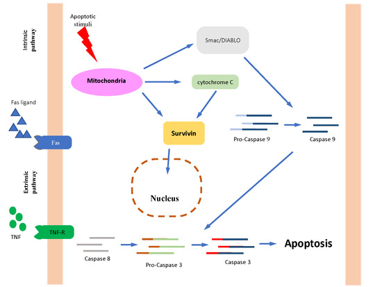
1. Introduction
2. Materials and Methods
2.1. Participants
2.2. Sample Collection
2.3. Hormonal Measurement
2.4. RT-qPCR
2.5. Statistical Analyses
3. Results
4. Discussion
5. Conclusions
Author Contributions
Funding
Acknowledgments
Conflicts of Interest
References
- Chapron, C.; Marcellin, L.; Borghese, B.; Santulli, P. Rethinking mechanisms, diagnosis and management of endometriosis. Nat. Rev. Endocrinol. 2019, 15, 666–682. [Google Scholar] [CrossRef]
- Zondervan, K.T.; Becker, C.M.; Koga, K.; Missmer, S.A.; Taylor, R.N.; Viganò, P. Endometriosis. Nat. Rev. Dis. Primers 2018, 4, 9. [Google Scholar] [CrossRef]
- Kiesel, L.; Sourouni, M. Diagnosis of endometriosis in the 21st century. Climacteric 2019, 22, 296–302. [Google Scholar] [CrossRef] [PubMed]
- Barbosa, C.P.; Souza, A.M.; Bianco, B.; Christofolini, D.; Bach, F.A.; Lima, G.R. Frequency of endometriotic lesions in peritoneum samples from asymptomatic fertile women and correlation with CA125 values. Sao Paulo Med. J. 2009, 127, 342–345. [Google Scholar] [CrossRef]
- Bast, R.C., Jr.; Badgwell, D.; Lu, Z.; Marquez, R.; Rosen, D.; Liu, J.; Baggerly, K.A.; Atkinson, E.N.; Skates, S.; Zhang, Z.; et al. New tumor markers: CA125 and beyond. Int. J. Gynecol. Cancer 2005, 15 (Suppl. 3), 274–281. [Google Scholar] [CrossRef]
- Dunselman, G.A.; Vermeulen, N.; Becker, C.; Calhaz-Jorge, C.; D’Hooghe, T.; De Bie, B.; Heikinheimo, O.; Horne, A.W.; Kiesel, L.; Nap, A.; et al. European Society of Human Reproduction and Embryology. ESHRE guideline: Management of women with endometriosis. Hum. Reprod. 2014, 29, 400–412. [Google Scholar] [CrossRef]
- Barra, F.; Biscaldi, E.; Scala, C.; Laganà, A.S.; Vellone, V.G.; Stabilini, C.; Ghezzi, F.; Ferrero, S. A Prospective Study Comparing Three-Dimensional Rectal Water Contrast Transvaginal Ultrasonography and Computed Tomographic Colonography in the Diagnosis of Rectosigmoid Endometriosis. Diagnostics 2020, 10, 252. [Google Scholar] [CrossRef]
- Laganà, A.S.; Garzon, S.; Götte, M.; Viganò, P.; Franchi, M.; Ghezzi, F.; Martin, D.C. The Pathogenesis of Endometriosis: Molecular and Cell Biology Insights. Int. J. Mol. Sci. 2019, 20, 5615. [Google Scholar] [CrossRef]
- Sofo, V.; Götte, M.; Laganà, A.S.; Salmeri, F.M.; Triolo, O.; Sturlese, E.; Retto, G.; Alfa, M.; Granese, R.; Abrão, M.S. Correlation between dioxin and endometriosis: An epigenetic route to unravel the pathogenesis of the disease. Arch. Gynecol. Obstet. 2015, 292, 973–986. [Google Scholar] [CrossRef] [PubMed]
- Wang, Y.; Nicholes, K.; Shih, I.M. The Origin and Pathogenesis of Endometriosis. Annu. Rev. Pathol. 2020, 15, 71–95. [Google Scholar] [CrossRef]
- Alkatout, İ.; Meinhold-Heerlein, I.; Keckstein, J.; Mettler, L. Endometriosis: A concise practical guide to current diagnosis and treatment. J. Turk. Ger. Gynecol. Assoc. 2018, 19, 173–175. [Google Scholar] [CrossRef] [PubMed]
- Alderman, M.H.; Yoder, N.; Taylor, H.S. The Systemic Effects of Endometriosis. Semin. Reprod. Med. 2017, 35, 263–270. [Google Scholar] [CrossRef]
- Freytag, D.; Mettler, L.; Maass, N.; Günther, V.; Alkatout, I. Uterine anomalies and endometriosis. Minerva Med. 2020, 111, 33–49. [Google Scholar] [CrossRef]
- Melin, A.; Sparén, P.; Persson, I.; Bergqvist, A. Endometriosis and the risk of cancer with special emphasis on ovarian cancer. Hum. Reprod. 2006, 21, 1237–1242. [Google Scholar] [CrossRef]
- Wang, C.; Liang, Z.; Liu, X.; Zhang, Q.; Li, S. The Association between Endometriosis, Tubal Ligation, Hysterectomy and Epithelial Ovarian Cancer: Meta-Analyses. Int. J. Environ. Res. Public Health 2016, 13, 1138. [Google Scholar] [CrossRef]
- Králíčková, M.; Laganà, A.S.; Ghezzi, F.; Vetvicka, V. Endometriosis and risk of ovarian cancer: What do we know? Arch. Gynecol. Obstet. 2020, 301, 1–10. [Google Scholar] [CrossRef]
- Moga, M.A.; Bălan, A.; Dimienescu, O.G.; Burtea, V.; Dragomir, R.M.; Anastasiu, C.V. Circulating miRNAs as Biomarkers for Endometriosis and Endometriosis-Related Ovarian Cancer-An Overview. J. Clin. Med. 2019, 8, 735. [Google Scholar] [CrossRef]
- Karnezis, A.N.; Leung, S.; Magrill, J.; McConechy, M.K.; Yang, W.; Chow, C.; Kobel, M.; Lee, C.H.; Huntsman, D.G.; Talhouk, A.; et al. Evaluation of endometrial carcinoma prognostic immunohistochemistry markers in the context of molecular classification. J. Pathol. Clin. Res. 2017, 3, 279–293. [Google Scholar] [CrossRef]
- Wheatley, S.P.; McNeish, I.A. Survivin: A protein with dual roles in mitosis and apoptosis. Int. Rev. Cytol. 2005, 247, 35–88. [Google Scholar] [CrossRef]
- Zafari, P.; Rafiei, A.; Esmaeili, S.A.; Moonesi, M.; Taghadosi, M. Survivin a pivotal antiapoptotic protein in rheumatoid arthritis. J. Cell. Physiol. 2019, 234, 21575–21587. [Google Scholar] [CrossRef]
- Marsicano, S.R.; Kuniyoshi, R.K.; Gehrke, F.S.; Alves, B.C.; Azzalis, L.A.; Fonseca, F.L. Survinin expression in patients with breast cancer during chemotherapy. Tumour Biol. 2015, 36, 3441–3445. [Google Scholar] [CrossRef] [PubMed]
- Rivadeneira, D.B.; Caino, M.C.; Seo, J.H.; Angelin, A.; Wallace, D.C.; Languino, L.R.; Altieri, D.C. Survivin promotes oxidative phosphorylation, subcellular mitochondrial repositioning, and tumor cell invasion. Sci. Signal. 2015, 8, ra80. [Google Scholar] [CrossRef]
- Ausserlechner, M.J.; Hagenbuchner, J. Mitochondrial survivin–an Achilles’ heel in cancer chemoresistance. Mol. Cell Oncol. 2015, 3, e1076589. [Google Scholar] [CrossRef]
- Galbo, P.M., Jr.; Ciesielski, M.J.; Figel, S.; Maguire, O.; Qiu, J.; Wiltsie, L.; Minderman, H.; Fenstermaker, R.A. Circulating CD9+/GFAP+/survivin+ exosomes in malignant glioma patients following survivin vaccination. Oncotarget 2017, 8, 114722–114735. [Google Scholar] [CrossRef]
- Wheatley, S.P.; Altieri, D.C. Survivin at a glance. J. Cell Sci. 2019, 132, jcs223826. [Google Scholar] [CrossRef]
- Konno, R.; Yamakawa, H.; Utsunomiya, H.; Ito, K.; Sato, S.; Yajima, A. Expression of survivin and Bcl-2 in the normal human endometrium. Mol. Hum. Reprod. 2000, 6, 529–534. [Google Scholar] [CrossRef]
- Lehner, R.; Enomoto, T.; McGregor, J.A.; Shroyer, L.; Haugen, B.R.; Pugazhenthi, U.; Shroyer, K.R. Correlation of survivin mRNA detection with histologic diagnosis in normal endometrium and endometrial carcinoma. Acta Obstet. Gynecol. Scand. 2002, 81, 162–167. [Google Scholar] [CrossRef]
- Zhang, H.; Li, M.; Zheng, X.; Sun, Y.; Wen, Z.; Zhao, X. Endometriotic stromal cells lose the ability to regulate cell-survival signaling in endometrial epithelial cells in vitro. Mol. Hum. Reprod. 2009, 15, 653–663. [Google Scholar] [CrossRef]
- Takai, N.; Miyazaki, T.; Nishida, M.; Nasu, K.; Miyakawa, I. Survivin expression correlates with clinical stage, histological grade, invasive behavior and survival rate in endometrial carcinoma. Cancer Lett. 2002, 184, 105–116. [Google Scholar] [CrossRef]
- Pallares, J.; Martínez-Guitarte, J.L.; Dolcet, X.; Llobet, D.; Rue, M.; Palacios, J.; Prat, J.; Matias-Guiu, X. Survivin expression in endometrial carcinoma: A tissue microarray study with correlation with PTEN and STAT-3. Int. J. Gynecol. Pathol. 2005, 24, 247–253. [Google Scholar] [CrossRef]
- Canis, M.; Donnez, J.G.; Guzick, D.S.; Halme, J.K.; Rock, J.A.; Schenken, R.S.; Vernon, M.W. Revised American Society for Reproductive Medicine classification of endometriosis: 1996. Fertil. Steril. 1997, 67, 817–821. [Google Scholar] [CrossRef]
- Burney, R.O.; Giudice, L.C. Pathogenesis and pathophysiology of endometriosis. Fertil. Steril. 2012, 98, 511–519. [Google Scholar] [CrossRef] [PubMed]
- Vetvicka, V.; Laganà, A.S.; Salmeri, F.M.; Triolo, O.; Palmara, V.I.; Vitale, S.G.; Sofo, V.; Králíčková, M. Regulation of apoptotic pathways during endometriosis: From the molecular basis to the future perspectives. Arch. Gynecol. Obstet. 2016, 294, 897–904. [Google Scholar] [CrossRef]
- Kokawa, K.; Shikone, T.; Nakano, R. Apoptosis in the human uterine endometrium during the menstrual cycle. J. Clin. Endocrinol. Metab. 1996, 81, 4144–4147. [Google Scholar]
- Gebel, H.M.; Braun, D.P.; Tambur, A.; Frame, D.; Rana, N.; Dmowski, W.P. Spontaneous apoptosis of endometrial tissue is impaired in women with endometriosis. Fertil. Steril. 1998, 69, 1042–1047. [Google Scholar] [CrossRef]
- Vaskivuo, T.E.; Stenbäck, F.; Karhumaa, P.; Risteli, J.; Dunkel, L.; Tapanainen, J.S. Apoptosis and apoptosis-related proteins in human endometrium. Mol. Cell. Endocrinol. 2000, 165, 75–83. [Google Scholar] [CrossRef]
- Uegaki, T.; Taniguchi, F.; Nakamura, K.; Osaki, M.; Okada, F.; Yamamoto, O.; Harada, T. Inhibitor of apoptosis proteins (IAPs) may be effective therapeutic targets for treating endometriosis. Hum. Reprod. 2015, 30, 149–158. [Google Scholar] [CrossRef]
- Vallée, A.; Lecarpentier, Y. Curcumin and Endometriosis. Int. J. Mol. Sci. 2020, 21, 2440. [Google Scholar] [CrossRef]
- Riedl, S.J.; Shi, Y. Molecular mechanisms of caspase regulation during apoptosis. Nat. Rev. Mol. Cell Biol. 2004, 5, 897–907. [Google Scholar] [CrossRef]
- Okada, H.; Mak, T.W. Pathways of apoptotic and non-apoptotic death in tumour cells. Nat. Rev. Cancer 2004, 4, 592–603. [Google Scholar] [CrossRef]
- Altieri, D.C. Survivin, versatile modulation of cell division and apoptosis in cancer. Oncogene 2003, 22, 8581–8589. [Google Scholar] [CrossRef] [PubMed]
- Altznauer, F.; Martinelli, S.; Yousefi, S.; Thürig, C.; Schmid, I.; Conway, E.M.; Schöni, M.H.; Vogt, P.; Mueller, C.; Fey, M.F.; et al. Inflammation-associated cell cycle-independent block of apoptosis by survivin in terminally differentiated neutrophils. J. Exp. Med. 2004, 199, 1343–1354. [Google Scholar] [CrossRef] [PubMed]
- Bianco, B.; Filipchiuk, C.; Christofolini, D.M.; Barbosa, C.P.; Montagna, E. The role of survivin in the pathogenesis of endometriosis. Minerva Med. 2020, 111, 21–32. [Google Scholar] [CrossRef]
- Matarese, G.; De Placido, G.; Nikas, Y.; Alviggi, C. Pathogenesis of endometriosis: Natural immunity dysfunction or autoimune disease? Trends Mol. Med. 2003, 9, 223–228. [Google Scholar] [CrossRef]
- Zwerts, F.; Lupu, F.; De Vriese, A.; Pollefeyt, S.; Moons, L.; Altura, R.A.; Jiang, Y.; Maxwell, P.H.; Hill, P.; Oh, H.; et al. Lack of endothelial cell survivin causes embryonic defects in angiogenesis, cardiogenesis, and neural tube closure. Blood 2007, 109, 4742–4752. [Google Scholar] [CrossRef]
- Lee, M.O.; Moon, S.H.; Jeong, H.C.; Yi, J.-Y.; Lee, T.-H.; Shim, S.H.; Rhee, Y.-H.; Lee, S.-H.; Oh, S.-J.; Lee, M.-Y.; et al. Inhibition of pluripotent stem cell-derived teratoma formation by small molecules. Proc. Natl. Acad. Sci. USA 2013, 110, E3281–E3290. [Google Scholar] [CrossRef]
- Gil-Kulik, P.; Krzyżanowski, A.; Dudzińska, E.; Karwat, J.; Chomik, P.; Świstowska, M.; Kondracka, A.; Kwaśniewska, A.; Cioch, M.; Jojczuk, M.; et al. Potential Involvement of BIRC5 in Maintaining Pluripotency and Cell Differentiation of Human Stem Cells. Oxid. Med. Cell. Longev. 2019, 2019, 8727925. [Google Scholar] [CrossRef]
- Mull, A.N.; Klar, A.; Navara, C.S. Differential localization and high expression of SURVIVIN splice variants in human embryonic stem cells but not in differentiated cells implicate a role for SURVIVIN in pluripotency. Stem Cell Res. 2014, 12, 539–549. [Google Scholar] [CrossRef]
- Altieri, D.C. Survivin–The inconvenient IAP. Semin. Cell Dev. Biol 2015, 39, 91–96. [Google Scholar] [CrossRef]
- Maijenburg, M.W.; Kleijer, M.; Vermeul, K.; Mul, E.P.J.; van Alphen, F.P.J.; van der Schoot, C.E.; Voermans, C. The composition of the mesenchymal stromal cell compartment in human bone marrow changes during development and aging. Haematologica 2012, 97, 179–183. [Google Scholar] [CrossRef]
- Reagan, M.R.; Rosen, C.J. Navigating the bone marrow niche: Translational insights and cancer-driven dysfunction. Nat. Rev. Rheumatol. 2016, 12, 154–168. [Google Scholar] [CrossRef] [PubMed]
- Laganà, A.S.; Salmeri, F.M.; Vitale, S.G.; Triolo, O.; Götte, M. Stem Cell Trafficking During Endometriosis: May Epigenetics Play a Pivotal Role? Reprod. Sci. 2018, 25, 978–979. [Google Scholar] [CrossRef] [PubMed]
- Koninckx, P.R.; Ussia, A.; Adamyan, L.; Wattiez, A.; Gomel, V.; Martin, D.C. Pathogenesis of endometriosis: The genetic/epigenetic theory. Fertil. Steril. 2019, 111, 327–340. [Google Scholar] [CrossRef] [PubMed]
- Alkatout, I. Laparoscopic hysterectomy: Total or subtotal?–Functional and didactic aspects. Minim. Invasive Ther. Allied Technol. 2020. [Google Scholar] [CrossRef]
- García-Gómez, E.; Vázquez-Martínez, E.R.; Reyes-Mayoral, C.; Cruz-Orozco, O.P.; Camacho-Arroyo, I.; Cerbón, M. Regulation of Inflammation Pathways and Inflammasome by Sex Steroid Hormones in Endometriosis. Front. Endocrinol. 2020, 10, 935. [Google Scholar] [CrossRef]
- Laschke, M.W.; Menger, M.D. Anti-angiogenic treatment strategies for the therapy of endometriosis. Hum. Reprod. Update 2012, 18, 682–702. [Google Scholar] [CrossRef]
- Acimovic, M.; Vidakovic, S.; Milic, N.; Jeremic, K.; Markovic, M.; Milosevic-Djeric, A.; Lazovic-Radonjic, G. Survivin and VEGF as Novel Biomarkers in Diagnosis of Endometriosis. J. Med. Biochem. 2016, 35, 63–68. [Google Scholar]
- Mirabi, P.; Alamolhoda, S.H.; Golsorkhtabaramiri, M.; Namdari, M.; Esmaeilzadeh, S. Prolactin concentration in various stages of endometriosis in infertile women. JBRA Assist. Reprod. 2019, 23, 225–229. [Google Scholar] [CrossRef]
- Marschalek, J.; Ott, J.; Husslein, H.; Kuessel, L.; Elhenicky, M.; Mayerhofer, K.; Franz, M.B. The impact of GnRH agonists in patients with endometriosis on prolactin and sex hormone levels: A pilot study. Eur. J. Obstet. Gynecol. Reprod. Biol. 2015, 195, 156–159. [Google Scholar] [CrossRef]


| Variable * | Endometriosis (n = 36) | Controls (n = 10) | p ** |
|---|---|---|---|
| Age (years) | 35 (33.0–38.0) | 33 (32–34.5) | 0.933 |
| BMI (kg/m2) | 24.3 (23.1–25.4) | 24.7 (23.8–25.7) | 0.800 |
| CA125 (mUI/mL) | 49.8 (22.6–67.6) | 18.9 (15.2–36.3) | <0.001 |
| FSH (mUI/mL) | 7.2 (6.8–8.2) | 6.4 (6.1–6.9) | <0.001 |
| LH (mUI/mL) | 6.3 (4.3–8.3) | 6.7 (5.0–8.3) | 0.838 |
| Progesterone (ng/mL) | 8.9 (6.9–11.0) | 5.9 (2.9–8.9) | 0.061 |
| Prolactin (ng/mL) | 17.1 (11.9–22.6) | 8.5 (6.5–15.1) | 0.010 |
| rho * | p | |
|---|---|---|
| CA125 | −0.191 | 0.265 |
| FSH | 0.276 | 0.115 |
| LH | 0.274 | 0.117 |
| Progesterone | 0.382 | 0.045 |
| Prolactin | −0.030 | 0.873 |
© 2020 by the authors. Licensee MDPI, Basel, Switzerland. This article is an open access article distributed under the terms and conditions of the Creative Commons Attribution (CC BY) license (http://creativecommons.org/licenses/by/4.0/).
Share and Cite
Filipchiuk, C.; Laganà, A.S.; Beteli, R.; Ponce, T.G.; Christofolini, D.M.; Martins Trevisan, C.; Fonseca, F.L.A.; Barbosa, C.P.; Bianco, B. BIRC5/Survivin Expression as a Non-Invasive Biomarker of Endometriosis. Diagnostics 2020, 10, 533. https://doi.org/10.3390/diagnostics10080533
Filipchiuk C, Laganà AS, Beteli R, Ponce TG, Christofolini DM, Martins Trevisan C, Fonseca FLA, Barbosa CP, Bianco B. BIRC5/Survivin Expression as a Non-Invasive Biomarker of Endometriosis. Diagnostics. 2020; 10(8):533. https://doi.org/10.3390/diagnostics10080533
Chicago/Turabian StyleFilipchiuk, Carolina, Antonio Simone Laganà, Rubia Beteli, Tatiana Guida Ponce, Denise Maria Christofolini, Camila Martins Trevisan, Fernando Luiz Affonso Fonseca, Caio Parente Barbosa, and Bianca Bianco. 2020. "BIRC5/Survivin Expression as a Non-Invasive Biomarker of Endometriosis" Diagnostics 10, no. 8: 533. https://doi.org/10.3390/diagnostics10080533
APA StyleFilipchiuk, C., Laganà, A. S., Beteli, R., Ponce, T. G., Christofolini, D. M., Martins Trevisan, C., Fonseca, F. L. A., Barbosa, C. P., & Bianco, B. (2020). BIRC5/Survivin Expression as a Non-Invasive Biomarker of Endometriosis. Diagnostics, 10(8), 533. https://doi.org/10.3390/diagnostics10080533