Diagnostics 2020, 10(8), 541; https://doi.org/10.3390/diagnostics10080541 - 31 Jul 2020
Cited by 15 | Viewed by 4110
Abstract
The prevention of sudden cardiac death (SCD) in cardiomyopathies (CM) remains a challenge. The current guidelines still favor the implantation of devices for the primary prevention of SCD only in patients with severely reduced left ventricular ejection fraction (LVEF) and heart failure (HF)
[...] Read more.
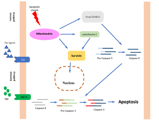
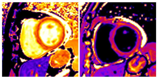
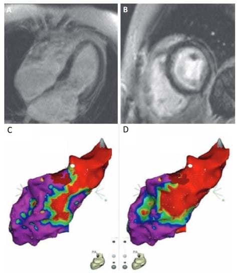
The prevention of sudden cardiac death (SCD) in cardiomyopathies (CM) remains a challenge. The current guidelines still favor the implantation of devices for the primary prevention of SCD only in patients with severely reduced left ventricular ejection fraction (LVEF) and heart failure (HF) symptoms. The implantation of an implantable cardioverter-defibrillator (ICD) is a protective barrier against arrhythmic events in CMs, but the benefit does not outweigh the cost in low risk patients. The identification of high risk patients is the key to an individualized prevention strategy. Cardiac magnetic resonance (CMR) provides reliable and reproducible information about biventricular function and tissue characterization. Furthermore, late gadolinium enhancement (LGE) quantification and pattern of distribution, as well as abnormal T1 mapping and extracellular volume (ECV), representing indices of diffuse fibrosis, can enhance our ability to detect high risk patients. CMR can also complement electro-anatomical mapping (EAM), a technique already applied in the risk evaluation and in the ventricular arrhythmias ablation therapy of CM patients, providing a more accurate assessment of fibrosis and arrhythmic corridors. As a result, CMR provides a new insight into the pathological substrate of CM. CMR may help identify high risk CM patients and, combined with EAM, can provide an integrated evaluation of scar and arrhythmic corridors in the ablative therapy of ventricular arrhythmias.
Full article
(This article belongs to the Special Issue Imaging Cardiac Arrhythmia/Sudden Cardiac Death)
►
Show Figures
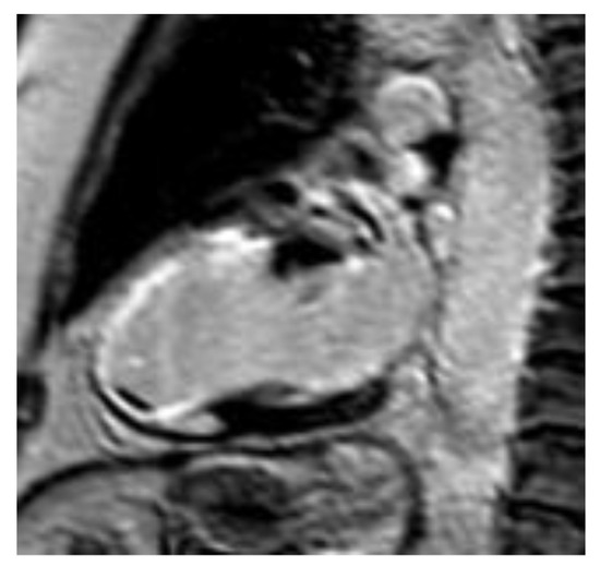
Figure 1


