Artifacts in Musculoskeletal Ultrasonography: From Physics to Clinics


 , ,
, , 
Abstract
:1. Introduction
2. Improper Adjustment of the Focal Zone
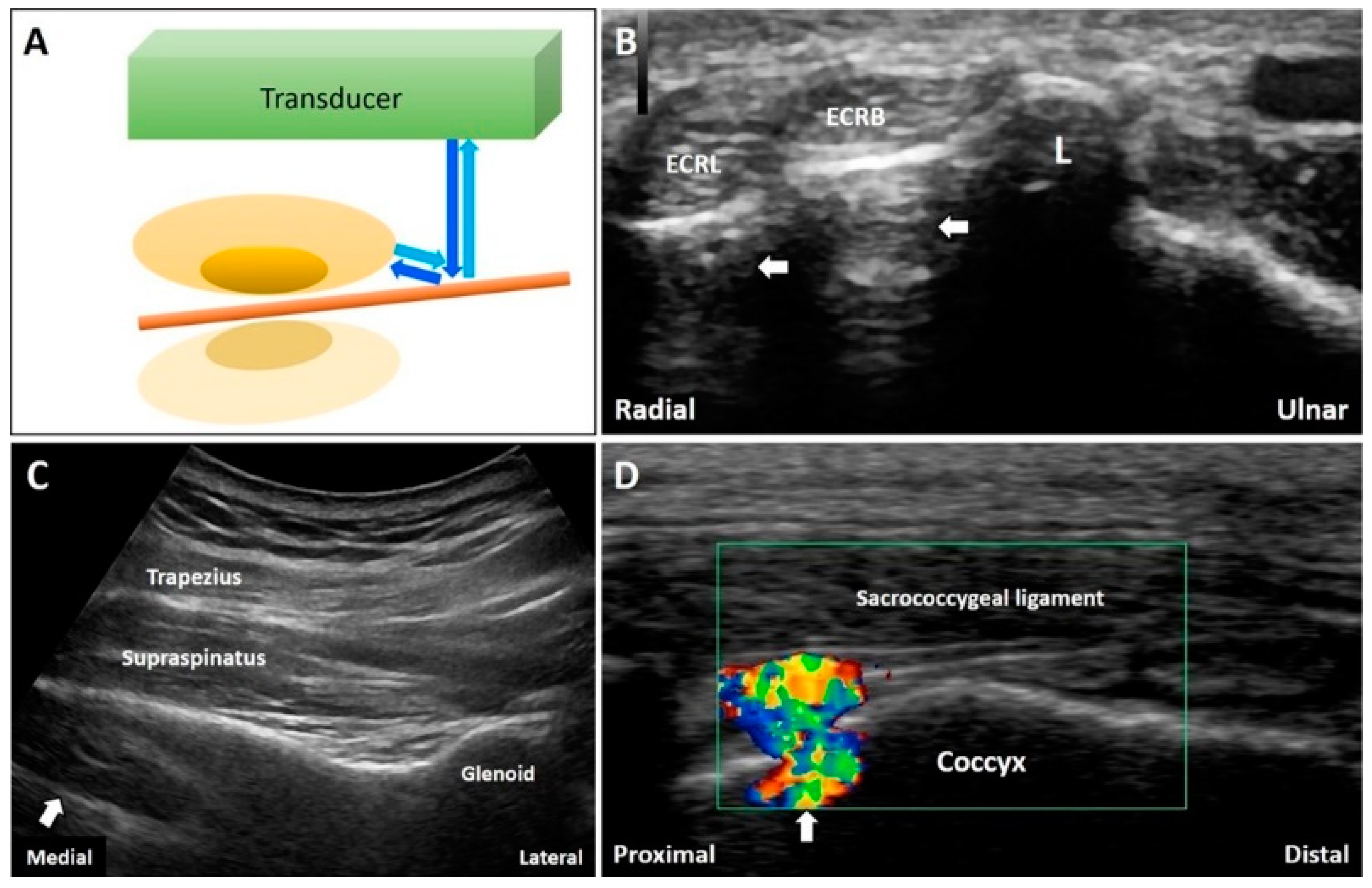
2.1. Focal Zone Artifact
2.1.1. Physics
2.1.2. Clinical Examples
3. Attenuation of Ultrasound Signals
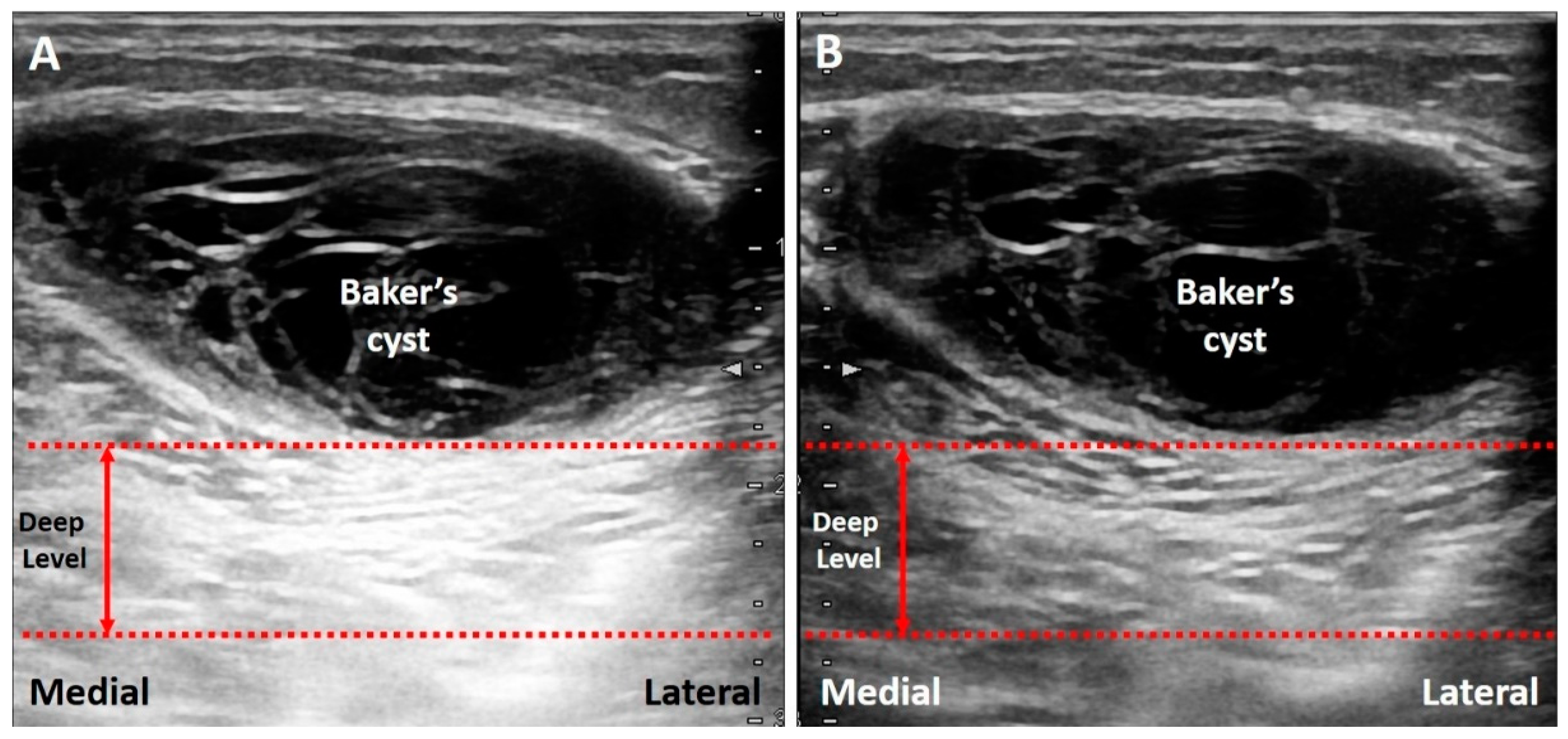
3.1. Posterior Enhancement
3.1.1. Physics
3.1.2. Clinical Examples
3.2. Acoustic Shadowing
3.2.1. Physics
3.2.2. Clinical Examples
3.3. Reverberation
3.3.1. Physics
3.3.2. Clinical Examples
3.4. Ring Down Artifact
3.4.1. Physics
3.4.2. Clinical Examples
3.5. Mirror Image Artifact
3.5.1. Physics
3.5.2. Clinical Examples
3.6. Anisotropy
3.6.1. Physics
3.6.2. Clinical Examples
3.7. Side Lobe Artifact
3.7.1. Physics
3.7.2. Clinical Examples
4. Speed of the Sound
4.1. Refraction Artifact
4.1.1. Physics
4.1.2. Clinical Examples
4.2. Edge Artifact
4.2.1. Physics
4.2.2. Clinical Examples
4.3. Range Ambiguity Artifact
4.3.1. Physics
4.3.2. Clinical Examples
5. Conclusions
Author Contributions
Funding
Conflicts of Interest
References
- Chang, K.-V.; Wu, W.-T.; Hsu, P.-C.; Lew, H.L.; Özçakar, L. Clinical Tests of the Shoulder. Am. J. Phys. Med. Rehabil. 2020, 99, 161–169. [Google Scholar] [CrossRef] [PubMed]
- Bianchi, S.; Hoffman, D.F.; Tamborrini, G.; Poletti, P. Ultrasound Findings in Less Frequent Causes of Carpal Tunnel Syndrome. J. Ultrasound Med. 2020. [Google Scholar] [CrossRef] [PubMed]
- Bianchi, S.; Beaulieu, J.-Y.; Poletti, P.-A. Ultrasound of the ulnar-palmar region of the wrist: Normal anatomy and anatomic variations. J. Ultrasound 2020, 1–14. [Google Scholar] [CrossRef]
- Ciloglu, O.; Karaali, E.; Gorgulu, F.F.; Ekiz, T. Ultrasonographic evaluation of the patellar tendon length and elasticity after open-wedge high tibial osteotomy: A comparison with radiological and clinical parameters. Knee 2020, 27, 1128–1134. [Google Scholar] [CrossRef]
- Wu, W.-T.; Chang, K.-V.; Mezian, K.; Naňka, O.; Lin, C.-P.; Özçakar, L. Basis of Shoulder Nerve Entrapment Syndrome: An Ultrasonographic Study Exploring Factors Influencing Cross-Sectional Area of the Suprascapular Nerve. Front. Neurol. 2018, 9, 902. [Google Scholar] [CrossRef] [Green Version]
- Wu, W.-T.; Chang, K.-V.; Mezian, K.; Nanka, O.; Yang, Y.-C.; Hsu, Y.-C.; Hsu, P.-C.; Özçakar, L. Ulnar Wrist Pain Revisited: Ultrasound Diagnosis and Guided Injection for Triangular Fibrocartilage Complex Injuries. J. Clin. Med. 2019, 8, 1540. [Google Scholar] [CrossRef] [Green Version]
- Sudoł-Szopińska, I.; Afonso, P.D.; Jacobson, J.A.; Teh, J. Imaging of gout: Findings and pitfalls. A pictorial review. Acta Reumatol. Port. 2020, 45, 20–25. [Google Scholar]
- Wang, J.-C.; Chang, K.-V.; Wu, W.-T.; Han, D.-S.; Özçakar, L. Ultrasound-Guided Standard vs. Dual-Target Subacromial Corticosteroid Injections for Shoulder Impingement Syndrome: A Randomized Controlled Trial. Arch. Phys. Med. Rehabil. 2019, 100, 2119–2128. [Google Scholar] [CrossRef]
- Bignotti, B.; Martinoli, C.; Tagliafico, A. Update on Ultrasound-Guided Interventional Procedures on Peripheral Nerves. Semin. Musculoskelet. Radiol. 2016, 20, 453–460. [Google Scholar] [CrossRef]
- Hung, C.-Y.; Chang, K.-V.; Mezian, K.; Nanka, O.; Wu, W.-T.; Hsu, P.-C.; Özçakar, L. Advanced Ankle and Foot Sonoanatomy: Imaging Beyond the Basics. Diagnostics 2020, 10, 160. [Google Scholar] [CrossRef] [Green Version]
- Rossi, F.; Zaottini, F.; Picasso, R.; Martinoli, C.; Tagliafico, A. Ankle and Foot Ultrasound: Reliability of Side-to-Side Comparison of Small Anatomic Structures. J. Ultrasound Med. 2018, 38, 2143–2153. [Google Scholar] [CrossRef] [PubMed]
- Feldman, M.K.; Katyal, S.; Blackwood, M.S. US Artifacts. Radiographics 2009, 29, 1179–1189. [Google Scholar] [CrossRef] [PubMed]
- Ricci, V.; Soylu, A.R.; Özçakar, L. Artifacts and Artistic Facts. Am. J. Phys. Med. Rehabil. 2019, 98, 521–525. [Google Scholar] [CrossRef]
- Baad, M.; Lu, Z.F.; Reiser, I.; Paushter, D. Clinical Significance of US Artifacts. Radiographics 2017, 37, 1408–1423. [Google Scholar] [CrossRef] [PubMed]
- Brown, J.M.; Yablon, C.M.; Morag, Y.; Brandon, C.J.; Jacobson, J.A. US of the Peripheral Nerves of the Upper Extremity: A Landmark Approach. Radiographics 2016, 36, 452–463. [Google Scholar] [CrossRef] [Green Version]
- Melville, D.; Scalcione, L.; Gimber, L.; Lorenz, E.; Witte, R.; Taljanovic, M. Artifacts in Musculoskeletal Ultrasonography. Semin. Musculoskelet. Radiol. 2014, 18, 3–11. [Google Scholar] [CrossRef] [Green Version]
- Gimber, L.; Melville, D.M.; Klauser, A.; Witte, R.S.; Arif-Tiwari, H.; Taljanovic, M.S. Artifacts at Musculoskeletal US: Resident and Fellow Education Feature. Radiographics 2016, 36, 479–480. [Google Scholar] [CrossRef] [Green Version]
- Blome, A.; Harrigan, R.; Goett, H.; Costantino, T.; Gibbons, R. Ultrasonographic Characteristics of Baker’s Cysts: The Sonographic Foucher’s Sign. J. Emerg. Med. 2017, 53, 753–755. [Google Scholar] [CrossRef]
- Griffith, J.F. Top-Ten Pitfalls in Rotator Cuff Ultrasound. Semin. Musculoskelet. Radiol. 2019, 23, 429–435. [Google Scholar] [CrossRef]
- Jacobson, J.A.; Lancaster, S.; Prasad, A.; Van Holsbeeck, M.T.; Craig, J.G.; Kolowich, P. Full-Thickness and Partial-Thickness Supraspinatus Tendon Tears: Value of US Signs in Diagnosis. Radiology 2004, 230, 234–242. [Google Scholar] [CrossRef] [Green Version]
- Bianchi, S. Ultrasound and bone: A pictorial review. J. Ultrasound 2020, 23, 227–257. [Google Scholar] [CrossRef] [PubMed]
- Rubin, J.M.; Adler, R.S.; Bude, R.O.; Fowlkes, J.B.; Carson, P.L. Clean and dirty shadowing at US: A reappraisal. Radiology 1991, 181, 231–236. [Google Scholar] [CrossRef] [PubMed]
- Quien, M.M.; Saric, M. Ultrasound imaging artifacts: How to recognize them and how to avoid them. Echocardiography 2018, 35, 1388–1401. [Google Scholar] [CrossRef]
- Kim, S.M.; Brigido, M.K.; Jacobson, J.A. Ultrasound-Guided Percutaneous Tenotomy. Semin. Musculoskelet. Radiol. 2016, 20, 414–421. [Google Scholar] [CrossRef]
- Avruch, L.; Cooperberg, P.L. The ring-down artifact. J. Ultrasound Med. 1985, 4, 21–28. [Google Scholar] [CrossRef] [PubMed]
- Magazzeni, P.; Jochum, D.; Iohom, G.; Mekler, G.; Albuisson, E.; Bouaziz, H. Ultrasound-Guided Selective Versus Conventional Block of the Medial Brachial Cutaneous and the Intercostobrachial Nerves: A Randomized Clinical Trial. Reg. Anesthesia Pain Med. 2018, 43, 832–837. [Google Scholar] [CrossRef]
- Tuma, J.; Jenssen, C.; Möller, K.; Cui, X.; Kinkel, H.; Uebel, S.; Dietrich, C. Ultrasound artifacts and their diagnostic significance in internal medicine and gastroenterology-Part 1: B-mode artifacts. Z. Gastroenterol. 2016, 54, 433–450. [Google Scholar] [CrossRef]
- Kremkau, F.W.; Taylor, K.J. Artifacts in ultrasound imaging. J. Ultrasound Med. 1986, 5, 227–237. [Google Scholar] [CrossRef]
- Aydın, T.; Şen, E.I.; Yardımcı, M.Y.; Kesiktaş, F.N.; Öneş, K.; Paker, N. Efficacy of ultrasound-guided suprascapular nerve block treatment in patients with painful hemiplegic shoulder. Neurol. Sci. 2019, 40, 985–991. [Google Scholar] [CrossRef]
- Van Holsbeeck, M.T.; Soliman, S.; Van Kerkhove, F.; Craig, J. Advanced Musculoskeletal Ultrasound Techniques: What are the applications? Am. J. Roentgenol. 2020. [Google Scholar] [CrossRef]
- Chianca, V.; Zappia, M.; Oliva, F.; Luca, B.; Maffulli, N. Post-operative MRI and US appearance of the Achilles tendons. J. Ultrasound 2020. [Google Scholar] [CrossRef] [PubMed]
- Paul, Y.; Barthez, D.; Léveillé, R.; Peter, V.; Scrivani, D. SIDE LOBES AND GRATING LOBES ARTIFACTS IN ULTRASOUND IMAGING. Veter-Radiol. Ultrasound 1997, 38, 387–393. [Google Scholar] [CrossRef] [PubMed]
- Bönhof, J.; McLaughlin, G. Artifacts in Sonography—Part 3. Ultraschall der Med. Eur. J. Ultrasound 2018, 39, 260–283. [Google Scholar] [CrossRef] [PubMed]
- Yablon, C.M.; Hammer, M.R.; Morag, Y.; Brandon, C.J.; Fessell, D.P.; Jacobson, J.A. US of the Peripheral Nerves of the Lower Extremity: A Landmark Approach. Radiographics 2016, 36, 464–478. [Google Scholar] [CrossRef]


© 2020 by the authors. Licensee MDPI, Basel, Switzerland. This article is an open access article distributed under the terms and conditions of the Creative Commons Attribution (CC BY) license (http://creativecommons.org/licenses/by/4.0/).
Share and Cite
Wu, W.-T.; Chang, K.-V.; Hsu, Y.-C.; Hsu, P.-C.; Ricci, V.; Özçakar, L. Artifacts in Musculoskeletal Ultrasonography: From Physics to Clinics. Diagnostics 2020, 10, 645. https://doi.org/10.3390/diagnostics10090645
Wu W-T, Chang K-V, Hsu Y-C, Hsu P-C, Ricci V, Özçakar L. Artifacts in Musculoskeletal Ultrasonography: From Physics to Clinics. Diagnostics. 2020; 10(9):645. https://doi.org/10.3390/diagnostics10090645
Chicago/Turabian StyleWu, Wei-Ting, Ke-Vin Chang, Yu-Chun Hsu, Po-Cheng Hsu, Vincenzo Ricci, and Levent Özçakar. 2020. "Artifacts in Musculoskeletal Ultrasonography: From Physics to Clinics" Diagnostics 10, no. 9: 645. https://doi.org/10.3390/diagnostics10090645