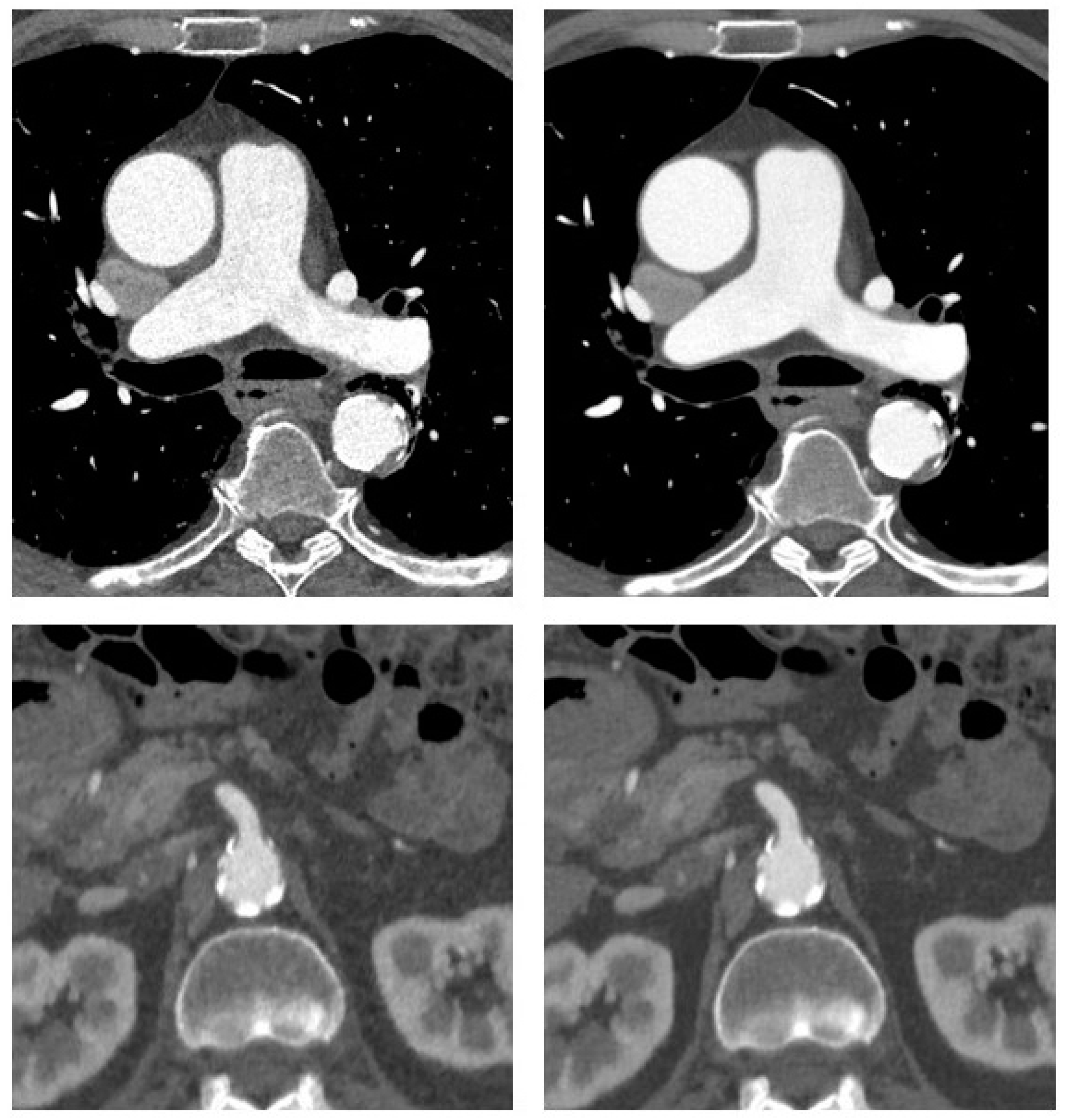
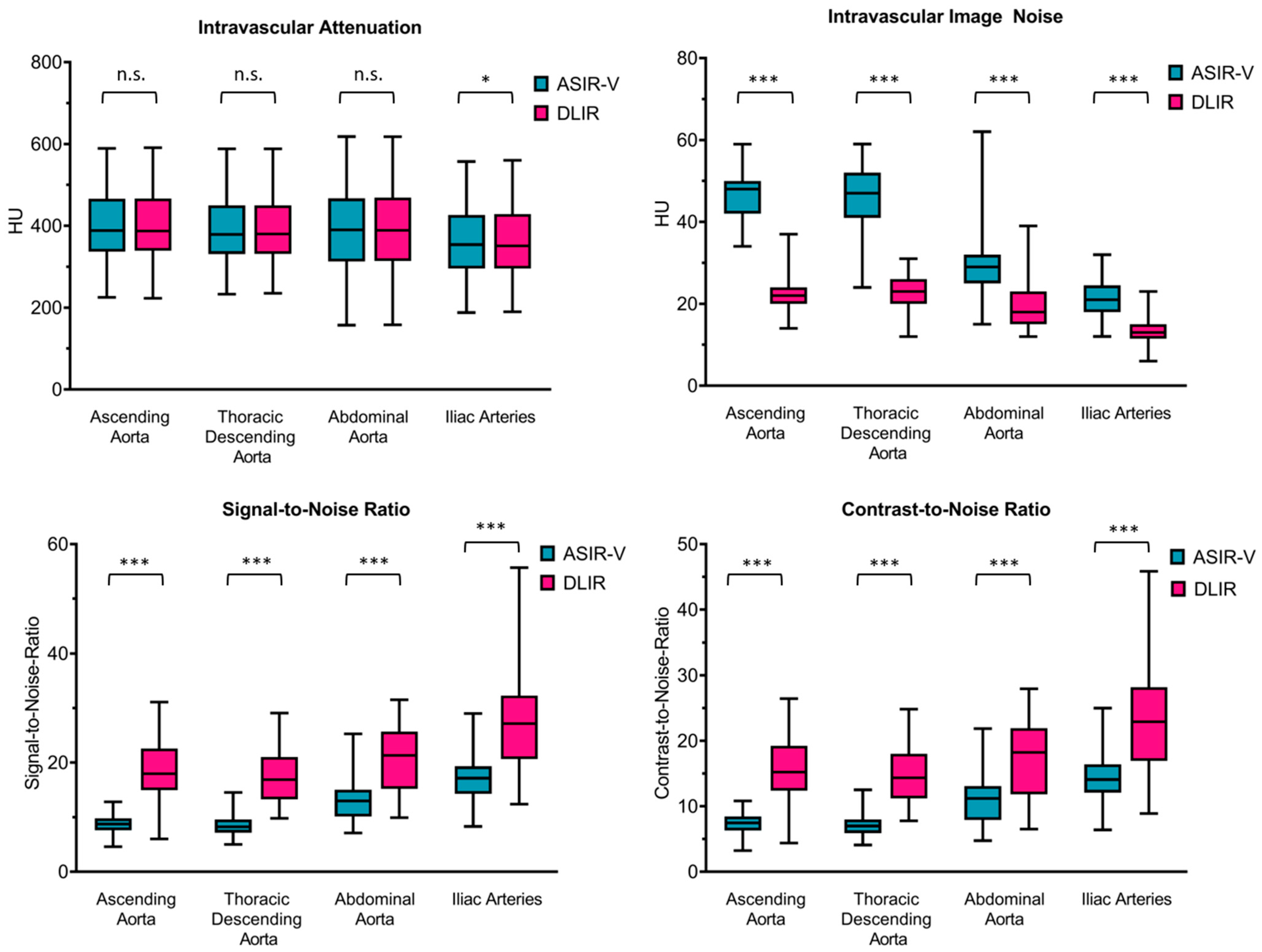
Deep Learning-Based Image Reconstruction for CT Angiography of the Aorta
Abstract
1. Introduction
2. Materials and Methods
2.1. Patient Selection and Study Design
2.2. CT Acquisition Protocol
2.3. CT Image Reconstruction
2.4. Radiation Metrics and Image Reconstruction
2.5. Analysis of Objective Image Quality and Dose Efficiency
2.6. Subjective Assessment of Diagnostic Confidence and Motion Artifacts
2.7. Statistical Analyses
3. Results
3.1. Patient Characteristics
3.2. Spectrum of Indications
3.3. Radiation Dose
3.4. Objective Image Quality
3.5. Subjective Image Quality
4. Discussion
5. Conclusions
Author Contributions
Funding
Institutional Review Board Statement
Informed Consent Statement
Data Availability Statement
Conflicts of Interest
References
- Rubin, G.D.; Leipsic, J.; Joseph Schoepf, U.; Fleischmann, D.; Napel, S. CT angiography after 20 years: A transformation in cardiovascular disease characterization continues to advance. Radiology 2014, 271, 633–652. [Google Scholar] [CrossRef]
- Meinel, F.G.; Nance, J.W.; Harris, B.S.; de Cecco, C.N.; Costello, P.; Schoepf, U.J. Radiation risks from cardiovascular imaging tests. Circulation 2014, 130, 442–445. [Google Scholar] [CrossRef]
- Tatsugami, F.; Higaki, T.; Nakamura, Y.; Yu, Z.; Zhou, J.; Lu, Y.; Fujioka, C.; Kitagawa, T.; Kihara, Y.; Iida, M.; et al. Deep learning-based image restoration algorithm for coronary CT angiography. Eur. Radiol. 2019, 29, 5322–5329. [Google Scholar] [CrossRef] [PubMed]
- Qiu, D.; Seeram, E. Does Iterative Reconstruction Improve Image Quality and Reduce Dose in Computed Tomography? Radiol. Open J. 2016, 1, 42–54. [Google Scholar] [CrossRef]
- Geyer, L.L.; Schoepf, U.J.; Meinel, F.G.; Nance, J.W.; Bastarrika, G.; Leipsic, J.A.; Paul, N.S.; Rengo, M.; Laghi, A.; de Cecco, C.N. State of the Art: Iterative CT Reconstruction Techniques. Radiology 2015, 276, 339–357. [Google Scholar] [CrossRef]
- Akagi, M.; Nakamura, Y.; Higaki, T.; Narita, K.; Honda, Y.; Zhou, J.; Yu, Z.; Akino, N.; Awai, K. Deep learning reconstruction improves image quality of abdominal ultra-high-resolution CT. Eur. Radiol. 2019, 29, 6163–6171. [Google Scholar] [CrossRef] [PubMed]
- Lim, K.; Kwon, H.; Cho, J.; Oh, J.; Yoon, S.; Kang, M.; Ha, D.; Lee, J.; Kang, E. Initial phantom study comparing image quality in computed tomography using adaptive statistical iterative reconstruction and new adaptive statistical iterative reconstruction v. J. Comput. Assist. Tomogr. 2015, 39, 443–448. [Google Scholar] [CrossRef] [PubMed]
- Kwon, H.; Cho, J.; Oh, J.; Kim, D.; Cho, J.; Kim, S.; Lee, S.; Lee, J. The adaptive statistical iterative reconstruction-V technique for radiation dose reduction in abdominal CT: Comparison with the adaptive statistical iterative reconstruction technique. Br. J. Radiol. 2015, 88, 20150463. [Google Scholar] [CrossRef]
- Fan, J.; Yue, M.; Melnyk, R. Benefits of ASiR-V reconstruction for reducing patient radiation dose and preserving diagnostic quality in CT exams. In White Paper; GE Healthcare: North GrandviewWaukesha, WI, USA, 2014. [Google Scholar]
- Arndt, C.; Güttler, F.; Heinrich, A.; Bürckenmeyer, F.; Diamantis, I.; Teichgräber, U. Deep Learning CT Image Reconstruction in Clinical Practice. Rofo 2021, 193, 252–261. [Google Scholar] [CrossRef] [PubMed]
- Jensen, C.T.; Liu, X.; Tamm, E.P.; Chandler, A.G.; Sun, J.; Morani, A.C.; Javadi, S.; Wagner-Bartak, N.A. Image Quality Assessment of Abdominal CT by Use of New Deep Learning Image Reconstruction: Initial Experience. AJR Am. J. Roentgenol. 2020, 215, 50–57. [Google Scholar] [CrossRef]
- Hsieh, J.; Liu, E.; Nett, B.; Tang, J.; Thibault, J.B.; Sahney, S. A New Era of Image Reconstruction: TrueFidelity—Technical White Paper on Deep Learning Image Reconstruction; GE Healthcare: North GrandviewWaukesha, WI, USA, 2019. [Google Scholar]
- Higaki, T.; Nakamura, Y.; Zhou, J.; Yu, Z.; Nemoto, T.; Tatsugami, F.; Awai, K. Deep Learning Reconstruction at CT: Phantom Study of the Image Characteristics. Acad. Radiol. 2020, 27, 82–87. [Google Scholar] [CrossRef]
- Funama, Y.; Takahashi, H.; Goto, T.; Aoki, Y.; Yoshida, R.; Kumagai, Y.; Awai, K. Improving Low-contrast Detectability and Noise Texture Pattern for Computed Tomography Using Iterative Reconstruction Accelerated with Machine Learning Method: A Phantom Study. Acad. Radiol. 2020, 27, 929–936. [Google Scholar] [CrossRef] [PubMed]
- Brady, S.L.; Trout, A.T.; Somasundaram, E.; Anton, C.G.; Li, Y.; Dillman, J.R. Improving Image Quality and Reducing Radiation Dose for Pediatric CT by Using Deep Learning Reconstruction. Radiology 2021, 298, 180–188. [Google Scholar] [CrossRef] [PubMed]
- Akagi, M.; Nakamura, Y.; Higaki, T.; Narita, K.; Honda, Y.; Awai, K. Deep learning reconstruction of equilibrium phase CT images in obese patients. Eur. J. Radiol. 2020, 133, 109349. [Google Scholar] [CrossRef] [PubMed]
- Narita, K.; Nakamura, Y.; Higaki, T.; Akagi, M.; Honda, Y.; Awai, K. Deep learning reconstruction of drip-infusion cholangiography acquired with ultra-high-resolution computed tomography. Abdom. Radiol. 2020, 45, 2698–2704. [Google Scholar] [CrossRef] [PubMed]
- Benz, D.C.; Benetos, G.; Rampidis, G.; von, F.E.; Bakula, A.; Sustar, A.; Kudura, K.; Messerli, M.; Fuchs, T.A.; Gebhard, C.; et al. Validation of deep-learning image reconstruction for coronary computed tomography angiography: Impact on noise, image quality and diagnostic accuracy. J. Cardiovasc. Comput. Tomogr. 2020, 14, 444–451. [Google Scholar] [CrossRef] [PubMed]
- Liu, Y.; Xu, J.; Li, J.; Ren, J.; Liu, H.; Xu, J.; Wei, M.; Hao, Y.; Zheng, M. The ascending aortic image quality and the whole aortic radiation dose of high-pitch dual-source CT angiography. J. Cardiothorac. Surg. 2013, 8, 228. [Google Scholar] [CrossRef] [PubMed]
- Schegerer, A.; Loose, R.; Heuser, L.J.; Brix, G. Diagnostische Referenzwerte für diagnostische und interventionelle Röntgenanwendungen in Deutschland: Aktualisierung und Handhabung. Rofo 2019, 191, 739–751. [Google Scholar] [CrossRef]
- Brix, G.; Lechel, U.; Petersheim, M.; Krissak, R.; Fink, C. Dynamic contrast-enhanced CT studies: Balancing patient exposure and image noise. Investig. Radiol. 2011, 46, 64–70. [Google Scholar] [CrossRef]
- Moro, L.; Panizza, D.; D’Ambrosio, D.; Carne, I. Considerations on an automatic computed tomography tube current modulation system. Radiat. Prot. Dosimetry 2013, 156, 525–530. [Google Scholar] [CrossRef]
- Seehofnerová, A.; Kok, M.; Mihl, C.; Douwes, D.; Sailer, A.; Nijssen, E.; de Haan, M.J.W.; Wildberger, J.E.; Das, M. Feasibility of low contrast media volume in CT angiography of the aorta. Eur. J. Radiol. Open 2015, 2, 58–65. [Google Scholar] [CrossRef] [PubMed][Green Version]
- Shen, Y.; Sun, Z.; Xu, L.; Li, Y.; Zhang, N.; Yan, Z.; Fan, Z. High-pitch, low-voltage and low-iodine-concentration CT angiography of aorta: Assessment of image quality and radiation dose with iterative reconstruction. PLoS ONE 2015, 10, e0117469. [Google Scholar] [CrossRef] [PubMed]
- Vardhanabhuti, V.; Nicol, E.; Morgan-Hughes, G.; Roobottom, C.A.; Roditi, G.; Hamilton, M.C.K.; Bull, R.K.; Pugliese, F.; Williams, M.C.; Stirrup, J.; et al. Recommendations for accurate CT diagnosis of suspected acute aortic syndrome (AAS)—On behalf of the British Society of Cardiovascular Imaging (BSCI)/British Society of Cardiovascular CT (BSCCT). Br. J. Radiol. 2016, 89, 20150705. [Google Scholar] [CrossRef]
- Vasconcelos, R.; Vrtiska, T.J.; Foley, T.A.; Macedo, T.A.; Cardona, J.C.M.; Williamson, E.E.; McCollough, C.H.; Fletcher, J.G. Reducing Iodine Contrast Volume in CT Angiography of the Abdominal Aorta Using Integrated Tube Potential Selection and Weight-Based Method Without Compromising Image Quality. AJR Am. J. Roentgenol. 2017, 208, 552–563. [Google Scholar] [CrossRef] [PubMed]
- De Marco, P.; Origgi, D. New adaptive statistical iterative reconstruction ASiR-V: Assessment of noise performance in comparison to ASiR. J. Appl. Clin. Med. Phys. 2018, 19, 275–286. [Google Scholar] [CrossRef]


| Parameter | Value | |
|---|---|---|
| Regions covered | ||
| Thoracic and abdominal aorta | 43 patients | |
| Thoracic aorta only | 8 patients | |
| Acquisition parameters | ||
| Tube voltage | 100 kV | |
| Tube current | tube current modulation | |
| Reference noise index | 25 | |
| ECG-triggering | 75% of RR-interval (chest only) | |
| Contrast protocol | ||
| Contrast volume | 80 mL | |
| Contrast concentration | 400 mg/mL | |
| Flow rate | 4 mL/s | |
| Saline chaser | 40 mL with 4 mL/s | |
| Reconstruction parameters | ||
| Reconstruction method | ASIR-V 60% | DLIR-H |
| Reconstruction kernel | HD Stnd | HD Stnd/Stnd |
| Slice thickness | 0.625 mm | 0.625 mm |
| Slice increment | 0.625 mm | 0.625 mm |
| Radiation metrics | ||
| CTDIvol chest | 5.5 (5.3–6.8) mGy | |
| CTDIvol abdomen | 6.4 (6.3–6.4) mGy | |
| DLP chest | 126 (125–153) mGy*cm | |
| DLP abdomen | 265 (246–298) mGy*cm | |
| Total DLP | 389 (344–418) mGy*cm | |
| All Patients | Men | Women | |
|---|---|---|---|
| No. of patients | 51 | 32 | 19 |
| Age (years) | 69 (57–74) | 64 (54–72) | 70 (65–79) |
| Body weight (kg) | 89 (72–100) | 94 (84–106) | 73 (61–81) |
| BMI (kg/m2) | 27.4 (23.9–31.0) | 27.8 (24.6–31.0) | 26.1 (21.7–30.9) |
| Main indication for CTA | |||
| Aneurysm follow-up | 9 | 7 | 2 |
| Dissection | 3 | 1 | 2 |
| preoperative | 9 | 7 | 2 |
| postop (incl. TEVAR) | 19 | 10 | 9 |
| Acute aortic syndrome | 9 | 5 | 4 |
| Trauma | 2 | 2 | 0 |
| Obese Patients (n = 17) | Normal-Weight Patients (n = 17) | |||||
|---|---|---|---|---|---|---|
| Contrast-to-Noise-Ratio | ASIR-V | DLIR | p-Value | ASIR-V | DLIR | p-Value |
| Ascending Aorta | 7 (6–8) | 14 (12–16) | <0.0001 | 8 (8–9) | 19 (15–22) | <0.0001 |
| Thoracic descending aorta | 7 (6–7) | 13 (11–15) | <0.0001 | 8 (7–9) | 18 (13–19) | <0.0001 |
| Abdominal aorta | 12 (8–13) | 19 (15–24) | <0.0001 | 9 (7–12) | 16 (11–21) | <0.0001 |
| Iliac arteries | 14 (12–15) | 21 (19–25) | <0.0001 | 15 (13–16) | 24 (19–28) | 0.001 |
| ASIR-V | DLIR | |
|---|---|---|
| Ascending aorta | 0.659 | 0.509 |
| Thoracic descending aorta | 0.630 | 0.807 |
| Abdominal aorta | 0.552 | 0.456 |
| Iliac arteries | 0.706 | 0.822 |
| ASIR-V | DLIR | p-Value | |
|---|---|---|---|
| Reader 1 | |||
| Ascending aorta | 4 (4–5) | 5 (5–5) | <0.0001 |
| Thoracic descending aorta | 4 (4–5) | 5 (5–5) | <0.0001 |
| Abdominal aorta | 4 (4–4) | 5 (4–5) | <0.0001 |
| Iliac arteries | 5 (4–5) | 5 (5–5) | <0.0001 |
| Reader 2 | |||
| Ascending Aorta | 4 (4–4) | 5 (4–5) | <0.0001 |
| Thoracic descending aorta | 4 (4–4) | 5 (5–5) | <0.0001 |
| Abdominal aorta | 4 (3–4) | 5 (4–5) | <0.0001 |
| Iliac arteries | 4 (4–5) | 5 (5–5) | <0.0001 |
Publisher’s Note: MDPI stays neutral with regard to jurisdictional claims in published maps and institutional affiliations. |
© 2021 by the authors. Licensee MDPI, Basel, Switzerland. This article is an open access article distributed under the terms and conditions of the Creative Commons Attribution (CC BY) license (https://creativecommons.org/licenses/by/4.0/).
Share and Cite
Heinrich, A.; Streckenbach, F.; Beller, E.; Groß, J.; Weber, M.-A.; Meinel, F.G. Deep Learning-Based Image Reconstruction for CT Angiography of the Aorta. Diagnostics 2021, 11, 2037. https://doi.org/10.3390/diagnostics11112037
Heinrich A, Streckenbach F, Beller E, Groß J, Weber M-A, Meinel FG. Deep Learning-Based Image Reconstruction for CT Angiography of the Aorta. Diagnostics. 2021; 11(11):2037. https://doi.org/10.3390/diagnostics11112037
Chicago/Turabian StyleHeinrich, Andra, Felix Streckenbach, Ebba Beller, Justus Groß, Marc-André Weber, and Felix G. Meinel. 2021. "Deep Learning-Based Image Reconstruction for CT Angiography of the Aorta" Diagnostics 11, no. 11: 2037. https://doi.org/10.3390/diagnostics11112037
APA StyleHeinrich, A., Streckenbach, F., Beller, E., Groß, J., Weber, M.-A., & Meinel, F. G. (2021). Deep Learning-Based Image Reconstruction for CT Angiography of the Aorta. Diagnostics, 11(11), 2037. https://doi.org/10.3390/diagnostics11112037


