
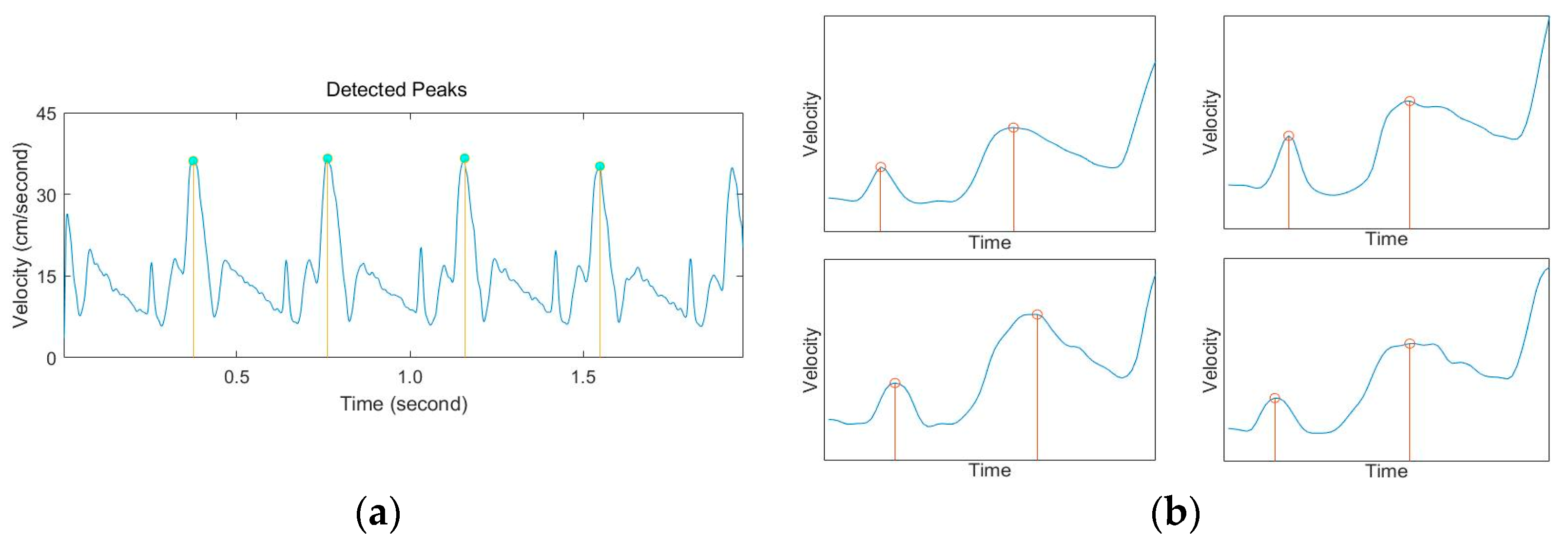
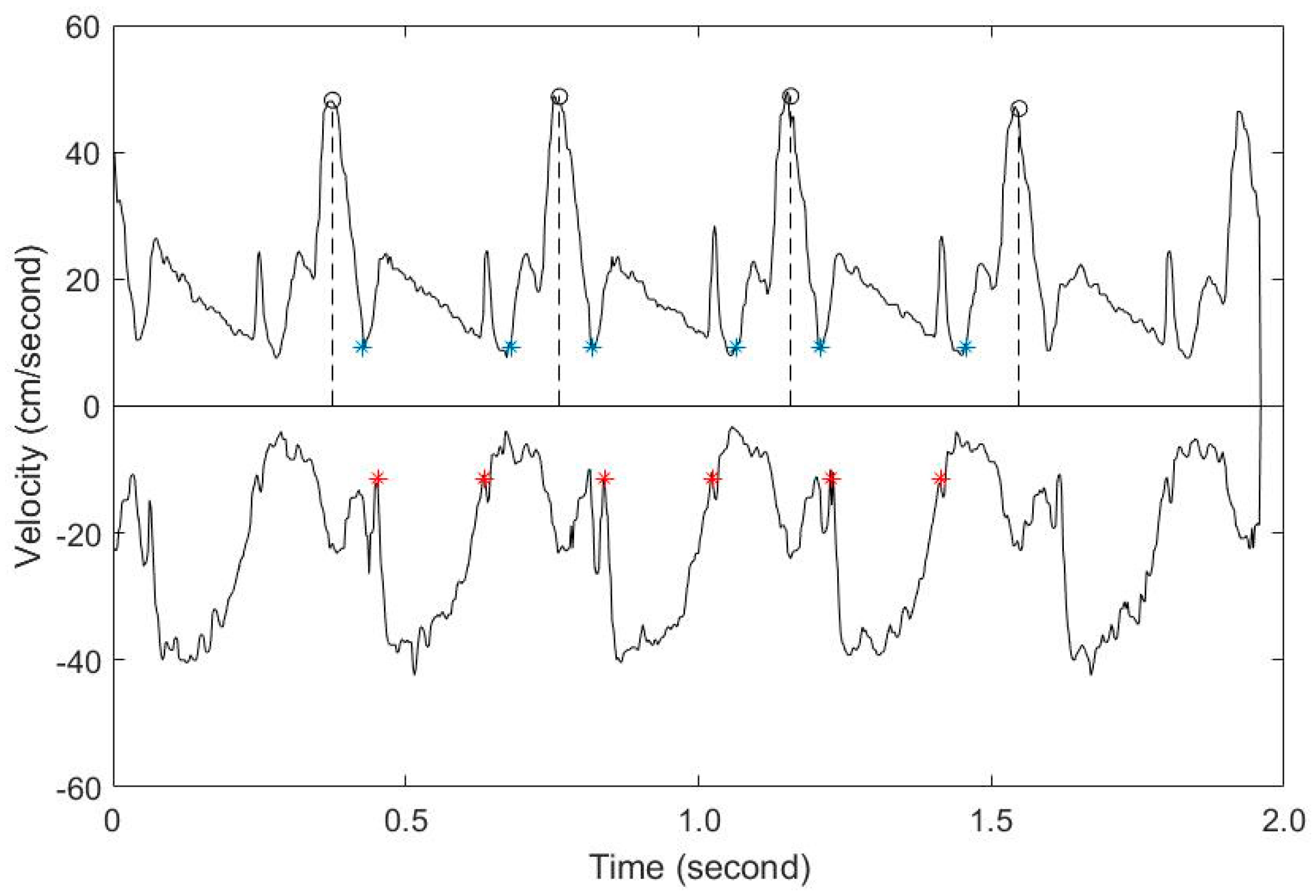
Evaluation of the Fetal Left Ventricular Myocardial Performance Index (MPI) by Using an Automated Measurement of Doppler Signals in Normal Pregnancies


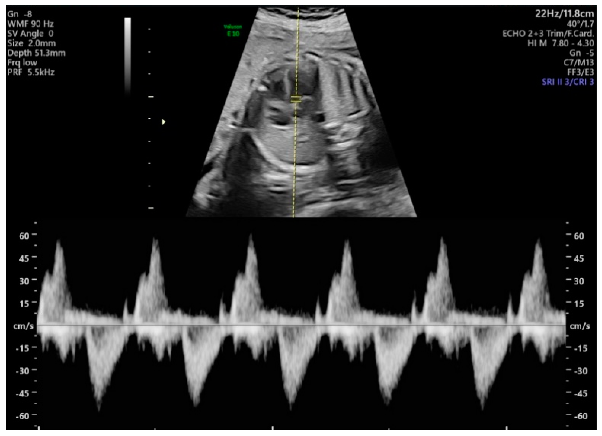
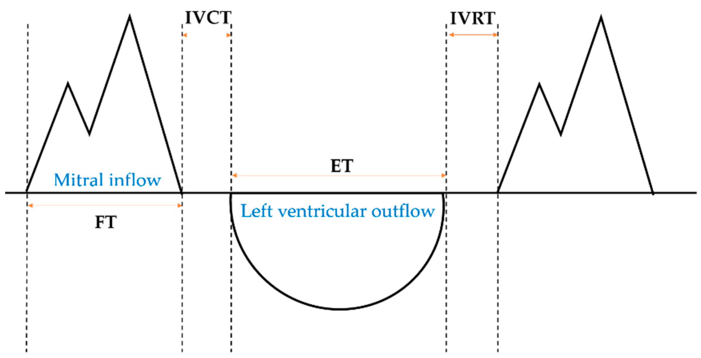
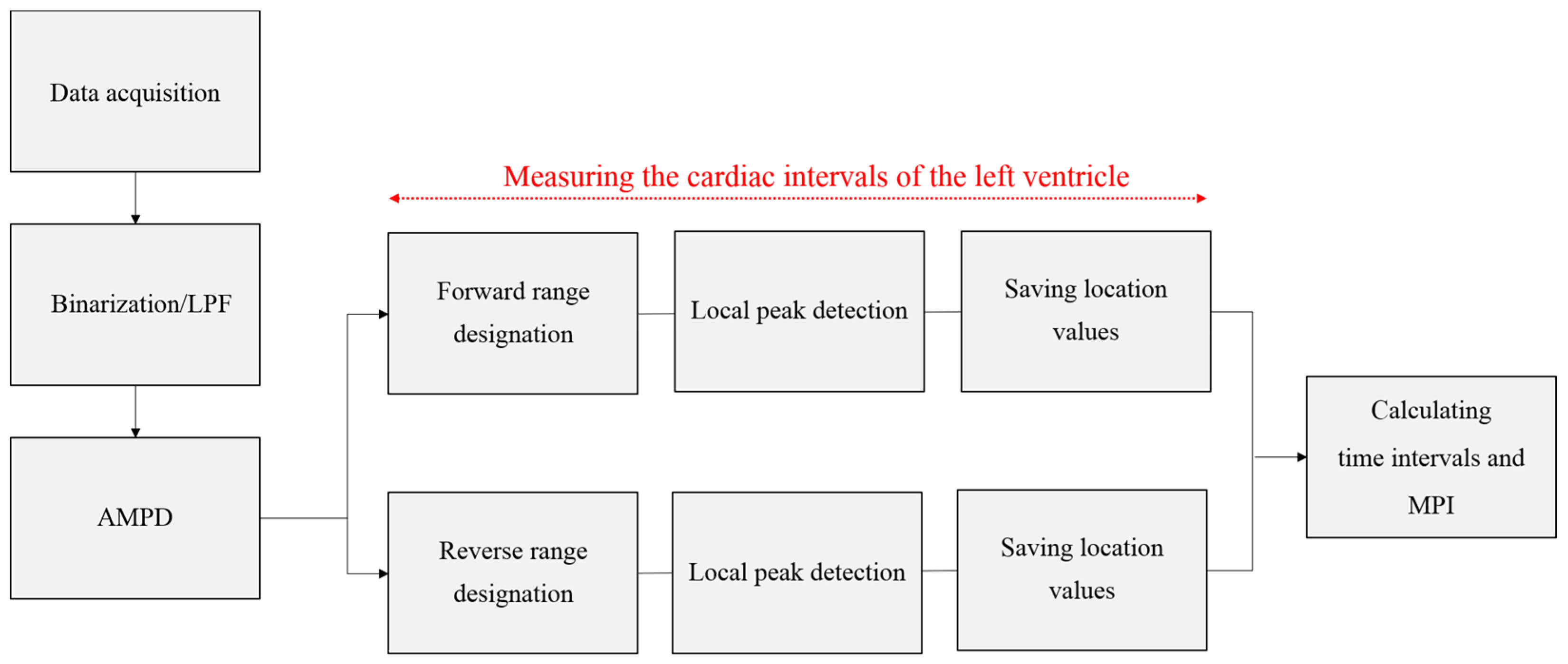
Abstract
Share and Cite
Kim, S.-M.; Ye, S.-Y. Evaluation of the Fetal Left Ventricular Myocardial Performance Index (MPI) by Using an Automated Measurement of Doppler Signals in Normal Pregnancies. Diagnostics 2021, 11, 358. https://doi.org/10.3390/diagnostics11020358
Kim S-M, Ye S-Y. Evaluation of the Fetal Left Ventricular Myocardial Performance Index (MPI) by Using an Automated Measurement of Doppler Signals in Normal Pregnancies. Diagnostics. 2021; 11(2):358. https://doi.org/10.3390/diagnostics11020358
Chicago/Turabian StyleKim, Su-Min, and Soo-Young Ye. 2021. "Evaluation of the Fetal Left Ventricular Myocardial Performance Index (MPI) by Using an Automated Measurement of Doppler Signals in Normal Pregnancies" Diagnostics 11, no. 2: 358. https://doi.org/10.3390/diagnostics11020358
APA StyleKim, S.-M., & Ye, S.-Y. (2021). Evaluation of the Fetal Left Ventricular Myocardial Performance Index (MPI) by Using an Automated Measurement of Doppler Signals in Normal Pregnancies. Diagnostics, 11(2), 358. https://doi.org/10.3390/diagnostics11020358