
The Use of Imaging Techniques in Chronic Kidney Disease-Mineral and Bone Disorders (CKD-MBD)—A Systematic Review

 and
and
Abstract
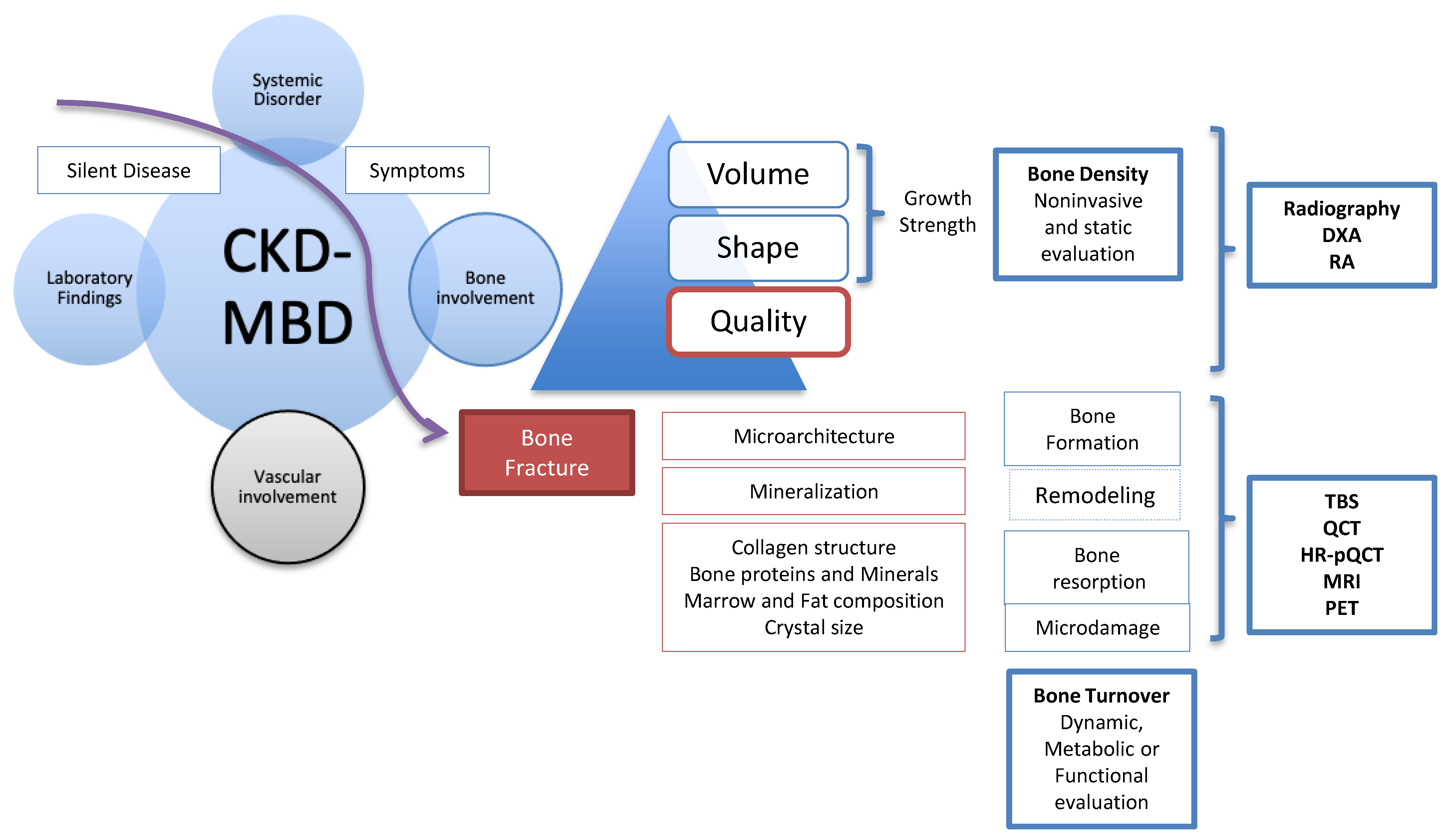
:1. Introduction and Pathophysiology
2. Characteristics of Bone Structure
3. Bone and Soft Tissue Imaging
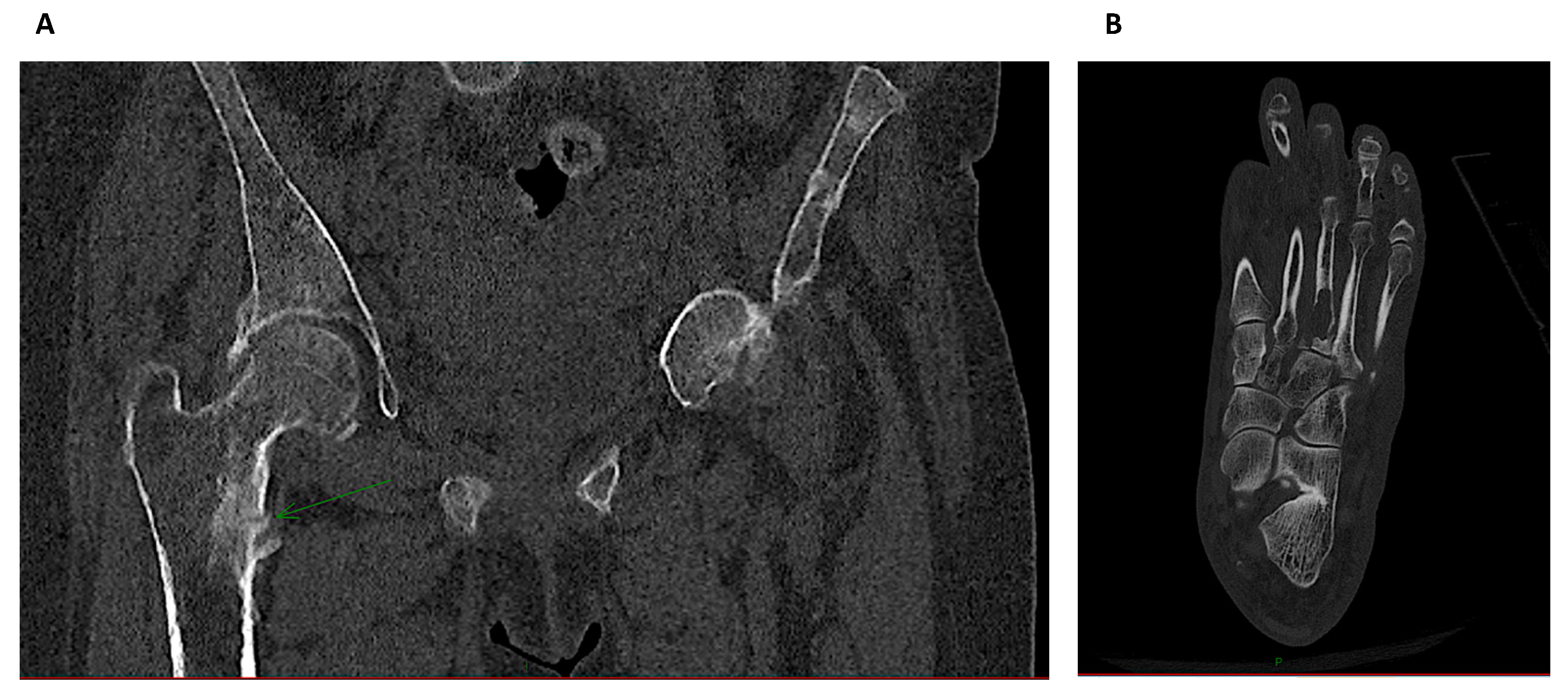
3.1. Conventional Radiography
3.2. Dual-Energy X-ray Absorptiometry
3.3. DXA-Derived Trabecular Bone Score (TBS)
3.4. Radiographic Absorptiometry (RA)
3.5. Quantitative Computerized Tomography
3.6. High Resolution-Peripheral Quantitative Computerized Tomography (HR-pQCT)
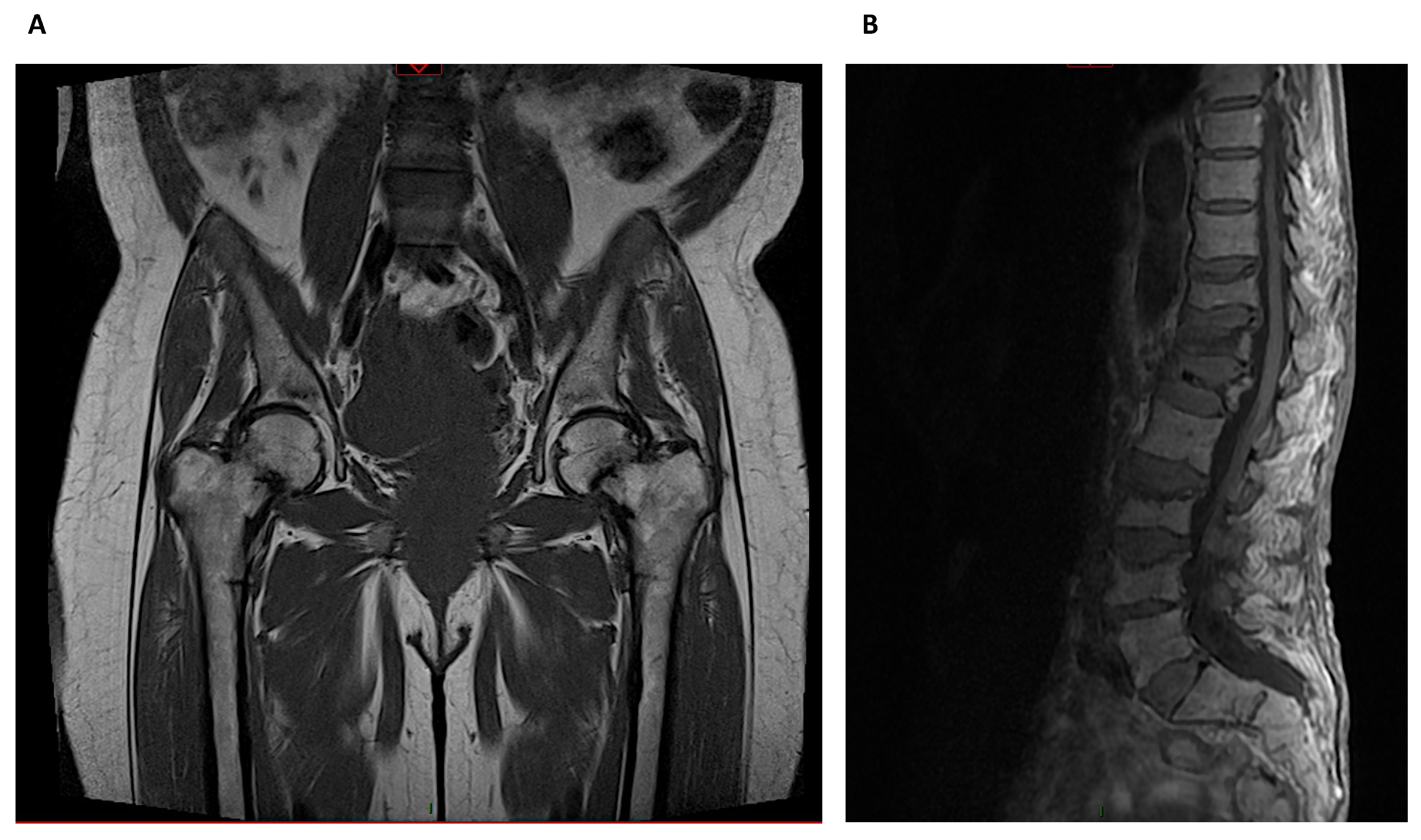
3.7. Magnetic Resonance Imaging (MRI)
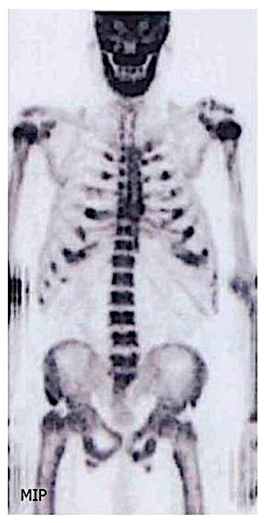
3.8. Other Imaging Techniques
4. Cardiovascular Calcifications
5. Conclusions
Funding
Institutional Review Board Statement
Informed Consent Statement
Data Availability Statement
Conflicts of Interest
References
- Isakova, T. An Introduction to PTH, Phosphate and Vitamin D: Current Issues and Concerns. Semin. Dial. 2015, 28, 563. [Google Scholar] [CrossRef]
- Jager, K.J.; Kovesdy, C.; Langham, R.; Rosenberg, M.; Jha, V.; Zoccali, C. A single number for advocacy and communication—worldwide more than 850 million individuals have kidney diseases. Kidney Int. 2019, 96, 1048–1050. [Google Scholar] [CrossRef]
- Covic, A.; Vervloet, M.; Massy, Z.A.; Torres, P.U.; Goldsmith, D.; Brandenburg, V.; Mazzaferro, S.; Evenepoel, P.; Bover, J.; Apetrii, M.; et al. Bone and mineral disorders in chronic kidney disease: Implications for cardiovascular health and ageing in the general population. Lancet Diabetes Endocrinol. 2018, 6, 319–331. [Google Scholar] [CrossRef]
- KDIGO. KDIGO clinical practice guideline for the diagnosis, evaluation, prevention, and treatment of Chronic Kidney Disease-Mineral and Bone Disorder (CKD-MBD). Kidney Int. 2009, 113, S1–S130. [Google Scholar]
- Malluche, H.H.; Mawad, H.W.; Monier-Faugere, M.C. Renal osteodystrophy in the first decade of the new millennium: Analysis of 630 bone biopsies in black and white patients. J. Bone Miner. Res. 2011, 26, 1368–1376. [Google Scholar] [CrossRef] [PubMed] [Green Version]
- Robertson, L.; Black, C.; Fluck, N.; Gordon, S.; Hollick, R.; Nguyen, H.; Prescott, G.; Marks, A. Hip fracture incidence and mortality in chronic kidney disease: The GLOMMS-II record linkage cohort study. BMJ Open 2018, 8, e020312. [Google Scholar] [CrossRef] [Green Version]
- Nitsch, D.; Mylne, A.; Roderick, P.J.; Smeeth, L.; Hubbard, R.; Fletcher, A. Chronic kidney disease and hip fracture-related mortality in older people in the UK. Nephrol. Dial. Transpl. 2009, 24, 1539–1544. [Google Scholar] [CrossRef] [Green Version]
- Aggarwal, H.K.; Jain, D.; Yadav, S.; Kaverappa, V. Bone mineral density in patients with predialysis chronic kidney disease. Ren. Fail. 2013, 35, 1105–1111. [Google Scholar] [CrossRef] [PubMed]
- Mittalhenkle, A.; Gillen, D.L.; Stehman-Breen, C.O. Increased risk of mortality associated with hip fracture in the dialysis population. Am. J. Kidney Dis. 2004, 44, 672–679. [Google Scholar] [CrossRef]
- Tentori, F.; McCullough, K.; Kilpatrick, R.D.; Bradbury, B.D.; Robinson, B.M.; Kerr, P.G.; Pisoni, R.L. High rates of death and hospitalization follow bone fracture among hemodialysis patients. Kidney Int. 2014, 85, 166–173. [Google Scholar] [CrossRef] [PubMed] [Green Version]
- Nair, S.S.; Lenihan, C.R.; Montez-Rath, M.E.; Lowenberg, D.W.; Chertow, G.M.; Winkelmayer, W.C. Temporal trends in the incidence, treatment and outcomes of hip fracture after first kidney transplantation in the United States. Am. J. Transpl. 2014, 14, 943–951. [Google Scholar] [CrossRef] [Green Version]
- Kauppila, L.I.; Polak, J.F.; Cupples, L.A.; Hannan, M.T.; Kiel, D.P.; Wilson, P.W. New indices to classify location, severity and progression of calcific lesions in the abdominal aorta: A 25-year follow-up study. Atherosclerosis 1997, 132, 245–250. [Google Scholar] [CrossRef]
- McCloskey, E.V.; Vasireddy, S.; Threlkeld, J.; Eastaugh, J.; Parry, A.; Bonnet, N.; Beneton, M.; Kanis, J.A.; Charlesworth, D. Vertebral Fracture Assessment (VFA) With a Densitometer Predicts Future Fractures in Elderly Women Unselected for Osteoporosis. J. Bone Miner. Res. 2008, 23, 1561–1568. [Google Scholar] [CrossRef] [PubMed]
- Schousboe, J.T.; Vokes, T.; Broy, S.B.; Ferrar, L.; McKiernan, F.; Roux, C.; Binkley, N. Vertebral Fracture Assessment: The 2007 ISCD Official Positions. J. Clin. Densitom. 2008, 11, 92–108. [Google Scholar] [CrossRef]
- Evenepoel, P.; Cunningham, J.; Ferrari, S.; Haarhaus, M.; Javaid, M.K.; Lafage-Proust, M.H.; Prieto-Alhambra, D.; Torres, P.U.; Cannata-Andia, J. European Renal Osteodystrophy workgroup aiotCKDMBDwgotERAE, the committee of Scientific A. In European Consensus Statement on the Diagnosis and Management of Osteoporosis in Chronic Kidney Disease Stages G4-G5D, Nephrol Dial Transplant; National Societies of the IOF: Karlstad, Sweden, 2020. [Google Scholar]
- Ketteler, M.; Block, G.A.; Evenepoel, P.; Fukagawa, M.; Herzog, C.A.; McCann, L.; Moe, S.M.; Shroff, R.; Tonelli, M.A.; Toussaint, N.D.; et al. Executive summary of the 2017 KDIGO Chronic Kidney Disease–Mineral and Bone Disorder (CKD-MBD) Guideline Update: What’s changed and why it matters. Kidney Int. 2017, 92, 26–36. [Google Scholar] [CrossRef] [PubMed] [Green Version]
- Griffith, J.F.; Genant, H.K. New imaging modalities in bone. Curr. Rheumatol. Rep. 2011, 13, 241–250. [Google Scholar] [CrossRef] [Green Version]
- Clarke, B. Normal Bone Anatomy and Physiology. Clin. J. Am. Soc. Nephrol. 2008, 3 (Suppl. 3), S131–S139. [Google Scholar] [CrossRef] [Green Version]
- Parfitt, A.M. Misconceptions (2): Turnover is always higher in cancellous than in cortical bone. Bone 2002, 30, 807–809. [Google Scholar] [CrossRef]
- Ott, S.M. Cortical or Trabecular Bone: What’s the Difference? Am. J. Nephrol. 2018, 47, 373–375. [Google Scholar] [CrossRef]
- Prescott, J.W. Quantitative Imaging Biomarkers: The Application of Advanced Image Processing and Analysis to Clinical and Preclinical Decision Making. J. Digit. Imaging 2013, 26, 97–108. [Google Scholar] [CrossRef] [Green Version]
- Lim, C.Y.; Ong, K.O. Various musculoskeletal manifestations of chronic renal insufficiency. Clin. Radiol. 2013, 68, e397–e411. [Google Scholar] [CrossRef] [PubMed]
- Alexander, A.J.; Jahangir, D.; Lazarus, M.; Sprague, S.M. Imaging in Chronic Kidney Disease-Metabolic Bone Disease. Semin. Dial. 2017, 30, 361–368. [Google Scholar] [CrossRef]
- Jevtic, V. Imaging of renal osteodystrophy. Eur. J. Radiol. 2003, 46, 85–95. [Google Scholar] [CrossRef]
- Murphey, M.D.; Sartoris, D.J.; Quale, J.L.; Pathria, M.N.; Martin, N.L. Musculoskeletal manifestations of chronic renal insufficiency. Radiographics 1993, 13, 357–379. [Google Scholar] [CrossRef]
- Ketteler, M.; Block, G.A.; Evenepoel, P.; Fukagawa, M.; Herzog, C.A.; McCann, L.; Moe, S.M.; Shroff, R.; Tonelli, M.A.; Toussaint, N.D.; et al. Diagnosis, Evaluation, Prevention, and Treatment of Chronic Kidney Disease–Mineral and Bone Disorder: Synopsis of the Kidney Disease: Improving Global Outcomes 2017 Clinical Practice Guideline Update. Ann. Intern. Med. 2018, 168, 422–430. [Google Scholar] [CrossRef]
- Adragao, T.; Pires, A.; Lucas, C.; Birne, R.; Magalhaes, L.; Goncalves, M.; Negrao, A.P. A simple vascular calcification score predicts cardiovascular risk in haemodialysis patients. Nephrol. Dial. Transpl. 2004, 19, 1480–1488. [Google Scholar] [CrossRef] [PubMed] [Green Version]
- Siakallis, L.; Tziakouri-Shiakalli, C.; Georgiades, C.S. Erratum to “Amyloidosis: Review and Imaging Findings” [Seminars in Ultrasound, CT, and MRI 2014;35(3):225-239]. Semin. Ultrasound CT MRI 2015, 36, 216. [Google Scholar] [CrossRef]
- Kiss, E.; Keusch, G.; Zanetti, M.; Jung, T.; Schwarz, A.; Schocke, M.; Jaschke, W.; Czermak, B.V. Dialysis-Related Amyloidosis Revisited. Am. J. Roentgenol. 2005, 185, 1460–1467. [Google Scholar] [CrossRef] [PubMed]
- Cummings, S.R.; Bates, D.; Black, D.M. Clinical use of bone densitometry: Scientific review. JAMA 2002, 288, 1889–1897. [Google Scholar] [CrossRef] [Green Version]
- Mazzaferro, S.; Diacinti, D.; Proietti, E.; Barresi, G.; Baldinelli, M.; Pisani, D.; D’Erasmo, E.; Pugliese, F. Morphometric X-ray absorptiometry in the assessment of vertebral fractures in renal transplant patients. Nephrol. Dial. Transpl. 2006, 21, 466–471. [Google Scholar] [CrossRef] [Green Version]
- Wiklund, P.; Toss, F.; Weinehall, L.; Hallmans, G.; Franks, P.W.; Nordström, A.; Nordström, P. Abdominal and Gynoid Fat Mass Are Associated with Cardiovascular Risk Factors in Men and Women. J. Clin. Endocrinol. Metab. 2008, 93, 4360–4366. [Google Scholar] [CrossRef] [Green Version]
- Lewis, J.R.; Wong, G.; Taverniti, A.; Vucak-Dzumhur, M.; Elder, G.J. Association between Aortic Calcification, Cardiovascular Events, and Mortality in Kidney and Pancreas-Kidney Transplant Recipients. Am. J. Nephrol. 2019, 50, 177–186. [Google Scholar] [CrossRef] [PubMed] [Green Version]
- Taal, M.W.; Roe, S.; Masud, T.; Green, D.; Porter, C.; Cassidy, M.J. Total hip bone mass predicts survival in chronic hemodialysis patients. Kidney Int. 2003, 63, 1116–1120. [Google Scholar] [CrossRef] [PubMed] [Green Version]
- Iimori, S.; Mori, Y.; Akita, W.; Kuyama, T.; Takada, S.; Asai, T.; Kuwahara, M.; Sasaki, S.; Tsukamoto, Y. Diagnostic usefulness of bone mineral density and biochemical markers of bone turnover in predicting fracture in CKD stage 5D patients--a single-center cohort study. Nephrol. Dial. Transpl. 2012, 27, 345–351. [Google Scholar] [CrossRef] [Green Version]
- Naylor, K.L.; Garg, A.X.; Zou, G.; Langsetmo, L.; Leslie, W.D.; Fraser, L.A.; Adachi, J.D.; Morin, S.; Goltzman, D.; Lentle, B.; et al. Comparison of Fracture Risk Prediction among Individuals with Reduced and Normal Kidney Function. Clin. J. Am. Soc. Nephrol. 2015, 10, 646–653. [Google Scholar] [CrossRef] [Green Version]
- West, S.L.; Lok, C.E.; Langsetmo, L.; Cheung, A.M.; Szabo, E.; Pearce, D.; Fusaro, M.; Wald, R.; Weinstein, J.; Jamal, S.A. Bone Mineral Density Predicts Fractures in Chronic Kidney Disease. J. Bone Miner. Res. 2015, 30, 913–919. [Google Scholar] [CrossRef] [PubMed]
- Carvalho, C.; Magalhães, J.; Neto, R.; Pereira, L.; Branco, P.; Adragão, T.; Frazão, J.M. Cortical bone analysis in a predialysis population: A comparison with a dialysis population. J. Bone Miner. Metab. 2017, 35, 513–521. [Google Scholar] [CrossRef] [PubMed]
- Matsubara, K.; Suliman, M.E.; Qureshi, A.R.; Axelsson, J.; Martola, L.; Heimbürger, O.; Barany, P.; Stenvinkel, P.; Lindholm, B. Bone Mineral Density in End-Stage Renal Disease Patients: Association with Wasting, Cardiovascular Disease and Mortality. Blood Purif. 2008, 26, 284–290. [Google Scholar] [CrossRef]
- Park, S.H.; Jia, T.; Qureshi, A.R.; Bárány, P.; Heimbürger, O.; Larsson, T.E.; Axelsson, J.; Stenvinkel, P.; Lindholm, B. Determinants and survival implications of low bone mineral density in end-stage renal disease patients. J. Nephrol. 2013, 26, 485–494. [Google Scholar] [CrossRef]
- Iseri, K.; Qureshi, A.R.; Dai, L.; Ripsweden, J.; Heimbürger, O.; Barany, P.; Bergström, I.; Stenvinkel, P.; Brismar, T.B.; Lindholm, B. Bone mineral density at different sites and 5 years mortality in end-stage renal disease patients: A cohort study. Bone 2020, 130, 115075. [Google Scholar] [CrossRef]
- Nickolas, T.L.; Stein, E.M.; Dworakowski, E.; Nishiyama, K.K.; Komandah-Kosseh, M.; Zhang, C.A.; McMahon, D.J.; Liu, X.S.; Boutroy, S.; Cremers, S.; et al. Rapid cortical bone loss in patients with chronic kidney disease. J. Bone Miner. Res. 2013, 28, 1811–1820. [Google Scholar] [CrossRef]
- El Maghraoui, A.; Roux, C. DXA scanning in clinical practice. QJM Int. J. Med. 2008, 101, 605–617. [Google Scholar] [CrossRef] [Green Version]
- Silva, B.C.; Leslie, W.D.; Resch, H.; Lamy, O.; Lesnyak, O.; Binkley, N.; McCloskey, E.V.; Kanis, J.A.; Bilezikian, J.P. Trabecular Bone Score: A Noninvasive Analytical Method Based Upon the DXA Image. J. Bone Miner. Res. 2014, 29, 518–530. [Google Scholar] [CrossRef] [PubMed]
- McCloskey, E.V.; Odén, A.; Harvey, N.C.; Leslie, W.D.; Hans, D.; Johansson, H.; Barkmann, R.; Boutroy, S.; Brown, J.; Chapurlat, R.; et al. A Meta-Analysis of Trabecular Bone Score in Fracture Risk Prediction and Its Relationship to FRAX. J. Bone Miner. Res. 2016, 31, 940–948. [Google Scholar] [CrossRef] [PubMed]
- Yavropoulou, M.P.; Vaios, V.; Pikilidou, M.; Chryssogonidis, I.; Sachinidou, M.; Tournis, S.; Makris, K.; Kotsa, K.; Daniilidis, M.; Haritanti, A.; et al. Bone Quality Assessment as Measured by Trabecular Bone Score in Patients With End-Stage Renal Disease on Dialysis. J. Clin. Densitom. 2017, 20, 490–497. [Google Scholar] [CrossRef]
- Brunerová, L.; Ronová, P.; Verešová, J.; Beranová, P.; Potoèková, J.; Kasalický, P.; Rychlík, I. Osteoporosis and Impaired Trabecular Bone Score in Hemodialysis Patients. Kidney Blood Press. Res. 2016, 41, 345–354. [Google Scholar] [CrossRef] [PubMed]
- Naylor, K.L.; Prior, J.; Garg, A.X.; Berger, C.; Langsetmo, L.; Adachi, J.D.; Goltzman, D.; Kovacs, C.S.; Josse, R.G.; Leslie, W.D. Trabecular Bone Score and Incident Fragility Fracture Risk in Adults with Reduced Kidney Function. Clin. J. Am. Soc. Nephrol. 2016, 11, 2032–2040. [Google Scholar] [CrossRef] [PubMed]
- Aleksova, J.; Kurniawan, S.; Elder, G.J. The trabecular bone score is associated with bone mineral density, markers of bone turnover and prevalent fracture in patients with end stage kidney disease. Osteoporos. Int. 2018, 29, 1447–1455. [Google Scholar] [CrossRef]
- Pérez-Sáez, M.J.; Herrera, S.; Prieto-Alhambra, D.; Nogués, X.; Vera, M.; Redondo-Pachón, D.; Mir, M.; Güerri, R.; Crespo, M.; Díez-Pérez, A.; et al. Bone Density, Microarchitecture, and Tissue Quality Long-term After Kidney Transplant. Transplantation 2017, 101, 1290–1294. [Google Scholar] [CrossRef] [PubMed]
- Bover, J.; Bailone, L.; López-Báez, V.; Benito, S.; Ciceri, P.; Galassi, A.; Cozzolino, M. Osteoporosis, bone mineral density and CKD–MBD: Treatment considerations. J. Nephrol. 2017, 30, 677–687. [Google Scholar] [CrossRef]
- Ramalho, J.; Marques, I.D.B.; Hans, D.; Dempster, D.; Zhou, H.; Patel, P.; Pereira, R.M.R.; Jorgetti, V.; Moyses, R.M.A.; Nickolas, T.L. The trabecular bone score: Relationships with trabecular and cortical microarchitecture measured by HR-pQCT and histomorphometry in patients with chronic kidney disease. Bone 2018, 116, 215–220. [Google Scholar] [CrossRef]
- Hayashi, Y.; Yamamoto, K.; Fukunaga, M.; Ishibashi, T.; Takahashi, K.; Nishii, Y. Assessment of bone mass by image analysis of metacarpal bone roentgenograms: A quantitative digital image processing (DIP) method. Radiat. Med. 1990, 8, 173–178. [Google Scholar]
- Ross, P.D. Radiographic absorptiometry for measuring bone mass. Osteoporos. Int. 1997, 7 (Suppl. 3), S103–S107. [Google Scholar] [CrossRef]
- Dey, A.; McCloskey, E.V.; Taube, T.; Cox, R.; Pande, K.C.; Ashford, R.U.; Forster, M.; de Takats, D.; Kanis, J.A. Metacarpal Morphometry Using a Semi-automated Technique in the Assessment of Osteoporosis and Vertebral Fracture Risk. Osteoporos. Int. 2000, 11, 953–958. [Google Scholar] [CrossRef]
- Nakagawa, Y.; Komaba, H.; Hamano, N.; Wada, T.; Hida, M.; Suga, T.; Kakuta, T.; Fukagawa, M. Metacarpal bone mineral density by radiographic absorptiometry predicts fracture risk in patients undergoing maintenance hemodialysis. Kidney Int. 2020, 98, 970–978. [Google Scholar] [CrossRef] [PubMed]
- Cejka, D.; Patsch, J.M.; Weber, M.; Diarra, D.; Riegersperger, M.; Kikic, Z.; Krestan, C.; Schueller-Weidekamm, C.; Kainberger, F.; Haas, M. Bone microarchitecture in hemodialysis patients assessed by HR-pQCT. Clin. J. Am. Soc. Nephrol. 2011, 6, 2264–2271. [Google Scholar] [CrossRef] [PubMed] [Green Version]
- Negri, A.L.; Del Valle, E.E.; Zanchetta, M.B.; Nobaru, M.; Silveira, F.; Puddu, M.; Barone, R.; Bogado, C.E.; Zanchetta, J.R. Evaluation of bone microarchitecture by high-resolution peripheral quantitative computed tomography (HR-pQCT) in hemodialysis patients. Osteoporos. Int. 2012, 23, 2543–2550. [Google Scholar] [CrossRef]
- Sharma, A.K.; Toussaint, N.D.; Masterson, R.; Holt, S.G.; Rajapakse, C.S.; Ebeling, P.R.; Mohanty, S.T.; Baldock, P.; Elder, G.J. Deterioration of Cortical Bone Microarchitecture: Critical Component of Renal Osteodystrophy Evaluation. Am. J. Nephrol. 2018, 47, 376–384. [Google Scholar] [CrossRef]
- Chun, K.J. Bone Densitometry. Semin. Nucl. Med. 2011, 41, 220–228. [Google Scholar] [CrossRef] [PubMed]
- Griffith, J.F.; Yeung, D.K.; Leung, J.C.; Kwok, T.C.; Leung, P.C. Prediction of bone loss in elderly female subjects by MR perfusion imaging and spectroscopy. Eur. Radiol. 2011, 21, 1160–1169. [Google Scholar] [CrossRef] [PubMed]
- Moorthi, R.N.; Fadel, W.; Eckert, G.J.; Ponsler-Sipes, K.; Moe, S.M.; Lin, C. Bone marrow fat is increased in chronic kidney disease by magnetic resonance spectroscopy. Osteoporos. Int. 2015, 26, 1801–1807. [Google Scholar] [CrossRef] [Green Version]
- Sharma, A.; Burkitt-Wright, E.; Rustom, R. Cinacalcet as an adjunct in the successful treatment of calciphylaxis. Br. J. Dermatol. 2006, 155, 1295–1297. [Google Scholar] [CrossRef]
- Van den Wyngaert, T.; Strobel, K.; Kampen, W.U.; Kuwert, T.; van der Bruggen, W.; Mohan, H.K.; Gnanasegaran, G.; Delgado-Bolton, R.; Weber, W.A.; Beheshti, M.; et al. The EANM practice guidelines for bone scintigraphy. Eur. J. Nucl. Med. Mol. Imaging 2016, 43, 1723–1738. [Google Scholar] [CrossRef] [Green Version]
- Abdelrazek, S.; Szumowski, P.; Rogowski, F.; Kociura-Sawicka, A.; Mojsak, M.; Szorc, M. Bone scan in metabolic bone diseases. Review. Nucl. Med. Rev. 2012, 15, 124–131. [Google Scholar]
- Hazenberg, B.P.; Van Rijswijk, M.H.; Piers, D.A.; Hooge, M.N.L.-D.; Vellenga, E.; Haagsma, E.B.; Hawkins, P.N.; Jager, P.L. Diagnostic Performance of 123I-Labeled Serum Amyloid P Component Scintigraphy in Patients with Amyloidosis. Am. J. Med. 2006, 119, 355.e15–355.e24. [Google Scholar] [CrossRef]
- Floege, J.; Burchert, W.; Brandis, A.; Gielow, P.; Nonnast-Daniel, B.; Spindler, E.; Hundeshagen, H.; Shaldon, S.; Koch, K.M. Imaging of dialysis-related amyloid (AB-amyloid) deposits with 131I-beta 2-microglobulin. Kidney Int. 1990, 38, 1169–1176. [Google Scholar] [CrossRef] [Green Version]
- Aaltonen, L.; Koivuviita, N.; Seppänen, M.; Tong, X.; Kröger, H.; Löyttyniemi, E.; Metsärinne, K. Correlation between 18F-Sodium Fluoride positron emission tomography and bone histomorphometry in dialysis patients. Bone 2020, 134, 115267. [Google Scholar] [CrossRef] [PubMed]
- Messa, C.; Goodman, W.G.; Hoh, C.K.; Choi, Y.; Nissenson, A.R.; Salusky, I.B.; Phelps, M.E.; Hawkins, R.A. Bone metabolic activity measured with positron emission tomography and [18F]fluoride ion in renal osteodystrophy: Correlation with bone histomorphometry. J. Clin. Endocrinol. Metab. 1993, 77, 949–955. [Google Scholar] [CrossRef] [PubMed]
- Wittich, A.; Vega, E.; Casco, C.; Marini, A.; Forlano, C.; Segovia, F.; Nadal, M.; Mautalen, C. Ultrasound Velocity of the Tibia in Patients on Hemodialysis. J. Clin. Densitom. 1998, 1, 157–163. [Google Scholar] [CrossRef]
- Goldenstein, P.T.; Jamal, S.A.; Moyses, R.M. Fractures in chronic kidney disease: Pursuing the best screening and management. Curr. Opin. Nephrol. Hypertens. 2015, 24, 317–323. [Google Scholar] [CrossRef]
- Nicoll, R.; Henein, M. Arterial calcification: A new perspective? Int. J. Cardiol. 2017, 228, 11–22. [Google Scholar] [CrossRef] [PubMed]
- Go, A.S.; Chertow, G.M.; Fan, D.; McCulloch, C.E.; Hsu, C.Y. Chronic Kidney Disease and the Risks of Death, Cardiovascular Events, and Hospitalization. N. Engl. J. Med. 2004, 351, 1296–1305. [Google Scholar] [CrossRef] [PubMed]
- Braun, J.; Oldendorf, M.; Moshage, W.; Heidler, R.; Zeitler, E.; Luft, F.C. Electron beam computed tomography in the evaluation of cardiac calcifications in chronic dialysis patients. Am. J. Kidney Dis. 1996, 27, 394–401. [Google Scholar] [CrossRef]
- Raggi, P.; Chertow, G.M.; Torres, P.U.; Csiky, B.; Naso, A.; Nossuli, K.; Moustafa, M.; Goodman, W.G.; Lopez, N.; Downey, G.; et al. The ADVANCE study: A randomized study to evaluate the effects of cinacalcet plus low-dose vitamin D on vascular calcification in patients on hemodialysis. Nephrol. Dial. Transpl. 2011, 26, 1327–1339. [Google Scholar] [CrossRef] [Green Version]
- Chen, J.; Budoff, M.J.; Reilly, M.P.; Yang, W.; Rosas, S.E.; Rahman, M.; Zhang, X.; Roy, J.A.; Lustigova, E.; Nessel, L.; et al. Coronary Artery Calcification and Risk of Cardiovascular Disease and Death Among Patients With Chronic Kidney Disease. JAMA Cardiol. 2017, 2, 635–643. [Google Scholar] [CrossRef] [Green Version]
- Okuno, S.; Ishimura, E.; Kitatani, K.; Fujino, Y.; Kohno, K.; Maeno, Y.; Maekawa, K.; Yamakawa, T.; Imanishi, Y.; Inaba, M.; et al. Presence of Abdominal Aortic Calcification Is Significantly Associated With All-Cause and Cardiovascular Mortality in Maintenance Hemodialysis Patients. Am. J. Kidney Dis. 2007, 49, 417–425. [Google Scholar] [CrossRef]
- Adragao, T.; Pires, A.; Birne, R.; Curto, J.D.; Lucas, C.; Gonçalves, M.; Negrao, A.P. A plain X-ray vascular calcification score is associated with arterial stiffness and mortality in dialysis patients. Nephrol. Dial. Transpl. 2009, 24, 997–1002. [Google Scholar] [CrossRef]
- Nelson, A.J.; Raggi, P.; Wolf, M.; Gold, A.M.; Chertow, G.M.; Roe, M.T. Targeting Vascular Calcification in Chronic Kidney Disease. JACC: Basic Transl. Sci. 2020, 5, 398–412. [Google Scholar] [CrossRef]
- Bover, J.; Górriz, J.L.; Ureña-Torres, P.; Lloret, M.J.; Ruiz-García, C.; DaSilva, I.; Chang, P.; Rodríguez, M.; Ballarín, J. Detection of cardiovascular calcifications: Is it a useful tool for nephrologists? Nefrología 2016, 36, 587–596. [Google Scholar] [CrossRef] [Green Version]
- Bover, J.; Ureña-Torres, P.; Górriz, J.L.; Lloret, M.J.; Da Silva, I.; Ruiz-García, C.; Chang, P.; Rodríguez, M.; Ballarín, J. Cardiovascular calcifications in chronic kidney disease: Potential therapeutic implications. Nefrología (Engl. Ed.) 2016, 36, 597–608. [Google Scholar] [CrossRef]
- Doris, M.K.; Newby, D.E. Identification of early vascular calcification with 18f-sodium fluoride: Potential clinical application. Expert Rev. Cardiovasc. Ther. 2016, 14, 691–701. [Google Scholar] [CrossRef] [PubMed]
- Fernández-Friera, L.; Fuster, V.; López-Melgar, B.; Oliva, B.; Sánchez-González, J.; Macías, A.; Pérez-Asenjo, B.; Zamudio, D.; Alonso-Farto, J.C.; España, S.; et al. Vascular Inflammation in Subclinical Atherosclerosis Detected by Hybrid PET/MRI. J. Am. Coll. Cardiol. 2019, 73, 1371–1382. [Google Scholar] [CrossRef] [PubMed]
- Robson, P.M.; Dey, D.; Newby, D.E.; Berman, D.; Li, D.; Fayad, Z.A.; Dweck, M.R. MR/PET Imaging of the Cardiovascular System. JACC Cardiovasc. Imaging 2017, 10, 1165–1179. [Google Scholar] [CrossRef] [PubMed]


| Type | Mechanism | Skeletal Site | Type of Bone Disease | CKD Stage |
|---|---|---|---|---|
| Plain Radiography | Bone resorption lesions Bone cysts Fractures | Sub-periosteal Subchondral Sub-tendinous Extra-skeletal calcifications All skeleton | Secondary Hyperparathyroidism Multiple Myeloma Amyloidosis Osteonecrosis Osteoporosis Calcific Uremic Arteriolopathy | All |
| DXA | Areal BMD measurements | Hip, distal radius, lumbar spine, whole body | Osteopenia Osteoporosis | All |
| Vertebral Assessment Fracture (VAF) | Vertebral deformities | Spine | Vertebral fractures | All |
| HR-pQCT | Trabecular architecture Volumetric BMD | Hip, distal radius, distal tibia | Secondary Hyperparathyroidism | All and research |
| Bone Scintigraphy | Tracer accumulation occurs in osteoblastic activity, and to a lesser extent, skeletal vascularity; Systemic amyloid burden; | Whole body | Osteoarthritis Metabolic Bone Disease: -Hyperparathyroidism and vitamin D deficiency -Osteomalacia; Fractures Enthesopathies Osteonecrosis Rare Osteoarticular Diseases: Sarcoidosis with bone involvement; Amyloidosis: 123I SAP scintigraphy if available—assess amyloid deposition in liver, spleen, kidneys, adrenals, localized soft tissue deposits and bones 131I-β2M amyloidosis | 3–5 |
| MRI | Cortical porosity Marrow fat content and composition Marrow perfusion, and molecular diffusion | Distal radius, distal tibia, calcaneus, hip, spine Whole skeleton | Secondary Hyperparathyroidism | Research |
| PET | Bone formation rate, osteoclast, osteoblast, erosion and mineralized surfaces | Lumbar region | Low or high bone turnover disease | All |
| US | Cortical deterioration | Tibia | Secondary Hyperparathyroidism | Research |
| Vascular Involvement | Clinical Imaging | Research Imaging | Clinical Outcomes |
|---|---|---|---|
| Carotid calcification | Echography and Döppler CT scan | Pulse wave velocity | Stroke Arterial stiffness |
| Agatston CAC score and Volume CAC score | CT scan Multi-slide CT Electron beam CT | PET scan | CV mortality and all-cause mortality Atherosclerotic events Stroke |
| AAC | Plain radiography CT scan Vertebral Assessment Fracture | - | Iliofemoral: renal graft failure Arterial stiffness |
| Valvular Calcification | Echocardiography and Döppler | - | Aortic stenosis Mitral stenosis |
| UCA and other calcifications | Plain radiography Echography and doppler | - | Peripheral arterial disease Arterio-venous fistula failure |
| Cardiac valves Coronary arteries Central and peripheral arteries | PET/MRI | PET/MRI | Detection of microcalcification within the aortic valve, great vessels, and vulnerable coronary plaque |
Publisher’s Note: MDPI stays neutral with regard to jurisdictional claims in published maps and institutional affiliations. |
© 2021 by the authors. Licensee MDPI, Basel, Switzerland. This article is an open access article distributed under the terms and conditions of the Creative Commons Attribution (CC BY) license (https://creativecommons.org/licenses/by/4.0/).
Share and Cite
Pimentel, A.; Bover, J.; Elder, G.; Cohen-Solal, M.; Ureña-Torres, P.A. The Use of Imaging Techniques in Chronic Kidney Disease-Mineral and Bone Disorders (CKD-MBD)—A Systematic Review. Diagnostics 2021, 11, 772. https://doi.org/10.3390/diagnostics11050772
Pimentel A, Bover J, Elder G, Cohen-Solal M, Ureña-Torres PA. The Use of Imaging Techniques in Chronic Kidney Disease-Mineral and Bone Disorders (CKD-MBD)—A Systematic Review. Diagnostics. 2021; 11(5):772. https://doi.org/10.3390/diagnostics11050772
Chicago/Turabian StylePimentel, Ana, Jordi Bover, Grahame Elder, Martine Cohen-Solal, and Pablo Antonio Ureña-Torres. 2021. "The Use of Imaging Techniques in Chronic Kidney Disease-Mineral and Bone Disorders (CKD-MBD)—A Systematic Review" Diagnostics 11, no. 5: 772. https://doi.org/10.3390/diagnostics11050772
APA StylePimentel, A., Bover, J., Elder, G., Cohen-Solal, M., & Ureña-Torres, P. A. (2021). The Use of Imaging Techniques in Chronic Kidney Disease-Mineral and Bone Disorders (CKD-MBD)—A Systematic Review. Diagnostics, 11(5), 772. https://doi.org/10.3390/diagnostics11050772