
Identifying Early Diagnostic Biomarkers Associated with Neonatal Hypoxic-Ischemic Encephalopathy

Abstract
:1. Introduction
1.1. Side Effects of Hypothermia Rescue Therapy
1.2. Biomarkers Associated with Outcomes
1.3. Magnetic Resonance Imaging and Outcomes
2. Materials and Methods
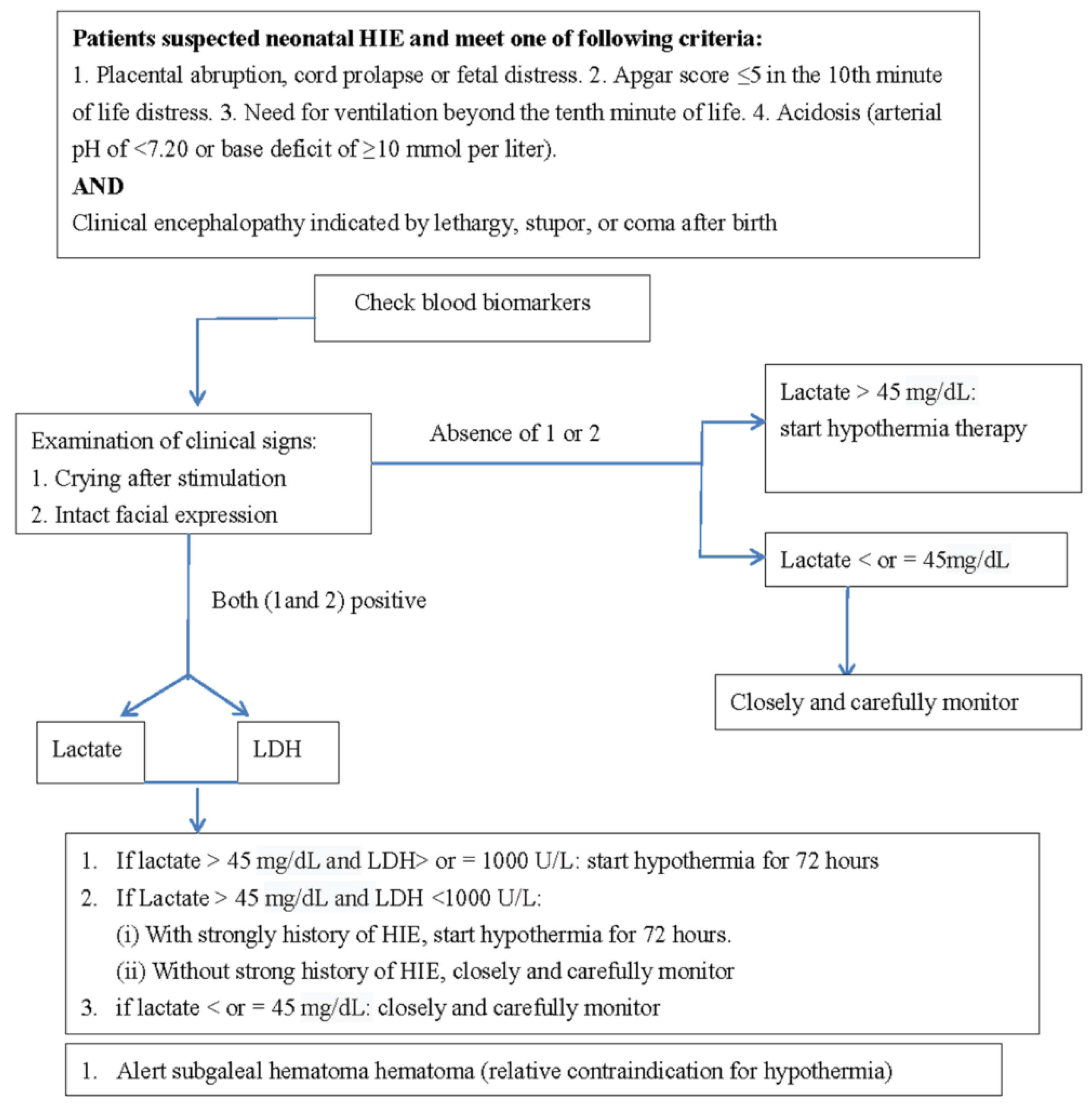
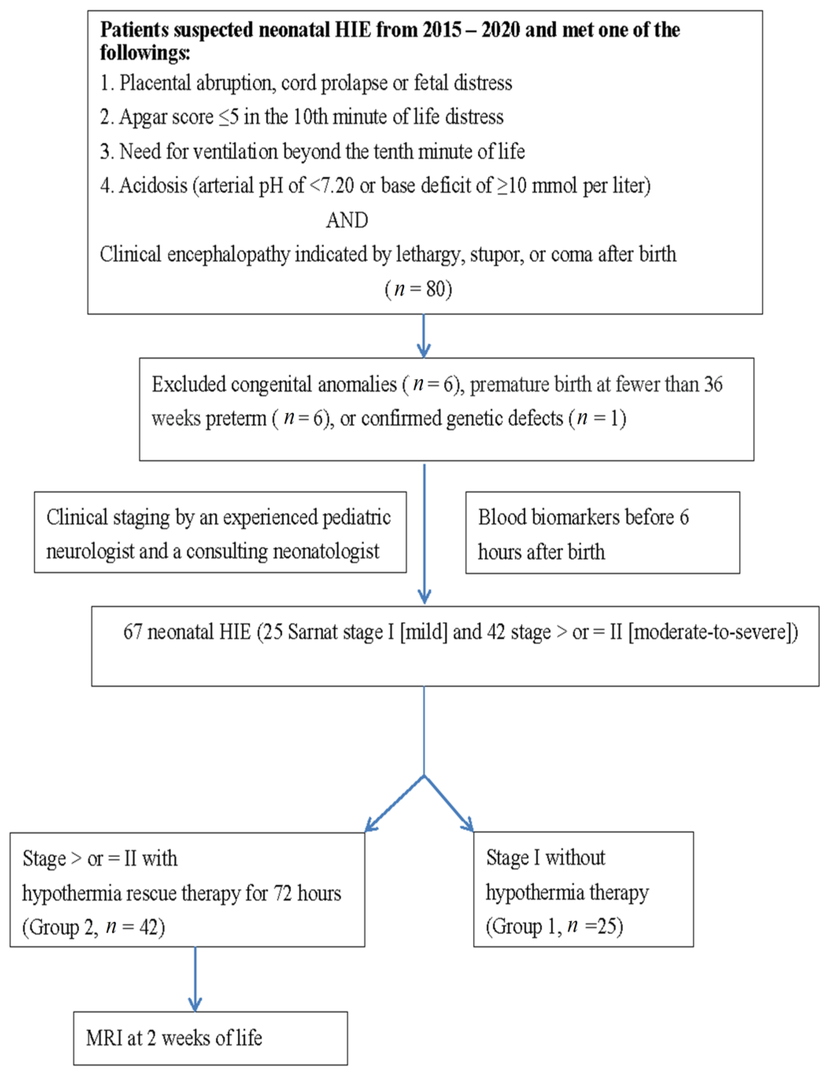
2.1. Patients (Newborns at High Risk of Encephalopathies)
2.2. Severe and Mild MRI Changes Correlated with Outcomes
2.3. Evaluation of Neurodevelopmental Outcomes
2.4. Statistical Analysis
3. Results
3.1. Demographic Data
3.2. Differences in Blood Biomarkers between Groups 1 and 2
3.3. MRI Findings for Patients in Stages II and III after Hypothermia Rescue Therapy
3.4. Differences in Biomarkers Correlated with MRI Results after Hypothermia Rescue Therapy
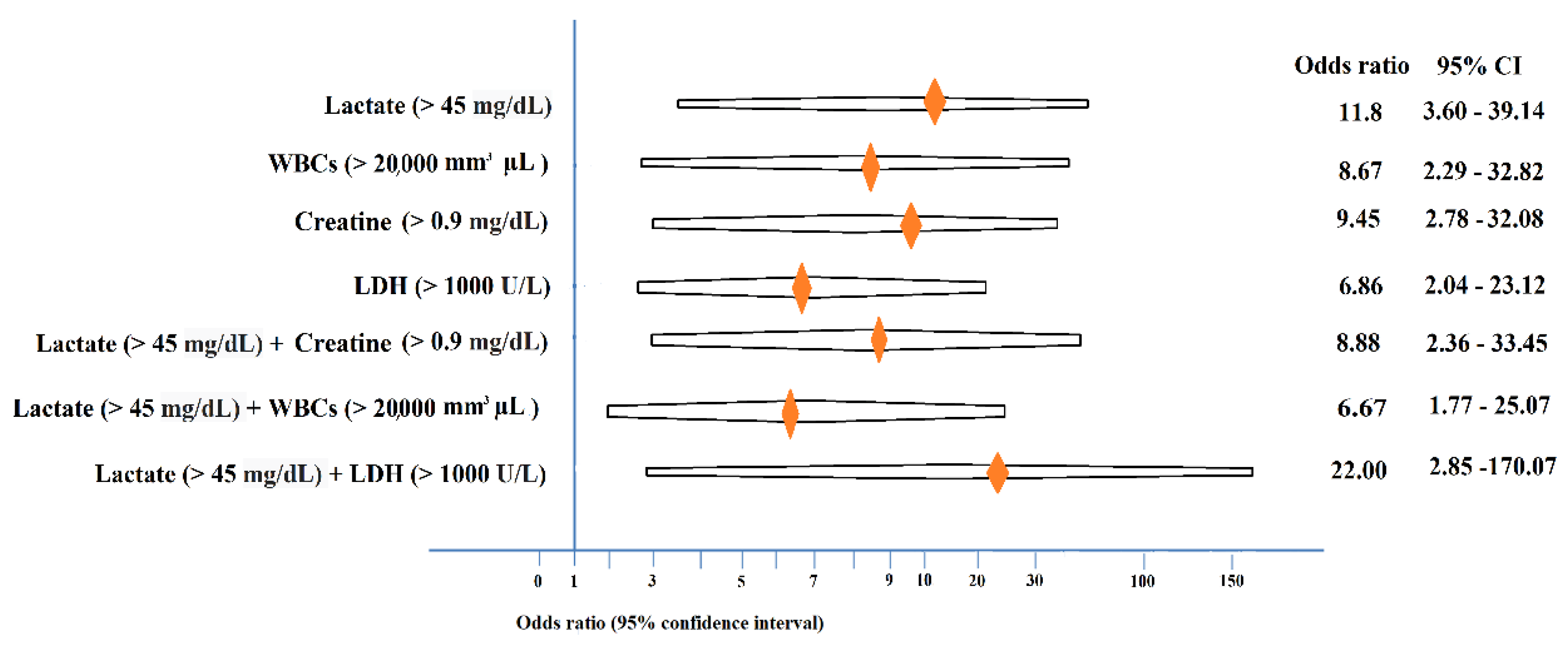
3.5. Identifying Clinically useful Biomarkers
4. Discussion
5. Conclusions
Author Contributions
Funding
Institutional Review Board Statement
Informed Consent Statement
Data Availability Statement
Acknowledgments
Conflicts of Interest
References
- Badurdeen, S.; Roberts, C.; Blank, D.; Miller, S.; Stojanovska, V.; Davis, P.; Hooper, S.; Polglase, G. Haemodynamic Instability and Brain Injury in Neonates Exposed to Hypoxia(-)Ischaemia. Brain Sci. 2019, 9, 49. [Google Scholar] [CrossRef] [Green Version]
- Armstrong, K.; Franklin, O.; Sweetman, D.; Molloy, E.J. Cardiovascular dysfunction in infants with neonatal encephalopathy. Arch. Dis. Child. 2012, 97, 372–375. [Google Scholar] [CrossRef]
- Saugstad, O.D.; Rootwelt, T.; Aalen, O. Resuscitation of asphyxiated newborn infants with room air or oxygen: An international controlled trial: The Resair 2 study. Pediatrics 1998, 102, e1. [Google Scholar] [CrossRef] [PubMed] [Green Version]
- Deorari, A.K.; Broor, S.; Maitreyi, R.S.; Agarwal, D.; Kumar, H.; Paul, V.K.; Singh, M. Incidence, clinical spectrum, and outcome of intrauterine infections in neonates. J. Trop. Pediatr. 2000, 46, 155–159. [Google Scholar] [CrossRef] [PubMed] [Green Version]
- Azzopardi, D.V.; Strohm, B.; Edwards, A.D.; Dyet, L.; Halliday, H.L.; Juszczak, E.; Kapellou, O.; Levene, M.; Marlow, N.; Porter, E.; et al. Moderate hypothermia to treat perinatal asphyxial encephalopathy. N. Engl. J. Med. 2009, 361, 1349–1358. [Google Scholar] [CrossRef] [Green Version]
- Edwards, A.D.; Brocklehurst, P.; Gunn, A.J.; Halliday, H.; Juszczak, E.; Levene, M.; Strohm, B.; Thoresen, M.; Whitelaw, A.; Azzopardi, D. Neurological outcomes at 18 months of age after moderate hypothermia for perinatal hypoxic ischaemic encephalopathy: Synthesis and meta-analysis of trial data. BMJ 2010, 340, c363. [Google Scholar] [CrossRef] [PubMed] [Green Version]
- Nakamura, T.; Asanuma, H.; Kusuda, S.; Imai, K.; Hosono, S.; Kato, R.; Suzuki, S.; Yokoi, K.; Kokubo, M.; Yamada, S.; et al. Multicenter study for brain/body hypothermia for hypoxic-ischemic encephalopathy: Changes in HMGB-1. Pediatr. Int. 2017, 59, 1074–1079. [Google Scholar] [CrossRef] [PubMed]
- Laptook, A.; Tyson, J.; Shankaran, S.; McDonald, S.; Ehrenkranz, R.; Fanaroff, A.; Donovan, E.; Goldberg, R.; O’Shea, T.M.; Higgins, R.D.; et al. Elevated temperature after hypoxic-ischemic encephalopathy: Risk factor for adverse outcomes. Pediatrics 2008, 122, 491–499. [Google Scholar] [CrossRef] [PubMed] [Green Version]
- Murray, D.M. Biomarkers in neonatal hypoxic-ischemic encephalopathy-Review of the literature to date and future directions for research. Handb. Clin. Neurol. 2019, 162, 281–293. [Google Scholar] [PubMed]
- Chakkarapani, E.; Davis, J.; Thoresen, M. Therapeutic hypothermia delays the C-reactive protein response and suppresses white blood cell and platelet count in infants with neonatal encephalopathy. Arch. Dis. Child. Fetal Neonatal Ed. 2014, 99, F458–F463. [Google Scholar] [CrossRef] [PubMed]
- El-Dib, M.; Parziale, M.P.; Johnson, L.; Benson, C.B.; Grant, P.E.; Robinson, J.; Volpe, J.J.; Inder, T. Encephalopathy in neonates with subgaleal hemorrhage is a keybpredictor of outcome. Pediatr. Res. 2019, 86, 234–241. [Google Scholar] [CrossRef]
- Goswami, I.R.; Whyte, H.; Wintermark, P.; Mohammad, K.; Shivananda, S.; Louis, D.; Yoon, E.W.; Shah, P.S. Characteristics and short-term outcomes of neonates with mild hypoxic-ischemic encephalopathy treated with hypothermia. J. Perinatol. 2020, 40, 275–283. [Google Scholar] [CrossRef]
- Saw, C.L.; Rakshasbhuvankar, A.; Rao, S.; Bulsara, M.; Patole, S. Current Practice of Therapeutic Hypothermia for Mild Hypoxic Ischemic Encephalopathy. J. Child. Neurol. 2019, 34, 402–409. [Google Scholar] [CrossRef] [PubMed]
- DuPont, T.L.; Chalak, L.F.; Morriss, M.C.; Burchfield, P.J.; Christie, L.; Sanchez, P.J. Short-term outcomes of newborns with perinatal acidemia who are not eligible for systemic hypothermia therapy. J. Pediatr. 2013, 162, 35–41. [Google Scholar] [CrossRef] [PubMed] [Green Version]
- Chiang, M.C.; Lien, R.; Chu, S.M.; Yang, P.H.; Lin, J.J.; Hsu, J.F.; Fu, R.H.; Lin, K.L. Serum Lactate, Brain Magnetic Resonance Imaging and Outcome of Neonatal Hypoxic Ischemic Encephalopathy after Therapeutic Hypothermia. Pediatr. Neonatol. 2016, 57, 35–40. [Google Scholar] [CrossRef] [PubMed] [Green Version]
- Celik, Y.; Atici, A.; Gulasi, S.; Makharoblidze, K.; Eskandari, G.; Sungur, M.A.; Akbayir, S. The effects of selective head cooling versus whole-body cooling on some neural and inflammatory biomarkers: A randomized controlled pilot study. Ital. J. Pediatr. 2015, 41, 79. [Google Scholar] [CrossRef] [PubMed] [Green Version]
- López-Suárez, O.; Concheiro-Guisán, A.; Sánchez-Pintos, P.; Cocho, J.A.; Lorenzo, J.R.F.; Couce, M.L. Acylcarnitine profile in neonatal hypoxic-ischemic encephalopathy: The value of butyrylcarnitine as a prognostic marker. Medicine 2019, 98, e15221. [Google Scholar] [CrossRef] [PubMed]
- Polackova, R.; Salounova, D.; Kantor, L. Lactate as an early predictor of psychomotor development in neonates with asphyxia receiving therapeutic hypothermia. Biomed. Pap. 2018, 162, 144–148. [Google Scholar] [CrossRef] [Green Version]
- Abiramalatha, T.; Kumar, M.; Chandran, S.; Sudhakar, Y.; Thenmozhi, M.; Thomas, N. Troponin-T as a biomarker in neonates with perinatal asphyxia. J. Neonatal Perinatal Med. 2017, 10, 275–280. [Google Scholar] [CrossRef] [PubMed]
- Munshi, U.K.; Brown, M.M.; Tauber, K.A.; Horgan, M.J. Early Troponin I Levels in Newborns Undergoing Therapeutic Hypothermia for Hypoxic Ischemic Encephalopathy and Residual Encephalopathy at Discharge. Am. J. Perinatol. 2020. [Google Scholar] [CrossRef]
- Haga, M.; Kawabata, K.; Sumiya, W.; Kurita, S.; Imanishi, T.; Kanno, C.; Kanno, M.; Shimizu, M. The Relationship between Serum Total Bilirubin and Severity of Hypoxic Injury in Neonatal Hypoxic-Ischemic Encephalopathy. Am. J. Perinatol. 2020. [Google Scholar] [CrossRef] [PubMed]
- Yang, L.; Li, D.; Chen, S. Hydrogen water reduces NSE, IL-6, and TNF-α levels in hypoxic-ischemic encephalopathy. Open Med. (Wars) 2016, 11, 399–406. [Google Scholar] [CrossRef]
- Massaro, A.N.; Wu, Y.W.; Bammler, T.K.; Comstock, B.; Mathur, A.; McKinstry, R.C.; Chang, T.; Mayock, D.E.; Mulkey, S.B.; Van Meurs, K.; et al. Plasma Biomarkers of Brain Injury in Neonatal Hypoxic-Ischemic Encephalopathy. J. Pediatr. 2018, 194, 67–75. [Google Scholar] [CrossRef] [PubMed]
- Graham, E.M.; Everett, A.D.; Delpech, J.C.; Northington, F.J. Blood biomarkers for evaluation of perinatal encephalopathy: State of the art. Curr. Opin. Pediatr. 2018, 30, 199–203. [Google Scholar] [CrossRef] [Green Version]
- Zhu, Y.; Yun, Y.; Jin, M.; Li, G.; Li, H.; Miao, P.; Ding, X.; Feng, X.; Xu, L.; Sun, B. Identification of novel biomarkers for neonatal hypoxic-ischemic encephalopathy using iTRAQ. Ital. J. Pediatr. 2020, 46, 67. [Google Scholar] [CrossRef] [PubMed]
- Sarnat, H.B.; Sarnat, M.S. Neonatal encephalopathy following fetal distress. A clinical and electroencephalographic study. Arch. Neurol. 1976, 33, 696–705. [Google Scholar] [CrossRef]
- Michniewicz, B.; Szpecht, D.; Sowińska, A.; Sibiak, R.; Szymankiewicz, M.; Gadzinowski, J. Biomarkers in newborns with hypoxic-ischemic encephalopathy treated with therapeutic hypothermia. Child’s Nerv. Syst. 2020, 36, 2981–2988. [Google Scholar] [CrossRef]
- Lin, Y.K.; Hwang-Bo, S.; Seo, Y.M.; Youn, Y.A. Clinical seizures and unfavorable brain MRI patterns in neonates with hypoxic ischemic encephalopathy. Medicine (Baltimore) 2021, 100, e25118. [Google Scholar]
- Chang, P.D.; Chow, D.S.; Alber, A.; Lin, Y.K.; Youn, Y.A. Predictive Values of Location and Volumetric MRI Injury Patterns for Neurodevelopmental Outcomes in Hypoxic-Ischemic Encephalopathy Neonates. Brain Sci. 2020, 10, 991. [Google Scholar] [CrossRef] [PubMed]


| Variables | Hypoxic-Ischemic Encephalopathy, Stage I; (n = 25) | Hypoxic-Ischemic Encephalopathy Stage II and III, (n = 42) | p # |
|---|---|---|---|
| Gestational age (weeks) | 39.3 ± 1.7 | 39.7 ± 1.3 | NS |
| Birth weight (gm) | 3421 ± 520 | 3530 ± 576 | NS |
| Gender | NS | ||
| Male | 15 (60.0%) | 26 (61.9%) | |
| Female | 10 (40.0%) | 16 (38.1%) | |
| Transfer mode | NS | ||
| Inborn | 9 (36.0%) | 15 (35.7%) | |
| Outborn | 16 (64.0%) | 27 (64.3%) | |
| Method of delivery | NS | ||
| Cesarean section | 11 (44.0%) | 15 (35.8%) | |
| Vaginal delivery | 14 (56.0%) | 27 (64.2%) |
| Biomarkers | Group 1 | ST | Group 2 | ST | p Value |
|---|---|---|---|---|---|
| WBCs * (9100–34,000 mm3 µL) | 16,941.0 | 4967.0 | 23,096.9 | 10,585.4 | 0.014 * |
| Platelet (84–478 mm3 µL) | 240,523.8 | 86,145.0 | 225,285.7 | 73,186.8 | 0.466 |
| Hemoglobin (13.88 ± 1.34 g/dL) | 16.4 | 2.2 | 17.7 | 7.0 | 0.426 |
| SGOT (30–100 U/L) | 72.1 | 34.6 | 225.6 | 319.8 | 0.062 |
| SGPT (6–40 U/L) | 18.0 | 10.7 | 68.9 | 104.6 | 0.051 |
| BUN (3–12 mg/dL) | 9.6 | 2.9 | 11.4 | 3.9 | 0.077 |
| Creatinine * (0.03–0.50 mg/dL) | 0.8 | 0.2 | 1.0 | 0.2 | 0.005 * |
| Lactate ** (4.4 to 14.4 mg/dL) | 47.0 | 25.2 | 91.4 | 52.9 | 0.001 ** |
| LDH * (170–580 U/L) | 681.5 | 399.6 | 1557.8 | 1787.8 | 0.040 * |
| PT (13.0 ± 1.43 s) | 14.3 | 3.6 | 18.2 | 9.0 | 0.071 |
| aPTT (42.9 ± 5.80 s) | 56.0 | 19.2 | 63.3 | 28.2 | 0.312 |
| Albumin (2.5–3.4 g/dL) | 3.5 | 0.8 | 3.5 | 0.5 | 0.706 |
| Glucose (40–60 mg/dL) | 97.1 | 30.1 | 131.5 | 81.5 | 0.067 |
| Na (133–146 mmol/L) | 135.9 | 3.6 | 135.6 | 3.6 | 0.769 |
| K (3.2–5.5 mmol/L) | 4.1 | 0.5 | 4.1 | 0.8 | 0.939 |
| CK (39–308 U/L) | 1355.7 | 981.0 | 3250.5 | 4454.9 | 0.090 |
| CK-MB (0–4.5 ng/mL) | 39.5 | 27.2 | 73.5 | 99.2 | 0.236 |
| Brain Lesions on MRI | Stage II * (n = 28) | Stage III * (n = 14) | p Value # |
|---|---|---|---|
| Thalamus or basal ganglion | 4 (14.3%) | 12 (85.7%) | 0.000 ** |
| Brain stem (one of midbrain, pons, and medulla) | 0 (0.0%) | 2 (14.3%) | NS |
| Multicystic encephalomalacia | 1 (3.6%) | 4 (28.8%) | 0.038 * |
| Diffuse white matter injury | 3 (10.7%) | 3 (21.4%) | NS |
| Small parenchymal lesion (infarction/hemorrhage) | 6 (21.4%) | 1 (7.1%) | NS |
| Subdural hemorrhage | 7 (25.0%) | 1 (7.1%) | NS |
| Unremarkable | 12 (42.9%) | 0 (0.0%) | 0.003 ** |
| Biomarkers | Group 3 ± (n = 22) | Standard Deviation | Group 4 ± (n = 20) | Standard Deviation | p Value |
|---|---|---|---|---|---|
| WBCs | 21,744.1 | 8130.7 | 24,584.9 | 12,817.2 | 0.392 |
| Platelet | 232,454.5 | 73,365.2 | 217,400.0 | 74,056.5 | 0.512 |
| Hemoglobin | 16.9 | 2.1 | 18.5 | 10.0 | 0.454 |
| SGOT | 164.6 | 197.7 | 292.8 | 410.4 | 0.198 |
| SGPT | 54.7 | 92.2 | 84.5 | 117.2 | 0.364 |
| BUN | 10.8 | 3.1 | 12.1 | 4.7 | 0.304 |
| Creatinine | 1.0 | 0.2 | 1.0 | 0.2 | 0.974 |
| Lactate * | 72.0 | 40.4 | 112.7 | 57.6 | 0.011 * |
| LDH | 1439.3 | 1625.5 | 1682.2 | 1978.9 | 0.669 |
| PT * | 15.4 | 4.0 | 21.4 | 11.7 | 0.031 * |
| aPTT | 60.9 | 21.2 | 65.8 | 34.8 | 0.580 |
| Albumin * | 3.7 | 0.4 | 3.3 | 0.5 | 0.035 * |
| Glucose | 109.5 | 65.7 | 154.7 | 91.3 | 0.075 |
| Na | 135.9 | 3.7 | 135.3 | 3.6 | 0.538 |
| K | 4.2 | 0.8 | 4.0 | 0.8 | 0.444 |
| CK | 4179.9 | 5502.5 | 2174.4 | 2549.8 | 0.153 |
| CK-MB | 68.5 | 82.4 | 78.5 | 116.9 | 0.802 |
| Blood Biomarkers | 95% CI | |||||||
|---|---|---|---|---|---|---|---|---|
| PPV (%) | NPV (%) | Specificity (%) | Sensitivity (%) | p Values | Odds Ratio | Lower | Upper | |
| Lactate (>45 mg/dL) (n = 45) | 82.6 | 71.4 | 65.2 | 86.4 | 0.000 | 11.88 | 3.60 | 39.14 |
| WBC (>20,000 mm3 µL) (n = 29) | 89.7 | 50.0 | 86.4 | 57.8 | 0.001 | 8.67 | 2.29 | 32.82 |
| Creatinine (>0.9 mg/dL) (n = 31) | 87.1 | 58.3 | 84.0 | 64.3 | 0.000 | 9.45 | 2.78 | 32.08 |
| LDH (>1000 U/L) (n = 30) | 86.7 | 51.4 | 82.6 | 59.1 | 0.001 | 6.86 | 2.04 | 23.12 |
| Lactate (>45 mg/dL) + Creatinine (>0.9 mg/dL) (n = 26) | 88.5 | 52.5 | 87.5 | 54.8 | 0.000 | 8.88 | 2.36 | 33.45 |
| Lactate (>45 mg/dL) + WBCs (>20,000 mm3 µL) (n = 25) | 88.0 | 47.6 | 87.0 | 50.0 | 0.003 | 6.67 | 1.77 | 25.07 |
| Lactate (>45 mg/dL) + LDH (>1000 U/L) (n = 23) | 95.7 | 50.0 | 95.7 | 50.0 | 0.000 | 22.00 | 2.85 | 170.07 |
Publisher’s Note: MDPI stays neutral with regard to jurisdictional claims in published maps and institutional affiliations. |
© 2021 by the authors. Licensee MDPI, Basel, Switzerland. This article is an open access article distributed under the terms and conditions of the Creative Commons Attribution (CC BY) license (https://creativecommons.org/licenses/by/4.0/).
Share and Cite
Lee, I.-C.; Wong, S.-H.; Wang, X.-A.; Yu, C.-S. Identifying Early Diagnostic Biomarkers Associated with Neonatal Hypoxic-Ischemic Encephalopathy. Diagnostics 2021, 11, 897. https://doi.org/10.3390/diagnostics11050897
Lee I-C, Wong S-H, Wang X-A, Yu C-S. Identifying Early Diagnostic Biomarkers Associated with Neonatal Hypoxic-Ischemic Encephalopathy. Diagnostics. 2021; 11(5):897. https://doi.org/10.3390/diagnostics11050897
Chicago/Turabian StyleLee, Inn-Chi, Swee-Hee Wong, Xing-An Wang, and Chin-Sheng Yu. 2021. "Identifying Early Diagnostic Biomarkers Associated with Neonatal Hypoxic-Ischemic Encephalopathy" Diagnostics 11, no. 5: 897. https://doi.org/10.3390/diagnostics11050897
APA StyleLee, I.-C., Wong, S.-H., Wang, X.-A., & Yu, C.-S. (2021). Identifying Early Diagnostic Biomarkers Associated with Neonatal Hypoxic-Ischemic Encephalopathy. Diagnostics, 11(5), 897. https://doi.org/10.3390/diagnostics11050897