
Clinical Characteristics and Outcome of Patients with Suspected COVID-19 in Emergency Department (RESILIENCY Study II)

 ,
,  , ,
, ,  , and
, and 
Abstract
:1. Introduction
2. Materials and Methods
2.1. Data Collection and Definitions
2.2. Main Outcome Measures
2.3. Microbiology
2.4. Statistical Analysis
3. Results
4. Discussion
5. Conclusions
Author Contributions
Funding
Institutional Review Board Statement
Informed Consent Statement
Data Availability Statement
Conflicts of Interest
References
- Huang, C.; Wang, Y.; Li, X.; Ren, L.; Zhao, J.; Hu, Y.; Zhang., L.; Fan, G.; Xu, J.; Gu, X.; et al. Clinical features of patients infected with 2019 novel coronavirus in Wuhan, China. Lancet 2020, 395, 497–506. [Google Scholar] [CrossRef] [Green Version]
- Bassetti, M.; Vena, A.; Giacobbe, D.R. The novel Chinese coronavirus (2019-nCoV) infections: Challenges for fighting the storm. Eur. J. Clin. Investig. 2020, 50, e13209. [Google Scholar] [CrossRef] [Green Version]
- Zhou, F.; Yu, T.; Du, R.; Fan, G.; Liu, Y.; Liu, Z.; Xiang, J.; Wang, Y.; Song, B.; Gu, X.; et al. Clinical course and risk factors for mortality of adult inpatients with COVID-19 in Wuhan, China: A retrospective cohort study. Lancet 2020, 395, 1054–1062. [Google Scholar] [CrossRef]
- Vena, A.; Giacobbe, D.R.; Di Biagio, A.; Mikulska, M.; Taramasso, L.; De Maria, A.; Ball, L.; Brunetti, I.; Loconte, M.; Patroniti, N.A.; et al. Clinical characteristics, management and in-hospital mortality of patients with coronavirus disease 2019 in Genoa, Italy. Clin. Microbiol. Infect. 2020, 26, 1537–1544. [Google Scholar] [CrossRef] [PubMed]
- Bartoletti, M.; Giannella, M.; Scudeller, L.; Tedeschi, S.; Rinaldi, M.; Bussini, L.; Fornaro, G.; Pancaldi, L.; Pasquini, Z.; Trapani, F.; et al. Development and validation of a prediction model for severe respiratory failure in hospitalized patients with SARS-CoV-2 infection: A multicentre cohort study (PREDI-CO study). Clin. Microbiol. Infect. 2020, 26, 1545–1553. [Google Scholar] [CrossRef] [PubMed]
- Liang, W.H.; Guan, W.J.; Li, C.C.; Li, Y.M.; Liang, H.R.; Zhao, Y.; Liu, X.; Sang, L.; Chen, R.; Tang, C.; et al. Clinical characteristics and outcomes of hospitalised patients with COVID-19 treated in Hubei (epicenter) and outside Hubei (non-epicenter): A Nationwide Analysis of China. Eur. Respir. J. 2020, 55, 2000562. [Google Scholar] [CrossRef] [Green Version]
- Grasselli, G.; Zangrillo, A.; Zanella, A.; Antonelli, M.; Cabrini, L.; Castelli, A.; Cereda, D.; Coluccelli, A.; Foti, G.; Fumagalli, R.; et al. Network, Baseline Characteristics and Outcomes of 1591 Patients Infected with SARS-CoV-2 Admitted to ICUs of the Lombardy Region, Italy. JAMA 2020, 323, 1574–1581. [Google Scholar] [CrossRef] [Green Version]
- Russo, A.; Bellelli, V.; Ceccarelli, G.; Marincola Cattaneo, F.; Bianchi, L.; Pierro, R.; Russo, R.; Steffanina, A.; Pugliese, F.; Mastroianni, C.M.; et al. Comparison Between Hospitalized Patients Affected or Not Affected by Coronavirus Disease 2019. Clin. Infect. Dis. 2021, 72, e1158–e1159. [Google Scholar] [CrossRef]
- Safavian, S.R.; Landgrebe, D. A survey of decision tree classifier methodology. IEEE Trans. Syst. Man Cybern. 1991, 21, 660–674. [Google Scholar] [CrossRef] [Green Version]
- Kononenko, I. Machine learning for medical diagnosis: History, state of the art and perspective. Artif. Intell. Med. 2001, 23, 89–109. [Google Scholar] [CrossRef] [Green Version]
- Knight, S.R.; Ho, A.; Pius, R.; Buchan, I.; Carson, G.; Drake, T.M.; Dunning, J.; Fairfield, C.J.; Gamble, C.; Green, C.A.; et al. Risk stratification of patients admitted to hospital with covid-19 using the ISARIC WHO Clinical Characterisation Protocol: Development and validation of the 4C Mortality Score. BMJ 2020, 370, m3339, Erratum in BMJ 2020, 371, m4334. [Google Scholar] [CrossRef]
- Gozzo, L.; Viale, P.; Longo, L.; Vitale, D.C.; Drago, F. The Potential Role of Heparin in Patients with COVID-19: Beyond the Anticoagulant Effect. A Review. Front. Pharm. 2020, 11, 1307. [Google Scholar] [CrossRef]
- Paolisso, P.; Bergamaschi, L.; D’Angelo, E.C.; Donati, F.; Giannella, M.; Tedeschi, S.; Pascale, R.; Bartoletti, M.; Tesini, G.; Biffi, M.; et al. Preliminary Experience with Low Molecular Weight Heparin Strategy in COVID-19 Patients. Front. Pharm. 2020, 11, 1124. [Google Scholar] [CrossRef] [PubMed]
- Mehta, P.; McAuley, D.F.; Brown, M.; Sanchez, E.; Tattersall, R.S.; Manson, J.J.U.K. Hlh Across Speciality Collaboration, COVID-19: Consider cytokine storm syndromes and immunosuppression. Lancet 2020, 395, 1033–1034. [Google Scholar] [CrossRef]
- Iftimie, S.; López-Azcona, A.F.; Vallverdú, I.; Hernández-Flix, S.; de Febrer, G.; Parra, S.; Hernández-Aguilera, A.; Riu, F.; Joven, J.; Andreychuk, N.; et al. First and second waves of coronavirus disease-19: A comparative study in hospitalized patients in Reus, Spain. PLoS ONE 2021, 16, e0248029. [Google Scholar] [CrossRef] [PubMed]
- Kontis, V.; Bennett, J.E.; Rashid, T.; Parks, R.M.; Pearson-Stuttard, J.; Guillot, M.; Asaria, P.; Zhou, B.; Battaglini, M.; Corsetti, G.; et al. Magnitude, demographics and dynamics of the effect of the first wave of the COVID-19 pandemic on all-cause mortality in 21 industrialized countries. Nat. Med. 2020, 26, 1919–1928, Erratum in Nat Med. 2021, 27, 562. [Google Scholar] [CrossRef] [PubMed]
- Munblit, D.; Nekliudov, N.A.; Bugaeva, P.; Blyuss, O.; Kislova, M.; Listovskaya, E.; Gamirova, A.; Shikhaleva, A.; Belyaev, V.; Timashev, P.; et al. StopCOVID cohort: An observational study of 3,480 patients admitted to the Sechenov University hospital network in Moscow city for suspected COVID-19 infection. Clin. Infect. Dis. 2020, 73, ciaa1535. [Google Scholar] [CrossRef] [PubMed]
- Docherty, A.B.; Harrison, E.M.; Green, C.A.; Hardwick, H.E.; Pius, R.; Norman, L.; Holden, K.A.; Read, J.M.; Dondelinger, F.; Carson, G.; et al. Features of 20 133 UK patients in hospital with covid-19 using the ISARIC WHO Clinical Characterisation Protocol: Prospective observational cohort study. BMJ 2020, 369, m1985. [Google Scholar] [CrossRef] [PubMed]
- Guan, W.J.; Ni, Z.Y.; Hu, Y.; Liang, W.; Ou, C.; He, J.; Liu, L.; Shan, H.; Lei, C.; Hui, D.S.C.; et al. Clinical Characteristics of Coronavirus Disease 2019 in China. N. Engl. J. Med. 2020, 382, 1708–1720. [Google Scholar] [CrossRef] [PubMed]
- Chan, A.S.; Rout, A. Use of Neutrophil-to-Lymphocyte and Platelet-to-Lymphocyte Ratios in COVID-19. J. Clin. Med. Res. 2020, 12, 448–453. [Google Scholar] [CrossRef]
- Carubbi, F.; Salvati, L.; Alunno, A.; Maggi, F.; Borghi, E.; Mariani, R.; Mai, F.; Paoloni, M.; Ferri, C.; Desideri, G.; et al. Ferritin is associated with the severity of lung involvement but not with worse prognosis in patients with COVID-19: Data from two Italian COVID-19 units. Sci. Rep. 2021, 11, 4863. [Google Scholar] [CrossRef] [PubMed]
- Heldt, F.S.; Vizcaychipi, M.P.; Peacock, S.; Cinelli, M.; McLachlan, L.; Andreotti, F.; Jovanović, S.; Dürichen, R.; Lipunova, N.; Fletcher, R.A.; et al. Early risk assessment for COVID-19 patients from emergency department data using machine learning. Sci. Rep. 2021, 11, 4200. [Google Scholar] [CrossRef] [PubMed]
- Harmon, S.A.; Sanford, T.H.; Xu, S.; Turkbey, E.B.; Roth, H.; Xu, Z.; Yang, D.; Myronenko, A.; Anderson, V.; Amalou, A.; et al. Artificial intelligence for the detection of COVID-19 pneumonia on chest CT using multinational datasets. Nat. Commun. 2020, 11, 4080. [Google Scholar] [CrossRef] [PubMed]
- Zhang, J.J.; Cao, Y.Y.; Dong, X.; Wang, B.; Liao, M.; Lin, J.; Yan, Y.; Akdis, C.A.; Gao, Y. Distinct characteristics of COVID-19 patients with initial rRT-PCR-positive and rRT-PCR-negative results for SARS-CoV-2. Allergy 2020, 75, 1809–1812. [Google Scholar] [CrossRef] [PubMed] [Green Version]
- Yoo, S.H.; Geng, H.; Chiu, T.L.; Yu, S.K.; Cho, D.C.; Heo, J.; Choi, M.S.; Choi, I.H.; Cung Van, C.; Nhung, N.V.; et al. Deep Learning-Based Decision-Tree Classifier for COVID-19 Diagnosis From Chest X-ray Imaging. Front. Med. 2020, 7, 427. [Google Scholar] [CrossRef]
- Pourbagheri-Sigaroodi, A.; Bashash, D.; Fateh, F.; Abolghasemi, H. Laboratory findings in COVID-19 diagnosis and prognosis. Clin. Chim. Acta. 2020, 510, 475–482. [Google Scholar] [CrossRef]
- Sánchez-Montañés, M.; Rodríguez-Belenguer, P.; Serrano-López, A.J.; Soria-Olivas, E.; Alakhdar-Mohmara, Y. Machine Learning for Mortality Analysis in Patients with COVID-19. Int. J. Environ. Res. Public Health. 2020, 17, 8386. [Google Scholar] [CrossRef]
- Domingo, P.; Pomar, V.; Mur, I.; Castellví, I.; Corominas, H.; de Benito, N. Not all COVID-19 pandemic waves are alike. Clin. Microbiol. Infect. 2021, 27, 1040.e7–1040.e10. [Google Scholar] [CrossRef] [PubMed]
- Welte, T.; Ambrose, L.J.; Sibbring, G.C.; Sheikh, S.; Müllerová, H.; Sabir, I. Current evidence for COVID-19 therapies: A systematic literature review. Eur. Respir. Rev. 2021, 30, 200384. [Google Scholar] [CrossRef] [PubMed]
- Ko, J.J.; Wu, C.; Mehta, N.; Wald-Dickler, N.; Yang, W.; Qiao, R. A Comparison of Methylprednisolone and Dexamethasone in Intensive Care Patients With COVID-19. J. Intensive Care Med. 2021, 36, 673–680. [Google Scholar] [CrossRef]
- Falcone, M.; Tiseo, G.; Barbieri, G.; Galfo, V.; Russo, A.; Virdis, A.; Forfori, F.; Corradi, F.; Guarracino, F.; Carrozzi, L.; et al. Role of Low-Molecular-Weight Heparin in Hospitalized Patients With Severe Acute Respiratory Syndrome Coronavirus 2 Pneumonia: A Prospective Observational Study. Open Forum Infect. Dis. 2020, 7, ofaa563. [Google Scholar] [CrossRef]
- Cuker, A.; Tseng, E.K.; Nieuwlaat, R.; Angchaisuksiri, P.; Blair, C.; Dane, K.; Davila, J.; DeSancho, M.T.; Diuguid, D.; Griffin, D.O.; et al. American Society of Hematology 2021 guidelines on the use of anticoagulation for thromboprophylaxis in patients with COVID-19. Blood Adv. 2021, 5, 872–888. [Google Scholar] [CrossRef]
- Olender, S.A.; Perez, K.K.; Go, A.S.; Balani, B.; Price-Haywood, E.G.; Shah, N.S.; Wang, S.; Walunas, T.L.; Swaminathan, S.; Slim, J.; et al. Remdesivir for Severe COVID-19 versus a Cohort Receiving Standard of Care. Clin. Infect. Dis. 2020, ciaa1041. [Google Scholar] [CrossRef] [PubMed]
- WHO Solidarity Trial Consortium; Pan, H.; Peto, R.; Henao-Restrepo, A.M.; Preziosi, M.P.; Sathiyamoorthy, V.; Abdool Karim, Q.; Alejandria, M.M.; Hernán-dez García, C.; Kieny, M.P.; et al. Repurposed Antiviral Drugs for Covid-19—Interim WHO Solidarity Trial Results. N. Engl. J. Med. 2021, 384, 497–511. [Google Scholar]
- Alhazzani, W.; Moller, M.H.; Arabi, Y.M.; Loeb, M.; Gong, M.N.; Fan, E.; Oczkowski, S.; Levy, M.M.; Derde, L.; Dzierba, A.; et al. Surviving Sepsis Campaign: Guidelines on the Management of Critically Ill Adults with Coronavirus Disease 2019 (COVID-19). Crit. Care Med. 2020, 48, e440–e469. [Google Scholar] [CrossRef] [PubMed]
- Bassetti, M.; Giacobbe, D.R.; Aliberti, S.; Barisione, E.; Centanni, S.; De Rosa, F.G.; Di Marco, F.; Gori, A.; Granata, G.; Mikulska, M.; et al. Balancing evidence and frontline experience in the early phases of the COVID-19 pandemic: Current position of the Italian Society of Anti-Infective Therapy (SITA) and the Italian Society of Pulmonology (SIP). Clin. Microbiol. Infect. 2020, 26, 880–894. [Google Scholar] [CrossRef] [PubMed]
- Sinha, P.; Calfee, C.S.; Cherian, S.; Brealey, D.; Cutler, S.; King, C.; Killick, C.; Richards, O.; Cheema, Y.; Bailey, C.; et al. Prevalence of phenotypes of acute respiratory distress syndrome in critically ill patients with COVID-19: A prospective observational study. Lancet Respir. Med. 2020, 8, 1209–1218. [Google Scholar] [CrossRef]

| Variables | Non-COVID-19 n = 319 | COVID-19 n = 717 | p-Value |
|---|---|---|---|
| Male sex | 199 (62%) | 446 (62%) | 0.956 |
| Age (years), median (IQR: 25–75%) ± SD | 75.3 ± 14.4 | 64.1 ± 17.1 | <0.001 |
| Days from symptoms to RT-PCR test, median (IQR: 25–75%) | 2 (1–5) | 4 (1–7) | 0.002 |
| Coexisting comorbidities, n (%) | 286 (89.6%) | 206 (28.7%) | <0.001 |
| Cardiovascular disease, n (%) | 248 (78%) | 57 (8%) | <0.001 |
| COPD, n (%) | 163 (51%) | 60 (8%) | <0.001 |
| Chronic renal disease, n (%) | 100 (31%) | 37 (5%) | <0.001 |
| Cirrhosis, n (%) | 64 (20%) | 17 (2%) | <0.001 |
| Diabetes, n (%) | 27 (8%) | 77 (11%) | <0.001 |
| Solid lung cancer, n (%) | 61 (19%) | 6 (1%) | <0.001 |
| Clinical features and radiological findings on admission | |||
| Fever > 3 days, n (%) | 39 (12%) | 554 (77%) | <0.001 |
| Dry cough, n (%) | 51 (16%) | 334 (47%) | <0.001 |
| Acute dyspnea, n (%) | 90 (28%) | 372 (52%) | <0.002 |
| Gastrointestinal symptoms (diarrhea, abdominal discomfort, nausea, vomiting) | 44 (14%) | 107 (15%) | 0.675 |
| Fatigue, n (%) | 189 (59%) | 109 (15%) | <0.001 |
| Pharyngodynia, n (%) | 16 (5%) | 38 (5%) | 1.000 |
| Rhinitis, n (%) | 184 (58%) | 259 (36%) | <0.001 |
| Arthralgia/myalgia, n (%) | 16 (5%) | 73 (10%) | 0.007 |
| Anosmia, n (%) | 7 (2%) | 31 (4%) | 0.101 |
| Conjunctivitis, n (%) | 0 (0%) | 4 (1%) | 0.073 |
| Chest pain, n (%) | 18 (6%) | 33 (5%) | 0.508 |
| Signs of overload (limb edema and/or pulmonary stasis), n (%) | 37 (12%) | 17 (2%) | <0.001 |
| Parenchymal thickening, n (%) | 66 (21%) | 344 (48%) | <0.001 |
| Interstitial lung disease, n (%) | 16 (5%) | 31 (4%) | 0.464 |
| Pleural effusion, n (%) | 110 (34%) | 191 (27%) | 0.022 |
| Cardiomegaly, n (%) | 99 (31%) | 232 (32%) | 0.750 |
| Bronchiectasis/emphysema, n (%) | 27 (24%) | 50 (15%) | 0.013 |
| Laboratory findings | |||
| WBC (×103/µL), median (IQR: 25–75%) | 7.5 (6.4–12.3) | 6.1 (4.5–8.8) | <0.001 |
| Neutrophils ×103/µL, median (IQR: 25–75%) | 5.6 (3.8–9.4) | 4.4 (3–7.1) | 0.368 |
| Lymphocytes ×103/µL, median (IQR: 25–75%) | 1.1 (0.8–1.8) | 0.9 (0.6–1.2) | <0.001 |
| Platelets ×103/µL, median (IQR: 25–75%) | 232 (192–305) | 205 (159–266) | <0.001 |
| D-dimer ng/mL, median (IQR: 25–75%) | 820 (367–1322) | 631 (340–1242) | 0.178 |
| Serum ferritin ng/mL, median (IQR: 25–75%) | 343 (120–811) | 458 (219–813) | 0.013 |
| Procalcitonin ng/mL, median (IQR: 25–75%) | 2.6 (0.4–3) | 0.9 (0.1–1.5) | 0.002 |
| LDH mU/mL, mean ± SD | 404 ± 191 | 317 ± 147 | <0.001 |
| CPK U/L, median (IQR: 25–75%) | 74 (48–170) | 89 (52–160) | 0.010 |
| Lactate mmol/L, mean ± SD | 1.8 ± 1.4 | 1.3 ± 0.8 | <0.001 |
| C-reactive protein mg/dL, median (IQR: 25–75%) | 10.3 (6.5–13) | 4 (1.3–9.8) | 0.022 |
| Alanine aminotransferase U/L, median (IQR: 25–75%) | 28 (20–46) | 29 (20–42) | 0.258 |
| Aspartate aminotransferase U/L, median (IQR: 25–75%) | 26 (18–45) | 22 (16–34) | 0.015 |
| PaO2/FiO2, mean ± SD | 326 ± 106 | 317 ± 114 | 0.010 |
| Bacterial co-infection, n (%) | 89 (28%) | 144 (20%) | 0.004 |
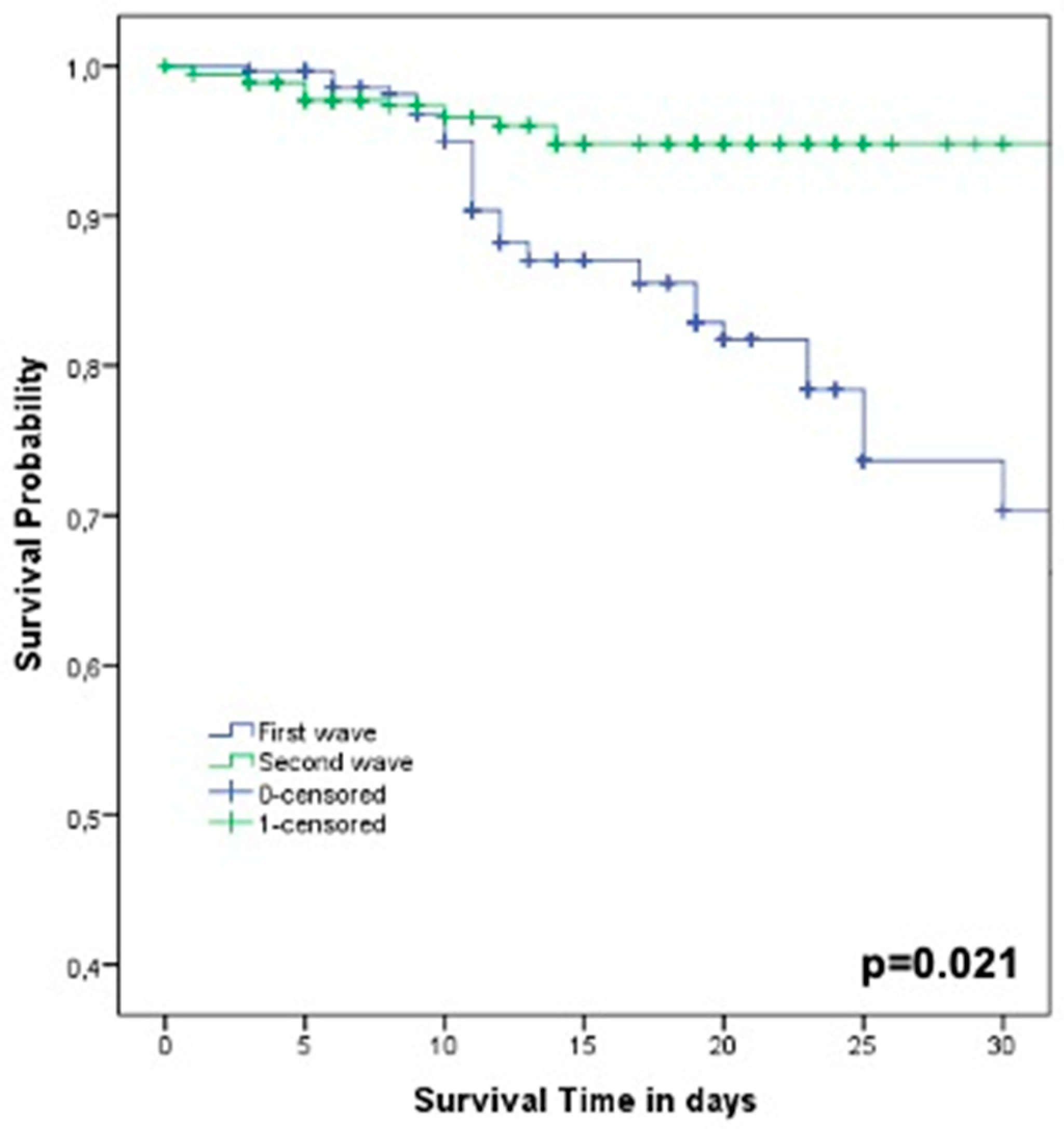
| Days of hospitalization, median (IQR: 25–75%) | 12 (9–19) | 12 (8–19) | 0.376 |
| Days to RT-PCR negative test, median (IQR: 25–75%) | - | 14 (11–23) | - |
| Variables | Non-COVID-19 n = 319 | COVID-19 n = 717 | p-Value |
|---|---|---|---|
| Invasive ventilation, n (%) | 27 (8%) | 36 (5%) | 0.059 |
| Low oxygen flow or room air, n (%) | 167 (52%) | 480 (67%) | <0.001 |
| HFNC/NIV, n (%) | 126 (39%) | 127 (18%) | <0.001 |
| 30-day mortality, n (%) | 111 (35%) | 61 (9%) | <0.001 |
| Variables | OR | CI95% | p-Value |
|---|---|---|---|
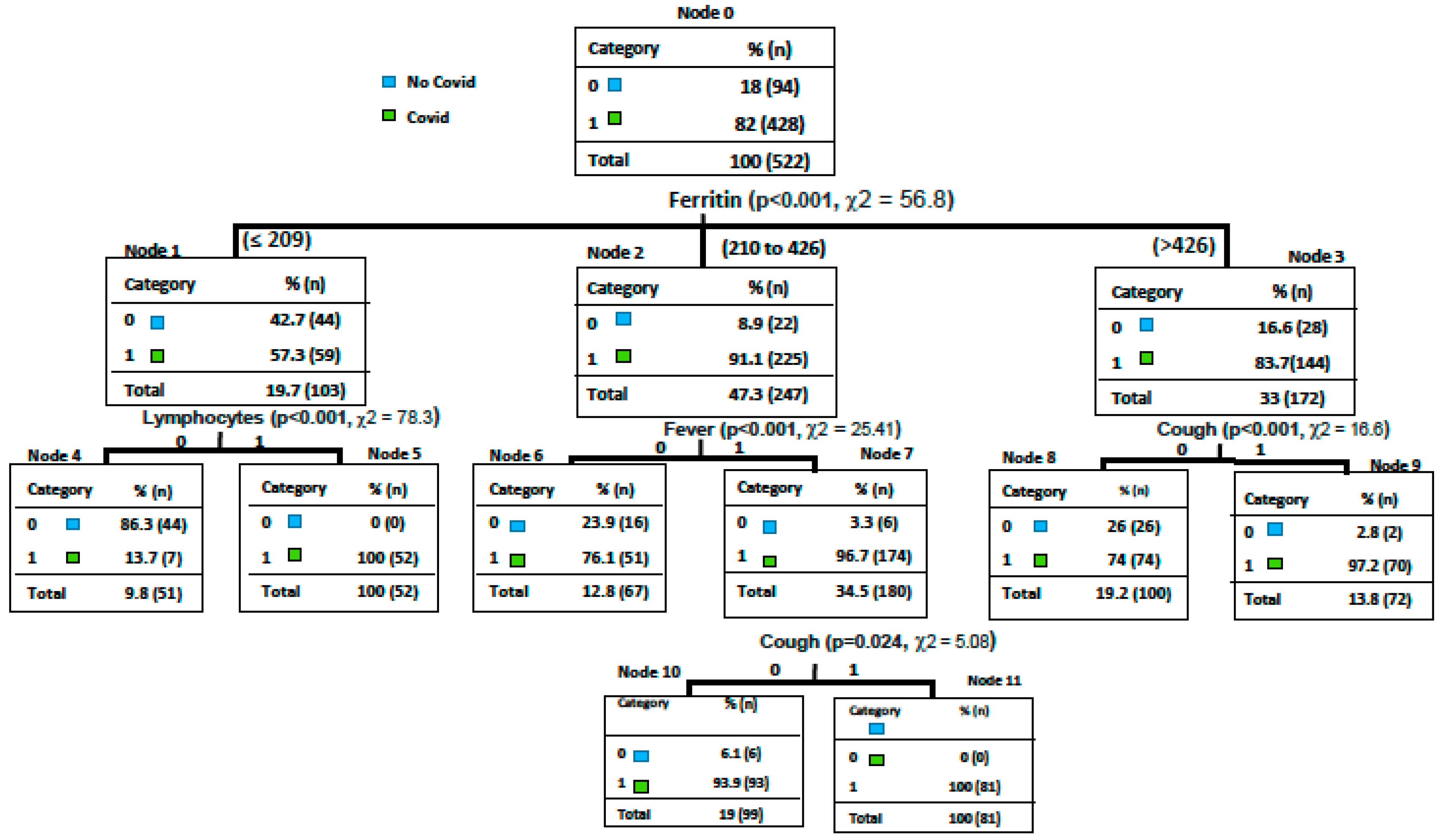
| Fever > 3 days | 14 | 9.06–20.07 | <0.001 |
| Dry cough | 4.06 | 3.03–6.05 | <0.001 |
| Acute dyspnea | 2.08 | 2.02–3.07 | <0.001 |
| Lymphocytes < 1000 × 103/µL | 1.05 | 1.01–2 | 0.027 |
| Ferritin > 250 ng/mL | 1.05 | 1.02–1.08 | 0.039 |
| Variables | OR | CI95% | p-Value |
|---|---|---|---|
| Age ≥ 65 years | 4.23 | 2.83–6.33 | <0.001 |
| No comorbidities | 0.03 | 0.02–0.04 | <0.001 |
| Steroids | 0.16 | 0.1–0.25 | <0.001 |
| LMWH | 0.2 | 0.12–0.32 | <0.001 |
| Remdesivir | 0.26 | 0.15–0.43 | <0.001 |
| ICU admission | 2.51 | 1.44–4.4 | 0.001 |
Publisher’s Note: MDPI stays neutral with regard to jurisdictional claims in published maps and institutional affiliations. |
© 2021 by the authors. Licensee MDPI, Basel, Switzerland. This article is an open access article distributed under the terms and conditions of the Creative Commons Attribution (CC BY) license (https://creativecommons.org/licenses/by/4.0/).
Share and Cite
Russo, A.; Gentilini Cacciola, E.; Borrazzo, C.; Filippi, V.; Bucci, T.; Vullo, F.; Celani, L.; Binetti, E.; Battistini, L.; Ceccarelli, G.; et al. Clinical Characteristics and Outcome of Patients with Suspected COVID-19 in Emergency Department (RESILIENCY Study II). Diagnostics 2021, 11, 1368. https://doi.org/10.3390/diagnostics11081368
Russo A, Gentilini Cacciola E, Borrazzo C, Filippi V, Bucci T, Vullo F, Celani L, Binetti E, Battistini L, Ceccarelli G, et al. Clinical Characteristics and Outcome of Patients with Suspected COVID-19 in Emergency Department (RESILIENCY Study II). Diagnostics. 2021; 11(8):1368. https://doi.org/10.3390/diagnostics11081368
Chicago/Turabian StyleRusso, Alessandro, Elio Gentilini Cacciola, Cristian Borrazzo, Valeria Filippi, Tommaso Bucci, Francesco Vullo, Luigi Celani, Erica Binetti, Luigi Battistini, Giancarlo Ceccarelli, and et al. 2021. "Clinical Characteristics and Outcome of Patients with Suspected COVID-19 in Emergency Department (RESILIENCY Study II)" Diagnostics 11, no. 8: 1368. https://doi.org/10.3390/diagnostics11081368
APA StyleRusso, A., Gentilini Cacciola, E., Borrazzo, C., Filippi, V., Bucci, T., Vullo, F., Celani, L., Binetti, E., Battistini, L., Ceccarelli, G., Alessandroni, M., Galardo, G., Mastroianni, C. M., & d’Ettorre, G. (2021). Clinical Characteristics and Outcome of Patients with Suspected COVID-19 in Emergency Department (RESILIENCY Study II). Diagnostics, 11(8), 1368. https://doi.org/10.3390/diagnostics11081368