Feasibility and Implementation of a Deep Learning MR Reconstruction for TSE Sequences in Musculoskeletal Imaging
Abstract
:1. Introduction
2. Materials and Methods
2.1. Study Design
2.2. Deep Learning Image Reconstruction
2.3. Implementation of DL Image Reconstruction in Clinical Workflow
2.4. Image Analysis
2.5. Statistical Analysis
3. Results
3.1. Assessment of Image Quality
3.2. Assessment of Anatomical Structures
4. Discussion
Author Contributions
Funding
Institutional Review Board Statement
Informed Consent Statement
Data Availability Statement
Acknowledgments
Conflicts of Interest
References
- Vanderby, S.; Badea, A.; Sánchez, J.N.P.; Kalra, N.; Babyn, P. Variations in Magnetic Resonance Imaging Provision and Processes among Canadian Academic Centres. Can. Assoc. Radiol. J. 2017, 68, 56–65. [Google Scholar] [CrossRef] [PubMed]
- Deshmane, A.; Gulani, V.; Griswold, M.A.; Seiberlich, N. Parallel mr imaging. J. Magn. Reson. Imaging 2012, 36, 55–72. [Google Scholar] [CrossRef] [Green Version]
- Gersing, A.S.; Bodden, J.; Neumann, J.; Diefenbach, M.N.; Kronthaler, S.; Pfeiffer, D.; Knebel, C.; Baum, T.; Schwaiger, B.J.; Hock, A.; et al. Accelerating anatomical 2D turbo spin echo imaging of the ankle using compressed sensing. Eur. J. Radiol. 2019, 118, 277–284. [Google Scholar] [CrossRef]
- Kreitner, K.-F.; Romaneehsen, B.; Krummenauer, F.; Oberholzer, K.; Müller, L.P.; Düber, C. Fast magnetic resonance imaging of the knee using a parallel acquisition technique (mSENSE): A prospective performance evaluation. Eur. Radiol. 2006, 16, 1659–1666. [Google Scholar] [CrossRef] [PubMed]
- Zuo, J.; Li, X.; Be, S.B.; Han, E.; Majumdar, S. Parallel imaging of knee cartilage at 3 Tesla. J. Magn. Reson. Imaging 2007, 26, 1001–1009. [Google Scholar] [CrossRef] [PubMed]
- Iuga, A.-I.; Abdullayev, N.; Weiss, K.; Haneder, S.; Brüggemann-Bratke, L.; Maintz, D.; Rau, R.; Bratke, G. Accelerated MRI of the knee. Quality and efficiency of compressed sensing. Eur. J. Radiol. 2020, 132, 109273. [Google Scholar] [CrossRef] [PubMed]
- Jaspan, O.N.; Fleysher, R.; Lipton, M.L. Compressed sensing MRI: A review of the clinical literature. Br. J. Radiol. 2015, 88, 20150487. [Google Scholar] [CrossRef]
- Matcuk, G.R.; Gross, J.S.; Fields, B.; Cen, S. Compressed Sensing MR Imaging (CS-MRI) of the Knee: Assessment of Quality, Inter-reader Agreement, and Acquisition Time. Magn. Reson. Med. Sci. 2020, 19, 254–258. [Google Scholar] [CrossRef] [Green Version]
- Zibetti, M.V.W.; Baboli, R.; Chang, G.; Otazo, R.; Regatte, R. Rapid compositional mapping of knee cartilage with compressed sensing MRI. J. Magn. Reson. Imaging 2018, 48, 1185–1198. [Google Scholar] [CrossRef]
- Jang, W.; Song, J.; Kim, S.; Yang, J. Comparison of Compressed Sensing and Gradient and Spin-Echo in Breath-Hold 3D MR Cholangiopancreatography: Qualitative and Quantitative Analysis. Diagnostics 2021, 11, 634. [Google Scholar] [CrossRef]
- Sodickson, D.; Manning, W.J. Simultaneous acquisition of spatial harmonics (SMASH): Fast imaging with radiofrequency coil arrays. Magn. Reson. Med. 1997, 38, 591–603. [Google Scholar] [CrossRef]
- Pruessmann, K.P.; Weiger, M.; Scheidegger, M.B.; Boesiger, P. Sense: Sensitivity encoding for fast mri. Magn. Reson. Med. Off. J. Int. Soc. Magn. Reson. Med. 1999, 42, 952–962. [Google Scholar] [CrossRef]
- Griswold, M.A.; Jakob, P.M.; Heidemann, R.M.; Nittka, M.; Jellus, V.; Wang, J.; Kiefer, B.; Haase, A. Generalized autocalibrating partially parallel acquisitions (grappa). Magn. Reson. Med. Off. J. Int. Soc. Magn. Reson. Med. 2002, 47, 1202–1210. [Google Scholar] [CrossRef] [PubMed] [Green Version]
- Lustig, M.; Donoho, D.; Pauly, J.M. Sparse MRI: The application of compressed sensing for rapid MR imaging. Magn. Reson. Med. 2007, 58, 1182–1195. [Google Scholar] [CrossRef] [PubMed]
- Knoll, F.; Hammernik, K.; Zhang, C.; Moeller, S.; Pock, T.; Sodickson, D.; Akcakaya, M. Deep-Learning Methods for Parallel Magnetic Resonance Imaging Reconstruction: A Survey of the Current Approaches, Trends, and Issues. IEEE Signal Process. Mag. 2020, 37, 128–140. [Google Scholar] [CrossRef]
- Lin, D.; Johnson, P.M.; Knoll, F.; Lui, Y.W. Artificial Intelligence for MR Image Reconstruction: An Overview for Clinicians. J. Magn. Reson. Imaging 2020, 53, 1015–1028. [Google Scholar] [CrossRef]
- Hyun, C.M.; Kim, H.P.; Lee, S.M.; Lee, S.; Seo, J.K. Deep learning for undersampled mri reconstruction. Phys. Med. Biol. 2018, 63, 135007. [Google Scholar] [CrossRef] [PubMed]
- Arel, I.; Rose, D.C.; Karnowski, T.P. Deep Machine Learning—A New Frontier in Artificial Intelligence Research [Research Frontier]. IEEE Comput. Intell. Mag. 2010, 5, 13–18. [Google Scholar] [CrossRef]
- Wang, G. A Perspective on Deep Imaging. IEEE Access 2016, 4, 8914–8924. [Google Scholar] [CrossRef]
- Mardani, M.; Gong, E.; Cheng, J.Y.; Vasanawala, S.S.; Zaharchuk, G.; Xing, L.; Pauly, J.M. Deep Generative Adversarial Neural Networks for Compressive Sensing MRI. IEEE Trans. Med. Imaging 2018, 38, 167–179. [Google Scholar] [CrossRef]
- Chen, F.; Taviani, V.; Malkiel, I.; Cheng, J.Y.; Tamir, J.I.; Shaikh, J.; Chang, S.T.; Hardy, C.J.; Pauly, J.M.; Vasanawala, S.S. Variable-Density Single-Shot Fast Spin-Echo MRI with Deep Learning Reconstruction by Using Variational Networks. Radiology 2018, 289, 366–373. [Google Scholar] [CrossRef] [Green Version]
- Lundervold, A.; Lundervold, A. An overview of deep learning in medical imaging focusing on MRI. Zeitschrift für Medizinische Physik 2019, 29, 102–127. [Google Scholar] [CrossRef]
- Recht, M.P.; Zbontar, J.; Sodickson, D.K.; Knoll, F.; Yakubova, N.; Sriram, A.; Murrell, T.; Defazio, A.; Rabbat, M.; Rybak, L.; et al. Using Deep Learning to Accelerate Knee MRI at 3 T: Results of an Interchangeability Study. Am. J. Roentgenol. 2020, 215, 1421–1429. [Google Scholar] [CrossRef] [PubMed]
- Knoll, F.; Hammernik, K.; Kobler, E.; Pock, T.; Recht, M.P.; Sodickson, D. Assessment of the generalization of learned image reconstruction and the potential for transfer learning. Magn. Reson. Med. 2018, 81, 116–128. [Google Scholar] [CrossRef] [PubMed]
- Hammernik, K.; Klatzer, T.; Kobler, E.; Recht, M.P.; Sodickson, D.; Pock, T.; Knoll, F. Learning a variational network for reconstruction of accelerated MRI data. Magn. Reson. Med. 2017, 79, 3055–3071. [Google Scholar] [CrossRef]
- Schlemper, J.; Caballero, J.; Hajnal, J.; Price, A.; Rueckert, D. A Deep Cascade of Convolutional Neural Networks for Dynamic MR Image Reconstruction. IEEE Trans. Med. Imaging 2017, 37, 491–503. [Google Scholar] [CrossRef] [Green Version]
- Yu, S.; Park, B.; Jeong, J. Deep iterative down-up cnn for image denoising. In Proceedings of the IEEE/CVF Conference on Computer Vision and Pattern Recognition Workshops, Long Beach, CA, USA, 6–20 June 2019. [Google Scholar]
- Ronneberger, O.; Fischer, P.; Brox, T. U-net: Convolutional networks for biomedical image segmentation. In Proceedings of the Medical Image Computing and Computer-Assisted Intervention—MICCAI 2015, Munich, Germany, 5–9 October 2015; Springer: Berlin/Heidelberg, Germany, 2015; pp. 234–241. [Google Scholar]
- Wang, Z.; Simoncelli, E.P.; Bovik, A.C. Multiscale structural similarity for image quality assessment. In Proceedings of the Thirty-Seventh Asilomar Conference on Signals, Systems & Computers, Pacific Grove, CA, USA, 9–12 November 2003; pp. 1398–1402. [Google Scholar]
- Robson, P.M.; Grant, A.K.; Madhuranthakam, A.J.; Lattanzi, R.; Sodickson, D.; McKenzie, C. Comprehensive quantification of signal-to-noise ratio andg-factor for image-based andk-space-based parallel imaging reconstructions. Magn. Reson. Med. 2008, 60, 895–907. [Google Scholar] [CrossRef] [Green Version]
- LeCun, Y.; Bengio, Y.; Hinton, G. Deep learning. Nat. Cell Biol. 2015, 521, 436–444. [Google Scholar] [CrossRef] [PubMed]
- Aggarwal, H.K.; Mani, M.P.; Jacob, M. MoDL: Model-Based Deep Learning Architecture for Inverse Problems. IEEE Trans. Med. Imaging 2018, 38, 394–405. [Google Scholar] [CrossRef] [PubMed]
- Küstner, T.; Fuin, N.; Hammernik, K.; Bustin, A.; Qi, H.; Hajhosseiny, R.; Masci, P.G.; Neji, R.; Rueckert, D.; Botnar, R.M.; et al. CINENet: Deep learning-based 3D cardiac CINE MRI reconstruction with multi-coil complex-valued 4D spatio-temporal convolutions. Sci. Rep. 2020, 10, 13710. [Google Scholar] [CrossRef] [PubMed]
- Eo, T.; Jun, Y.; Kim, T.; Jang, J.; Lee, H.; Hwang, D. KIKI -net: Cross-domain convolutional neural networks for reconstructing undersampled magnetic resonance images. Magn. Reson. Med. 2018, 80, 2188–2201. [Google Scholar] [CrossRef]
- Wang, S.; Su, Z.; Ying, L.; Peng, X.; Zhu, S.; Liang, F.; Feng, D.; Liang, D. Accelerating magnetic resonance imaging via deep learning. In Proceedings of the 2016 IEEE 13th International Symposium on Biomedical Imaging (ISBI), Prague, Czech Republic, 13–16 April 2016; pp. 514–517. [Google Scholar] [CrossRef]
- Han, Y.; Sunwoo, L.; Ye, J.C. K-space deep learning for accelerated MRI. IEEE Trans. Med. Imaging 2019, 39, 377–386. [Google Scholar] [CrossRef] [Green Version]
- Lee, D.; Yoo, J.; Tak, S.; Ye, J.C. Deep Residual Learning for Accelerated MRI Using Magnitude and Phase Networks. IEEE Trans. Biomed. Eng. 2018, 65, 1985–1995. [Google Scholar] [CrossRef] [Green Version]
- Qin, C.; Schlemper, J.; Caballero, J.; Price, A.N.; Hajnal, J.V.; Rueckert, D. Convolutional Recurrent Neural Networks for Dynamic MR Image Reconstruction. IEEE Trans. Med. Imaging 2018, 38, 280–290. [Google Scholar] [CrossRef] [Green Version]
- Yang, G.; Yu, S.; Dong, H.; Slabaugh, G.G.; Dragotti, P.L.; Ye, X.; Liu, F.; Arridge, S.R.; Keegan, J.; Guo, Y.; et al. DAGAN: Deep De-Aliasing Generative Adversarial Networks for Fast Compressed Sensing MRI Reconstruction. IEEE Trans. Med. Imaging 2017, 37, 1310–1321. [Google Scholar] [CrossRef] [PubMed] [Green Version]
- Zhu, B.; Liu, J.Z.; Cauley, S.F.; Rosen, B.R.; Rosen, M. Image reconstruction by domain-transform manifold learning. Nat. Cell Biol. 2018, 555, 487–492. [Google Scholar] [CrossRef] [PubMed] [Green Version]
- Sandino, C.M.; Lai, P.; Vasanawala, S.S.; Cheng, J.Y. Accelerating cardiac cine MRI using a deep learning-based ESPIRiT reconstruction. Magn. Reson. Med. 2020, 85, 152–167. [Google Scholar] [CrossRef] [PubMed]
- Hosseini, S.A.H.; Zhang, C.; Weingärtner, S.; Moeller, S.; Stuber, M.; Ugurbil, K.; Akçakaya, M. Accelerated coronary MRI with sRAKI: A database-free self-consistent neural network k-space reconstruction for arbitrary undersampling. PLoS ONE 2020, 15, e0229418. [Google Scholar] [CrossRef] [Green Version]
- Fuin, N.; Bustin, A.; Küstner, T.; Oksuz, I.; Clough, J.; King, A.P.; Schnabel, J.A.; Botnar, R.M.; Prieto, C. A multi-scale variational neural network for accelerating motion-compensated whole-heart 3D coronary MR angiography. Magn. Reson. Imaging 2020, 70, 155–167. [Google Scholar] [CrossRef] [PubMed]
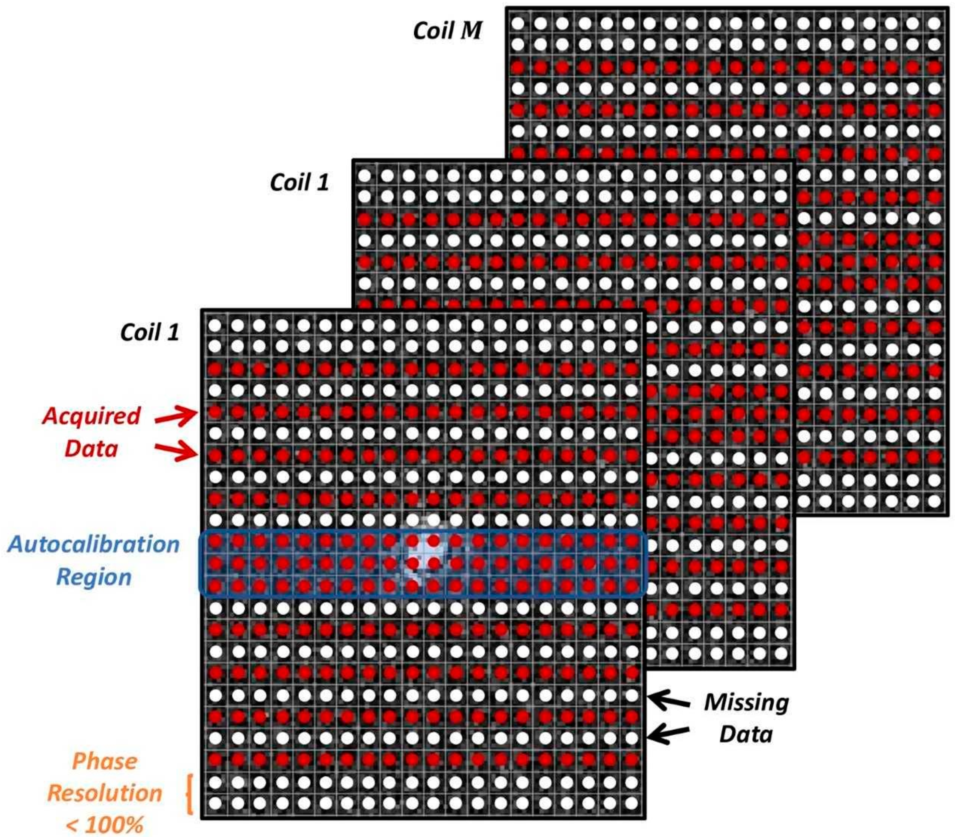


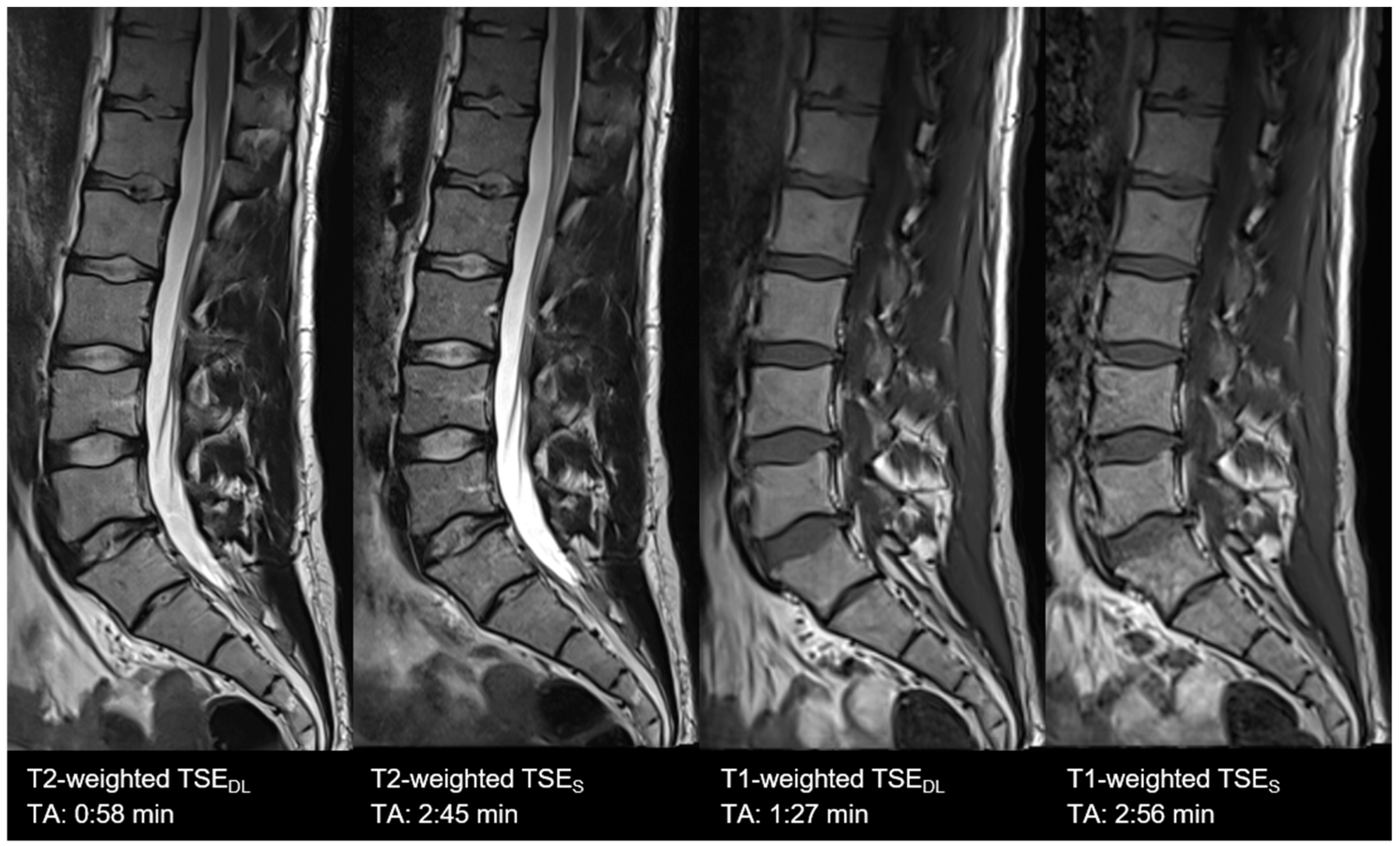

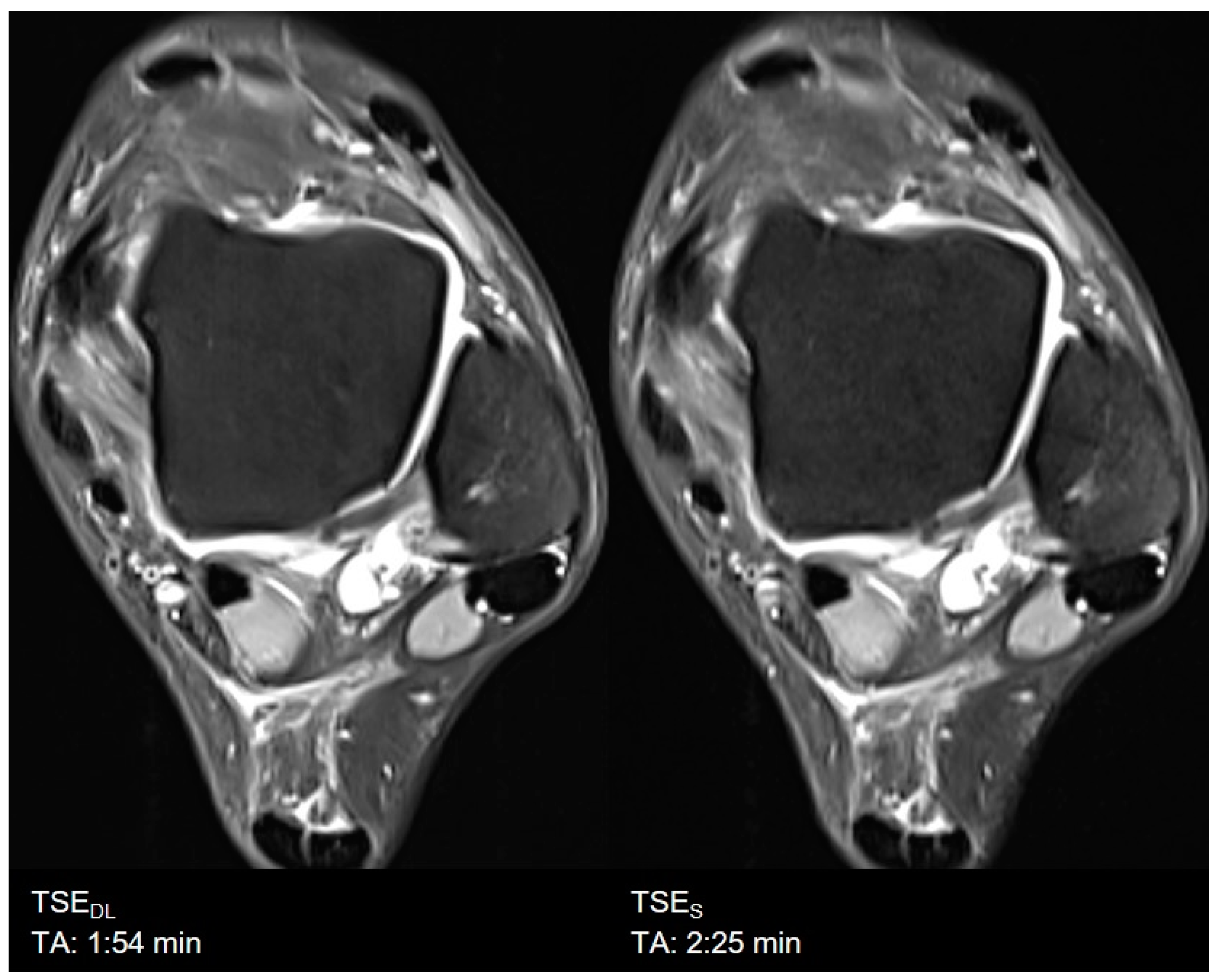
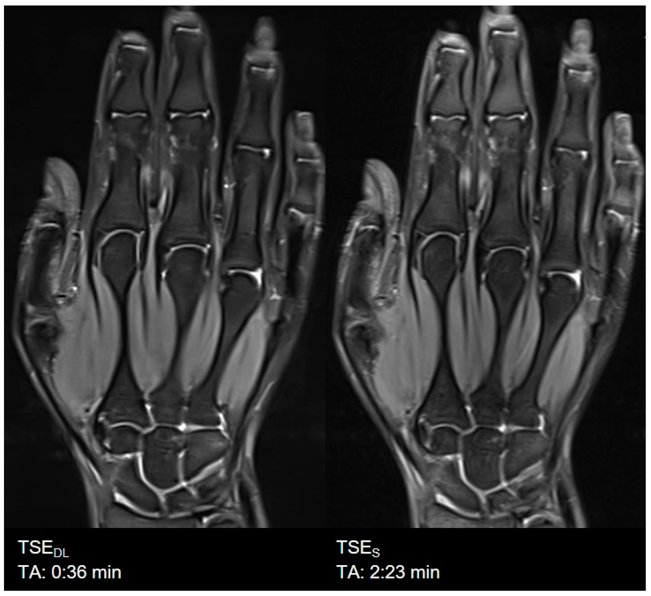
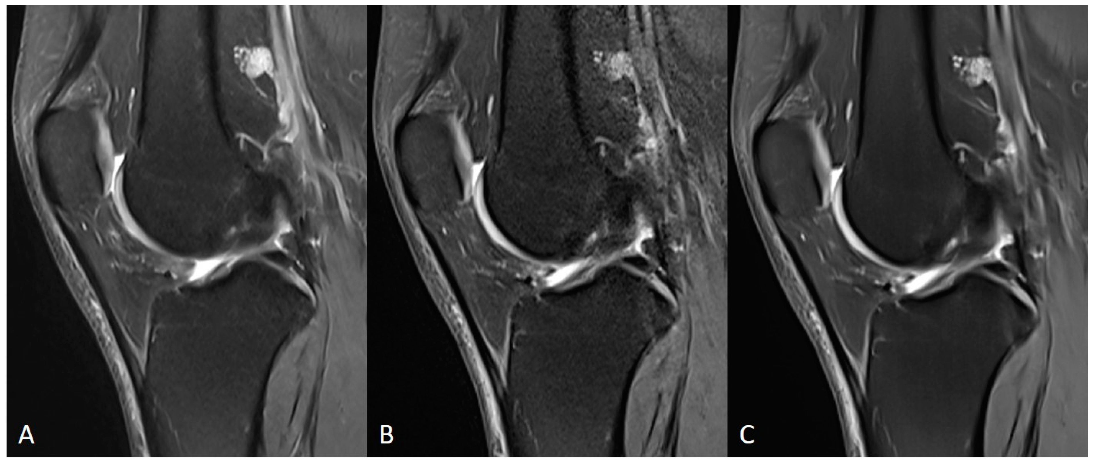
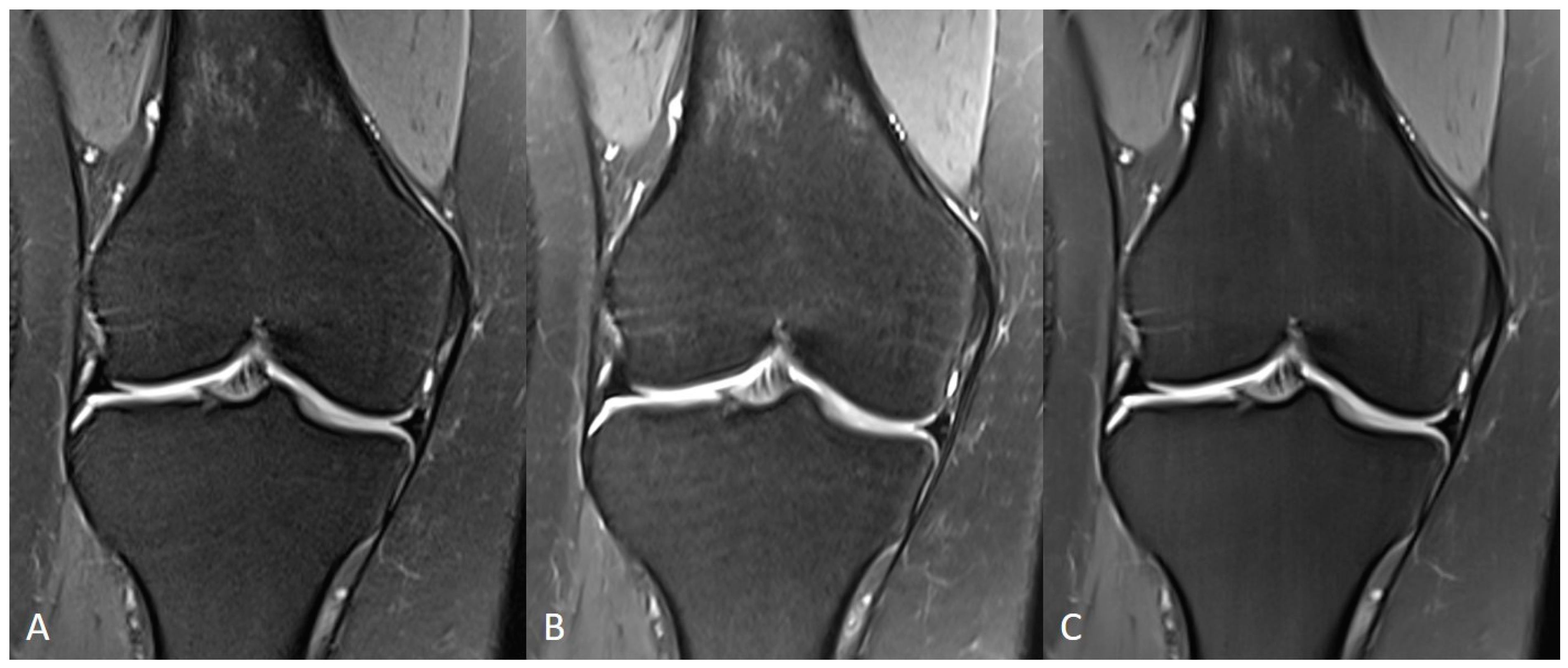
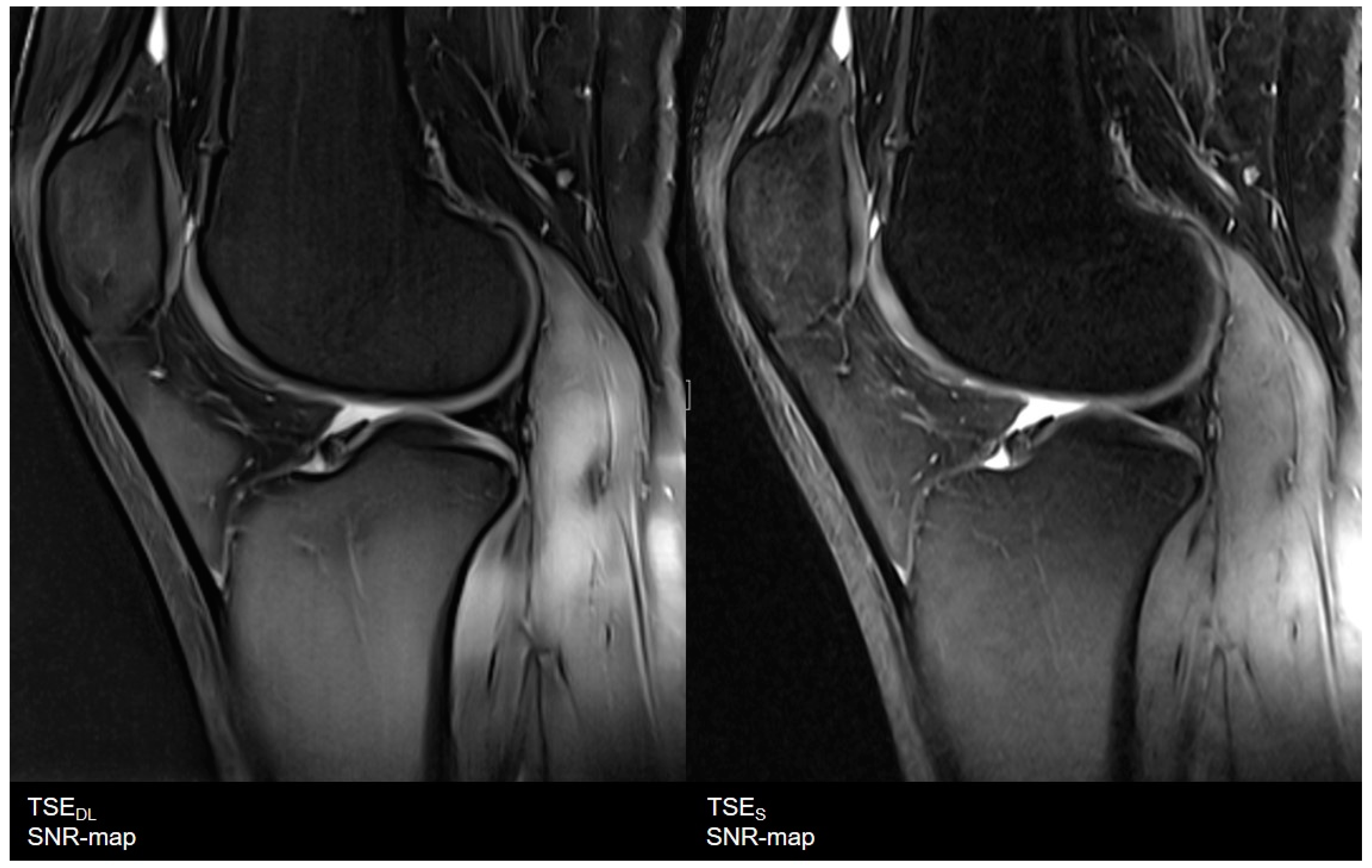
| Sequence | Orientation | TA, min | FOV, mm | Voxel Size, mm | A | C | PAT | TR, ms | TE, ms | FA, Degree | Bandwith, Hz/Px | Echo Spacing, ms | ||
|---|---|---|---|---|---|---|---|---|---|---|---|---|---|---|
| Shoulder | TSES | TSE PD FS | axial | 2:14 | 180 | 0.6 × 0.6 × 3.0 | 1 | 2 | 2 | 3000 | 44 | 150 | 180 | 10.9 |
| coronal | 2:53 | 180 | 0.6 × 0.6 × 3.0 | 2 | 1 | 2 | 3300 | 42 | 150 | 191 | 10.6 | |||
| TSEDL | TSE PD FS | axial | 1:10 | 180 | 0.6 × 0.6 × 3.0 | 1 | 1 | 3 | 3520 | 44 | 150 | 180 | 10.9 | |
| coronal | 1:09 | 180 | 0.6 × 0.6 × 3.0 | 1 | 1 | 3 | 3000 | 42 | 150 | 191 | 10.6 | |||
| Knee | TSES | TSE PD FS | coronal | 3:11 | 150 | 0.2 × 0.2 × 3.0 | 2 | 1 | 3 | 3790 | 44 | 150 | 100 | 14.6 |
| sagittal | 3:11 | 150 | 0.2 × 0.2 × 3.0 | 2 | 1 | 3 | 3790 | 44 | 150 | 100 | 14.6 | |||
| TSEDL | TSE PD FS | coronal | 1:33 | 150 | 0.5 × 0.5 × 3.0 | 1 | 1 | 3 | 3580 | 41 | 150 | 120 | 13.7 | |
| sagittal | 1:33 | 150 | 0.5 × 0.5 × 3.0 | 1 | 1 | 3 | 3580 | 41 | 150 | 120 | 13.7 | |||
| Lumbar spine | TSES | T1 TSE | sagittal | 2:56 | 300 | 0.9 × 0.9 × 3.0 | 1 | 2 | 0 | 562 | 10 | 150 | 180 | 10.4 |
| T2 TSE FS | sagittal | 2:45 | 300 | 0.7 × 0.7 × 3.0 | 2 | 1 | 2 | 6040 | 102 | 150 | 189 | 11.3 | ||
| TSEDL | T1 TSE | sagittal | 1:27 | 300 | 0.9 × 0.9 × 3.0 | 1 | 2 | 3 | 462 | 10 | 150 | 180 | 10.4 | |
| T2 TSE FS | sagittal | 0:58 | 300 | 0.7 × 0.7 × 3.0 | 1 | 1 | 3 | 4470 | 105 | 150 | 189 | 10.5 | ||
| Hip | TSES | TSE PD FS | axial | 3:02 | 200 | 0.3 × 0.3 × 3.0 | 1 | 1 | 0 | 3410 | 42 | 150 | 100 | 14.1 |
| coronal | 2:01 | 200 | 0.3 × 0.3 × 3.0 | 1 | 1 | 2 | 3410 | 42 | 150 | 100 | 14.1 | |||
| TSEDL | TSE PD FS | axial | 1:32 | 200 | 0.6 × 0.6 × 3.0 | 1 | 1 | 3 | 3069 | 42 | 150 | 120 | 13.1 | |
| coronal | 1:33 | 200 | 0.6 × 0.6 × 3.0 | 1 | 1 | 3 | 3000 | 41 | 150 | 120 | 13.7 | |||
| Ankle | TSES | TSE PD FS | axial | 2:25 | 150 | 0.4 × 0.4 × 3.0 | 1 | 1 | 2 | 3340 | 17 | 150 | 90 | 17.1 |
| sagittal | 1:47 | 160 | 0.2 × 0.2 × 3.0 | 1 | 1 | 3 | 3000 | 32 | 150 | 100 | 15.9 | |||
| TSEDL | TSE PD FS | axial | 1:54 | 150 | 0.4 × 0.4 × 3.0 | 1 | 1 | 3 | 3000 | 17 | 150 | 90 | 16.9 | |
| sagittal | 1:45 | 160 | 0.4 × 0.4 × 3.0 | 1 | 1 | 3 | 3000 | 31 | 150 | 100 | 15.7 | |||
| Hand | TSES | TSE PD FS | coronal | 2:23 | 200 | 0.5 × 0.5 × 2.0 | 2 | 1 | 0 | 3000 | 41 | 150 | 121 | 13.6 |
| axial | 4:40 | 180 | 0.5 × 0.5 × 2.0 | 2 | 2 | 0 | 3310 | 42 | 150 | 121 | 13.9 | |||
| TSEDL | TSE PD FS | coronal | 0:36 | 200 | 0.5 × 0.5 × 2.0 | 1 | 1 | 3 | 3000 | 44 | 150 | 119 | 14.7 | |
| axial | 1:23 | 180 | 0.5 × 0.5 × 2.0 | 1 | 2 | 2 | 3190 | 42 | 150 | 119 | 14.1 |
| Variables | |
|---|---|
| Total (male/female), n | 60 (37/23) |
| Age, mean ± SD (range), y | total: 26 ± 7 (20–55) |
| knee: 25 ± 4 (20–31) | |
| ankle: 26 ± 5 (20–35) | |
| hip: 26 ± 5 (20–35) | |
| shoulder: 27 ± 10 (20–55) | |
| wrist: 27 ± 8 (20–44) | |
| lumbar spine: 29 ± 11 (20–55) |
| TSES | TSEDL | TSES vs. TSEDL | ||||||
|---|---|---|---|---|---|---|---|---|
| R1 m (IQR) | R2 m (IQR) | κ | R1 m (IQR) | R2 m (IQR) | κ | R1 | R2 | |
| IQ | 4 (4−4) | 4 (3−4) | 0.697 | 4 (4−4) | 4 (4−4) | 0.634 | 0.002 | 0.013 |
| Artifacts | 4 (4−4) | 4 (4−4) | 0.649 | 4 (4−4) | 4 (4−4) | 0.700 | 0.180 | 0.157 |
| Edge sharpness | 4 (3−4) | 4 (3−4) | 0.883 | 4 (4−4) | 4 (4−4) | 0.792 | <0.001 | <0.001 |
| Contrast resolution | 4 (4−4) | 4 (4−4) | 0.741 | 4 (4−4) | 4 (4−4) | 0.649 | 0.257 | 0.157 |
| Noise | 4 (3−4) | 4 (3−4) | 0.897 | 4 (4−4) | 4 (4−4) | 0.651 | <0.001 | <0.001 |
| Clarity of anatomic structures | 4 (4−4) | 4 (4−4) | 0.747 | 4 (4−4) | 4 (4−4) | 0.889 | 0.317 | 0.564 |
| Bone | 4 (4−4) | 4 (4−4) | 0.896 | 4 (4−4) | 4 (4−4) | 0.741 | 0.014 | 0.025 |
| Articular cartilage | 4 (4−4) | 4 (4−4) | 0.739 | 4 (4−4) | 4 (4−4) | 0.773 | 0.157 | 0.705 |
| Ligaments | 4 (4−4) | 4 (4−4) | 0.732 | 4 (4−4) | 4 (4−4) | 0.643 | 0.705 | 0.480 |
| Tendons | 4 (4−4) | 4 (4−4) | 0.640 | 4 (4−4) | 4 (4−4) | 0.659 | 0.180 | 0.083 |
| Diagnostic confidence | 4 (4−4) | 4 (4−4) | 0.773 | 4 (4−4) | 4 (4−4) | 0.651 | 0.102 | 0.096 |
| Image impression | 4 (4−4) | 4 (4−4) | 0.848 | 4 (3−4) | 4 (3−4) | 0.923 | <0.001 | <0.001 |
Publisher’s Note: MDPI stays neutral with regard to jurisdictional claims in published maps and institutional affiliations. |
© 2021 by the authors. Licensee MDPI, Basel, Switzerland. This article is an open access article distributed under the terms and conditions of the Creative Commons Attribution (CC BY) license (https://creativecommons.org/licenses/by/4.0/).
Share and Cite
Herrmann, J.; Koerzdoerfer, G.; Nickel, D.; Mostapha, M.; Nadar, M.; Gassenmaier, S.; Kuestner, T.; Othman, A.E. Feasibility and Implementation of a Deep Learning MR Reconstruction for TSE Sequences in Musculoskeletal Imaging. Diagnostics 2021, 11, 1484. https://doi.org/10.3390/diagnostics11081484
Herrmann J, Koerzdoerfer G, Nickel D, Mostapha M, Nadar M, Gassenmaier S, Kuestner T, Othman AE. Feasibility and Implementation of a Deep Learning MR Reconstruction for TSE Sequences in Musculoskeletal Imaging. Diagnostics. 2021; 11(8):1484. https://doi.org/10.3390/diagnostics11081484
Chicago/Turabian StyleHerrmann, Judith, Gregor Koerzdoerfer, Dominik Nickel, Mahmoud Mostapha, Mariappan Nadar, Sebastian Gassenmaier, Thomas Kuestner, and Ahmed E. Othman. 2021. "Feasibility and Implementation of a Deep Learning MR Reconstruction for TSE Sequences in Musculoskeletal Imaging" Diagnostics 11, no. 8: 1484. https://doi.org/10.3390/diagnostics11081484
APA StyleHerrmann, J., Koerzdoerfer, G., Nickel, D., Mostapha, M., Nadar, M., Gassenmaier, S., Kuestner, T., & Othman, A. E. (2021). Feasibility and Implementation of a Deep Learning MR Reconstruction for TSE Sequences in Musculoskeletal Imaging. Diagnostics, 11(8), 1484. https://doi.org/10.3390/diagnostics11081484


