
Prevalence and Clinical Significance of Residual or Reconverted Red Bone Marrow on Knee MRI


 , , , ,
Abstract
:1. Introduction
2. Methods
2.1. Study Design
2.2. Participants
2.3. Demographics and Lifestyle Factors
2.4. Knee Symptom Measurements
2.5. MRI Measurements
2.6. Statistical Analysis
3. Results
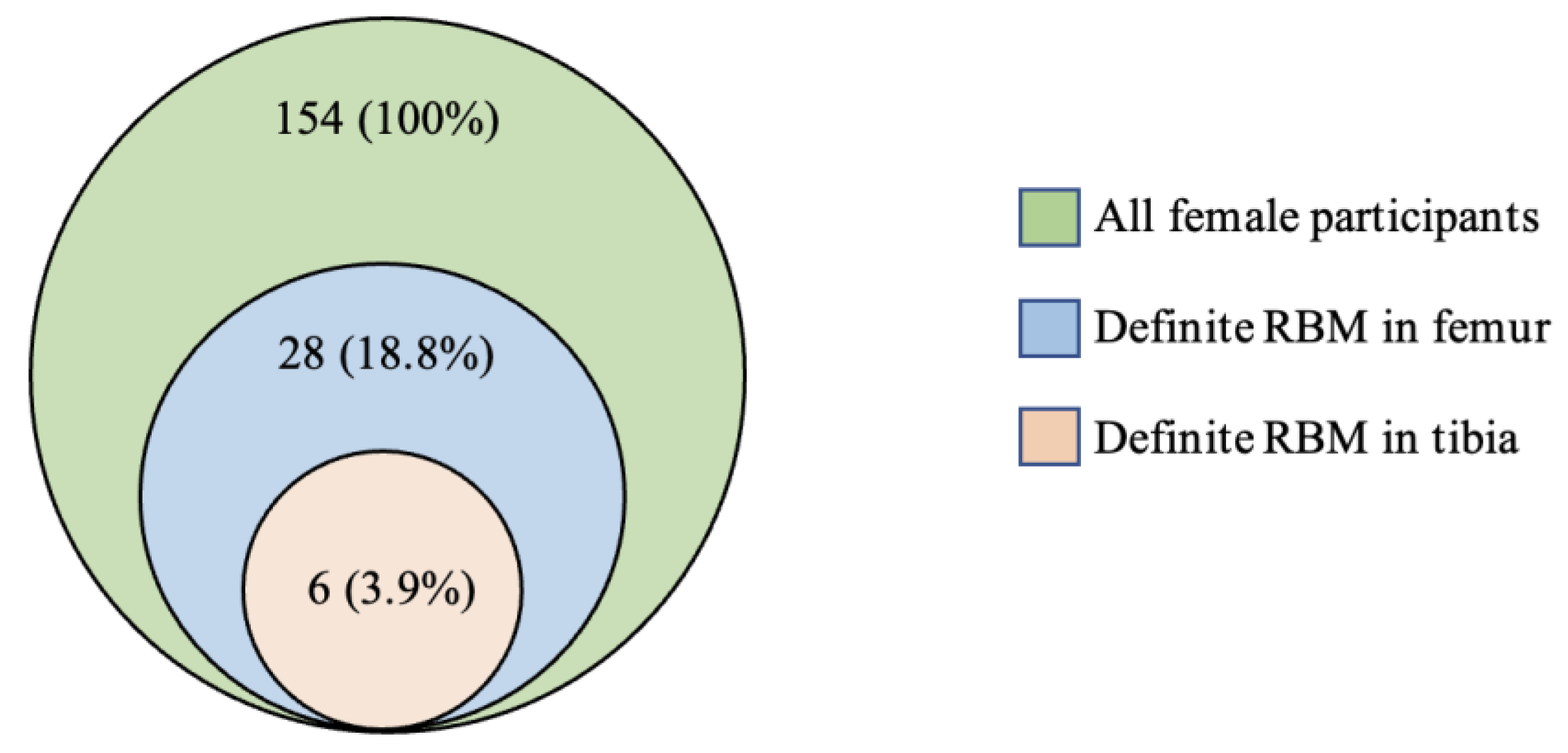
3.1. Prevalence
3.2. Pattern of Distribution
3.3. Associations with Lifestyle Factors
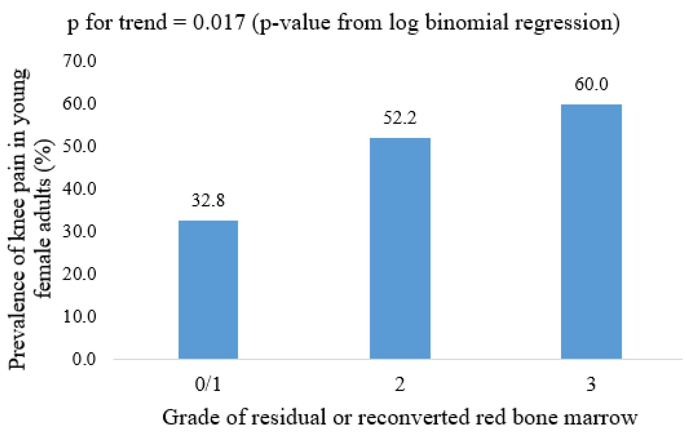
3.4. Associations with Knee Symptoms
3.5. Associations with Degenerative Knee Structural Abnormalities
4. Discussion
5. Conclusions
Supplementary Materials
Author Contributions
Funding
Institutional Review Board Statement
Informed Consent Statement
Data Availability Statement
Acknowledgments
Conflicts of Interest
References
- Piney, A. The anatomy of the bone marrow: With special reference to the distribution of the red marrow. Br. Med. J. 1922, 2, 792–795. [Google Scholar]
- Guillerman, R.P. Marrow: Red, yellow and bad. Pediatric Radiol. 2013, 43, 181–192. [Google Scholar] [CrossRef] [PubMed]
- Cabrita, G.J.; Ferreira, B.S.; Da Silva, C.L.; Goncalves, R.; Almeida-Porada, G.; Cabral, J.M. Hematopoietic stem cells: From the bone to the bioreactor. Trends Biotechnol. 2003, 21, 233–240. [Google Scholar] [CrossRef]
- Riley, R.S.; Williams, D.; Ross, M.; Zhao, S.; Chesney, A.; Clark, B.D.; Ben-Ezra, J.M. Bone marrow aspirate and biopsy: A pathologist’s perspective. II. interpretation of the bone marrow aspirate and biopsy. J. Clin. Lab. Anal. 2009, 23, 259–307. [Google Scholar] [CrossRef]
- Elgazzar, A.H. Bone marrow imaging. In Orthopedic Nuclear Medicine; Springer Nature Switzerland: Chem, Switzerland, 2017; pp. 307–322. [Google Scholar]
- Foster, K.; Chapman, S.; Johnson, K. MRI of the marrow in the paediatric skeleton. Clin. Radiol. 2004, 59, 651–673. [Google Scholar] [CrossRef] [PubMed]
- Blebea, J.S.; Houseni, M.; Torigian, D.A.; Fan, C.; Mavi, A.; Zhuge, Y.; Iwanaga, T.; Mishra, S.; Udupa, J.; Zhuang, J.; et al. Structural and Functional Imaging of Normal Bone Marrow and Evaluation of Its Age-Related Changes. Semin. Nucl. Med. 2007, 37, 185–194. [Google Scholar] [CrossRef] [PubMed]
- Małkiewicz, A.; Dziedzic, M. Bone marrow reconversion–imaging of physiological changes in bone marrow. Pol. J. Radiol. 2012, 77, 45. [Google Scholar] [CrossRef]
- Andrews, C.L. Evaluation of the marrow space in the adult hip. Radiographics 2000, 20 (Suppl.1), S27–S42. [Google Scholar] [CrossRef]
- Hynes, J.P.; Hughes, N.; Cunningham, P.; Kavanagh, E.C.; Eustace, S.J. Whole-body MRI of bone marrow: A review. J. Magn. Reson. Imaging 2019, 50, 1687–1701. [Google Scholar] [CrossRef]
- Ricci, C.; Cova, M.; Kang, Y.S.; Yang, A.; Rahmouni, A.; Scott, W.W.; A Zerhouni, E. Normal age-related patterns of cellular and fatty bone marrow distribution in the axial skeleton: MR imaging study. Radiology 1990, 177, 83–88. [Google Scholar] [CrossRef]
- Laor, T.; Jaramillo, D. MR imaging insights into skeletal maturation: What is normal? Radiology 2009, 250, 28–38. [Google Scholar] [CrossRef] [Green Version]
- Vogler, J.B., 3rd; Murphy, W.A. Bone marrow imaging. Radiology 1988, 168, 679–693. [Google Scholar] [CrossRef]
- Emery, J.L.; Follett, G.F. Regression of bone-marrow haemopoiesis from the terminal digits in the foetus and infant. Br. J. Haematol. 1964, 10, 485–489. [Google Scholar] [CrossRef]
- Taccone, A.; Oddone, M.; Dell’Acqua, A.; Occhi, M.; Ciccone, M.A. MRI “road-map” of normal age-related bone marrow. Pediatric Radiol. 1995, 25, 596–606. [Google Scholar] [CrossRef]
- Moore, S.G.; Dawson, K.L. Red and yellow marrow in the femur: Age-related changes in appearance at MR imaging. Radiology 1990, 175, 219–223. [Google Scholar] [CrossRef]
- Deutsch, A.L.; Mink, J.H.; Rosenfelt, F.P.; Waxman, A.D. Incidental detection of hematopoietic hyperplasia on routine knee MR imaging. Am. J. Roentgenol. 1989, 152, 333–336. [Google Scholar] [CrossRef] [PubMed]
- Shellock, F.G.; Morris, E.; Deutsch, A.L.; Mink, J.H.; Kerr, R.; Boden, S.D. Hematopoietic bone marrow hyperplasia: High prevalence on MR images of the knee in asymptomatic marathon runners. AJR Am. J. Roentgenol. 1992, 158, 335–338. [Google Scholar] [CrossRef] [PubMed] [Green Version]
- Poulton, T.B.; Murphy, W.D.; Duerk, J.L.; Chapek, C.C.; Feiglin, D.H. Bone marrow reconversion in adults who are smokers: MR Imaging findings. AJR Am. J. Roentgenol. 1993, 161, 1217–1221. [Google Scholar] [CrossRef] [PubMed]
- Wilson, A.J.; Hodge, J.C.; Pilgram, T.K.; Kang, E.H.; Murphy Jr, W.A. Prevalence of red marrow around the knee joint in adults as demonstrated on magnetic resonance imaging. Acad. Radiol. 1996, 3, 550–555. [Google Scholar] [CrossRef]
- Lang, P.; Fritz, R.; Majumdar, S.; Vahlensieck, M.; Peterfy, C.; Genant, H.K. Hematopoietic bone marrow in the adult knee: Spin-echo and opposed-phase gradient-echo MR imaging. Skelet. Radiol. 1993, 22, 95–103. [Google Scholar] [CrossRef]
- Arslan, G.; Ozmen, E.; Soyturk, M. MRI of residual red bone marrow in the distal femur of healthy subjects. Pol. J. Radiol. 2015, 80, 300. [Google Scholar]
- Gonzalez, F.M.; Mitchell, J.; Monfred, E.; Anguh, T.; Mulligan, M. Knee MRI patterns of bone marrow reconversion and relationship to anemia. Acta Radiol. 2016, 57, 964–970. [Google Scholar] [CrossRef]
- Carroll, K.W.; Feller, J.F.; Tirman, P.F.J. Useful internal standards for distinguishing infiltrative marrow pathology from hematopoietic marrow at MRI. J. Magn. Reson. Imaging 1997, 7, 394–398. [Google Scholar] [CrossRef]
- Von Elm, E.; Altman, D.G.; Egger, M.; Pocock, S.J.; Gøtzsche, P.C.; Vandenbroucke, J.P. Strengthening the Reporting of Observational Studies in Epidemiology (STROBE) statement: Guidelines for reporting observational studies. BMJ 2007, 335, 806–808. [Google Scholar] [CrossRef] [Green Version]
- Antony, B.S.E.; Jones, G.; Venn, A.; Cicuttini, F.; March, L.; Blizzard, L.; Dwyer, T.; Cross, M.; Ding, C. Childhood Physical Performance Measures and Adulthood Knee Cartilage Volume and Bone Area: A 25-Year Cohort Study. Arthritis Rheum. 2015, 67, 1263–1271. [Google Scholar] [CrossRef] [Green Version]
- Antony, B.S.E.; Jones, G.; Venn, A.; Cicuttini, F.; March, L.; Blizzard, L.; Dwyer, T.; Cross, M.; Ding, C. Association between childhood overweight measures and adulthood knee pain, stiffness and dysfunction: A 25-year cohort study. Ann. Rheum. Dis. 2013, 74, 711–717. [Google Scholar] [CrossRef] [PubMed]
- Smith, R.D.; McHugh, G.A.; Quicke, J.G.; Dziedzic, K.S.; Healey, E.L. Comparison of reliability, construct validity and responsiveness of the IPAQ-SF and PASE in adults with osteoarthritis. Musculoskelet. Care 2021. [Google Scholar] [CrossRef]
- Tran, V.D.; Do, V.V.; Pham, N.M.; Nguyen, C.T.; Xuong, N.T.; Jancey, J.; Lee, A.H. Validity of the International Physical Activity Questionnaire–Short Form for Application in Asian Countries: A Study in Vietnam. Eval. Health Prof. 2020, 43, 105–109. [Google Scholar] [CrossRef] [PubMed]
- Craig, C.L.; Marshall, A.L.; Sjöström, M.; Bauman, A.E.; Booth, M.L.; Ainsworth, B.E.; Pratt, M.; Ekelund, U.L.F.; Yngve, A.; Sallis, J.F.; et al. International Physical Activity Questionnaire: 12-Country Reliability and Validity. Med. Sci. Sports Exerc. 2003, 35, 1381–1395. [Google Scholar] [CrossRef] [PubMed] [Green Version]
- Helou, K.; El Helou, N.; Mahfouz, M.; Mahfouz, Y.; Salameh, P.; Harmouche-Karaki, M. Validity and reliability of an adapted arabic version of the long international physical activity questionnaire. BMC Public Health 2017, 18, 49. [Google Scholar] [CrossRef]
- Heintjes, E.M.; Bierma-Zeinstra, S.M.A.; Berger, M.Y.; Koes, B.W. Lysholm scale and WOMAC index were responsive in prospective cohort of young general practice patients. J. Clin. Epidemiol. 2008, 61, 481–488. [Google Scholar] [CrossRef] [PubMed]
- Van de Graaf, V.A.; Wolterbeek, N.; Scholtes, V.A.; Mutsaerts, E.L.; Poolman, R.W. Reliability and Validity of the IKDC, KOOS, and WOMAC for Patients With Meniscal Injuries. Am. J. Sports Med. 2014, 42, 1408–1416. [Google Scholar] [CrossRef] [PubMed]
- McConnell, S.; Kolopack, P.; Davis, A.M. The Western Ontario and McMaster Universities Osteoarthritis Index (WOMAC): A review of its utility and measurement properties. Arthritis Rheum 2001, 45, 453–461. [Google Scholar] [CrossRef]
- Antony, B.; Venn, A.; Cicuttini, F.; March, L.; Blizzard, L.; Dwyer, T.; Halliday, A.; Cross, M.; Jones, G.; Ding, C. Correlates of knee bone marrow lesions in younger adults. Arthritis Res. 2016, 18, 1–9. [Google Scholar] [CrossRef] [PubMed] [Green Version]


| Male (n = 173)—Prevalence (Definite RBM, Grade ≥ 2) = * 0.0% | ||
|---|---|---|
| RBM Grade | Femur/n (%) | Tibia/n (%) |
| 0 | 170 (98.3%) | 173 (100%) |
| 1 | 3 (1.7%) | 0 (0.0%) |
| 2 | 0 (0.0%) | 0 (0.0%) |
| 3 | 0 (0.0%) | 0 (0.0%) |
| Female (n = 154)—Prevalence (Definite RBM, Grade ≥ 2) = * 18.8% | ||
| RBM Grade | Femur/n (%) | Tibia/n (%) |
| 0 | 94 (61.0%) | 133 (86.4%) |
| 1 | 31 (20.1%) | 15 (9.7%) |
| 2 | 23 (14.9%) | 6 (3.9%) |
| 3 | 6 (3.9%) | 0 (0.0%) |
| Prevalence of Red Bone Marrow | Definite RBM Absent (Grade ≤ 1) n = 125 | Definite RBM Present (Grade ≥ 2) n = 29 | p-Value + | |
|---|---|---|---|---|
| Demographic and lifestyle factors | ||||
| Age (years) | mean (s.d.) | 35.5 (2.7) | 35.1 (2.8) | 0.534 ^ |
| Knee injury (yes) | n (%) | 15 (12.3%) | 2 (7.1%) | 0.438 * |
| Smoking status (yes) | n (%) | 37 (31.6%) | 7 (25.0%) | 0.493 * |
| Weight (kg) | mean (s.d.) | 66.1 (10.6) | 71.5 (19.5) | 0.053 ^ |
| Overweight status (yes) | n (%) | 34 (28.6%) | 13 (52.0%) | 0.023 * |
| BMI (kgm−2) | mean (s.d.) | 24.1 (3.7) | 26.7 (6.8) | 0.008 ^ |
| Total physical activity (hours/week) | mean (s.d.) | 10.9 (6.8) | 9.4 (6.3) | 0.310 ^ |
| Knee symptoms at CDAH knee study | ||||
| WOMAC pain (yes) | n (%) | 40 (32.8%) | 15 (53.6%) | 0.040 * |
| WOMAC stiffness (yes) | n (%) | 30 (24.6%) | 11 (39.3%) | 0.116 * |
| WOMAC dysfunction (yes) | n (%) | 47 (38.5%) | 15 (53.6) | 0.145 * |
| Knee symptoms at CDAH-3 study | ||||
| WOMAC pain (yes) | n (%) | 31 (43.1%) | 10 (55.6%) | 0.341 * |
| WOMAC stiffness (yes) | n (%) | 26 (35.6%) | 10 (52.6%) | 0.176 * |
| WOMAC dysfunction (yes) | n (%) | 30 (41.1%) | 8 (42.1%) | 0.937 * |
| Knee structural abnormalities | ||||
| Cartilage defect (yes) | n (%) | 53 (43.4%) | 11 (37.9%) | 0.589 * |
| Subchondral BMLs (yes) | n (%) | 35 (28.0%) | 8 (27.6%) | 0.964 * |
| Meniscal tear (yes) | n (%) | 3 (2.5%) | 1 (3.5%) | 0.771 * |
| Meniscal extrusion (yes) | n (%) | 35 (28.9%) | 12 (41.4%) | 0.194 * |
| Univariable PR (95% CI) | Multivariable * PR (95% CI) | |
|---|---|---|
| Age (year) | 0.96 (0.85 to 1.09) | 0.97 (0.85 to 1.11) |
| Knee injury (yes) | 0.60 (0.16 to 2.32) | 0.47 (0.20 to 1.12) |
| Smoking (yes) | 0.77 (0.35 to 1.67) | 0.76 (0.34 to 1.71) |
| Weight (kg) | 1.02 (1.00 to 1.04) | 1.03 (1.01 to 1.06) |
| Overweight (yes) | 2.24 (1.10 to 4.53) | 2.15 (1.06 to 4.38) |
| BMI (kg/m2) | 1.08 (1.03 to 1.14) | 1.09 (1.03 to 1.16) |
| Total physical activity (hours/week) | 0.97 (0.92 to 1.03) | 0.97 (0.91 to 1.04) |
| Univariable PR (95% CI) | Multivariable * PR (95% CI) | |
|---|---|---|
| Cross-sectional association Predictor variable: RBM CDAH knee cartilage study (yes/no) Outcome variable: WOMAC at CDAH knee cartilage study (yes/no) | ||
| WOMAC pain (yes) | 1.66 (1.06 to 2.51) | 1.75 (1.11 to 2.74) |
| WOMAC stiffness (yes) | 1.60 (0.91 to 2.79) | 1.29 (0.64 to 2.58) |
| WOMAC dysfunction (yes) | 1.39 (0.92 to 2.10) | 1.33 (0.84 to 2.11) |
| Longitudinal association Predictor variable: RBM at CDAH knee cartilage study (yes/no) Outcome variable: WOMAC at CDAH-3 study (yes/no) | ||
| WOMAC pain (yes) | 1.29 (0.79 to 2.11) | 1.15 (0.66 to 2.00) |
| WOMAC stiffness (yes) | 1.48 (0.87 to 2.50) | 1.40 (0.78 to 2.50) |
| WOMAC dysfunction (yes) | 1.02 (0.56 to 1.86) | 0.85 (0.46 to 1.59) |
| Univariable PR (95% CI) | Multivariable * PR (95% CI) | |
|---|---|---|
| Cartilage defect (yes) | 0.83 (0.42 to 1.64) | 0.71 (0.33 to 1.53) |
| Subchondral BMLs (yes) | 0.98 (0.47 to 2.05) | 1.13 (0.51 to 2.51) |
| Meniscal tear (yes) | 1.30 (0.23 to 7.39) | 1.80 (0.25 to 12.81) |
| Meniscal extrusion (yes) | 1.55 (0.80 to 2.98) | 1.63 (0.80 to 3.34) |
Publisher’s Note: MDPI stays neutral with regard to jurisdictional claims in published maps and institutional affiliations. |
© 2021 by the authors. Licensee MDPI, Basel, Switzerland. This article is an open access article distributed under the terms and conditions of the Creative Commons Attribution (CC BY) license (https://creativecommons.org/licenses/by/4.0/).
Share and Cite
Vo, M.T.; Singh, A.; Meng, T.; Kaur, J.; Venn, A.; Cicuttini, F.; March, L.; Cross, M.; Dwyer, T.; Halliday, A.; et al. Prevalence and Clinical Significance of Residual or Reconverted Red Bone Marrow on Knee MRI. Diagnostics 2021, 11, 1531. https://doi.org/10.3390/diagnostics11091531
Vo MT, Singh A, Meng T, Kaur J, Venn A, Cicuttini F, March L, Cross M, Dwyer T, Halliday A, et al. Prevalence and Clinical Significance of Residual or Reconverted Red Bone Marrow on Knee MRI. Diagnostics. 2021; 11(9):1531. https://doi.org/10.3390/diagnostics11091531
Chicago/Turabian StyleVo, Minh Tu, Ambrish Singh, Tao Meng, Jasveen Kaur, Alison Venn, Flavia Cicuttini, Lyn March, Marita Cross, Terence Dwyer, Andrew Halliday, and et al. 2021. "Prevalence and Clinical Significance of Residual or Reconverted Red Bone Marrow on Knee MRI" Diagnostics 11, no. 9: 1531. https://doi.org/10.3390/diagnostics11091531
APA StyleVo, M. T., Singh, A., Meng, T., Kaur, J., Venn, A., Cicuttini, F., March, L., Cross, M., Dwyer, T., Halliday, A., Jones, G., Ding, C., & Antony, B. (2021). Prevalence and Clinical Significance of Residual or Reconverted Red Bone Marrow on Knee MRI. Diagnostics, 11(9), 1531. https://doi.org/10.3390/diagnostics11091531