
Pathological Nodal Staging Score for Gastric Signet Ring Cell Carcinoma: A Clinical Tool of Adequate Nodal Staging


Abstract
:1. Introduction
2. Materials and Methods
3. Results
3.1. Clinical Characterization of the Included GSRCC Patients
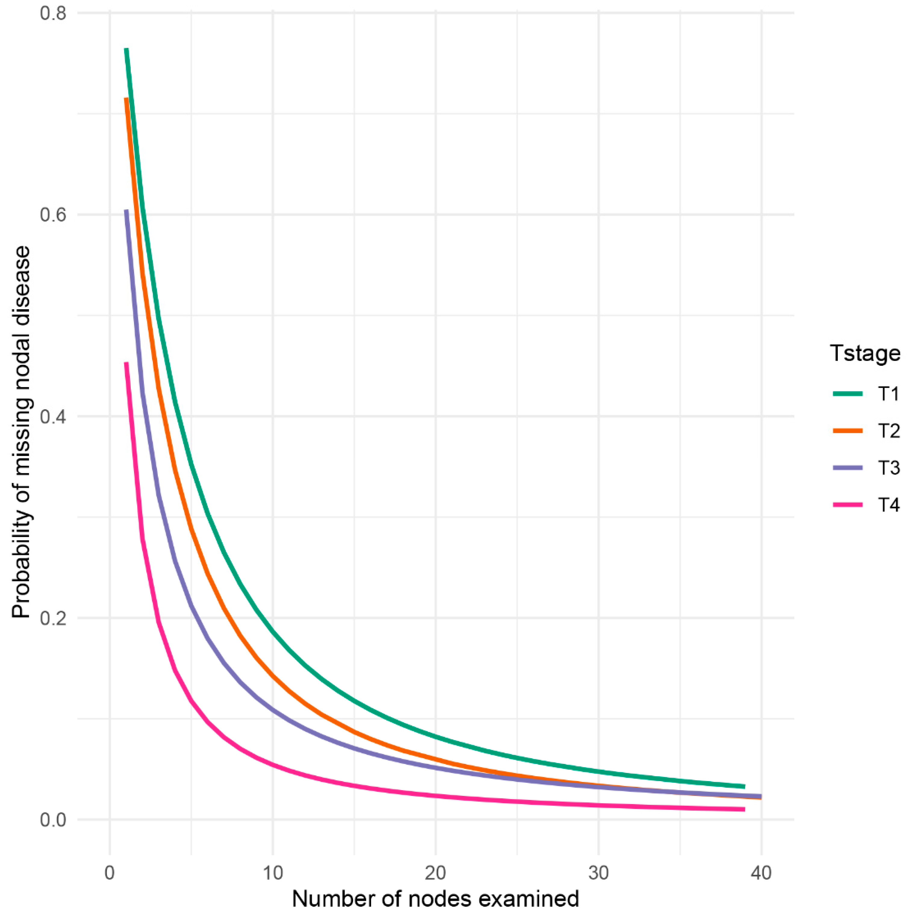
3.2. Compute the Probability of Missing Potential Metastatic Lymph Node
3.3. Compution of the NSS
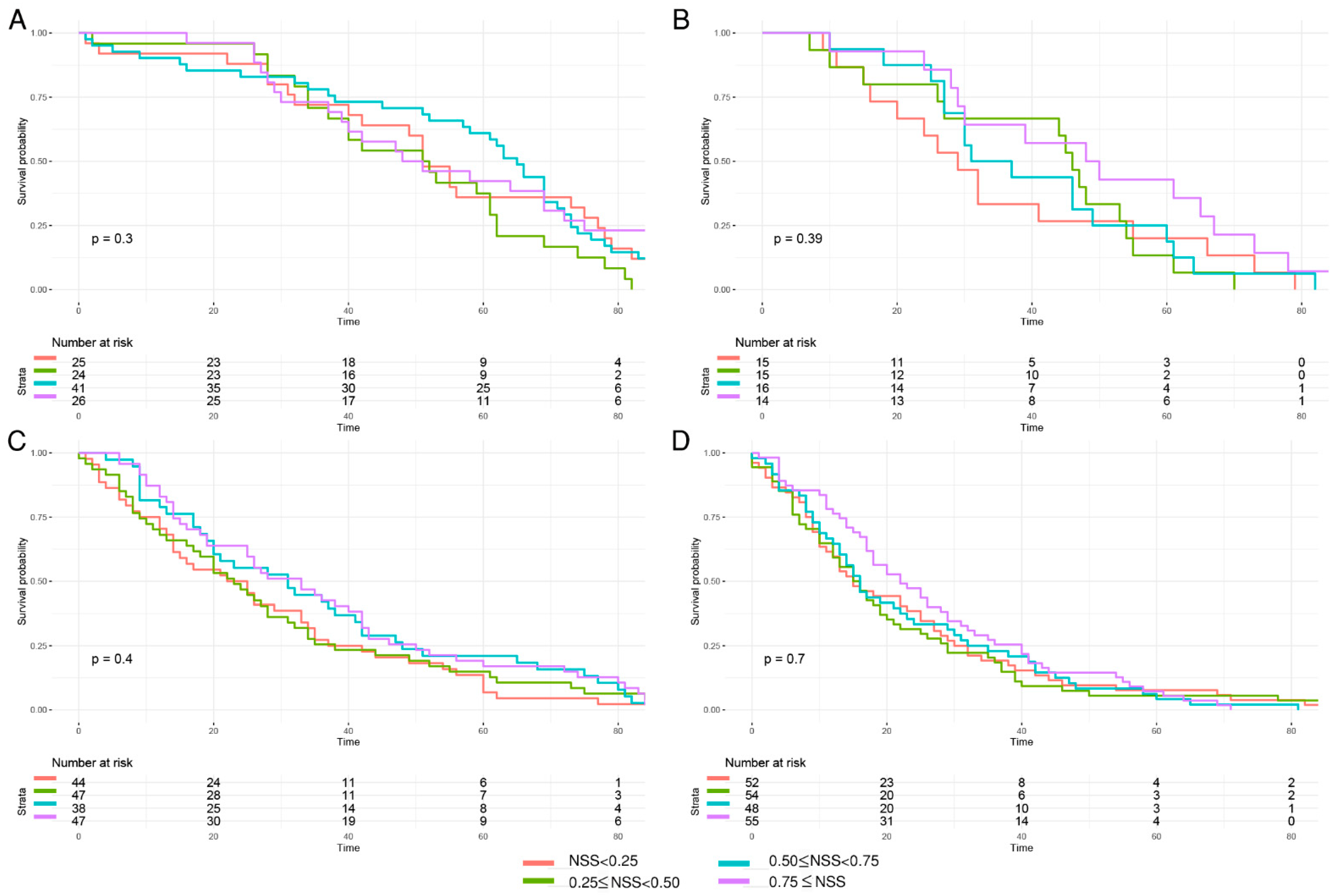
3.4. Prognostic Analysis of NSS-Based Group Classification
4. Discussion
5. Conclusions
Supplementary Materials
Author Contributions
Funding
Institutional Review Board Statement
Informed Consent Statement
Data Availability Statement
Conflicts of Interest
References
- Arai, T. Where does signet-ring cell carcinoma come from and where does it go? Gastric Cancer 2019, 22, 651–652. [Google Scholar] [PubMed]
- Murai, K.; Takizawa, K.; Shimoda, T.; Fujii, S.; Sugino, T.; Yoshida, M.; Kawata, N.; Tanaka, M.; Kakushima, N.; Terashima, M.; et al. Effect of double-layer structure in intramucosal gastric signet-ring cell carcinoma on lymph node metastasis: A retrospective, single-center study. Gastric Cancer 2019, 22, 751–758. [Google Scholar] [CrossRef] [PubMed]
- World Health Organization. World Health Organization Classification of Tumours of The Digestive System, 4th ed.; International Agency for Research on Cancer: Lyon, France, 2010; pp. 45–58.
- Efared, B.; Kadi, M.; Tahiri, L.; Lahmidani, N.; Hassani, K.I.M.; El Bouhaddouti, H.; Benbrahim, Z.; Adil, I.S.; Chbani, L. Gastric Signet Ring Cell Carcinoma: A Comparative Analysis of Clinicopathologic Features. Cancer Control 2020, 27, 1073274820976596. [Google Scholar] [CrossRef] [PubMed]
- Bamboat, Z.M.; Tang, L.H.; Vinuela, E.; Kuk, D.; Gonen, M.; Shah, M.A.; Brennan, M.F.; Coit, D.G.; Strong, V.E. Stage-stratified prognosis of signet ring cell histology in patients undergoing curative resection for gastric adenocarcinoma. Ann. Surg. Oncol. 2014, 21, 1678–1685. [Google Scholar] [CrossRef] [PubMed]
- Henson, D.E.; Dittus, C.; Younes, M.; Nguyen, H.; Albores-Saavedra, J. Differential trends in the intestinal and diffuse types of gastric carcinoma in the United States, 1973–2000: Increase in the signet ring cell type. Arch. Pathol. Lab. Med. 2004, 128, 765–770. [Google Scholar] [CrossRef]
- Xie, Y.; Song, X.; Dong, W.; Jin, H.; Ni, Z.; Li, X.; Huang, H. Anatomic Subsites and Prognosis of Gastric Signet Ring Cell Carcinoma: A SEER Population-Based 1:1 Propensity-Matched Study. Biomed Res. Int. 2022, 2022, 1565207. [Google Scholar] [CrossRef]
- Zhao, S.; Lv, L.; Zheng, K.; Tian, Y.; Zheng, J.C.; Jiang, C.G. Prognosis and Biological Behavior of Gastric Signet-Ring Cell Carcinoma Better or Worse: A Meta-Analysis. Front. Oncol. 2021, 11, 603070. [Google Scholar] [CrossRef]
- Dong, X.; Sun, G.; Qu, H.; He, Q.; Hao, Z. Prognostic Significance of Signet-Ring Cell Components in Patients with Gastric Carcinoma of Different Stages. Front. Surg. 2021, 8, 642468. [Google Scholar] [CrossRef]
- Machlowska, J.; Pucułek, M.; Sitarz, M.; Terlecki, P.; Maciejewski, R.; Sitarz, R. State of the art for gastric signet ring cell carcinoma: From classification, prognosis, and genomic characteristics to specified treatments. Cancer Manag. Res. 2019, 11, 2151–2161. [Google Scholar] [CrossRef]
- Jeong, S.H.; Kim, J.W.; Kim, H.M. Prevalence of Regional Lymph Node Metastasis of Mucosal Gastric Signet Ring Cell Carcinoma: Analysis of the Collaborative Stage Data Survey of the Korean Central Cancer Registry. Dig. Surg. 2021, 38, 330–336. [Google Scholar] [CrossRef]
- Wu, Z.; Qin, G.; Zhao, N.; Jia, H.; Zheng, X. Assessing the adequacy of lymph node yield for different tumor stages of colon cancer by nodal staging scores. BMC Cancer 2017, 17, 498. [Google Scholar] [CrossRef] [PubMed]
- Edge, S.; Byrd, D.; Compton, C.; Fritz, A.; Greene, F.; Trotti, A. The Eighth Edition AJCC Cancer Staging Manual: Continuing to build a bridge from a population-based to a more “personalized” approach to cancer staging. CA Cancer J. Clin. 2017, 67, 93–99. [Google Scholar]
- Mranda, G.M.; Xiang, Z.P.; Zhou, X.G.; Liu, J.J.; Xue, Y.; Wang, Y.; Wei, T.; Ding, Y.L. Revisiting the 8th AJCC system for gastric cancer: A review on validations, nomograms, lymph nodes impact, and proposed modifications. Ann. Med. Surg. 2022, 75, 103411. [Google Scholar] [CrossRef]
- Voron, T.; Messager, M.; Duhamel, A.; Lefevre, J.; Mabrut, J.Y.; Goere, D.; Meunier, B.; Brigand, C.; Hamy, A.; Glehen, O.; et al. Is signet-ring cell carcinoma a specific entity among gastric cancers? Gastric Cancer 2016, 19, 1027–1040. [Google Scholar] [CrossRef] [PubMed]
- Morkavuk, Ş.B.; Çulcu, S.; Tez, M.; Ünal, A.E. The efficiency of D1(+) lymphadenectomy in signet ring cell carcinoma: Comparison of postoperative early and late outcomes between standard lymphadenectomy and D1(+) lymphadenectomy. Libyan J. Med. 2021, 16, 1973761. [Google Scholar] [CrossRef] [PubMed]
- Ye, J.; Ren, Y.; Dai, W.; Chen, J.; Cai, S.; Tan, M.; He, Y.; Yuan, Y. Does Lymphadenectomy with at Least 15 Perigastric Lymph Nodes Retrieval Promise an Improved Survival for Gastric Cancer: A Retrospective Cohort Study in Southern China. J. Cancer 2019, 10, 1444–1452. [Google Scholar] [CrossRef] [PubMed]
- Chen, Y.-H.; Lu, J.; Nie, R.-C.; Liu, D.; Liu, A.-H.; Deng, Z.-J.; Chen, X.-J.; Xiang, J.; Chen, Y.-B.; Huang, C.-M.; et al. Retrieval of 30 Lymph Nodes Is Mandatory for Selected Stage II Gastric Cancer Patients. Front. Oncol. 2021, 11, 593470. [Google Scholar] [CrossRef]
- Cao, L.; Selby, L.V.; Hu, X.; Zhang, Y.; Janjigian, Y.Y.; Tang, L.; Coit, D.G.; Brennan, M.F.; Strong, V.E. Risk factors for recurrence in T1-2N0 gastric cancer in the United States and China. J. Surg. Oncol. 2016, 113, 745–749. [Google Scholar] [CrossRef]
- Jin, L.X.; Moses, L.E.; Squires, M.H.; Poultsides, G.A.; Votanopoulos, K.; Weber, S.M.; Bloomston, M.; Pawlik, T.M.; Hawkins, W.G.; Linehan, D.C.; et al. Factors Associated with Recurrence and Survival in Lymph Node-negative Gastric Adenocarcinoma: A 7-Institution Study of the US Gastric Cancer Collaborative. Ann. Surg. 2015, 262, 999–1005. [Google Scholar] [CrossRef]
- Pokala, S.K.; Zhang, C.; Chen, Z.; Gamboa, A.M.; Cristofaro, S.L.; Keilin, S.A.; Cai, Q.; Willingham, F.F. Incidence, survival, and predictors of lymph node involvement in early-stage gastric signet ring cell carcinoma in the US. J. Gastrointest. Surg. 2018, 22, 569–577. [Google Scholar] [CrossRef]
- Chen, H.M.; Feng, G. Nodal staging score and adequacy of nodal staging. OncoTargets Ther. 2019, 12, 449–455. [Google Scholar] [CrossRef] [PubMed] [Green Version]
- Gönen, M.; Schrag, D.; Weiser, M.R. Nodal staging score: A tool to assess adequate staging of node-negative colon cancer. J. Clin. Oncol. 2009, 27, 6166–6171. [Google Scholar] [CrossRef] [PubMed]
- Robinson, T.J.; Thomas, S.; Dinan, M.A.; Roman, S.; Sosa, J.A.; Hyslop, T. How Many Lymph Nodes Are Enough? Assessing the Adequacy of Lymph Node Yield for Papillary Thyroid Cancer. J. Clin. Oncol. 2016, 34, 3434–3439. [Google Scholar] [CrossRef]
- Ries, L.G.; Young, J.L.; Keel, G.E.; Eisner, M.P.; Lin, Y.D.; Horner, M.J. SEER survival monograph: Cancer survival among adults: US SEER program, 1988–2001, patient and tumor characteristics. Natl. Cancer Inst. SEER Program NIH Pub 2007, 7, 193–202. [Google Scholar]
- Ji, X.; Bu, Z.D.; Yan, Y.; Li, Z.Y.; Wu, A.W.; Zhang, L.H.; Zhang, J.; Wu, X.J.; Zong, X.L.; Li, S.X.; et al. The 8th edition of the American Joint Committee on Cancer tumor-node-metastasis staging system for gastric cancer is superior to the 7th edition: Results from a Chinese mono-institutional study of 1663 patients. Gastric. Cancer 2018, 21, 643–652. [Google Scholar] [CrossRef]
- Khanjani, N.; Mirzaei, S.; Nasrolahi, H.; Hamedi, S.H.; Mosalaei, A.; Omidvari, S.; Ahmadloo, N.; Ansari, M.; Sobhani, F.; Mohammadianpanah, M. Insufficient lymph node assessment in gastric adenocarcinoma. J. Egypt. Natl. Cancer Inst. 2019, 31, 2. [Google Scholar] [CrossRef]
- Woo, Y.; Goldner, B.; Ituarte, P.; Lee, B.; Melstrom, L.; Son, T.; Noh, S.H.; Fong, Y.; Hyung, W.J. Lymphadenectomy with Optimum of 29 Lymph Nodes Retrieved Associated with Improved Survival in Advanced Gastric Cancer: A 25,000-Patient International Database Study. J. Am. Coll. Surg. 2017, 224, 546–555. [Google Scholar] [CrossRef]
- Kim, Y.I. Is retrieval of at least 15 lymph nodes sufficient recommendation in early gastric cancer? Ann. Surg. Treat. Res. 2014, 87, 180–184. [Google Scholar] [CrossRef]
- Park, K.; Jang, G.; Baek, S.; Song, H. Usefulness of combined PET/CT to assess regional lymph node involvement in gastric cancer. Tumori 2014, 100, 201–206. [Google Scholar] [CrossRef]
- Wei, F.; Lyu, H.; Wang, S.; Chu, Y.; Chen, F. Positive lymph node ratio as a novel indicator of prognosis in gastric signet ring cell carcinoma: A population-based retrospective study. Transl. Cancer Res. 2020, 9, 3658–3668. [Google Scholar] [CrossRef]
- Berlth, F.; Chon, S.H.; Chevallay, M.; Jung, M.K.; Mönig, S.P. Preoperative staging of nodal status in gastric cancer. Transl. Gastroenterol. Hepatol. 2017, 2, 8. [Google Scholar] [CrossRef] [PubMed]
- Ahn, H.S.; Lee, H.J.; Yoo, M.W.; Kim, S.G.; Im, J.P.; Kim, S.H.; Kim, W.H.; Lee, K.U.; Yang, H.K. Diagnostic accuracy of T and N stages with endoscopy, stomach protocol CT, and endoscopic ultrasonography in early gastric cancer. J. Surg. Oncol. 2009, 99, 20–27. [Google Scholar] [CrossRef] [PubMed]
- Puli, S.R.; Batapati Krishna Reddy, J.; Bechtold, M.L.; Antillon, M.R.; Ibdah, J.A. How good is endoscopic ultrasound for TNM staging of gastric cancers? A meta-analysis and systematic review. World J. Gastroenterol. 2008, 14, 4011–4019. [Google Scholar] [CrossRef] [PubMed]
- Hasbahceci, M.; Akcakaya, A.; Memmi, N.; Turkmen, I.; Cipe, G.; Yildiz, P.; Arici, D.S.; Muslumanoglu, M. Diffusion MRI on lymph node staging of gastric adenocarcinoma. Quant. Imaging Med. Surg. 2015, 5, 392–400. [Google Scholar] [PubMed]
- Degiuli, M.; De Manzoni, G.; Di Leo, A.; D’Ugo, D.; Galasso, E.; Marrelli, D.; Petrioli, R.; Polom, K.; Roviello, F.; Santullo, F.; et al. Gastric cancer: Current status of lymph node dissection. World J. Gastroenterol. 2016, 22, 2875–2893. [Google Scholar] [CrossRef] [PubMed]


| LN Negative | LN Positive | p Value | ||
|---|---|---|---|---|
| n | 193 | 368 | ||
| T stage (%) | ||||
| T1 | 98 (50.8) | 18 (4.9) | <0.001 | |
| T2 | 25 (13.0) | 35 (9.5) | ||
| T3 | 48 (24.9) | 128 (34.8) | ||
| T4 | 22 (11.4) | 187 (50.8) | ||
| N stage (%) | ||||
| N0 | 193 (100.0) | 0 (0.0) | <0.001 | |
| N1 | 0 (0.0) | 95 (25.8) | ||
| N2 | 0 (0.0) | 89 (24.2) | ||
| N3 | 0 (0.0) | 184 (50.0) | ||
| M stage (%) | ||||
| M0 | 182 (94.3) | 308 (83.7) | 0.001 | |
| M1 | 11 (5.7) | 60 (16.3) | ||
| Sex (%) | ||||
| Male | 108 (56.0) | 179 (48.6) | 0.119 | |
| Female | 85 (44.0) | 189 (51.4) | ||
| Age (years) (%) | ||||
| <30 | 8 (4.1) | 7 (1.9) | 0.137 | |
| ≥70 | 70 (36.3) | 112 (30.4) | ||
| 30≤ <50 | 31 (16.1) | 58 (15.8) | ||
| 50≤ <70 | 84 (43.5) | 191 (51.9) | ||
| Tumor size (%) | ||||
| <3 cm | 89 (46.1) | 42 (11.4) | <0.001 | |
| 3 cm≤ <6 cm | 16 (8.3) | 152 (41.3) | ||
| ≥6 cm | 39 (20.2) | 125 (34.0) | ||
| unknown | 49 (25.4) | 49 (13.3) | ||
| Bone metastasis (%) | ||||
| No | 193 (100.0) | 364 (98.9) | 0.348 | |
| Yes | 0 (0.0) | 3 (0.8) | ||
| Unknown | 0 (0.0) | 1 (0.3) | ||
| Brain metastasis (%) | ||||
| No | 193 (100.0) | 366 (99.5) | 0.779 | |
| Yes | 0 (0.0) | 0 (0.0) | ||
| Unknown | 0 (0.0) | 2 (0.5) | ||
| Liver metastasis (%) | ||||
| No | 192 (99.5) | 367 (99.7) | 0.296 | |
| Yes | 1 (0.5) | 0 (0.0) | ||
| Unknown | 0 (0.0) | 1 (0.3) | ||
| Lung metastasis (%) | ||||
| No | 193 (100.0) | 366 (99.5) | 0.591 | |
| Yes | 0 (0.0) | 1 (0.3) | ||
| Unknown | 0 (0.0) | 1 (0.3) | ||
| Tumor position (%) | 0.007 | |||
| C16.1-Fundus of stomach | 7 (3.6) | 13 (3.5) | ||
| C16.2-Body of stomach | 24 (12.4) | 28 (7.6) | ||
| C16.3-Gastric antrum | 53 (27.5) | 120 (32.6) | ||
| C16.4-Pylorus | 7 (3.6) | 11 (3.0) | ||
| C16.5-Lesser curvature of stomach NOS | 17 (8.8) | 41 (11.1) | ||
| C16.6-Greater curvature of stomach NOS | 15 (7.8) | 15 (4.1) | ||
| C16.8-Overlapping lesion of stomach | 14 (7.3) | 60 (16.3) | ||
| C16.9-Stomach, NOS | 26 (13.5) | 28 (7.6) | ||
| C16.0-Cardia, NOS | 30 (15.5) | 52 (14.1) |
| T Stage | αT stage (95% CI) | βT stage (95% CI) |
|---|---|---|
| T1 | 0.06938207 (0.03686795–0.1235056) | 1.8877199 (1.2074176–4.266865) |
| T2 | 0.42578161 (0.26941777–0.7476104) | 2.2180490 (1.4385130–4.397587) |
| T3 | 0.48686973 (0.38153144–0.6422990) | 1.2430379 (0.9737503–1.666755) |
| T4 | 0.80543434 (0.64691993–1.0226410) | 0.8710085 (0.7142595–1.084519) |
| T Stage AJCC | Apparent Prevalence (%) | Adjusted Prevalence (%) |
|---|---|---|
| T1 | 0.173 | 0.155 |
| T2 | 0.690 | 0.583 |
| T3 | 0.807 | 0.727 |
| T4 | 0.941 | 0.895 |
Publisher’s Note: MDPI stays neutral with regard to jurisdictional claims in published maps and institutional affiliations. |
© 2022 by the authors. Licensee MDPI, Basel, Switzerland. This article is an open access article distributed under the terms and conditions of the Creative Commons Attribution (CC BY) license (https://creativecommons.org/licenses/by/4.0/).
Share and Cite
Yu, C.; Zhou, Z.; Liu, B.; Yao, D.; Huang, Y.; Wang, P.; Li, Y. Pathological Nodal Staging Score for Gastric Signet Ring Cell Carcinoma: A Clinical Tool of Adequate Nodal Staging. Diagnostics 2022, 12, 2289. https://doi.org/10.3390/diagnostics12102289
Yu C, Zhou Z, Liu B, Yao D, Huang Y, Wang P, Li Y. Pathological Nodal Staging Score for Gastric Signet Ring Cell Carcinoma: A Clinical Tool of Adequate Nodal Staging. Diagnostics. 2022; 12(10):2289. https://doi.org/10.3390/diagnostics12102289
Chicago/Turabian StyleYu, Chaoran, Zhiyuan Zhou, Bin Liu, Danhua Yao, Yuhua Huang, Pengfei Wang, and Yousheng Li. 2022. "Pathological Nodal Staging Score for Gastric Signet Ring Cell Carcinoma: A Clinical Tool of Adequate Nodal Staging" Diagnostics 12, no. 10: 2289. https://doi.org/10.3390/diagnostics12102289