
A Bipartite Obturator Artery with Multiple Pelvic Branching—A Gynecologic Approach

 ,
, 
Abstract
:1. Introduction
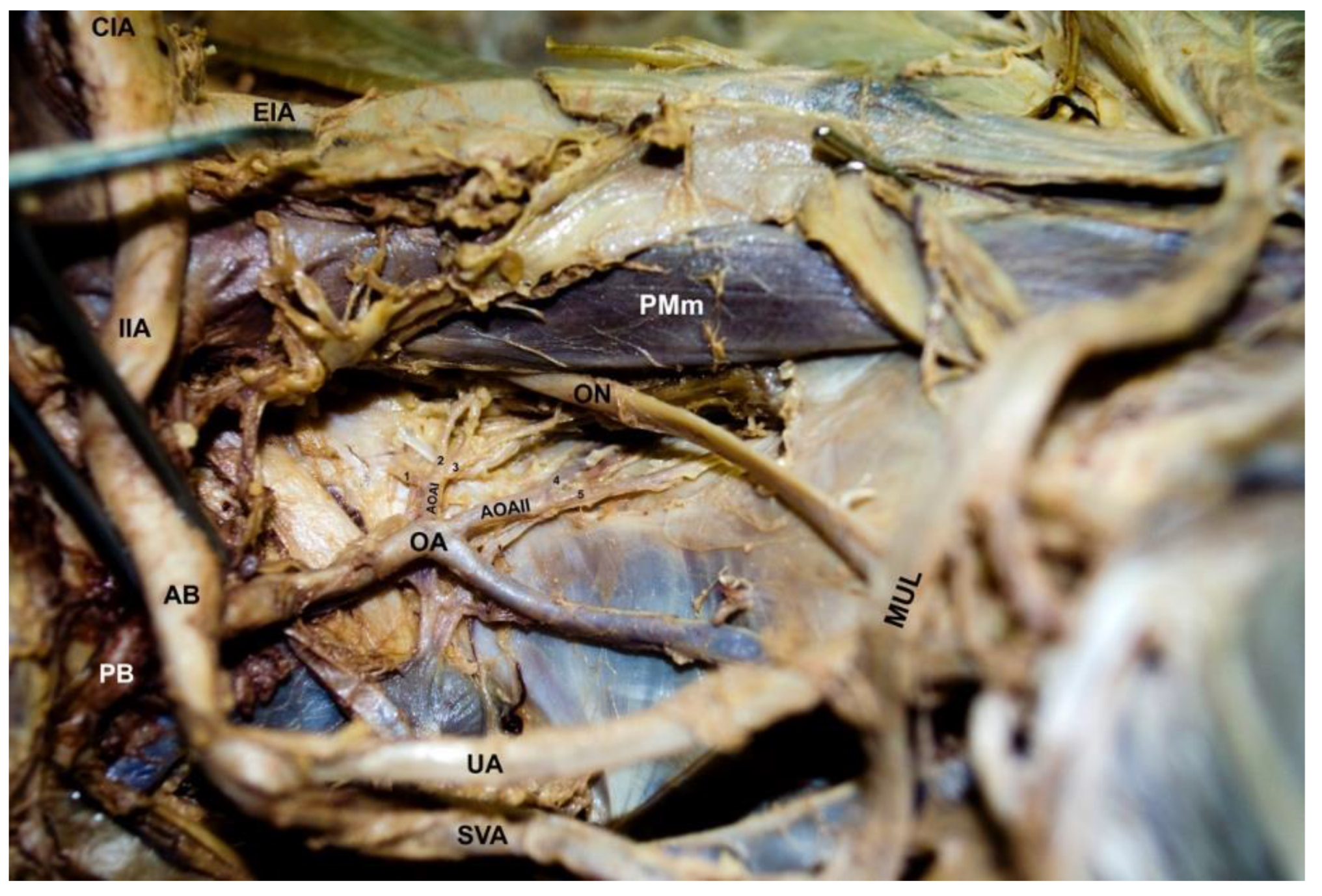
2. Case Report
3. Discussion
3.1. Embryological Basis
3.2. Clinical Significance
4. Conclusions
Author Contributions
Funding
Institutional Review Board Statement
Informed Consent Statement
Data Availability Statement
Acknowledgments
Conflicts of Interest
References
- Ates, M.; Kinaci, E.; Kose, E.; Soyer, V.; Sarici, B.; Cuglan, S.; Korkmaz, F.; Dirican, A. Corona mortis: In vivo anatomical knowledge and the risk of injury in totally extraperitoneal inguinal hernia repair. Hernia 2016, 20, 659–665. [Google Scholar] [CrossRef]
- Heichinger, R.; Pretterklieber, M.L.; Hammer, N.; Pretterklieber, B. The Corona mortis is similar in size to the regular obturator artery, but is highly variable at the level of origin: An anatomical study. Anat. Sci. Int. 2022, 1–11. [Google Scholar] [CrossRef] [PubMed]
- Perandini, S.; Perandini, A.; Puntel, G.; Puppini, G.; Montemezzi, S. Corona mortis variant of the obturator artery: A systematic study of 300 hemipelvises by means of computed tomography angiography. Pol. J. Radiol. 2018, 83, e519–e523. [Google Scholar] [CrossRef]
- Maraqa, T.I.; Shin, J.J.; Diallo, I.; Sachwani-Daswani, G.R.; Mercer, L.C. Penetrating Obturator Artery Injury after Gunshot Wounds: A Successful Multidisciplinary Trauma Team Approach to a Potentially Lethal Injury. Cureus 2017, 11, e1857. [Google Scholar] [CrossRef] [Green Version]
- Agur, A.M.; Dalley, A.F. Grants Atlas of Anatomy; Wolters Kluwer Health/Lippincott Williams & Wilkins: Philadelphia, PA, USA, 2013. [Google Scholar]
- Jusoh, A.R.; Abd Rahman, N.; Abd Latiff, A.; Othman, F.; Das, S.; Abd Ghafar, N.; Haji Suhaimi, F.; Hussan, F.; Maatoq Sulaiman, I. The anomalous origin and branches of the obturator artery with its clinical implications. Rom. J. Morphol. Embryol. 2010, 51, 163–166. [Google Scholar]
- Trikha, A.; Singh, P.M. Management of major obstetric haemorrhage. Indian J. Anaesth. 2018, 62, 698–703. [Google Scholar] [CrossRef]
- Kayem, G.; Kurinczuk, J.J.; Alfirevic, Z.; Spark, P.; Brocklehurst, P.; Knight, M. Specific second-line therapies for postpartum haemorrhage: A national cohort study. BJOG—Int. J. Obstet. Gynaecol. 2011, 118, 856–864. [Google Scholar] [CrossRef]
- McGlone, B.; Hamilton, S.; FitzGerald, M. Pelvic digit: An uncommon developmental anomaly. Eur. Radiol. 2000, 10, 89–91. [Google Scholar] [CrossRef]
- Qazi, E.; Wilting, J.; Patel, N.R.; Alenezi, A.O.; Kennedy, S.A.; Tan, K.T.; Jaberi, A.; Mafeld, S. Arteries of the Lower Limb-Embryology, Variations, and Clinical Significance. Can. Assoc. Radiol. J. 2021, 73, 259–270. [Google Scholar] [CrossRef] [PubMed]
- Hamabe, A.; Harino, T.; Ogino, T.; Tanida, T.; Noura, S.; Morita, S.; Dono, K. Analysis of anatomical variations of intrapelvic vessels for advanced pelvic surgery. BMC Surg. 2020, 20, 47. [Google Scholar] [CrossRef] [Green Version]
- Kostov, S.; Slavchev, S.; Dzhenkov, D.; Stoyanov, G.; Dimitrov, N.; Yordanov, A.D. Corona mortis, aberrant obturator vessels, accessory obturator vessels: Clinical applications in gynecology. Folia Morphol. 2020, 80, 776–785. [Google Scholar] [CrossRef]
- Suryavanshi, S.; Bhagel, P.; Sharma, D. Variability in the Origin of the Obturator Artery: A Descriptive Cross-Sectional Cadaveric Study. IJSS Surg. 2016, 2, 16–19. [Google Scholar]
- Pai, M.M.; Krishnamurthy, A.; Prabhu, L.V.; Pai, M.V.; Kumar, S.A.; Hadimani, G.A. Variability in the origin of the obturator artery. Clinics 2009, 64, 897–901. [Google Scholar] [CrossRef] [PubMed] [Green Version]
- Granite, G.; Meshida, K.; Wind, G. Frequency and clinical review of the aberrant obturator artery: A Cadaveric study. Diagnostics 2020, 10, 546. [Google Scholar] [CrossRef] [PubMed]
- Pillay, M. A study of variations in the origin of obturator artery and its clinical significance. JCDR—J. Clin. Diagn. Res. 2015, 9, AC12–AC15. [Google Scholar] [CrossRef]
- Sakthivel Priyadarshini, S. Variability of origin of obturator artery and its clinical significance. IJAR Int. J. Anat. Res. 2015, 3, 1704–1709. [Google Scholar] [CrossRef]
- Bosse, B.L.; Palacios, V.J.; Dutcher, D.W.; Etter, E.J.; Lim, P.C.; Cobine, C.A.; Moritz, G.L. Unconventional Obturator Artery Nutrient Branch: Image of an Anatomical Variation. Diagnostics 2019, 12, 2019. [Google Scholar] [CrossRef] [PubMed]
- Deshmukh, V.; Singh, S.; Sirohi, N.; Baruhee, D. Variation in the obturator vasculature during routine anatomy dissection of a cadaver. Sultan Qaboos Univ. Med. J. 2016, 16, e356–e358. [Google Scholar] [CrossRef]
- Pick, J.W.; Anson, B.J.; Ashley, F.L. The origin of the obturator artery. A study of 640 body-halves. Am. J. Anat. 1942, 70, 317–343. [Google Scholar] [CrossRef]
- Gyimadu, A.; Salman, M.C.; Karcaaltincaba, M.; Yuce, K. Retroperitoneal Vascular Aberrations Increase the risk of vascular injury during lymphadenectomy in gynecologic cancers. Arch. Gynecol. Obstet. 2012, 286, 449–455. [Google Scholar] [CrossRef] [PubMed]
- Lau, H.; Lee, F. A prospective endoscopic study of retropubic vascular anatomy in 121 patients undergoing endoscopic extraperitoneal inguinal hernioplasty. Surg. Endosc. 2003, 17, 1376–1379. [Google Scholar] [CrossRef] [PubMed]
- Skandalakis, L.J.; Skandalakis, P.N.; Colborn, G.L.; Skandalakis, J.E. Obturator hernia: Embryology, anatomy, surgery. Hernia 2000, 4, 121–128. [Google Scholar] [CrossRef]
- Sañudo, J.R.; Mirapeix, R.; Rodriguez-Niedenführ, M.; Maranillo, E.; Parkin, I.G.; Vázquez, T. Obturator artery revisited. Int. Urogynecol. J. 2011, 22, 1313. [Google Scholar] [CrossRef] [PubMed]
- Pisano, U.; Soon, V.L.; Douglas, P. Corona mortis injury causing delayed presentation of pelvic pseudoaneurysm. Radiol. Case Rep. 2021, 16, 1095–1098. [Google Scholar] [CrossRef] [PubMed]
- Noussios, G.; Galanis, N.; Chatzis, I.; Konstantinidis, S.; Filo, E.; Karavasilis, G.; Katsourakis, A. The anatomical characteristics of Corona Mortis: A systematic review of the literature and its clinical importance in hernia repair. J. Clin. Med. Res. 2020, 12, 108–114. [Google Scholar] [CrossRef] [PubMed] [Green Version]
- Darmanis, S.; Lewis, A.; Mansoor, A.; Bircher, M. Corona Mortis: An anatomical study with clinical implications in approaches to the pelvis and acetabulum. Clin. Anat. 2007, 20, 433–439. [Google Scholar] [CrossRef]
- Kostov, S.; Slavchev, S.; Dzhenkov, D.; Mitev, D.; Yordanov, A. Avascular Spaces of the Female Pelvis-Clinical Applications in Obstetrics and Gynecology. J. Clin. Med. 2020, 9, 1460. [Google Scholar] [CrossRef]
- Silva, K.C.D.P.; Samarakkody, S.N. Drive safely through the pelvis—Know your pelvic roads: The Vesico-Uterine Space. Sri Lanka J. Obstet. Gynaecol. 2019, 41, 89–93. [Google Scholar] [CrossRef] [Green Version]
- Lin, L.; Huang, M.-C.; Su, T.-H.; Lau, H.-H. Comparison between tension-free vaginal tape and transobturator tape in treating stress urinary incontinence after vaginal mesh surgery. Taiwan J. Obstet. Gynecol. 2018, 57, 528–531. [Google Scholar] [CrossRef] [PubMed]
- Sharma, J.B.; Thariani, K.; Deoghare, M.; Kumari, R. Autologous Fascial Slings for Surgical Management of Stress Urinary Incontinence: A Come Back. J. Obstet. Gynaecol. India 2021, 71, 106–114. [Google Scholar] [CrossRef]
- Veit-Rubin, N.; Dubuisson, J.; Ford, A.; Dubuisson, J.-B.; Mourad, S.; Digesu, A. Burch colposuspension. Neurourol. Urodyn. 2019, 38, 553–562. [Google Scholar] [CrossRef] [PubMed]
- Lo, T.-S.; Horng, S.-G.; Chang, C.-L.; Huang, H.-J.; Tseng, L.-H.; Liang, C.-C. Tension-free vaginal tape procedure after previous failure in incontinence surgery. Urology 2002, 60, 57–61. [Google Scholar] [CrossRef]
- Lo, T.S.; Wang, A.C.; Horng, S.G.; Liang, C.C.; Soong, Y.K. Ultrasonographic and urodynamic evaluation after tension free vagina tape procedure (TVT). Acta Obstet. Gynecol. Scand. 2001, 80, 65–70. [Google Scholar] [CrossRef]
- Ulmsten, U.; Henriksson, L.; Johnson, P.; Varhos, G. An ambulatory surgical procedure under local anesthesia for treatment of female urinary incontinence. Int. Urogynecol. J. Pelvic Floor Dysfunct. 1996, 7, 81–86. [Google Scholar] [CrossRef] [PubMed]
- Molden, S.M.; Lucente, V.R. New minimally invasive slings: TVT Secur. Curr. Urol. Rep. 2008, 9, 358–361. [Google Scholar] [CrossRef]
- Shrestha, R.; Shrestha, S.; Sitaula, S.; Basnet, P. Anatomy of Internal Iliac Artery and Its Ligation to Control Pelvic Hemorrhage. JNMA J. Nepal. Med. Assoc. 2020, 58, 826–830. [Google Scholar] [CrossRef]
- Selçuk, İ.; Uzuner, B.; Boduç, E.; Baykuş, Y.; Akar, B.; Güngör, T. Step-by-step ligation of the internal iliac artery. J. Turk. Ger. Gynecol. Assoc. 2019, 20, 123–128. [Google Scholar] [CrossRef]
- Zhang, X.Q.; Chen, X.T.; Zhang, Y.T.; Mai, C.X. The Emergent Pelvic Artery Embolization in the Management of Postpartum Hemorrhage: A Systematic Review and Meta-analysis. Obstet. Gynecol. Surv. 2021, 76, 234–244. [Google Scholar] [CrossRef] [PubMed]

Publisher’s Note: MDPI stays neutral with regard to jurisdictional claims in published maps and institutional affiliations. |
© 2022 by the authors. Licensee MDPI, Basel, Switzerland. This article is an open access article distributed under the terms and conditions of the Creative Commons Attribution (CC BY) license (https://creativecommons.org/licenses/by/4.0/).
Share and Cite
Quiñones-Rodríguez, J.I.; Acevedo-Arroyo, A.N.; Santiago-Negrón, C.L.; Garcés-Torres, L.F.; Fonseca-Salgado, C. A Bipartite Obturator Artery with Multiple Pelvic Branching—A Gynecologic Approach. Diagnostics 2022, 12, 2614. https://doi.org/10.3390/diagnostics12112614
Quiñones-Rodríguez JI, Acevedo-Arroyo AN, Santiago-Negrón CL, Garcés-Torres LF, Fonseca-Salgado C. A Bipartite Obturator Artery with Multiple Pelvic Branching—A Gynecologic Approach. Diagnostics. 2022; 12(11):2614. https://doi.org/10.3390/diagnostics12112614
Chicago/Turabian StyleQuiñones-Rodríguez, Jailenne I., Alexandra N. Acevedo-Arroyo, Camille L. Santiago-Negrón, Lucia F. Garcés-Torres, and Carlos Fonseca-Salgado. 2022. "A Bipartite Obturator Artery with Multiple Pelvic Branching—A Gynecologic Approach" Diagnostics 12, no. 11: 2614. https://doi.org/10.3390/diagnostics12112614
APA StyleQuiñones-Rodríguez, J. I., Acevedo-Arroyo, A. N., Santiago-Negrón, C. L., Garcés-Torres, L. F., & Fonseca-Salgado, C. (2022). A Bipartite Obturator Artery with Multiple Pelvic Branching—A Gynecologic Approach. Diagnostics, 12(11), 2614. https://doi.org/10.3390/diagnostics12112614