Somatostatin Receptor Targeted PET-Imaging for Diagnosis, Radiotherapy Planning and Theranostics of Meningiomas: A Systematic Review of the Literature
Abstract
:1. Introduction
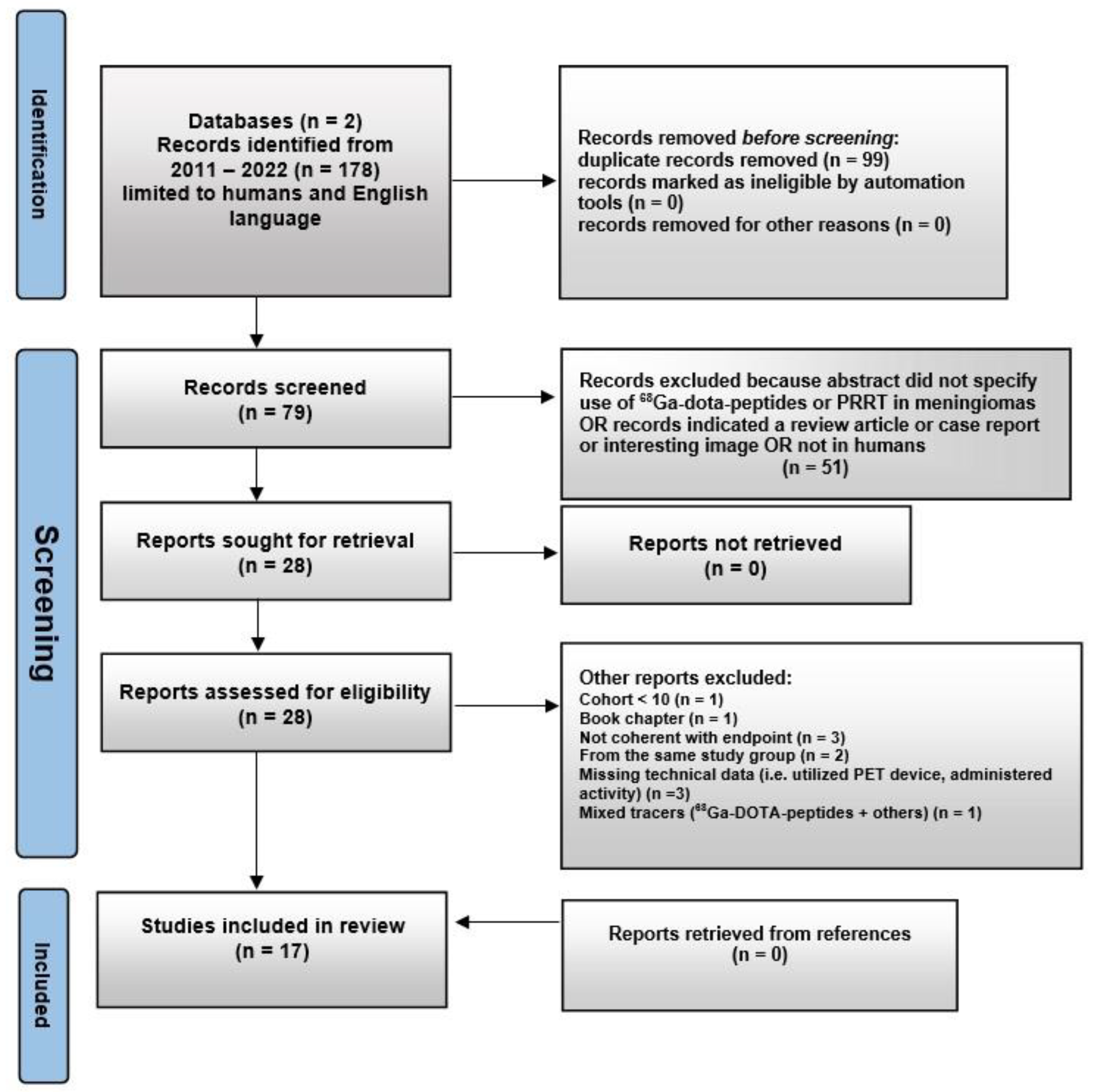
2. Materials and Methods
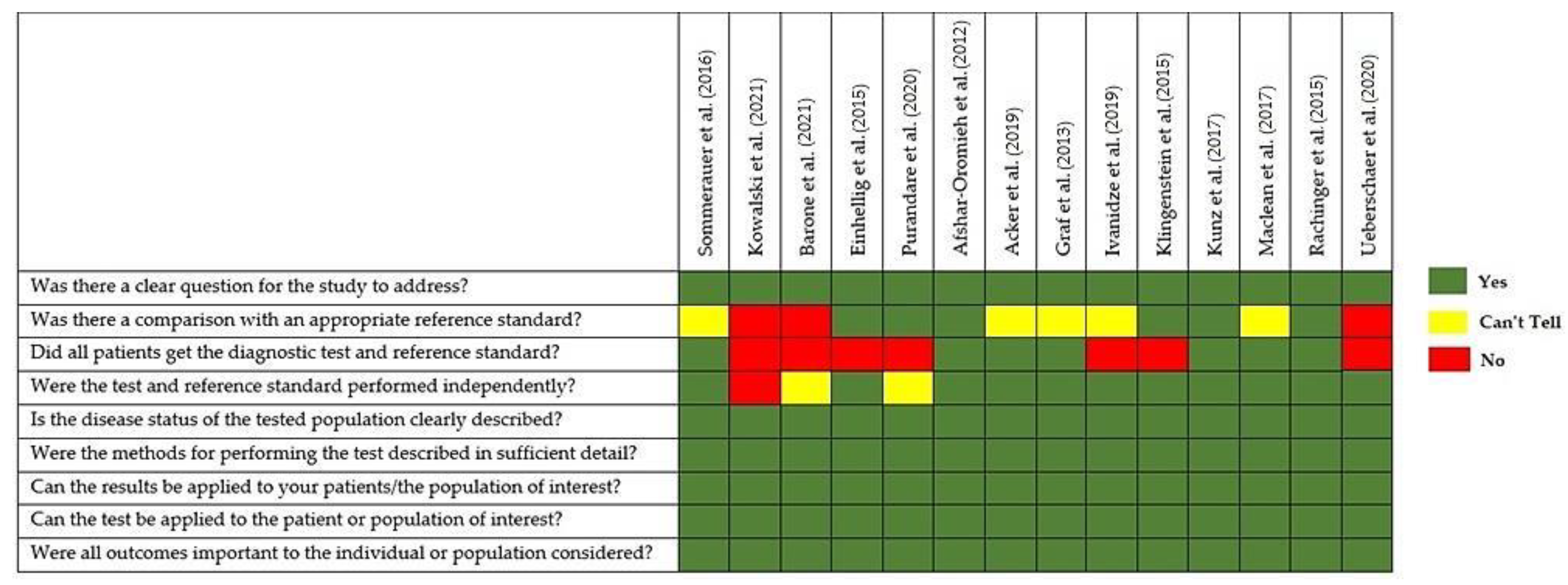
Quality of the Selected Papers
3. Results
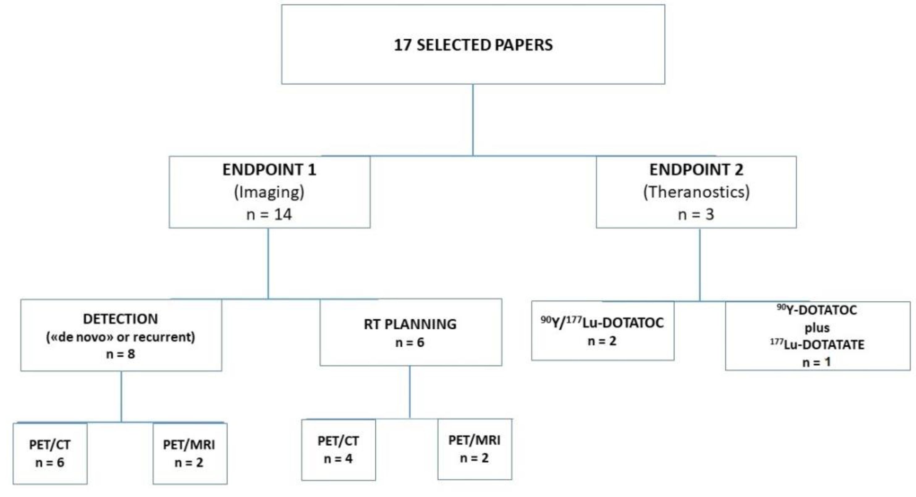
3.1. Analysis of the Evidence
3.2. Imaging
3.2.1. De Novo Diagnosis and Detection of Recurrence
3.2.2. Radiation Therapy Planning and Post-Treatment Assessment
3.3. Peptide Receptor Radionuclide Therapy
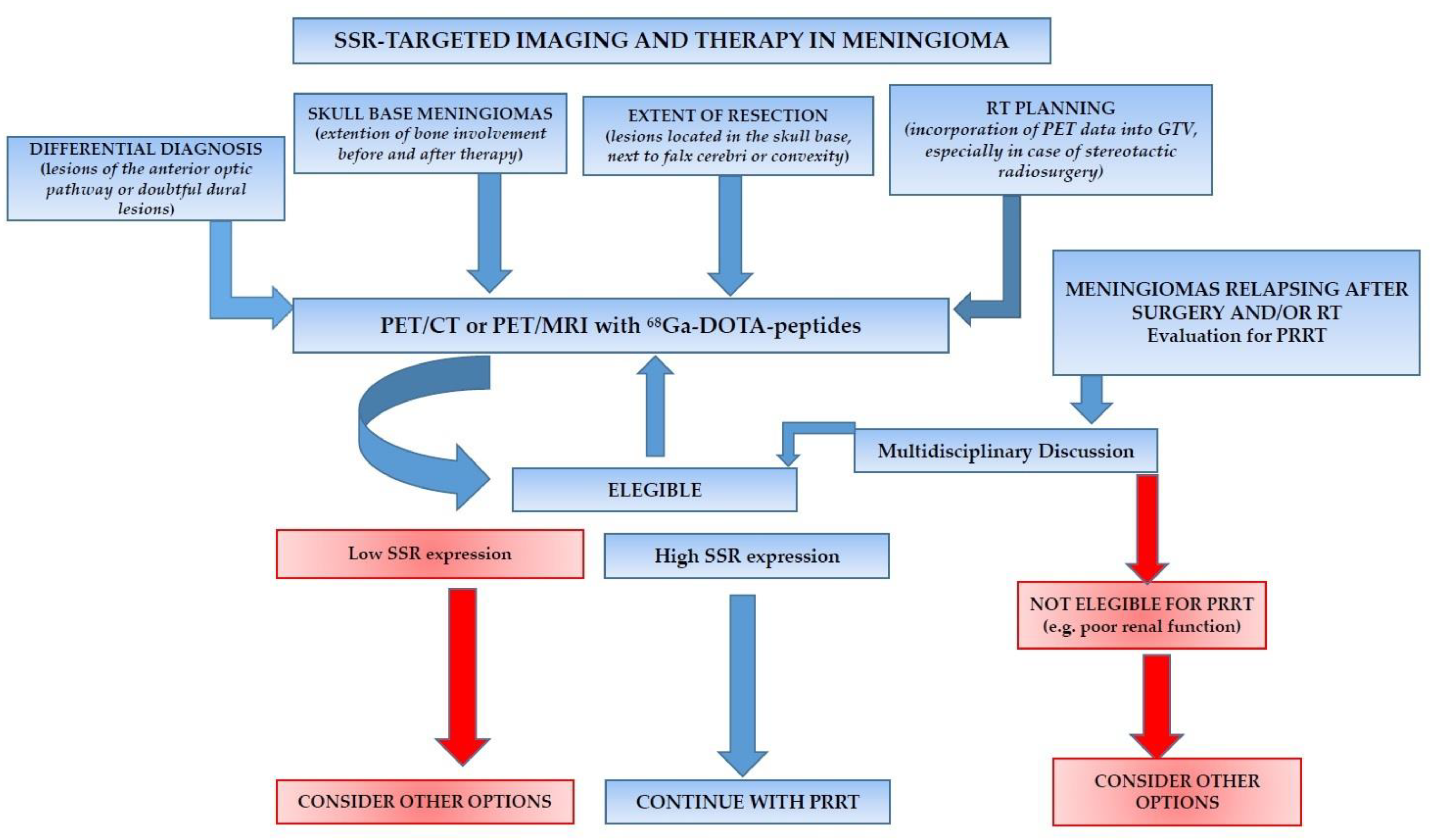
4. Discussion
5. Conclusions
Author Contributions
Funding
Conflicts of Interest
References
- Low, J.T.; Ostrom, Q.T.; Cioffi, G.; Neff, C.; Waite, K.A.; Kruchko, C.; Barnholtz-Sloan, J.S. Primary Brain and Other Central Nervous System Tumors in the United States (2014-2018): A Summary of the CBTRUS Statistical Report for Clinicians. Neurooncol. Pract. 2022, 9, 165–182. [Google Scholar] [CrossRef] [PubMed]
- Louis, D.N.; Perry, A.; Reifenberger, G.; von Deimling, A.; Figarella-Branger, D.; Cavenee, W.K.; Ohgaki, H.; Wiestler, O.D.; Kleihues, P.; Ellison, D.W. The 2016 World Health Organization Classification of Tumors of the Central Nervous System: A Summary. Acta Neuropathol. 2016, 131, 803–820. [Google Scholar] [CrossRef] [PubMed] [Green Version]
- Roehrkasse, A.M.; Peterson, J.E.G.; Fung, K.-M.; Pelargos, P.E.; Dunn, I.F. The Discrepancy Between Standard Histologic WHO Grading of Meningioma and Molecular Profile: A Single Institution Series. Front. Oncol. 2022, 12, 846232. [Google Scholar] [CrossRef] [PubMed]
- Huang, R.Y.; Bi, W.L.; Griffith, B.; Kaufmann, T.J.; la Fougère, C.; Schmidt, N.O.; Tonn, J.C.; Vogelbaum, M.A.; Wen, P.Y.; Aldape, K.; et al. Imaging and Diagnostic Advances for Intracranial Meningiomas. Neuro-Oncology 2019, 21, i44–i61. [Google Scholar] [CrossRef] [PubMed]
- Apra, C.; Peyre, M.; Kalamarides, M. Current Treatment Options for Meningioma. Expert Rev. Neurother. 2018, 18, 241–249. [Google Scholar] [CrossRef]
- Hemmati, S.M.; Ghadjar, P.; Grün, A.; Badakhshi, H.; Zschaeck, S.; Senger, C.; Acker, G.; Misch, M.; Budach, V.; Kaul, D. Adjuvant Radiotherapy Improves Progression-Free Survival in Intracranial Atypical Meningioma. Radiat. Oncol. 2019, 14, 160. [Google Scholar] [CrossRef] [Green Version]
- Theodoropoulou, M.; Stalla, G.K. Somatostatin Receptors: From Signaling to Clinical Practice. Front. Neuroendocrinol. 2013, 34, 228–252. [Google Scholar] [CrossRef]
- Angeletti, S.; Corleto, V.D.; Schillaci, O.; Marignani, M.; Annibale, B.; Moretti, A.; Silecchia, G.; Scopinaro, F.; Basso, N.; Bordi, C.; et al. Use of the Somatostatin Analogue Octreotide to Localise and Manage Somatostatin-Producing Tumours. Gut 1998, 42, 792–794. [Google Scholar] [CrossRef] [Green Version]
- Filippi, L.; Valentini, F.B.; Gossetti, B.; Gossetti, F.; De Vincentis, G.; Scopinaro, F.; Massa, R. Intraoperative Gamma Probe Detection of Head and Neck Paragangliomas with 111In-Pentetreotide: A Pilot Study. Tumori J. 2005, 91, 173–176. [Google Scholar] [CrossRef]
- Nicolato, A. 111Indium-Octreotide Brain Scintigraphy: A Prognostic Factor in Skull Base Meningiomas Treated with Gamma Knife Radiosurgery. Q. J. Nucl. Med. Mol. Imaging 2004, 48, 26–32. [Google Scholar]
- Israel, O.; Pellet, O.; Biassoni, L.; De Palma, D.; Estrada-Lobato, E.; Gnanasegaran, G.; Kuwert, T.; la Fougère, C.; Mariani, G.; Massalha, S.; et al. Two Decades of SPECT/CT—The Coming of Age of a Technology: An Updated Review of Literature Evidence. Eur. J. Nucl. Med. Mol. Imaging 2019, 46, 1990–2012. [Google Scholar] [CrossRef] [PubMed] [Green Version]
- Dore, F.; Filippi, L. Reply: Bone Scintigraphy and SPECT/CT in Bisphosphonate-Induced Osteonecrosis of the Jaw. J. Nucl. Med. 2009, 50, 30–35. [Google Scholar] [CrossRef] [PubMed] [Green Version]
- Bozkurt, M.F.; Virgolini, I.; Balogova, S.; Beheshti, M.; Rubello, D.; Decristoforo, C.; Ambrosini, V.; Kjaer, A.; Delgado-Bolton, R.; Kunikowska, J.; et al. Guideline for PET/CT Imaging of Neuroendocrine Neoplasms with 68Ga-DOTA-Conjugated Somatostatin Receptor Targeting Peptides and 18F–DOPA. Eur. J. Nucl. Med. Mol. Imaging 2017, 44, 1588–1601. [Google Scholar] [CrossRef] [PubMed]
- Laudicella, R.; Albano, D.; Annunziata, S.; Calabrò, D.; Argiroffi, G.; Abenavoli, E.; Linguanti, F.; Albano, D.; Vento, A.; Bruno, A.; et al. Theragnostic Use of Radiolabelled Dota-Peptides in Meningioma: From Clinical Demand to Future Applications. Cancers 2019, 11, 1412. [Google Scholar] [CrossRef] [Green Version]
- Mannheim, J.G.; Schmid, A.M.; Schwenck, J.; Katiyar, P.; Herfert, K.; Pichler, B.J.; Disselhorst, J.A. PET/MRI Hybrid Systems. Semin. Nucl. Med. 2018, 48, 332–347. [Google Scholar] [CrossRef]
- Delso, G.; ter Voert, E.; de Galiza Barbosa, F.; Veit-Haibach, P. Pitfalls and Limitations in Simultaneous PET/MRI. Semin. Nucl. Med. 2015, 45, 552–559. [Google Scholar] [CrossRef]
- Filippi, L.; Schillaci, O. Total-Body [18F]FDG PET/CT Scan Has Stepped into the Arena: The Faster, the Better. Is It Always True? Eur. J. Nucl. Med. Mol. Imaging 2022, 1–16. [Google Scholar] [CrossRef]
- Rodrigues, M.; Svirydenka, H.; Virgolini, I. Theragnostics in Neuroendocrine Tumors. PET Clin. 2021, 16, 365–373. [Google Scholar] [CrossRef] [PubMed]
- Ambrosini, V.; Kunikowska, J.; Baudin, E.; Bodei, L.; Bouvier, C.; Capdevila, J.; Cremonesi, M.; de Herder, W.W.; Dromain, C.; Falconi, M.; et al. Consensus on Molecular Imaging and Theranostics in Neuroendocrine Neoplasms. Eur. J. Cancer 2021, 146, 56–73. [Google Scholar] [CrossRef]
- Goldbrunner, R.; Weller, M.; Regis, J.; Lund-Johansen, M.; Stavrinou, P.; Reuss, D.; Evans, D.G.; Lefranc, F.; Sallabanda, K.; Falini, A.; et al. EANO Guideline on the Diagnosis and Treatment of Vestibular Schwannoma. Neuro-Oncology 2020, 22, 31–45. [Google Scholar] [CrossRef]
- Page, M.J.; McKenzie, J.E.; Bossuyt, P.M.; Boutron, I.; Hoffmann, T.C.; Mulrow, C.D.; Shamseer, L.; Tetzlaff, J.M.; Akl, E.A.; Brennan, S.E.; et al. The PRISMA 2020 Statement: An Updated Guideline for Reporting Systematic Reviews. BMJ 2021, 10, 1–11. [Google Scholar] [CrossRef]
- Afshar-Oromieh, A.; Giesel, F.L.; Linhart, H.G.; Haberkorn, U.; Haufe, S.; Combs, S.E.; Podlesek, D.; Eisenhut, M.; Kratochwil, C. Detection of Cranial Meningiomas: Comparison of 68Ga-DOTATOC PET/CT and Contrast-Enhanced MRI. Eur. J. Nucl. Med. Mol. Imaging 2012, 39, 1409–1415. [Google Scholar] [CrossRef] [PubMed]
- Graf, R.; Nyuyki, F.; Steffen, I.G.; Michel, R.; Fahdt, D.; Wust, P.; Brenner, W.; Budach, V.; Wurm, R.; Plotkin, M. Contribution of 68Ga-DOTATOC PET/CT to Target Volume Delineation of Skull Base Meningiomas Treated with Stereotactic Radiation Therapy. Int. J. Radiat. Oncol. Biol. Phys. 2013, 85, 68–73. [Google Scholar] [CrossRef]
- Klingenstein, A.; Haug, A.R.; Miller, C.; Hintschich, C. Ga-68-DOTA-TATE PET/CT for Discrimination of Tumors of the Optic Pathway. Orbit 2015, 34, 16–22. [Google Scholar] [CrossRef] [PubMed]
- Rachinger, W.; Stoecklein, V.M.; Terpolilli, N.A.; Haug, A.R.; Ertl, L.; Pöschl, J.; Schüller, U.; Schichor, C.; Thon, N.; Tonn, J.-C. Increased 68Ga-DOTATATE Uptake in PET Imaging Discriminates Meningioma and Tumor-Free Tissue. J. Nucl. Med. 2015, 56, 347–353. [Google Scholar] [CrossRef] [PubMed] [Green Version]
- Sommerauer, M.; Burkhardt, J.-K.; Frontzek, K.; Rushing, E.; Buck, A.; Krayenbuehl, N.; Weller, M.; Schaefer, N.; Kuhn, F.P. 68 Gallium-DOTATATE PET in Meningioma: A Reliable Predictor of Tumor Growth Rate? Neuro-Oncology 2016, 18, 1021–1027. [Google Scholar] [CrossRef] [Green Version]
- Maclean, J.; Fersht, N.; Sullivan, K.; Kayani, I.; Bomanji, J.; Dickson, J.; O’Meara, C.; Short, S. Simultaneous 68Ga DOTATATE Positron Emission Tomography/Magnetic Resonance Imaging in Meningioma Target Contouring: Feasibility and Impact Upon Interobserver Variability Versus Positron Emission Tomography/Computed Tomography and Computed Tomography/Magnetic Resonance Imaging. Clin. Oncol. (R. Coll. Radiol.) 2017, 29, 448–458. [Google Scholar] [CrossRef]
- Kunz, W.G.; Jungblut, L.M.; Kazmierczak, P.M.; Vettermann, F.J.; Bollenbacher, A.; Tonn, J.C.; Schichor, C.; Rominger, A.; Albert, N.L.; Bartenstein, P.; et al. Improved Detection of Transosseous Meningiomas Using 68Ga-DOTATATE PET/CT Compared with Contrast-Enhanced MRI. J. Nucl. Med. 2017, 58, 1580–1587. [Google Scholar] [CrossRef] [Green Version]
- Acker, G.; Kluge, A.; Lukas, M.; Conti, A.; Pasemann, D.; Meinert, F.; Anh Nguyen, P.T.; Jelgersma, C.; Loebel, F.; Budach, V.; et al. Impact of 68Ga-DOTATOC PET/MRI on Robotic Radiosurgery Treatment Planning in Meningioma Patients: First Experiences in a Single Institution. Neurosurg. Focus 2019, 46, E9. [Google Scholar] [CrossRef] [Green Version]
- Ivanidze, J.; Roytman, M.; Lin, E.; Magge, R.S.; Pisapia, D.J.; Liechty, B.; Karakatsanis, N.; Ramakrishna, R.; Knisely, J.; Schwartz, T.H.; et al. Gallium-68 DOTATATE PET in the Evaluation of Intracranial Meningiomas. J. Neuroimaging 2019, 29, 650–656. [Google Scholar] [CrossRef]
- Ueberschaer, M.; Vettermann, F.J.; Forbrig, R.; Unterrainer, M.; Siller, S.; Biczok, A.-M.; Thorsteinsdottir, J.; Cyran, C.C.; Bartenstein, P.; Tonn, J.-C.; et al. Simpson Grade Revisited-Intraoperative Estimation of the Extent of Resection in Meningiomas Versus Postoperative Somatostatin Receptor Positron Emission Tomography/Computed Tomography and Magnetic Resonance Imaging. Neurosurgery 2020, 88, 140–146. [Google Scholar] [CrossRef] [PubMed]
- Purandare, N.C.; Puranik, A.; Shah, S.; Agrawal, A.; Gupta, T.; Moiyadi, A.; Shetty, P.; Shridhar, E.; Patil, V.; Rangarajan, V. Differentiating Dural Metastases from Meningioma: Role of 68Ga DOTA-NOC PET/CT. Nucl. Med. Commun. 2020, 41, 356–362. [Google Scholar] [CrossRef] [PubMed]
- Kowalski, E.S.; Khairnar, R.; Gryaznov, A.A.; Kesari, V.; Koroulakis, A.; Raghavan, P.; Chen, W.; Woodworth, G.; Mishra, M. 68Ga-DOTATATE PET-CT as a Tool for Radiation Planning and Evaluating Treatment Responses in the Clinical Management of Meningiomas. Radiat. Oncol. 2021, 16, 151. [Google Scholar] [CrossRef] [PubMed]
- Barone, F.; Inserra, F.; Scalia, G.; Ippolito, M.; Cosentino, S.; Crea, A.; Sabini, M.G.; Valastro, L.; Patti, I.V.; Mele, S.; et al. 68Ga-DOTATOC PET/CT Follow Up after Single or Hypofractionated Gamma Knife ICON Radiosurgery for Meningioma Patients. Brain Sci. 2021, 11, 375. [Google Scholar] [CrossRef] [PubMed]
- Einhellig, H.C.; Siebert, E.; Bauknecht, H.-C.; Tietze, A.; Graef, J.; Furth, C.; Schulze, D.; Miszczuk, M.; Bohner, G.; Schatka, I.; et al. Comparison of Diagnostic Value of 68 Ga-DOTATOC PET/MRI and Standalone MRI for the Detection of Intracranial Meningiomas. Sci. Rep. 2021, 11, 9064. [Google Scholar] [CrossRef] [PubMed]
- Marincek, N.; Radojewski, P.; Dumont, R.A.; Brunner, P.; Müller-Brand, J.; Maecke, H.R.; Briel, M.; Walter, M.A. Somatostatin Receptor-Targeted Radiopeptide Therapy with 90Y-DOTATOC and 177Lu-DOTATOC in Progressive Meningioma: Long-Term Results of a Phase II Clinical Trial. J. Nucl. Med. 2015, 56, 171–176. [Google Scholar] [CrossRef] [Green Version]
- Gerster-Gilliéron, K.; Forrer, F.; Maecke, H.; Mueller-Brand, J.; Merlo, A.; Cordier, D. 90Y-DOTATOC as a Therapeutic Option for Complex Recurrent or Progressive Meningiomas. J. Nucl. Med. 2015, 56, 1748–1751. [Google Scholar] [CrossRef] [Green Version]
- Seystahl, K.; Stoecklein, V.; Schüller, U.; Rushing, E.; Nicolas, G.; Schäfer, N.; Ilhan, H.; Pangalu, A.; Weller, M.; Tonn, J.-C.; et al. Somatostatin Receptor-Targeted Radionuclide Therapy for Progressive Meningioma: Benefit Linked to 68Ga-DOTATATE/-TOC Uptake. Neuro-Oncology 2016, 18, 1538–1547. [Google Scholar] [CrossRef] [Green Version]
- Schwartz, T.H.; McDermott, M.W. The Simpson Grade: Abandon the Scale but Preserve the Message. J. Neurosurg. 2020, 35, 488–495. [Google Scholar] [CrossRef]
- Mirimanoff, R.O.; Dosoretz, D.E.; Linggood, R.M.; Ojemann, R.G.; Martuza, R.L. Meningioma: Analysis of Recurrence and Progression Following Neurosurgical Resection. J. Neurosurg. 1985, 62, 18–24. [Google Scholar] [CrossRef] [Green Version]
- Overcast, W.B.; Davis, K.M.; Ho, C.Y.; Hutchins, G.D.; Green, M.A.; Graner, B.D.; Veronesi, M.C. Advanced Imaging Techniques for Neuro-Oncologic Tumor Diagnosis, with an Emphasis on PET-MRI Imaging of Malignant Brain Tumors. Curr. Oncol. Rep. 2021, 23, 34. [Google Scholar] [CrossRef] [PubMed]
- Inserra, F.; Barone, F.; Palmisciano, P.; Scalia, G.; DA Ros, V.; Abdelsalam, A.; Crea, A.; Sabini, M.G.; Tomasi, S.O.; Ferini, G.; et al. Hypofractionated Gamma Knife Radiosurgery: Institutional Experience on Benign and Malignant Intracranial Tumors. Anticancer Res. 2022, 42, 1851–1858. [Google Scholar] [CrossRef] [PubMed]
- Galldiks, N.; Albert, N.L.; Sommerauer, M.; Grosu, A.L.; Ganswindt, U.; Law, I.; Preusser, M.; Le Rhun, E.; Vogelbaum, M.A.; Zadeh, G.; et al. PET Imaging in Patients with Meningioma-Report of the RANO/PET Group. Neuro-Oncology 2017, 19, 1576–1587. [Google Scholar] [CrossRef] [PubMed]
- Macdonald, D.R.; Cascino, T.L.; Schold, S.C.; Cairncross, J.G. Response Criteria for Phase II Studies of Supratentorial Malignant Glioma. JCO 1990, 8, 1277–1280. [Google Scholar] [CrossRef] [PubMed]
- Watts, J.; Box, G.; Galvin, A.; Brotchie, P.; Trost, N.; Sutherland, T. Magnetic Resonance Imaging of Meningiomas: A Pictorial Review. Insights Imaging 2014, 5, 113–122. [Google Scholar] [CrossRef] [PubMed] [Green Version]
- Lyndon, D.; Lansley, J.A.; Evanson, J.; Krishnan, A.S. Dural Masses: Meningiomas and Their Mimics. Insights Imaging 2019, 10, 11. [Google Scholar] [CrossRef]
- Ghosal, N.; Dadlani, R.; Gupta, K.; Furtado, S.V.; Hegde, A.S. A Clinicopathological Study of Diagnostically Challenging Meningioma Mimics. J. Neurooncol. 2012, 106, 339–352. [Google Scholar] [CrossRef]
- Yu, J.; Chen, F.; Zhang, H.; Zhang, H.; Luo, S.; Huang, G.; Lin, F.; Lei, Y.; Luo, L. Comparative Analysis of the MRI Characteristics of Meningiomas According to the 2016 WHO Pathological Classification. Technol. Cancer Res. Treat. 2020, 19, 153303382098328. [Google Scholar] [CrossRef]
- Zhang, H.-W.; Liu, X.-L.; Zhang, H.-B.; Li, Y.-Q.; Wang, Y.; Feng, Y.-N.; Deng, K.; Lei, Y.; Huang, B.; Lin, F. Differentiation of Meningiomas and Gliomas by Amide Proton Transfer Imaging: A Preliminary Study of Brain Tumour Infiltration. Front. Oncol. 2022, 12, 886968. [Google Scholar] [CrossRef]
- Musafargani, S.; Ghosh, K.K.; Mishra, S.; Mahalakshmi, P.; Padmanabhan, P.; Gulyás, B. PET/MRI: A Frontier in Era of Complementary Hybrid Imaging. Eur. J. Hybrid Imaging 2018, 2, 12. [Google Scholar] [CrossRef] [Green Version]
- Evangelista, L.; Urso, L.; Caracciolo, M.; Stracuzzi, F.; Panareo, S.; Cistaro, A.; Catalano, O. FDG PET/CT Volume-Based Quantitative Data and Survival Analysis in Breast Cancer Patients: A Systematic Review of the Literature. CMIR 2022. Epub ahead of printing. [Google Scholar] [CrossRef] [PubMed]
- Filippi, L.; Di Costanzo, G.G.; Tortora, R.; Pelle, G.; Saltarelli, A.; Marino Marsilia, G.; Cianni, R.; Schillaci, O.; Bagni, O. Prognostic Value of Neutrophil-to-Lymphocyte Ratio and Its Correlation with Fluorine-18-Fluorodeoxyglucose Metabolic Parameters in Intrahepatic Cholangiocarcinoma Submitted to 90Y-Radioembolization. Nucl. Med. Commun. 2020, 41, 78–86. [Google Scholar] [CrossRef] [PubMed]
- Durmo, R.; Filice, A.; Fioroni, F.; Cervati, V.; Finocchiaro, D.; Coruzzi, C.; Besutti, G.; Fanello, S.; Frasoldati, A.; Versari, A. Predictive and Prognostic Role of Pre-Therapy and Interim 68Ga-DOTATOC PET/CT Parameters in Metastatic Advanced Neuroendocrine Tumor Patients Treated with PRRT. Cancers 2022, 14, 592. [Google Scholar] [CrossRef] [PubMed]
- Poeppel, T.D.; Binse, I.; Petersenn, S.; Lahner, H.; Schott, M.; Antoch, G.; Brandau, W.; Bockisch, A.; Boy, C. 68 Ga-DOTATOC Versus 68 Ga-DOTATATE PET/CT in Functional Imaging of Neuroendocrine Tumors. J. Nucl. Med. 2011, 52, 1864–1870. [Google Scholar] [CrossRef] [PubMed] [Green Version]
- Hofman, M.S.; Lau, W.F.E.; Hicks, R.J. Somatostatin Receptor Imaging with 68 Ga DOTATATE PET/CT: Clinical Utility, Normal Patterns, Pearls, and Pitfalls in Interpretation. RadioGraphics 2015, 35, 500–516. [Google Scholar] [CrossRef] [Green Version]
- Frost, S.H.L.; Frayo, S.L.; Miller, B.W.; Orozco, J.J.; Booth, G.C.; Hylarides, M.D.; Lin, Y.; Green, D.J.; Gopal, A.K.; Pagel, J.M.; et al. Comparative Efficacy of 177Lu and 90Y for Anti-CD20 Pretargeted Radioimmunotherapy in Murine Lymphoma Xenograft Models. PLoS ONE 2015, 10, e0120561. [Google Scholar] [CrossRef] [Green Version]
- Vonken, E.-J.P.A.; Bruijnen, R.C.G.; Snijders, T.J.; Seute, T.; Lam, M.G.E.H.; de Keizer, B.; Braat, A.J.A.T. Intraarterial Administration Boosts 177 Lu-HA-DOTATATE Accumulation in Salvage Meningioma Patients. J. Nucl. Med. 2022, 63, 406–409. [Google Scholar] [CrossRef]
- D’Arienzo, M.; Pimpinella, M.; Capogni, M.; De Coste, V.; Filippi, L.; Spezi, E.; Patterson, N.; Mariotti, F.; Ferrari, P.; Chiaramida, P.; et al. Phantom Validation of Quantitative Y-90 PET/CT-Based Dosimetry in Liver Radioembolization. EJNMMI Res. 2017, 7, 94. [Google Scholar] [CrossRef] [Green Version]
- Verburg, F.A.; Wiessmann, M.; Neuloh, G.; Mottaghy, F.M.; Brockmann, M.-A. Intraindividual Comparison of Selective Intraarterial versus Systemic Intravenous 68Ga-DOTATATE PET/CT in Patients with Inoperable Meningioma. Nuklearmedizin 2019, 58, 23–27. [Google Scholar] [CrossRef]


| Authors | Year | Location | Type of Study | Pts | MG | Clinical Setting | Radiotracer/Ad. Activity | Device | Reference | Comment |
|---|---|---|---|---|---|---|---|---|---|---|
| Afshar-Oromieh et al. [22] | 2012 | Germany | R H-to-H | 134 | 190 | H-to-H comparison between PET/CT and MRI for RT planning | 68Ga-DOTATOC 139.6 MBq (55–307) | PET/CT (Biograph-6, Siemens) | MRI | PET/CT showed higher sensitivity than MRI for meningiomas’ detection, resulting particularly useful in case of location next the SB or falx cerebri, therefore influencing RT planning and follow-up strategy. |
| Graf et al. [23] | 2013 | Germany | R | 48 | 54 | Comparison among CT, MRI and PET for the definition of GTV in skull base meningiomas before stereotactic RT | 68Ga-DOTATOC 70–120 MBq | PET/CT (Biograph-16, Siemens) | Comparison among imaging modalities | PET/CT led to a modification in GTV size in 32 out of the 48 examined meningiomas, thus significantly influencing RT treatment planning. |
| Klingenstein et al. [24] | 2015 | Germany | R (CS) | 13 | 10 | Role of PET for the characterization of ambiguous lesions of the optic pathways | 68Ga-DOTATATE 210 MBq (175–254) | PET/CT (Biograph 64 TruePoint, Siemens) | Histology (n = 5) or follow-up | PET/CT resulted useful for correctly characterizing lesions of the anterior optic pathway and meaningfully influenced therapeutic decision. |
| Rachinger et al. [25] | 2015 | Germany | P H-to-H | 21 | 21 | H-to-H comparison between PET and MRI for detection of de novo or recurrent meningiomas | 68Ga-DOTATATE 150 MBq | PET/CT (Biograph-64, Siemens) | Histology | PET/CT accurately identified meningiomas’ tissue both in de novo and in recurrent patients, with higher sensitivity and similar specificity with respect to MRI. Furthermore, a correlation between SSR-2 expression and SUVmax calculated on PET images was found. |
| Sommerauer et al. [26] | 2016 | Switzerland | R | 23 | 64 | Assessment of correlation among SSR expression and TGR measured by serial MRIs | 68Ga-DOTATATE 150 MBq | PET/CT (Discovery VCT, GE Healthcare) | Follow-up | The authors found a correlation between SSR expression (measured by SUVmax) TGR in WHO grade I and II and transosseous meningiomas, limitedly to the intracranial compartment, while this correlation was not detected in WHO grade III lesions. |
| Maclean et al. [27] | 2017 | UK | P H-to-H | 10 | 10 | H-to-H comparison between PET/MRI and PET/CT for tumor contouring in meningiomas submitted to RT | 68Ga-DOTATATE 100 MBq | PET/MRI (Biograph-mMR, Siemens) vs. PET/CT (Biograph-HiRez, Siemens) | Not applicable | PET/MRI information did not significantly improve inter-observer variability when contouring meningiomas with respect to PET/CT. |
| Kunz et al. [28] | 2017 | Germany | R H-to-H | 82 | 82 | H-to-H comparison between PET and ce-MRI for the definition of transosseous meningiomas’ extent | 68Ga-DOTATATE 150 MBq [IQR] 129–187 | PET/CT (Biograph 64 TruePoint, Siemens) | Histology | PET/CT presented higher sensitivity and specificity than ce-MRI for the detection of meningiomas’ osseous involvement. PET-based volume resulted higher than MRI-based volume in transosseous meningiomas. |
| Acker et al. [29] | 2019 | Germany | R | 10 | 11 | Influence of PET on robotic radiosurgery treatment planning | 68Ga-DOTATOC 165 MBq; [IQR] 154–180 | PET/MRI (Biograph-mMR, Siemens) | PTV | Implementation of PET/MRI data meaningfully changes PTV for robotic radiosurgery; however, the impact is strictly dependent by operator’s expertise. |
| Ivanidze et al. [30] | 2019 | USA | R (CS) | 17 | 49 | Useful of PET for pretreatment assessment, detection of recurrence, identification of additional lesions with respect to MRI | 68Ga-DOTATATE 185 MBq | PET/MRI (Biograph-mMR, Siemens) or (SIGNATM, GE Healthcare,) | Histology or follow-up | PET/MRI was useful to detect meningiomas, also revealing additional focuses with respect to conventional MRI. Furthermore, PET helped differentiating between post treatment change and tumor recurrence. |
| Ueberschaer et al. [31] | 2020 | Germany | R + P | 49 | 52 | Accuracy of PET for determining the extent of EOR vs SG | 68Ga-DOTATATE 150 MBq | PET/CT (Biograph 64 TruePoint, Siemens) | Comparison among imaging modalities (PET/CT, MRI) | PET depicted tracer uptake indicating residual meningioma tissue after resection in 40.5% of cases classified as complete resection by neurosurgeon (SG I–II), especially in lesions next to falx and convexity. |
| Purandare et al. [32] | 2020 | India | R | 31 | 31 | Differential diagnosis between dural metastasis and meningiomas | 68Ga-DOTANOC 2.64MBq/Kg | PET/CT (Philips Astonish TF systems) | Histology or Follow-up | PET/CT proved capable to discriminate dural metastasis from meningiomas, taking into account the different expression of SSR. |
| Kowalski et al. [33] | 2021 | USA | R | 19 | 27 | PET for RT planning and for (n = 10) post RT evaluation | 68Ga-DOTATATE 185 MBq | PET/CT (Biograph mCT, Siemens) | MRI at 3 mo by RECIST criteria Clinical Follow-up | At pre-RT phase PET more clearly assessed tumor extent than MRI/CT and led to change in clinical management in 3 cases. In 10 pts examined pre and post-RT a decrease in PET-parameters were observed, in spite of stable MRI data. |
| Barone et al. [34] | 2021 | Italy | R (CS) | 20 | 20 | PET for planning of Gamma Knife (n = 12) post treatment assessment | 68Ga-DOTATOC 110 MBq. | PET/CT (Biograph Horizon 16, Siemens) or (GE Discovery 690, GE Healthcare,) | Follow-up | PET helped identifying meningiomas before Gamma Knife therapy; in patients performing pre and post therapy assessment a decrease in SUVmax was found in the majority of cases |
| Einhellig et al. [35] | 2021 | Germany | P H-to-H | 57 | 112 | Head-to-head comparison of PET/MRI vs MRI alone for meningiomas detection | 68Ga-DOTATOC 163.2 MBq; [IQR] 154.3–168 | PET/MRI (Biograph-mMR, Siemens) | Post treatment Histology | MRI alone can detect meningiomas with high sensitivity and specificity. PET/MRI can be helpful in case of small or difficult located lesions. |
| Authors | Year | Location | Type of Study | Pts | Clinical Setting | Theranostic Couple | Ad. Activity/ N. Cycles | Response Rate | Survival OS/PFS | Follow-Up | Comment |
|---|---|---|---|---|---|---|---|---|---|---|---|
| Marincek et al. [36] | 2015 | Switzerland | P | 34 | Primary endpoint: long-term outcome | 111In-pentetreotide/ 90Y-177Lu-DOTATOC | 7.4 GBqfoi each cycle/ 1–4 cycle per patient + Renal protection | SD (n = 23, i.e., 65.6%) PD (n = 11, i.e., 34.4%) | Mean OS was 8.6 years | Mean 21.8 mo (range, 1.0–137.4 mo) | PRRT may be a useful tool for treating progressive/therapy refractory meningiomas. Stable disease after PRRT and high tracer incorporation within tumors resulted predictive of more favorable outcome. |
| Gerster-Gilliéron et al. [37] | 2015 | Switzerland | Phase II | 15 | Primary endpoint was survival Secondary was toxicity | 111In-pentetreotide/ 90Y-DOTATOC | 3700 MBq/m2 for 2 cycles, with an 8-wk interval + Renal protection | SD (n = 13, i.e., 86.7%) PD (n = 2, i.e., 13.3%) MRI-based at 6–8 week after therapy | Median PFS was at least 24 mo. | Mean 49.7 mo (range, 12–137 mo) | PRRT represents a feasible approach for the management of recurrent meningiomas after surgery or RT, with moderate and transient hematological toxicity. |
| Seystahl et al. [38] | 2016 | Switzerland | R | 20 | Safety and efficacy of PRRT in progressive meningiomas | 111In-pentetreotide or 68Ga-DOTATATE-TOC/ 90Y-DOTATOC or 177Lu-DOTATATE or both | Range 3.4–7.648 GBq per each cycle/maximum 4 cycles + Renal protection | SD (n = 10, i.e., 50%) PD (n = 10, i.e., 50%), MRI-based | Median PFS was 5.4 mo, median OS not reached at follow-up | Median 20 mo (range n.a.) | PRRT in progressive meningiomas allowed disease stabilization in 50% of treated patients for a median of 17 mo. High SUVmean and low WHO grade were identified as independent prognostic factors correlated with disease control. |
Publisher’s Note: MDPI stays neutral with regard to jurisdictional claims in published maps and institutional affiliations. |
© 2022 by the authors. Licensee MDPI, Basel, Switzerland. This article is an open access article distributed under the terms and conditions of the Creative Commons Attribution (CC BY) license (https://creativecommons.org/licenses/by/4.0/).
Share and Cite
Filippi, L.; Palumbo, I.; Bagni, O.; Schillaci, O.; Aristei, C.; Palumbo, B. Somatostatin Receptor Targeted PET-Imaging for Diagnosis, Radiotherapy Planning and Theranostics of Meningiomas: A Systematic Review of the Literature. Diagnostics 2022, 12, 1666. https://doi.org/10.3390/diagnostics12071666
Filippi L, Palumbo I, Bagni O, Schillaci O, Aristei C, Palumbo B. Somatostatin Receptor Targeted PET-Imaging for Diagnosis, Radiotherapy Planning and Theranostics of Meningiomas: A Systematic Review of the Literature. Diagnostics. 2022; 12(7):1666. https://doi.org/10.3390/diagnostics12071666
Chicago/Turabian StyleFilippi, Luca, Isabella Palumbo, Oreste Bagni, Orazio Schillaci, Cynthia Aristei, and Barbara Palumbo. 2022. "Somatostatin Receptor Targeted PET-Imaging for Diagnosis, Radiotherapy Planning and Theranostics of Meningiomas: A Systematic Review of the Literature" Diagnostics 12, no. 7: 1666. https://doi.org/10.3390/diagnostics12071666
APA StyleFilippi, L., Palumbo, I., Bagni, O., Schillaci, O., Aristei, C., & Palumbo, B. (2022). Somatostatin Receptor Targeted PET-Imaging for Diagnosis, Radiotherapy Planning and Theranostics of Meningiomas: A Systematic Review of the Literature. Diagnostics, 12(7), 1666. https://doi.org/10.3390/diagnostics12071666


