Case Report

Pacinian Corpuscles as a Diagnostic Clue of Ledderhose Disease—A Case Report and Mapping of Pacinian Corpuscles of the Sole

by
Jorge Feito
1,2,*,
Ruth Esteban
1,
María Lourdes García-Martínez
3,
Francisco J. García-Alonso
4,
Raquel Rodríguez-Martín
2,
María Belén Rivas-Marcos
2,
Juan L. Cobo
1,
Benjamín Martín-Biedma
5,
Manuel Lahoz
6 and
José A. Vega
1,7
1
Grupo SINPOS, Departamento de Morfología y Biología Celular, Universidad de Oviedo, 33003 Oviedo, Asturias, Spain
2
Servicio de Anatomía Patológica, Instituto de Investigación Biomédica de Salamanca, Complejo Asistencial Universitario de Salamanca, 37007 Salamanca, Spain
3
Servicio de Cirugía Plástica, Complejo Asistencial Universitario de Salamanca, 37007 Salamanca, Spain
4
Servicio de Aparato Digestivo, Hospital Universitario Rio Hortega, 47012 Valladolid, Spain
5
Departamento de Cirugía y Especialidades Médico-Quirúrgicas, Universidad de Santiago de Compostela, 15705 Santiago de Compostela, Spain
6
Departamento de Anatomía e Histología, Universidad de Zaragoza, 50009 Zaragoza, Spain
7
Facultad de Ciencias de la Salud, Universidad Autónoma de Chile, Providencia 7500912, Región Metropolitana, Chile
*
Author to whom correspondence should be addressed.
Diagnostics 2022, 12(7), 1705; https://doi.org/10.3390/diagnostics12071705
Submission received: 6 June 2022 / Revised: 4 July 2022 / Accepted: 12 July 2022 / Published: 13 July 2022
(This article belongs to the Section Pathology and Molecular Diagnostics)

Abstract

:
Background: Plantar fibromatosis, known as Ledderhose disease, is a neoplastic disease characterized by a locally-aggressive bland fibroblastic proliferation. Although Pacinian corpuscles alterations are commonly described in palmar fibromatosis, there are still no references about Pacinian corpuscles alterations in the rarer plantar version. Methods: We present a case report where a wide cutaneous resection, including the plantar fascia was performed, allowing a detailed study of Pacinian corpuscles. Pacinian corpuscles were analyzed using immunohistochemistry for neurofilament proteins, S100 protein, CD34, vimentin, glucose transporter 1, epithelial membrane antigen, neural-cell adhesion molecule, actin, desmin, type IV collagen, and high-affinity neurotrophin Trk-receptors. Moreover, the density and the size of the corpuscles were determined. Results: A clear increase in the number (hyperplasia) of Pacinian corpuscles was evidenced in the Ledderhose disease plantar fascia in comparison with similarly aged normal subjects. Pacinian hypertrophy was not demonstrated, but a significant decrease in the number of corpuscular lamellae was noted, with a subsequent increase in the interlamellar spaces. Pacinian corpuscles from the pathological plantar fascia showed an abnormal structure and immunohistochemical profile, generally without identifiable axons, and also absence of an inner core or an intermediate layer. Moreover, other molecules related with trophic maintenance of corpuscles were also absent. Finally, a vascular proliferation was commonly noted in some corpuscles, which involved all corpuscular constituents. Conclusion: The observed Pacinian corpuscles hyperplasia could be considered a diagnostic clue of plantar fibromatosis.

1. Introduction

Fibromatosis is a well-stablished soft tissue neoplasm characterized by a locally infiltrative fibroblastic proliferation [1]. Under the name fibromatosis are encompassed two separate clinical entities: palmar/plantar fibromatosis and desmoid-type fibromatosis [2,3]. In addition to the conventional WHO-approved names, these entities are also known as superficial (fascial) and deep (musculoaponeurotic or deep fascial) fibromatosis [1]. Although desmoid-type fibromatosis has been described in the foot [4], the WHO tumours of soft tissue and bone texts [2,3] do not consider these regions as possible location of desmoid fibromatosis. An anatomical basis for the existence of both variants of fibromatosis exists, as long as there is actually a superficial and a deep fascia in the foot, frequently fused [5]. However, the distinction between both neoplasms is nearly impossible, as both may have local recurrence if not completely excised, may display overlapping histological features, and lack metastatic ability [1].
Palmar/plantar fibromatosis was proposed to be a reactive process rather than a clonal neoplasm [6], histologically characterized by a proliferation of homogeneous immature-appearing spindled cells in a dense stroma [2]. Furthermore, fibromatosis was described to involve Pacinian corpuscles, and in this respect hyperplasia [7,8,9,10,11,12], hyperplasia/hypertrophy [8], hypotrophy [7], no changes or even absence of Pacinian corpuscles were all noted regarding palmar fibromatosis [10].
Superficial plantar fibromatosis is more infrequent than the palmar, which has made the study of this rare disease difficult. Thus, palmar fibromatosis affects up to 2% of general population, while plantar fibromatosis is included in the National Institute of Health’s list of rare diseases, with an imprecise incidence, affecting in any case <200,000 people [13,14], and we have found no studies of plantar fibromatosis referring changes in Pacinian corpuscles. From an anatomical standpoint, the foot shows an abundance of Pacinian corpuscles, a higher concentration was already noted in the weight-bearing area below the transverse metatarsal ligament [15,16,17,18]. The analysis of vibrotactile sensitivity supports this predominant localization of plantar Pacinian corpuscles around the thenar eminence [19,20]. These findings were confirmed in recent histological [21,22] and radiological [23] studies.
Here we analyze the Pacinian corpuscles of one case of plantar fibromatosis, initially diagnosed as a sarcoma. The differential diagnosis between fibromatosis and sarcoma may be difficult in both directions [1,3,24], and in the presented case the erroneous wide excision permitted the exhaustive study of Pacinian corpuscles. We compared the morphology and the immunohistochemical profile with corpuscles from normal soles, to check if plantar fibromatosis shares the Pacinian corpuscles alterations with the palmar version.

2. Materials and Methods

A 67-year-old woman visited the Plastic Surgery Department of Donostia University Hospital due to a plantar retraction, which was previously biopsied, with an initial histopathologic diagnosis of low grade leiomyosarcoma. After the initial diagnosis, a thickening of the plantar fascia was apparent on an MRI, concordant with plantar fasciitis. Exploration showed a retracted and a firm plantar surface, without ulceration or deformities. Finally, the patient was submitted to surgery, and a wide excision was performed due to the suspicion of a sarcoma. The final diagnosis of the patient in the resection specimen was plantar fibromatosis. During two years of follow up, no recurrences were found. The excised specimen measured 11.5 × 5 × 2 cm, while the cutaneous surface measured 10.5 × 2.5 cm. The piece was fixed in 10% formaldehyde and completely embedded in 2–3 mm thick paraffin blocks.
Two control cases, covering most of the plantar surface and including epidermis, dermis, hypodermis, and fascia, were also completely included in paraffin blocks to compare both the morphology and the density of the corpuscles. Controls came from wide tumoral resection pieces due to malignant neoplasms in the heel, corresponding to Donostia University Hospital and Salamanca University Hospital. The first control was a plantar skin sample of 9.5 × 4 cm and the second control was a plantar skin sample of 20.5 × 6.5 cm, including the five fingers; these samples did not have neoplastic involvement, neither macroscopically nor microscopically. Patients were 60- and 62-year-old males. These samples followed the same technical procedures as fibromatosis, sampling the whole pieces in paraffin blocks.
When the material was embedded in paraffin, several diagrams were made correlating the topography of each tissue block in order to constitute comprehensive maps of the plantar surface. Each paraffin block was cut in 5 and 20 µm thick consecutive sections, covering an approximate thickness of 100 µm; the first and the last slide were stained with hematoxylin-eosin (HE), leaving several blank slides (10 of 5 µm, and 2 of 20 µm thickness) to perform immunohistochemistry later on. Any identifiable corpuscle in the HE slides was counted, based on its onion-like lamellar structure, and included in the map of the sole of each case. Each corpuscle had its maximum diameter measured and its lamellae manually counted under photomicrographs. Corpuscles from the toes and the ball of the foot were excluded for comparison purposes. Photographs were taken with a Nikon Eclipse Ci microscope (Nikon Instruments, Tokyo, Japan), equipped with Plan Apo λ objectives (Nikon Instruments, Tokyo, Japan) and a MD-U3-10 microphotography camera (Catchbest Vision, Beijing, China).
The automated diagnostic platform Roche-VentanaTM Benchmark Ultra (Tucson, AZ, USA) was used for immunohistochemistry for diagnostic purposes and also in the 5 µm slides with identifiable sensory corpuscles. Antibodies used for diagnostic purposes of fibromatosis were both types of actin, desmin, S100 and CD34, among others. Antibodies were also directed against the main corpuscular constituents [25,26], and they have been used in formalin-fixed, paraffin-embedded human tissues (Table 1). The results of the fibromatosis case and the normal profile of Pacinian corpuscles is summarized in Table 2 in Section 3.2 of the Results.

3. Results

In the fibromatosis specimen, a widespread fibrous paucicellular proliferation was present, involving the dermis and, particularly, the hypodermis (Figure 1a), consistent with the diagnosis of plantar fibromatosis. The fibrous areas displayed positivity for vimentin and both clones of actin, with a very focal expression of desmin (data not shown).

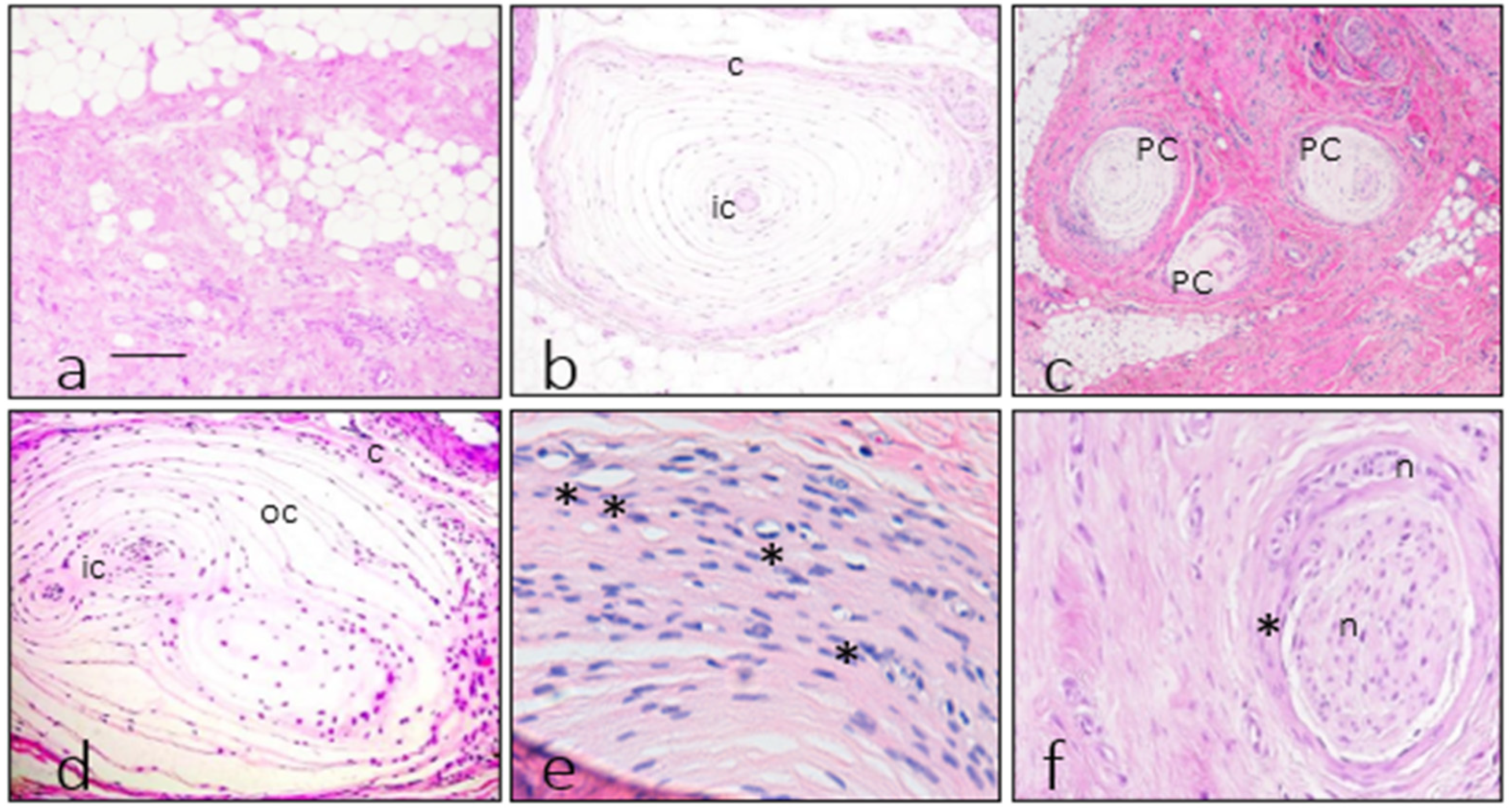
3.1. Morphology of Fibromatosis and Control Corpuscles

Pacinian corpuscles were identified in all the cases investigated. Corpuscles from control specimens were located in the subcutis and surrounded by fatty tissue, with a preserved structure that was consistent with normality (Figure 1b).
Comparing the distribution between normal and pathological conditions, a striking tendency to form clusters in the fibrous areas of the fibromatosis case (Figure 1b) was apparent, while the control cases displayed a more uniform distribution along the plantar surface. Generally, corpuscles from the fibromatosis case were located in fibrous areas (Figure 1c,d). Moreover, the outer core capsular system was thicker with respect to a normal condition, and it showed unusual density and placement of capillaries (Figure 1e). In addition, the proliferation was accompanied by a striking nerve sheath fibrous thickening (Figure 1f).

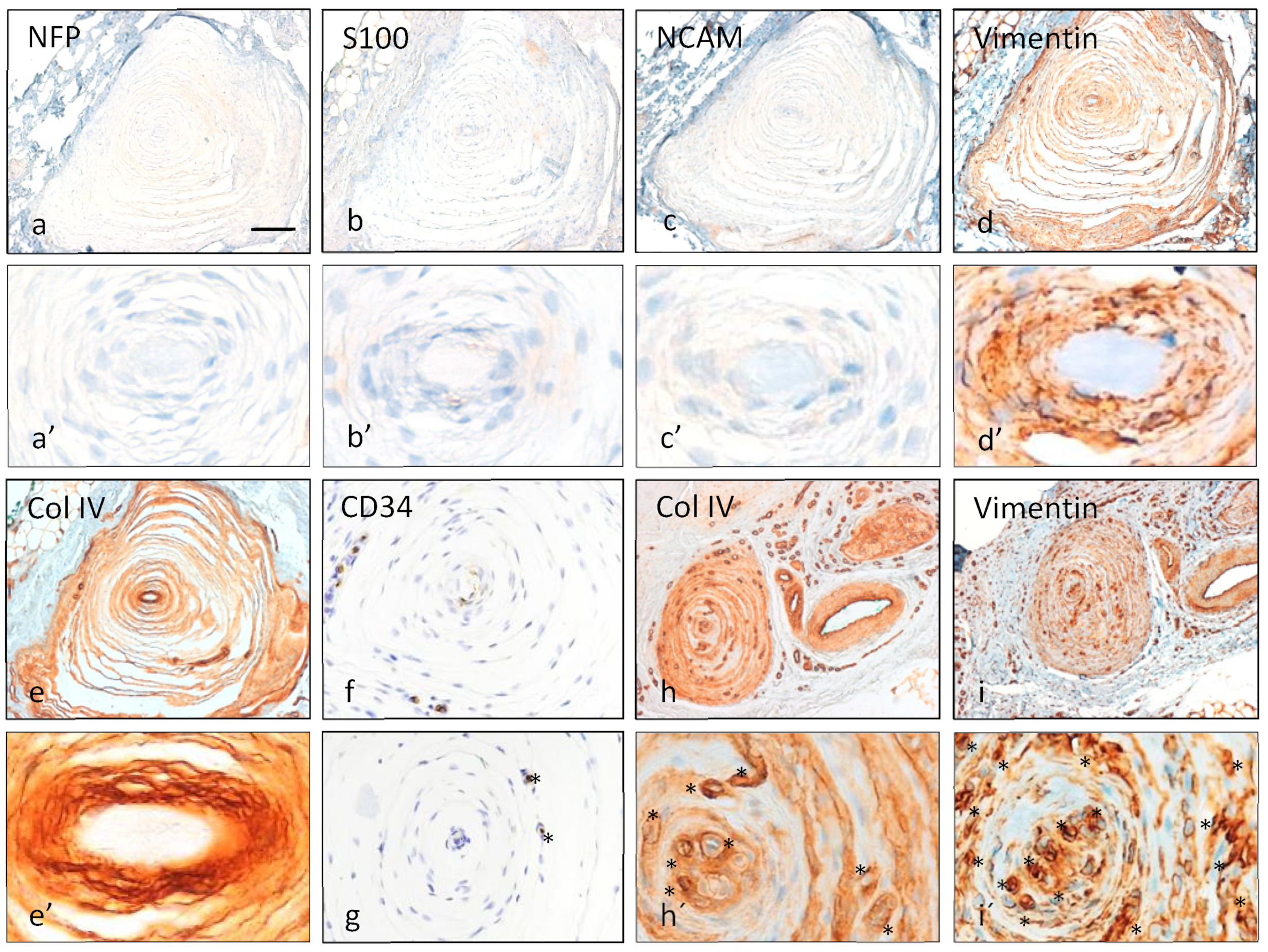
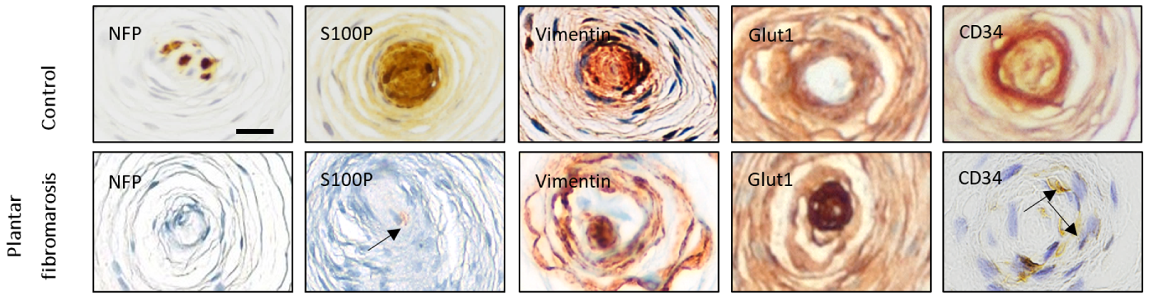
3.2. Immunohistochemistry of Pacinian Corpuscles

The arrangement of various corpuscular constituents, including central axon, inner core, intermediate layer, and outer core capsular complex, was severely altered in almost all corpuscles (approximately 90%), with an absence of immunostaining for NFP (characteristic of the axon, Figure 2a,a’), S100 protein (characteristic of the inner core, Figure 2b,b’), vimentin (characteristic of the inner and outer cores, Figure 2c,c’), NCAM (characteristic of the inner core, Figure 2d,d’), type IV collagen (characteristic of the interlamellar spaces, Figure 2e,e’), and CD34 (characteristic of the intermediate layer, also demonstrates vessels, Figure 2f,g). In fact, while in Pacinian corpuscles from control cases, capillaries were restricted to the capsule, in those from the fibromatosis specimen, capillaries were also found in the outer core and even in the inner core space (Figure 2e–i and Figure 3). NTrk expression was neither detected (data not shown). Apart from these findings, the immunohistochemical profiles were consistent with normality (Figure 4 and Table 2).

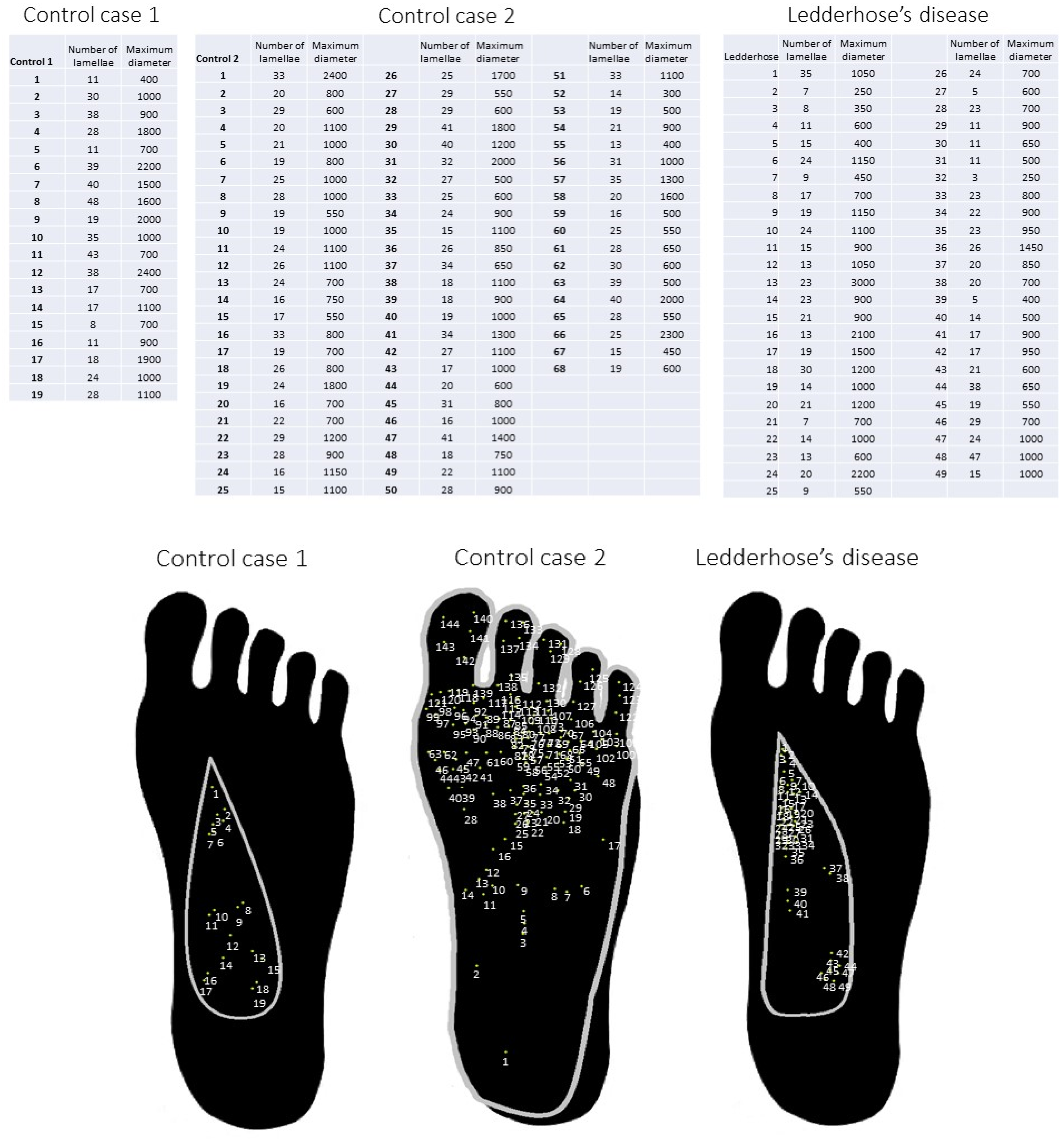
3.3. Measurements of Pacinian Corpuscles

Assuming that the sampling included all the corpuscles, the fibromatosis case had 1.87 corpuscles per cm2, the first control had 0.5 corpuscles per cm2, and the second control had a density of 0.6 corpuscles per cm2, excluding the toes and the ball of the foot (Figure 5).
All the measured corpuscles showed a variable size and number of lamellae, consistent with tangential cutting of various corpuscles (Figure 5). The mean size of Pacinian corpuscles in control cases was 1242 (C.I. 968.75–1515.25) µm and 963 (C.I. 857.69–1068.31) µm, respectively, and the mean number of lamellae was 26.47 (C.I. 20.61–32.33) and 24.63 (C.I. 22.92–26.34), respectively. The mean size of Pacinian corpuscles in the fibromatosis case was 902 (C.I. 15.8–20.6) µm, ranging between 250 µm (the smallest corpuscle of the studied cases) and 3000 µm (the biggest corpuscle of the studied cases). The mean number of lamellae in this case was 18.2 (C.I. 95% 15.7–20.7), ranging between 5 (the corpuscle with less lamellae) and 47 (the corpuscle with most lamellae).

3.4. Statistical Analysis

Statistical comparison was performed through Student’s t-test, considering one group the 49 Pacinian corpuscles of the fibromatosis case and the other group the total 87 corpuscles in the controls. For the corpuscular size, the difference between both groups was 122 (C.I.−52–296) µm, with a p = 0.17. Regarding the lamellae count, the fibromatosis case had a mean of 18,2 (C.I. 95% 15.7–20.7), while the controls had a combined mean of 25 (C.I. 95% 23.2–26.9), meaning a difference of 6.8 (C.I. 3.8–9.9) lamellae between both, with a p < 0.001.

4. Discussion

Our observations regarding the distribution of Pacinian corpuscles in normal plantar fascia and in plantar fibromatosis are in good agreement with the normal anatomy, previously described with different approaches [15,16,17,18,21,22,23]. In plantar fibromatosis, Pacinian corpuscles morphologically showed hyperplasia and a significant diminution in the number of lamellae, but without a statistically relevant hypertrophy or hypotrophy. The increase in corpuscular density may be related with the progression stage of superficial fibromatosis, which may thereafter be followed by a more severe phase showing a decrease or an absence of Pacinian corpuscles [27,28].
As far as we know, publications reporting histologic evaluations of plantar fibromatosis did not mention Pacinian corpuscles [29,30,31]. In addition, specific reviews of plantar fibromatosis did not report nervous disarrangements [28,32]. Conversely, there is abundant information about changes in Pacinian corpuscles in palmar fibromatosis [7,8,9,10,11,12,27], and therefore the discussion of the present results must necessarily be in relation to this pathology. Moreover, a relationship between superficial fibromatosis and the peripheral nervous system has been described recently, noting diminished mean sensitivity in palmar fibromatosis patients [33].
As it occurs in the Pacinian corpuscles of palmar fibromatosis [12], we observed radical changes in the neuronal compartment of these plantar corpuscles, with disarrangement and absence of immunoreactivity for conventional markers of the elements contained therein. In particular, the generalized absence of an NFP-immunoreactive axon, S100 protein positive inner core and CD34-expressing intermediate layer clearly suggests corpuscular denervation, also found in a large number of palmar fibromatosis Pacinian corpuscles [12]. The other main structural finding in Pacinian corpuscles from plantar fibromatosis was the proliferation of capillary vessels involving the capsule, the outer core, and the inner core. This has not been reported earlier, and this finding might be consistent with the filling of the space left by the absence of the corpuscular neural compartment.
To explain the peripheral nervous system changes in palmar fibromatosis, a hypothesis was proposed based on increased nerve growth factor levels [8,9,34]. To explore whether neurotrophins could be involved in the changes of Pacinian corpuscles from plantar fibromatosis, the expression of the pan-neurotrophin receptor NTrk was studied. With the complete negativity observed for NTrk, the neurotrophin hypothesis seems not applicable for plantar fibromatosis. Other proposed mechanisms for Pacinian hyperplasia, which might be applicable in plantar fibromatosis, are related with microtraumatism-related perineurial fibrosis or with mastocyte-driven inflammation [7,8,9,10,11,12,35].
Thanks to the improvement on imaging techniques, Pacinian corpuscles and Pacinian corpuscle hyperplasia can now be diagnosed by MRI [22,36]. This attribute of Pacinian corpuscles hyperplasia can, thus, become a diagnostic clue of plantar fibromatosis, helping in the diagnosis of this entity in addition to other features [14,37,38].

Author Contributions

Conceptualization, J.F.; methodology, R.R.-M. and M.B.R.-M.; software, F.J.G.-A.; validation, M.L. and B.M.-B.; investigation, J.F. and R.E.; resources, M.L.G.-M., J.L.C. and B.M.-B.; writing—original draft preparation, J.F.; writing—review and editing, J.A.V.; visualization, J.F. and R.E.; supervision, J.F. and J.A.V.; funding acquisition, J.F and B.M.-B. All authors have read and agreed to the published version of the manuscript.

Funding

This research was funded in part by Gerencia Regional de Salud de Castilla y León to JF (grants GRS2164/A/2020 and GRS2379/A/21).

Institutional Review Board Statement

The study was conducted in accordance with the Declaration of Helsinki and approved by the Ethics Committee for Biomedical Research of the Principality of Asturias, Spain (protocol code CElm, Past: Proyecto 266/18; 19 November 2018).

Informed Consent Statement

Patient consent was waived due to the absence of traceable clinical information of the patients either in the text or along the investigation procedures.

Data Availability Statement

Not applicable.

Acknowledgments

The authors would like to specially thank Raquel Martín Sanz for her critical review of the text from a clinical standpoint and the patients from whom samples were obtained. Results from this anatomical research can improve future patients’ diagnoses and, therefore, they deserve our highest gratitude.

Conflicts of Interest

The authors declare no conflict of interest.

References

  1. Glodblum, J.R.; Folpe, A.L.; Weiss, S.W. Benign fibroblastic/myofibroblastic proliferations, including superficial fibromatoses. In Enzinger and Weiss’s Soft Tissue Tumors; Elsevier: Philadelphia, PA, USA, 2020. [Google Scholar]
  2. Goldblum, J.R.; Fletcher, J.A. Palmar/plantar fibromatosis. In WHO Classification of Tumours of Soft Tissue and Bone; IARC Press: Lyon, France, 2013. [Google Scholar]
  3. Thway, K.; Nascimento, A.F. Palmar/plantar fibromatosis. In WHO Classification of Tumours. Soft Tissue and Bone Tumours; IARC Press: Lyon, France, 2020. [Google Scholar]
  4. Liu, Q.; Fang, L.; Li, B. Desmoid fibromatosis in the foot. Medicine 2018, 97, e13109. [Google Scholar] [CrossRef]
  5. Bourne, M.; Talkad, A.; Varacallo, M. Anatomy, Bony Pelvis and Lower Limb, Foot Fascia; StatPearls Publishing: Treasure Island, FL, USA, 2022. [Google Scholar]
  6. Wang, L.; Zhu, H. Clonal analysis of palmar fibromatosis: A study whether palmar fibromatosis is a real tumor. J. Transl. Med. 2006, 4, 21. [Google Scholar] [CrossRef] [Green Version]
  7. Akyürek, N.; Ataoğlu, O.; Cenetoğlu, S.; Ozmen, S.; Cavuşoğlu, T.; Yavuzer, R. Pacinian corpuscle hyperplasia coexisting with Dupuytren’s contracture. Ann. Plast. Surg. 2000, 45, 220–222. [Google Scholar] [PubMed]
  8. Ehrmantant, W.R.; Graham, W.P., 3rd; Towfighi, J.; Mackay, D.R.; Ehrlich, H.P. A histological and anatomical profile of pacinian corpuscles from Dupuytren’s contracture and the expression of nerve growth factor receptor. Plat. Reconstr. Surg. 2004, 114, 721–727. [Google Scholar] [CrossRef] [PubMed]
  9. Yenidunya, M.O.; Yenidunya, S.; Seven, E. Pacinian hypertrophy in a type 2A hand burn contracture and Pacinian hypertrophy and hyperplasia in a Dupuytren’s contracture. Burns 2009, 35, 446–450. [Google Scholar] [CrossRef] [PubMed]
  10. Von Campe, A.; Mende, K.; Omaren, H.; Meuli-Simmen, C. Painful nodules and cords in Dupuytren disease. J. Hand Surg. Am. 2012, 37, 1313–1318. [Google Scholar] [CrossRef]
  11. Stecco, C.; Macchi, V.; Barbieri, A.; Tiengo, C.; Porzionato, A.; De Caro, R. Hand fasciae innervation: The palmar aponeurosis. Clin. Anat. 2018, 31, 677–683. [Google Scholar] [CrossRef]
  12. García-Martínez, I.; García-Mesa, Y.; García-Piqueras, J.; Martínez-Pubil, A.; Cobo, J.L.; Feito, J.; García-Suárez, O.; Vega, J.A. Sensory innervation of the human palmar aponeurosis in healthy individuals and patients with palmar fibromatosis. J. Anat. 2022, 240, 972–984. [Google Scholar] [CrossRef]
  13. Young, J.R.; Sternbach, S.; Willinger, M.; Hutchinson, I.D.; Rosenbaum, A. The etiology, evaluation and management of plantar fibromatosis. Orthop. Res. Rev. 2018, 11, 1–7. [Google Scholar] [CrossRef] [Green Version]
  14. Stewart, D.; Nascimento, A.F. Palmar and plantar fibromatosis: A review. J. Pathol. Transl. Med. 2021, 55, 265–270. [Google Scholar] [CrossRef]
  15. Bojsen-Moller, F.; Flagstad, K.E. Plantar aponeurosis and internal architecture of the ball of the foot. J. Anat. 1976, 121, 599–611. [Google Scholar]
  16. Stecco, C.; Corradin, M.; Macchi, V.; Morra, A.; Porzionato, A.; Biz, C.; De Caro, R. Plantar fascia anatomy and its relationship with Achilles tendon and paratenon. J. Anat. 2013, 223, 665–676. [Google Scholar] [CrossRef]
  17. Strzalkowski, N.D.J.; Peters, R.M.; Inglis, J.T.; Bent, L.R. Cutaneous afferent innervation of the human foot sole: What can we learn from single-unit recordings? J. Neurophysiol. 2018, 120, 1233–1246. [Google Scholar] [CrossRef]
  18. Corniani, G.; Saal, H.P. Tactile innervation densities across the whole body. J. Neurophysiol. 2020, 124, 1229–1240. [Google Scholar] [CrossRef] [PubMed]
  19. Kowalzik, R.; Hermann, B.; Biedermann, H.; Peiper, U. Two-point discrimination of vibratory perception on the sole of the human foot. Foot Ankle Int. 1996, 17, 629–634. [Google Scholar] [CrossRef]
  20. Gu, C.; Griffin, M.J. Spatial summation of vibrotactile sensations at the foot. Med. Eng. Phys. 2013, 35, 1221–1227. [Google Scholar] [CrossRef] [Green Version]
  21. Jin, Z.W.; Cho, K.H.; Xu, D.Y.; You, Y.Q.; Kim, J.H.; Murakami, G.; Abe, H. Pacinian corpuscles in the human fetal foot: A study using 3D reconstruction and immunohistochemistry. Ann. Anat. 2020, 227, 151421. [Google Scholar] [CrossRef]
  22. Sugai, N.; Cho, K.H.; Murakami, G.; Abe, H.; Uchiyama, E.; Kura, H. Distribution of sole Pacinian corpuscles: A histological study using near-term human feet. Surg. Radiol. Anat. 2021, 43, 10311039. [Google Scholar] [CrossRef]
  23. Germann, C.; Sutter, R.; Nanz, D. Novel observations of Pacinian corpuscle distribution in the hands and feet based on high—Resolution 7-T MRI in healthy volunteers. Skeletal. Radiol. 2021, 50, 1249–1255. [Google Scholar] [CrossRef]
  24. Toepfer, A.; Harrasser, N.; Dreyer, F.; Mogler, C.; Walther, M.; von Eisenhart-Rothe, R. Epithelioid sarcoma of the plantar fascia mimicking morbus Ledderhose—A severe pitfall for clinical and histopathological misinterpretation. Foot Ankle Surg. 2017, 23, e25–e30. [Google Scholar] [CrossRef]
  25. Feito, J.; Ramos-García, J.L.; Gago, Á.; Cobo, J.L.; García-Suárez, O.; Junquera, L.M.; Vega, J.A. Pacinian Corpuscles in a Cervical Chondrocutaneous Remnant: A Case Report and Update of Pacinian Corpuscles. Am. J. Dermatopathol. 2016, 38, 231–235. [Google Scholar] [CrossRef]
  26. Cobo, R.; García-Piqueras, J.; Cobo, J.; Vega, J.A. The Human Cutaneous Sensory Corpuscles: An Update. J. Clin. Med. 2021, 10, 227. [Google Scholar] [CrossRef] [PubMed]
  27. Mikusev, I.E. New facts about the pathogenesis of Dupuytren’s contracture. Acta Chir. Plast. 1989, 31, 1–14. [Google Scholar] [PubMed]
  28. Carroll, P.; Henshaw, R.; Garwood, C.; Raspovic, K.; Kumar, D. Plantar fibromatosis: Pathophysiology, surgical and nonsurgical therapies: An evidence-based review. Foot Ankle Spec. 2018, 11, 168–176. [Google Scholar] [CrossRef]
  29. Fausto de Souza, D.; Micaelo, L.; Cuzzi, T.; Silva, M.R.E. Ledderhose disease: An unusual presentation. J. Clin. Aesthet. Dermatol. 2010, 3, 45–47. [Google Scholar]
  30. Miceli, A.J.; Junkins-Hopkins, J.M.; Polley, D.C.; Elston, D.M. Multiple nodules on the sole of the foot. Indian Dermatol. Online J. 2015, 6, 422–424. [Google Scholar] [CrossRef]
  31. Ocampo-Garza, J.; Cunha, R.A.; Pereira, C.S.; Sanderson, A.; Pinto-Desmond, K.; Machado-Filho, C.D.A.; Haneke, E.; de Carvalho, M. Plantar fibromatosis: Surgical approach of a giant bilateral case. Int. J. Dermatol. 2018, 57, 365–367. [Google Scholar] [CrossRef]
  32. Neagu, T.P.; Tiglis, M.; Popescu, A.; Enache, V.; Popescu, S.A.; Lascar, I. Clinical, histological and therapeutic modern approach of Ledderhose disease. Rom. J. Morphol. Embryol. 2018, 59, 691–697. [Google Scholar]
  33. Gerosa, T.; Pierrart, J.; Serane-Fresnel, J.; Amsallem, L.; Masmejean, E.H. Distal sensory disorders in Dupuytren’s disease. Orthop. Traumatol. Surg. Res. 2018, 104, 897–900. [Google Scholar] [CrossRef]
  34. Lubahn, J.D.; Pollard, M.; Cooney, T. Immunohistochemical evidence of nerve growth factor in Dupuytren’s disease palmar fascia. J. Hand Surg. Am. 2007, 32, 337–342. [Google Scholar] [CrossRef]
  35. Schubert, T.E.O.; Weidler, C.; Borisch, N.; Schubert, C.; Hofstädter, F.; Straub, R.H. Dupuytren’s contracture is associated with sprouting of substance P positive nerve fibres and infiltration by mast cells. Ann. Rheum. Dis. 2006, 65, 414–415. [Google Scholar] [CrossRef] [PubMed] [Green Version]
  36. Chambers, A.R.; Dreyer, M.A.; Song, K.Y.; Lam, K.K. Bilateral symptomatic Pacinian corpuscle hyperplasia of the adult foot. Foot 2020, 49, 101709. [Google Scholar] [CrossRef] [PubMed]
  37. Hughes, P.; Miranda, R.; Doyle, A.J. MRI imaging of soft tissue tumours of the foot and ankle. Insights Imaging 2019, 10, 60. [Google Scholar] [CrossRef] [PubMed]
  38. Ahlawat, S.; Fayad, L.M. Revisiting the WHO classification system of soft tissue tumours: Emphasis on advanced magnetic resonance imaging sequences. Part 1. Pol. J. Radiol. 2020, 85, e396–e408. [Google Scholar] [CrossRef]
Figure 1. Morphology found in the fibromatosis and control cases. The fibromatosis case had a fibrous proliferation apparent in the subcutaneous tissue with low cellularity but the presence of infiltrative tracts in the hypodermal adipose tissue (a). Control Pacinian corpuscles (b) had a thin capsule (c), making contrast with the peripheral fatty tissue and also had a more colored inner core (ic) with a solid central structure with no nuclei consistent with an axon. The neoplastic fibrous proliferation encompassed some Pacinian corpuscles (pc), with a clustered appearance at low power (c). Corpuscles from the fibromatosis case (d) didn’t displayed the solid inner core/axon complex, and in the central region corresponding with the inner core area (ic) it was often apparent capillaries. The capsule of various corpuscles from the fibromatosis case (e) displayed a thickened capsule with an abundance of small blood vessels (*). Peripheral nerves (f) found inside the neoplastic fibrous proliferation showed intense fibrosis (*) entrapping nerve fascicles (n). Images (e,f) demonstrate the perineurial nature of the nerve fibers fibrosis employing consecutive slides showing a thick CD34 negative layer surrounding the nerve fiber (e) and the same thick layer positive for Glut1 (f). Scale bar correspond to 50 µm in images (a,b,d), 100 µm in (c), 20 µm in (e), and 30 µm in (f).
Diagnostics 12 01705 g001
Figure 2. Immunohistochemical findings in consecutive sections of two different Pacinian corpuscles (af,h,i). The first corpuscle depicted (af) had no evidence of axonal profile innervating it due to an absent NFP expression ((a), detailed in (a’)); it had neither evidence of inner core, as long as there is no S100 ((b), detailed in (b’)) positivity and N-CAM was also negative ((c), detailed in (c’)); vimentin ((d), detailed in (d’)) left a central zone without expression in the region where the inner core should be. The outer core was well defined by type IV collagen ((e), detailed in (e’)). CD34 from different corpuscles was faintly present (f) or absent (g) surrounding the inner core region. The second corpuscle depicted in the bottom right (h,i) displayed a lamellar architecture of highlighted with Type IV collagen and vimentin expression; and a noticeable vascular proliferation (blood vessels highlighted with *) in the innermost and the capsular regions of the corpuscle was apparent in detail (h’,i’). Scale bar corresponds to 100 µm images (ae,h,i), to 60 µm in images (f,g), 30 µm in the detailed images (h’,i’), and 15 µm in (a’–e’).
Diagnostics 12 01705 g002
Figure 3. Capillaries in Pacinian corpuscles. The Pacinian corpuscle from Figure 2 is displayed with haematoxilin-eosin staining to identify the different corpuscular regions, including the capsule (c), outer core (oc), and inner core (ic); arrows indicate capillaries ((a), detailed in (b)). Typically, only the capsule of human Pacinian corpuscles contains capillaries, highlighted with type IV collagen immunostain in a typical control corpuscle, where only the peripheral capsule contains capillaries (c). Nevertheless, in those from fibromatosis disease, capillaries are present in all the corpuscular compartments (d).
Diagnostics 12 01705 g003
Figure 4. Representative images of the changes in the expression of some selected antigens in the Pacinian corpuscles of Ledderhose disease compared with controls. Scale bar = 25 µm.
Diagnostics 12 01705 g004
Figure 5. Schematic diagram of Pacinian corpuscle distribution in Ledderhose disease and in the control cases, including the measure and the lamellae count of each corpuscle. Total corpuscles in the first control case were numbered 1–19 for a total surface of 38 cm2. Total corpuscles in the second control case were numbered 1–68 for a total surface of 75 cm2, excluding the toes and the ball of the foot. Total corpuscles in the fibromatosis case were numbered 1–49 for a total surface of 26 cm2.
Diagnostics 12 01705 g005
Table 1. Primary antibodies used in the study.
AntigenOriginDilutionSupplier
Glut1 (polyclonal)RabbitpredilutedCell Marque 1
EMA (E29)MousepredilutedCell Marque 1
N-CAM (123C3)MousepredilutedVentana Medical Systems 2
NFP (2F11)MousepredilutedCell Marque 1
S-100 protein (polyclonal)RabbitpredilutedVentana Medical Systems 2
Vimentin (V9)MousepredilutedCell Marque 1
Actin, muscle specific (HHF35)MousepredilutedVentana Medical Systems 2
Actin, smooth muscle (1A4)MousepredilutedVentana Medical Systems 2
Desmin (D33)MousepredilutedCell Marque 1
Collagen type IV (CIV22)MousepredilutedVentana Medical Systems 2
CD34 (QBEnd/10)MousepredilutedMaster Diagnóstica 3
NTrk (EPR17341)MousepredilutedVentana Medical Systems 2
Glut1: glucose transporter 1; EMA: epithelial membrane antigen; N-CAM: neural-cell adhesion molecule; NFP: neurofilament protein; 1 Rocklin, California, USA; 2 Oro Valley, Arizona, USA; 3 Granada, Spain.
Table 2. Immunohistochemical profile of Pacinian corpuscles in normal plantar fascia (C) and plantar fibromatosis (PF). Quantification of the immunostain was performed considering absence of expression (−), faint expression (+), intermediate expression (++), and strong expression (+++).
AntigenAxonICILOCC
NFPControl+++
PF− (1)
S100PControl+++
PF− (1)
EMAControl++++
PF+/+++/++
CD34Control+++
PF−/+− (2)− (2)
VIMControl+++++++++++
PF−/+++ (3)+++++++++
ActinControl
PF
DesminControl
PF
IV COLControl++ (4)
PF−/+ (4)
Glut1Control++++
PF+++ (3)+++
N-CAMControl++
PF
NTrkControl+++++
PF
PF: Plantar Fibromatosis; C: capsule; IC: inner core, IL: intermediate layer; OC: outer core; (1) More than 90% of the corpuscles; (2) CD34 positivity in the endothelium; (3) less than 5% of the corpuscles; (4) pattern of basement membrane.
Publisher’s Note: MDPI stays neutral with regard to jurisdictional claims in published maps and institutional affiliations.

