
Virtual Monochromatic Images from Dual-Energy Computed Tomography Do Not Improve the Detection of Synovitis in Hand Arthritis

 , , , and
Abstract
:1. Introduction
2. Materials and Methods
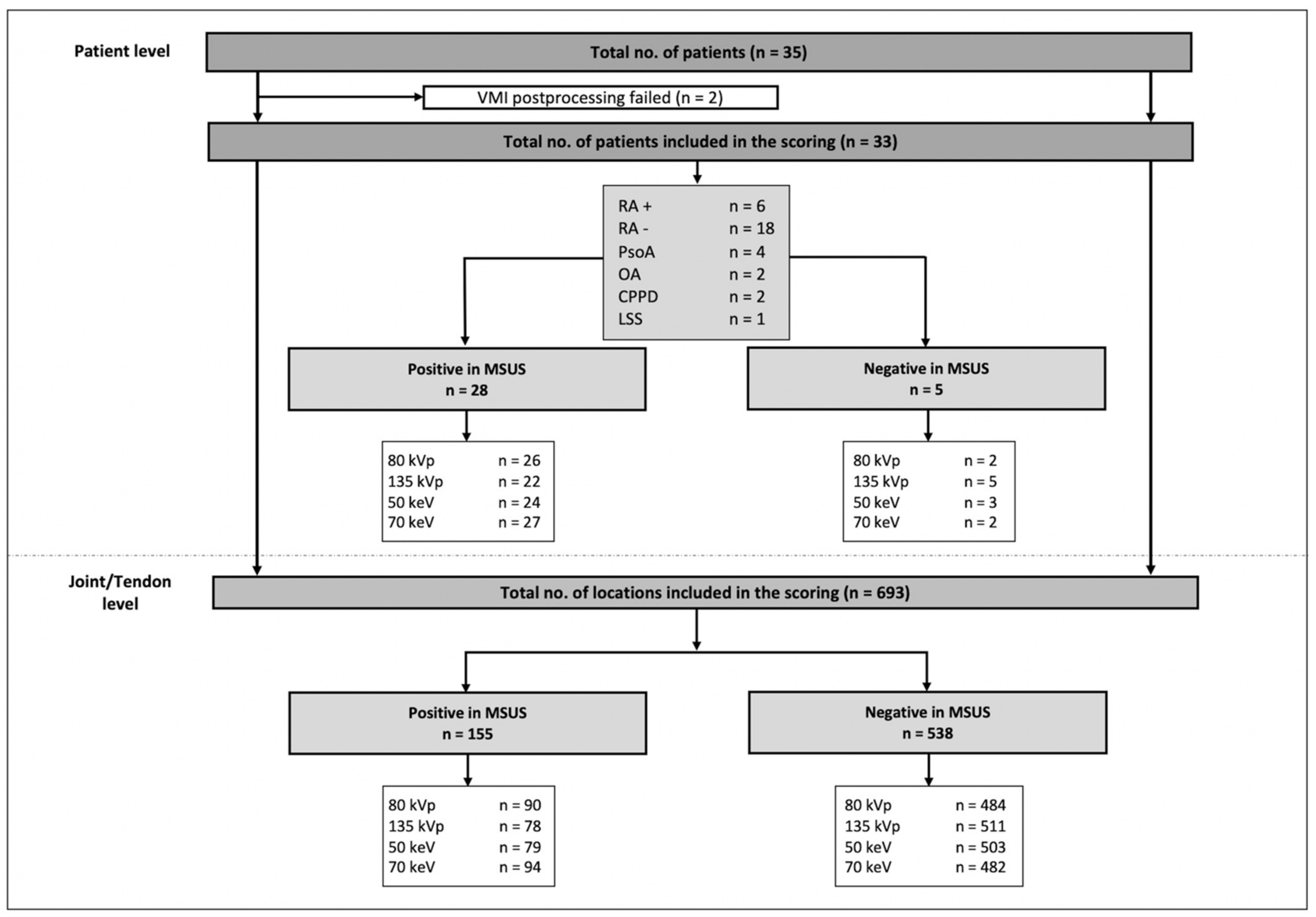
2.1. Subjects
2.2. DECT and MSUS Imaging Procedures
2.3. VMI and Subtraction
2.4. Image Reading
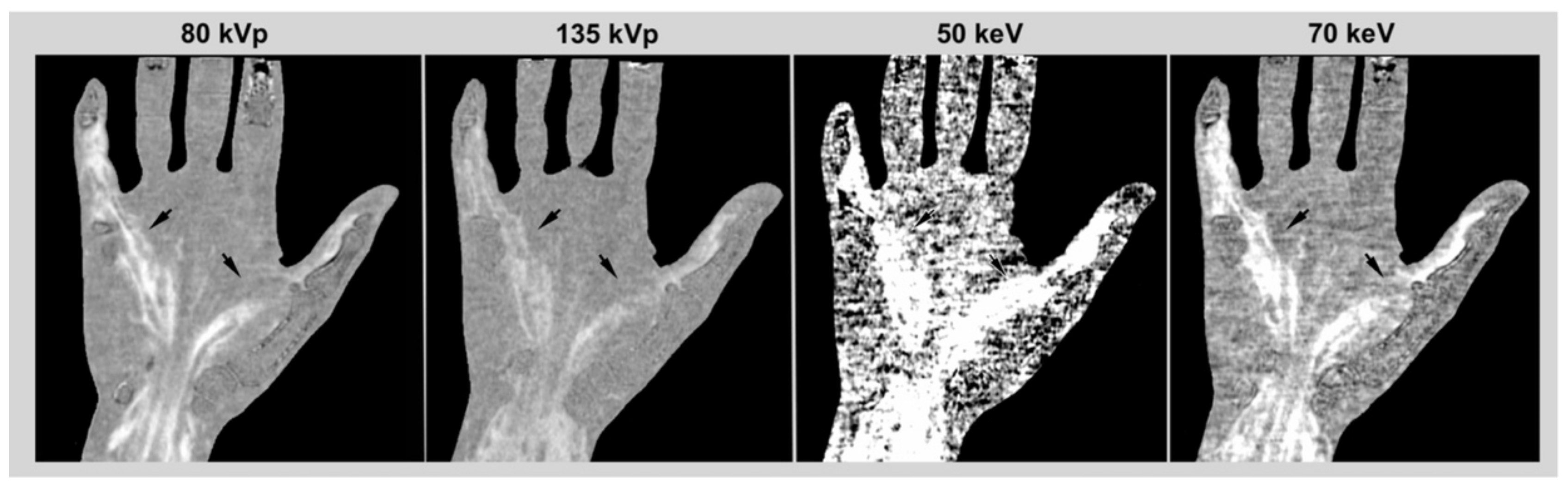
2.5. Image Quality
2.6. Radiation Exposure
2.7. Statistical Analysis
3. Results
3.1. Subjects
3.2. Image Reading and Statistical Analysis
3.3. Image Quality
3.4. Radiation Exposure
4. Discussion
5. Conclusions
Supplementary Materials
Author Contributions
Funding
Institutional Review Board Statement
Informed Consent Statement
Data Availability Statement
Conflicts of Interest
References
- Jans, L.; De Kock, I.; Herregods, N.; Verstraete, K.; Van den Bosch, F.; Carron, P.; Oei, E.H.; Elewaut, D.; Jacques, P. Dual-energy CT: A new imaging modality for bone marrow oedema in rheumatoid arthritis. Ann. Rheum. Dis. 2018, 77, 958–960. [Google Scholar] [CrossRef]
- Hu, H.J.; Liao, M.Y.; Xu, L.Y. Clinical utility of dual-energy CT for gout diagnosis. Clin. Imaging 2015, 39, 880–885. [Google Scholar] [CrossRef]
- Mallinson, P.I.; Coupal, T.M.; McLaughlin, P.D.; Nicolaou, S.; Munk, P.L.; Ouellette, H.A. Dual-Energy CT for the Musculoskeletal System. Radiology 2016, 281, 690–707. [Google Scholar] [CrossRef]
- Goo, H.W.; Goo, J.M. Dual-Energy CT: New Horizon in Medical Imaging. Korean J. Radiol. 2017, 18, 555–569. [Google Scholar] [CrossRef] [Green Version]
- Forghani, R.; De Man, B.; Gupta, R. Dual-Energy Computed Tomography: Physical Principles, Approaches to Scanning, Usage, and Implementation: Part 1. Neuroimaging Clin. N. Am. 2017, 27, 371–384. [Google Scholar] [CrossRef]
- Yu, L.; Leng, S.; McCollough, C.H. Dual-energy CT-based monochromatic imaging. AJR Am. J. Roentgenol. 2012, 199, S9–S15. [Google Scholar] [CrossRef]
- Fukuda, T.; Umezawa, Y.; Tojo, S.; Yonenaga, T.; Asahina, A.; Nakagawa, H.; Fukuda, K. Initial Experience of Using Dual-Energy CT with an Iodine Overlay Image for Hand Psoriatic Arthritis: Comparison Study with Contrast-enhanced MR Imaging. Radiology 2017, 284, 134–142. [Google Scholar] [CrossRef]
- Diekhoff, T.; Ulas, S.T.; Poddubnyy, D.; Schneider, U.; Hermann, S.; Biesen, R.; Burmester, G.R.; Hamm, B.; Hermann, K.G. Ultra-low-dose CT detects synovitis in patients with suspected rheumatoid arthritis. Ann. Rheum. Dis. 2019, 78, 31–35. [Google Scholar] [CrossRef] [Green Version]
- Ulas, S.T.; Hermann, K.G.; Makowski, M.R.; Biesen, R.; Proft, F.; Schilling, R.; Diekhoff, T. Perfusion in hand arthritis on dynamic contrast-enhanced computed tomography: A randomized prospective study using MRI as a standard of reference. Skelet. Radiol. 2021, 50, 59–68. [Google Scholar] [CrossRef]
- Dohn, U.M.; Terslev, L.; Szkudlarek, M.; Hansen, M.S.; Hetland, M.L.; Hansen, A.; Madsen, O.R.; Hasselquist, M.; Møller, J.; Østergaard, M. Detection, scoring and volume assessment of bone erosions by ultrasonography in rheumatoid arthritis: Comparison with CT. Ann. Rheum. Dis. 2013, 72, 530–534. [Google Scholar] [CrossRef] [PubMed]
- Fukuda, T.; Umezawa, Y.; Asahina, A.; Nakagawa, H.; Furuya, K.; Fukuda, K. Dual energy CT iodine map for delineating inflammation of inflammatory arthritis. Eur. Radiol. 2017, 27, 5034–5040. [Google Scholar] [CrossRef] [PubMed]
- Ulas, S.T.; Ziegeler, K.; Richter, S.T.; Ohrndorf, S.; Biesen, R.; Proft, F.; Poddubnyy, D.; Diekhoff, T. Contrast-enhanced CT techniques and MRI perform equally well in arthritis imaging of the hand: A prospective diagnostic accuracy study. Eur. Radiol. 2022, 32, 1–8. [Google Scholar] [CrossRef]
- Ostergaard, M.; Edmonds, J.; McQueen, F.; Peterfy, C.; Lassere, M.; Ejbjerg, B.; Bird, P.; Emery, P.; Genant, H.; Conaghan, P. An introduction to the EULAR-OMERACT rheumatoid arthritis MRI reference image atlas. Ann. Rheum. Dis. 2005, 64, i3–i7. [Google Scholar] [CrossRef] [Green Version]
- Polster, J.M.; Winalski, C.S.; Sundaram, M.; Lieber, M.L.; Schils, J.; Ilaslan, H.; Davros, W.; Husni, M.E. Rheumatoid arthritis: Evaluation with contrast-enhanced CT with digital bone masking. Radiology 2009, 252, 225–231. [Google Scholar] [CrossRef]
- McQueen, F.M. Imaging in early rheumatoid arthritis. Best. Pract. Res. Clin. Rheumatol. 2013, 27, 499–522. [Google Scholar] [CrossRef] [PubMed]
- Gerster, J.C.; Landry, M.; Dufresne, L.; Meuwly, J.Y. Imaging of tophaceous gout: Computed tomography provides specific images compared with magnetic resonance imaging and ultrasonography. Ann. Rheum. Dis. 2002, 61, 52–54. [Google Scholar] [CrossRef] [Green Version]
- Klauser, A.S.; Halpern, E.J.; Strobl, S.; Abd Ellah, M.M.; Gruber, J.; Bellmann-Weiler, R.; Auer, T.; Feuchtner, G.; Jaschke, W. Gout of hand and wrist: The value of US as compared with DECT. Eur. Radiol. 2018, 28, 4174–4181. [Google Scholar] [CrossRef] [Green Version]
- Choi, H.K.; Al-Arfaj, A.M.; Eftekhari, A.; Munk, P.L.; Shojania, K.; Reid, G.; Nicolaou, S. Dual energy computed tomography in tophaceous gout. Ann. Rheum. Dis. 2009, 68, 1609–1612. [Google Scholar] [CrossRef] [PubMed]
- Diekhoff, T.; Kiefer, T.; Stroux, A.; Pilhofer, I.; Juran, R.; Mews, J.; Hermann, K.G.A. Detection and characterization of crystal suspensions using single-source dual-energy computed tomography: A phantom model of crystal arthropathies. Investig. Radiol. 2015, 50, 255–260. [Google Scholar] [CrossRef] [PubMed]
- Bongartz, T.; Glazebrook, K.N.; Kavros, S.J.; Murthy, N.S.; Merry, S.P.; Franz, W.B., III; Michet, C.J.; Veetil, B.M.A.; Davis, J.M., III; Mason, T.G., II; et al. Dual-energy CT for the diagnosis of gout: An accuracy and diagnostic yield study. Ann. Rheum. Dis. 2015, 74, 1072–1077. [Google Scholar] [CrossRef] [PubMed] [Green Version]
- Patel, B.N.; Thomas, J.V.; Lockhart, M.E.; Berland, L.L.; Morgan, D.E. Single-source dual-energy spectral multidetector CT of pancreatic adenocarcinoma: Optimization of energy level viewing significantly increases lesion contrast. Clin. Radiol. 2013, 68, 148–154. [Google Scholar] [CrossRef] [PubMed]
- Lam, S.; Gupta, R.; Levental, M.; Yu, E.; Curtin, H.D.; Forghani, R. Optimal Virtual Monochromatic Images for Evaluation of Normal Tissues and Head and Neck Cancer Using Dual-Energy CT. AJNR Am. J. Neuroradiol. 2015, 36, 1518–1524. [Google Scholar] [CrossRef] [PubMed] [Green Version]
- Yu, L.; Christner, J.A.; Leng, S.; Wang, J.; Fletcher, J.G.; McCollough, C.H. Virtual monochromatic imaging in dual-source dual-energy CT: Radiation dose and image quality. Med. Phys. 2011, 38, 6371–6379. [Google Scholar] [CrossRef] [PubMed] [Green Version]
- Kayama, R.; Fukuda, T.; Ogiwara, S.; Momose, M.; Tokashiki, T.; Umezawa, Y.; Fukuda, K. Quantitative analysis of therapeutic response in psoriatic arthritis of digital joints with Dual-energy CT iodine maps. Sci. Rep. 2020, 10, 1225. [Google Scholar] [CrossRef] [PubMed] [Green Version]
- Ogiwara, S.; Fukuda, T.; Kawakami, R.; Ojiri, H.; Fukuda, K. Anatomical analysis of inflammation in hand psoriatic arthritis by Dual-Energy CT Iodine Map. Eur. J. Radiol. Open 2021, 8, 100383. [Google Scholar] [CrossRef]
- Baerends, E.; Oostveen, L.J.; Smit, C.T.; Das, M.; Sechopoulos, I.; Brink, M.; de Lange, F.; Prokop, M. Comparing dual energy CT and subtraction CT on a phantom: Which one provides the best contrast in iodine maps for sub-centimetre details? Eur. Radiol. 2018, 28, 5051–5059. [Google Scholar] [CrossRef] [Green Version]
- Gervaise, A.; Osemont, B.; Lecocq, S.; Noel, A.; Micard, E.; Felblinger, J.; Blum, A. CT image quality improvement using Adaptive Iterative Dose Reduction with wide-volume acquisition on 320-detector CT. Eur. Radiol. 2012, 22, 295–301. [Google Scholar] [CrossRef]
- Diekhoff, T.; Scheel, M.; Hermann, S.; Mews, J.; Hamm, B.; Hermann, K.A. Osteitis: A retrospective feasibility study comparing single-source dual-energy CT to MRI in selected patients with suspected acute gout. Skelet. Radiol. 2017, 46, 185–190. [Google Scholar] [CrossRef] [Green Version]
- Tortora, M.; Gemini, L.; D’Iglio, I.; Ugga, L.; Spadarella, G.; Cuocolo, R. Spectral Photon-Counting Computed Tomography: A Review on Technical Principles and Clinical Applications. J. Imaging 2022, 8, 112. [Google Scholar] [CrossRef]
- Combe, B.; Landewe, R.; Daien, C.I.; Hua, C.; Aletaha, D.; Álvaro-Gracia, J.M.; Bakkers, M.; Brodin, N.; Burmester, G.R.; Codreanu, C.; et al. 2016 update of the EULAR recommendations for the management of early arthritis. Ann. Rheum. Dis. 2017, 76, 948–959. [Google Scholar] [CrossRef] [Green Version]
- Moller, I.; Janta, I.; Backhaus, M.; Ohrndorf, S.; Bong, D.A.; Martinoli, C.; Filippucci, E.; Sconfienza, L.M.; Terslev, L.; Damjanov, N.; et al. The 2017 EULAR standardised procedures for ultrasound imaging in rheumatology. Ann. Rheum. Dis. 2017, 76, 1974–1979. [Google Scholar] [CrossRef]
- Hoving, J.L.; Buchbinder, R.; Hall, S.; Lawler, G.; Coombs, P.; McNealy, S.; Bird, P.; Connell, D. A comparison of magnetic resonance imaging, sonography, and radiography of the hand in patients with early rheumatoid arthritis. J. Rheumatol. 2004, 31, 663–675. [Google Scholar]


| Characteristics | |
|---|---|
| Number of patients (women/men) | 33 (21/12) |
| Mean age (y) (SD; range) | 55 (11.8; 23–75) |
| Mean symptom duration (y) (SD; median; range) | 1.7 (3.8; 0; 0–16) |
| Mean CRP (mg/L) (SD; median; range) | 18.3 (30.3; 3.4; 0.3–132.0) |
| ACPA-positive (>20 U/mL), n | 6 (18.2%) |
| RF-IgM-positive (>14 U/mL), n | 12 (36.4%) |
| Patient Level | SE (95% CI) | SP (95% CI) | PPV (95% CI) | NPV (95% CI) |
|---|---|---|---|---|
| 80 kVp | 0.93 (0.77–0.99) | 0.4 (0.07–0.77) | 0.90 (0.74–0.96) | 0.5 (0.09–0.91) |
| 135 kVp | 0.79 (0.60–0.90) | 1.0 (0.57–1.00) | 1.0 (0.85–1.0) | 0.45 (0.21–0.72) |
| 50 keV | 0.86 (0.69–0.94) | 0.6 (0.23–0.93) | 0.92 (0.76–0.99) | 0.43 (0.16–0.75) |
| 70 keV | 0.96 (0.83–0.99) | 0.4 (0.07–0.77) | 0.9 (0.74–0.97) | 0.67 (0.12–0.98) |
| Joint/Tendon Level | ||||
| 80 kVp | 0.58 (0.50–0.66) | 0.90 (0.87–0.92) | 0.63 (0.54–0.70) | 0.88 (0.85–0.90) |
| 135 kVp | 0.50 (0.43–0.58) | 0.95 (0.93–0.97) | 0.74 (0.65–0.82) | 0.87 (0.84–0.89) |
| 50 keV | 0.51 (0.43–0.59) | 0.93 (0.91–0.95) | 0.69 (0.60–0.77) | 0.87 (0.84–0.89) |
| 70 keV | 0.61 (0.53–0.68) | 0.90 (0.87–0.92) | 0.63 (0.55–0.70) | 0.89 (0.86–0.91) |
| Joint Level | ||||
| 80 kVp | 0.56 (0.47–0.64) | 0.89 (0.85–0.92) | 0.71 (0.61–0.79) | 0.81 (0.76–0.85) |
| 135 kVp | 0.47 (0.38–0.56) | 0.95 (0.91–0.97) | 0.81 (0.70–0.88) | 0.79 (0.74–0.83) |
| 50 keV | 0.50 (0.42–0.59) | 0.90 (0.85–0.93) | 0.70 (0.60–0.79) | 0.79 (0.74–0.84) |
| 70 keV | 0.62 (0.52–0.70) | 0.86 (0.81–0.90) | 0.67 (0.58–0.75) | 0.82 (0.77–0.87) |
| Tendon Level | ||||
| 80 kVp | 0.66 (0.50–0.79) | 0.91 (0.87–0.94) | 0.48 (0.35–0.61) | 0.95 (0.92–0.97) |
| 135 kVp | 0.61 (0.45–0.74) | 0.95 (0.92–0.97) | 0.62 (0.46–0.76) | 0.95 (0.92–0.97) |
| 50 keV | 0.53 (0.37–0.68) | 0.97 (0.94–0.98) | 0.67 (0.49–0.81) | 0.94 (0.91–0.96) |
| 70 keV | 0.58 (0.42–0.72) | 0.93 (0.89–0.95) | 0.51 (0.37–0.65) | 0.94 (0.91–0.97) |
Publisher’s Note: MDPI stays neutral with regard to jurisdictional claims in published maps and institutional affiliations. |
© 2022 by the authors. Licensee MDPI, Basel, Switzerland. This article is an open access article distributed under the terms and conditions of the Creative Commons Attribution (CC BY) license (https://creativecommons.org/licenses/by/4.0/).
Share and Cite
Ulas, S.T.; Ziegeler, K.; Richter, S.-T.; Ohrndorf, S.; Proft, F.; Poddubnyy, D.; Diekhoff, T. Virtual Monochromatic Images from Dual-Energy Computed Tomography Do Not Improve the Detection of Synovitis in Hand Arthritis. Diagnostics 2022, 12, 1891. https://doi.org/10.3390/diagnostics12081891
Ulas ST, Ziegeler K, Richter S-T, Ohrndorf S, Proft F, Poddubnyy D, Diekhoff T. Virtual Monochromatic Images from Dual-Energy Computed Tomography Do Not Improve the Detection of Synovitis in Hand Arthritis. Diagnostics. 2022; 12(8):1891. https://doi.org/10.3390/diagnostics12081891
Chicago/Turabian StyleUlas, Sevtap Tugce, Katharina Ziegeler, Sophia-Theresa Richter, Sarah Ohrndorf, Fabian Proft, Denis Poddubnyy, and Torsten Diekhoff. 2022. "Virtual Monochromatic Images from Dual-Energy Computed Tomography Do Not Improve the Detection of Synovitis in Hand Arthritis" Diagnostics 12, no. 8: 1891. https://doi.org/10.3390/diagnostics12081891
APA StyleUlas, S. T., Ziegeler, K., Richter, S.-T., Ohrndorf, S., Proft, F., Poddubnyy, D., & Diekhoff, T. (2022). Virtual Monochromatic Images from Dual-Energy Computed Tomography Do Not Improve the Detection of Synovitis in Hand Arthritis. Diagnostics, 12(8), 1891. https://doi.org/10.3390/diagnostics12081891