Article

Salivary Gland Toxicity of PSMA-Targeted Radioligand Therapy with 177Lu-PSMA and Combined 225Ac- and 177Lu-Labeled PSMA Ligands (TANDEM-PRLT) in Advanced Prostate Cancer: A Single-Center Systematic Investigation

by
Thomas Langbein
1,2,*,
Harshad R. Kulkarni
1,3,
Christiane Schuchardt
1,
Dirk Mueller
1,4,
Gerd Fabian Volk
5 and
Richard P. Baum
1,6
1
Theranostics Center for Molecular Radiotherapy and Molecular Imaging, Zentralklinik Bad Berka, 99438 Bad Berka, Germany
2
Department of Nuclear Medicine, Technical University of Munich, Klinikum Rechts der Isar, 81675 Munich, Germany
3
BAMF Health, Grand Rapids, MI 49503, USA
4
Department of Nuclear Medicine, University Hospital Ulm, 89081 Ulm, Germany
5
Department of Otorhinolaryngology, Facial-Nerve-Center Jena, Center for Rare Diseases Jena, Jena University Hospital, 07743 Jena, Germany
6
CURANOSTICUM Wiesbaden-Frankfurt, Center for Advanced Radiomolecular Precision Oncology, 65191 Wiesbaden, Germany
*
Author to whom correspondence should be addressed.
Diagnostics 2022, 12(8), 1926; https://doi.org/10.3390/diagnostics12081926
Submission received: 18 July 2022 / Revised: 6 August 2022 / Accepted: 8 August 2022 / Published: 10 August 2022
(This article belongs to the Special Issue Salivary Gland Disorders: Diagnosis, Prognosis, and Treatment)

Abstract

:
Purpose: PSMA-targeted radioligand therapy (PRLT) is a promising treatment option for patients with metastatic castration-resistant prostate cancer (mCRPC). However, a high uptake of the radiopharmaceutical in the salivary glands (SG) can lead to xerostomia and becomes dose-limiting for 225Ac-PSMA-617. This study investigated the sialotoxicity of 177Lu-PSMA-I&T/-617 monotherapy and co-administered 225Ac-PSMA-617 and 177Lu-PSMA-617 (Tandem-PPRLT). Methods: Three patient cohorts, that had undergone 177Lu-PSMA-I&T/-617 monotherapy or Tandem-PRLT, were retrospectively analyzed. In a short-term cohort (91 patients), a xerostomia assessment (CTCAE v.5.0), a standardized questionnaire (sXI), salivary gland scintigraphy (SGS), and SG SUVmax and the metabolic volume (MV) on 68Ga-PSMA-11-PET/CT were obtained before and after two cycles of 177Lu-PSMA-I&T/-617. In a long-term cohort, 40 patients were similarly examined. In a Tandem cohort, the same protocol was applied to 18 patients after one cycle of Tandem-PRLT. Results: Grade 1 xerostomia in the short-term follow-up was observed in 22 (24.2%) patients with a worsening of sXI from 7 to 8 at (p < 0.05). In the long-term cohort, xerostomia grades 1 to 2 occurred in 16 (40%) patients. SGS showed no significant changes, but there was a decline of the MV of all SGs. After Tandem-PRLT, 12/18 (66.7%) patients reported xerostomia grades 1 to 2, and the sXI significantly worsened from 9.5 to 14.0 (p = 0.005), with a significant reduction in the excretion fraction (EF) and MV of all SGs. Conclusion: 177Lu-PSMA-I&T/-617 causes only minor SG toxicity, while one cycle of Tandem-PRLT results in a significant SG impairment. This standardized protocol may help to objectify and quantify SG dysfunction.

1. Introduction

With approximately 1,276,000 new diagnoses and 359,000 deaths in 2018, prostate cancer (PC) is the second-most common and one of the deadliest cancers in men worldwide [1]. In 16% of the patients, metastases are detected within 10 years after definitive therapy, and about 3% of patients will be diagnosed with primary metastatic disease [2,3]. The 5-year survival rate drops distinctly from 98% for localized to 30% in the advanced disease [4]. After failing androgen deprivation therapy (ADT), patients develop metastatic castration-resistant prostate cancer (mCRPC), which is associated with a poor prognosis and a 5-year survival rate of 15% [4,5].The transmembrane protein (84 kDa) prostate-specific membrane antigen (PSMA) shows a distinct overexpression on PC cells, which increases with a higher Gleason score and metastatic disease [6,7,8,9]. Additionally, due to its continuous cell internalization, PSMA therefore became an excellent target for Theranostics in PC. However, physiological PSMA expression has also been detected in other organs (e.g., in the proximal renal tubule, duodenum, and parotid glands [6]). Since 2002, PSMA-targeting radiopharmaceuticals have been investigated [10], with 68Ga-PSMA-11 being the first PET tracer translated to human use [11,12].
PSMA-targeted radioligand therapy (PRLT) commonly uses the beta-emitter Lutetium-177 (177Lu) or the alpha-emitter Actinium-225 (225Ac). However, the first clinical endoradiotherapies were performed with 131I-MIP-1095, reporting significant rates of salivary gland (SG) toxicity [13]. The worldwide first Lutetium-PSMA radioligand therapy was performed in 2013 using PSMA-I&T at Zentralklinik Bad Berka. The initial results of 177Lu-PSMA-I&T/-617 PRLT showed promising response rates [14,15,16,17]. However, grade 1 dryness of the mouth occurred in 87% of the patients in a phase II study [18], and in a phase III trial that recently led to the FDA approval of 177Lu-PSMA-617, grade 1 to 2 xerostomia was observed in 38.8% of the patients [19]. Despite the high antitumor efficacy of 225Ac-PSMA-617 PRLT, the severity of xerostomia was much higher and became even the dose-limiting toxicity factor [20,21,22,23]. Therefore, Tandem-PRLT with the coadministration of 177Lu-PSMA-617 and lower activities of 225Ac-PSMA-617 was introduced, demonstrating comparable initial response rates [24]. Still, little is known about the underlying mechanism of the SG uptake of PSMA-targeting radiopharmaceuticals. In a preclinical study, 177Lu-PSMA-617 accumulation in the SG was considered partly PSMA-specific, with a high nonspecific uptake fraction [25], while other authors suggested a predominantly non-PSMA-mediated uptake [26].
The impact on quality of life due to severe xerostomia is known from, e.g., patients after radiotherapy for head and neck cancer or after radioiodine therapy for thyroid cancer [27,28,29]. Salivary gland scintigraphy (SGS) has been previously established to assess the post-therapeutic SG dysfunction [30].
In this retrospective analysis, we aimed to systematically investigate and objectify the short-term and long-term SG toxicity of 177Lu-PSMA-I&T/-617 PRLT in mCRPC patients by using a standardized protocol. For quantification of the SG dysfunction, a validated questionnaire, the SGS and PSMA-PET/CT parameters were used and predictors for SG hypofunction after PRLT were studied. In a sub-investigation, the early SG dysfunction of Tandem-PRLT was examined for comparison with the data of 225Ac-PSMA-617 monotherapy.

2. Material and Methods
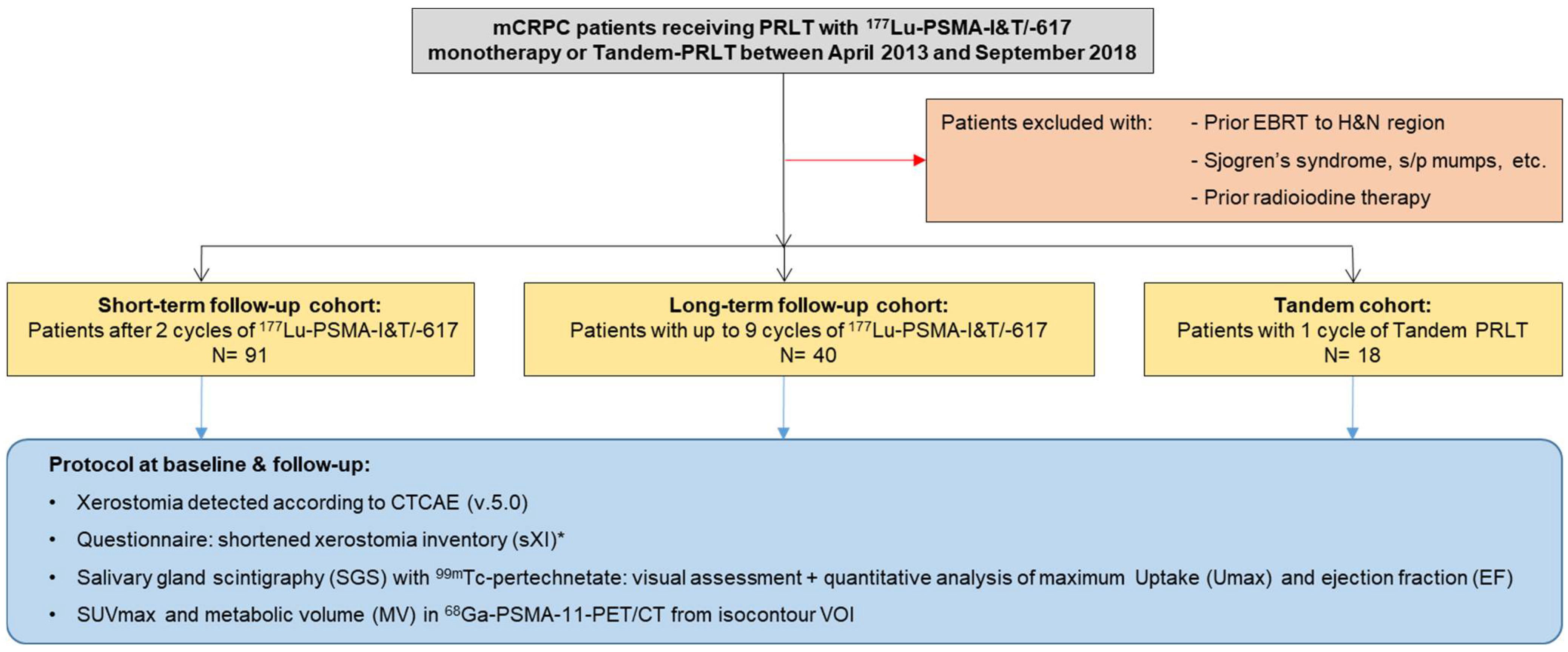
2.1. Study Design

Due to the retrospective data acquisition and analysis of this investigation, ethical review and approval were waived by the institutional review board. All patients gave their written informed consent. All procedures were in compliance with The German Medicinal Products Act, AMG §13 2b, the conditions of the Declaration of Helsinki article 37 “Unproven interventions in clinical practice“, and the responsible regulatory body (Government of Thuringia).
The institutional eligibility criteria for PRLT have been published before [14,31] and are outlined in Supplementary Table S1. Patients with mCRPC and prior taxane-based chemotherapy, ineligibility for chemotherapy, or explicit refusal of chemotherapy were suitable. A pretherapeutic 68Ga-PSMA-11-PET scan had confirmed intense PSMA expression of the tumor lesions. The dosing of 177Lu-177-PSMA-I&T/-617 and 225Ac-PSMA-617 PRLT were patient-individualized based on, e.g., the tumor burden, renal function, bone marrow reserve, and pretreatments. Tandem-PRLT with the combined administration of 225Ac- and 177Lu-PSMA-617 was indicated in patients with a critically high tumor load or after patients failed or became refractory to 177Lu-PSMA-I&T/-617 monotherapy, respectively [24]. The clinical management and follow-up of PRLT were based on national and international consensus recommendations and guidelines, respectively [32,33]. The radiolabeling and administration of 68Ga-PSMA-11, 177Lu-PSMA-I&T/-PSMA-617, and 225Ac-PSMA-617 were performed as previously published [12,15,21,34].

2.2. Patient Population

From the institutional database, three cohorts of patients receiving their first cycle of either 177Lu-PSMA-I&T/-617 monotherapy or Tandem-PRLT between April 2013 and September 2018 were identified. The exclusion criteria were prior external radiotherapy to the head and neck region; comorbidities known to affect the baseline salivary gland function (e.g., Sjögren’s syndrome, mumps, etc.); or prior radioiodine therapy (Figure 1).
In the first cohort (short-term cohort), 91 patients were included who underwent 2 cycles of 177Lu-PSMA-I&T/-617. The age at first cycle, initial Gleason score, tumor distribution on baseline PSMA-PET/CT, prior mCRPC treatment lines, and the cumulative dose of 177Lu-PSMA-I&T/-617 were acquired. Patients were categorized based on their visually assessed tumor burden on the baseline PSMA-PET/CT (low, moderate, or high; Supplementary Figure S1), as previously described [35]. Additionally, three evenly distributed, age-based subgroups were created (<65 years, 65–72 years, and >72 years at PRLT initiation) in this cohort. In a second cohort (long-term cohort), 40 mCRPC patients were included for analysis on a long-term follow-up after 177Lu-PSMA-I&T/-617 monotherapy. Similar to the short-term cohort, the baseline parameters and the cumulative dose of 177Lu-PSMA-I&T/-617 were obtained. The time between the first PRLT cycle and last follow-up was calculated. Three evenly distributed subgroups based on the cumulatively administered activity of 177Lu-PSMA-I&T/-617 were created (<30 GBq, 30–40 GBq, and >40 GBq).
The third cohort consisted of 18 patients with available follow-up data after one cycle of Tandem-PRLT (Tandem cohort). Similarly, the baseline parameters and cumulative administered activity of 177Lu-PSMA-I&T/-617 alone or of 225Ac- and 177Lu-PSMA-617 during Tandem-PRLT were collected.

2.3. Assessment of Salivary Gland Function

The workflow based on a protocol of parameters obtained from patient charts and diagnostic procedures in all three patient cohorts is outlined in Figure 1. Xerostomia was documented according to the CTCAE (version 5.0) and results of a validated questionnaire—the shortened xerostomia inventory (sXI, five xerostomia-related items, summated score between 5 and 25 points; Supplementary Table S2) [36,37]-were acquired. Dynamic salivary gland scintigraphy (SGS) was performed as previously described, using 70 ± 10 MBq 99mTc-pertechnetate [38,39]. To avoid premature stimulation of the SG, patients were asked to fast for at least 6 h prior to the SGS, brushing of their teeth, and additional potential salivary stimulators, e.g., chewing gum, were not allowed. A standardized excretion stimulus of 5 mL of lemon juice was administered orally 20 min after tracer injection. First, SGS were visually assessed based on established stages of SG dysfunction [30]. The SG function was graded from stage 0 (normal) to stage 3 (severely impaired) based on the qualitatively evaluated tracer uptake and excretion (Supplementary Table S3). Subsequently, the maximum tracer uptake (Umax) and the excretion fraction (EF) of the parotid (PG) and submandibular glands (SMG) were calculated using manually drawn regions of interest (ROI) (Figure 2). From PSMA-PET/CT (acquired 60–80 min after i.v. injection of 1.8–2.2 MBq 68Ga-68-PSMA-11 per kg bodyweight), the SUVmax and the metabolic volume (MV) of PG and SMG were acquired. For this purpose, standardized, 3-dimensional volumes of interest (VOIs) with an isocontour threshold of 20% of the SUVmax were used, as previously described [40,41]. To investigate influences on the baseline SG function and predictors for the SG toxicity of 177Lu-PSMA-I&T/-617, age at PRLT initiation, previous taxane-based chemotherapy, tumor burden on the baseline PSMA PET/CT, and the cumulatively administered activity of 177Lu-PSMA-I&T/-617 were assessed as independent factors.

2.4. Statistical Analysis

The results are reported as the frequency (percentage), median with 25–75th percentiles (IQR), median with range, mean ± standard deviation (SD), ranges, and/or 95% confidence intervals, as noted accordingly. In the case of normal distribution, a Student’s t-test and univariate analysis of variance (one-way ANOVA) with a post hoc analysis using Tukey’s HSD test were applied. In the case of non-normal distribution, the Wilcoxon’s signed ranks test, Mann–Whitney U test, and Kruskal–Wallis H test were performed.
For the univariate analysis, the mean values of all 4 investigated SG were calculated; a multivariate analysis of variance (one-way MANOVA) was conducted with a post hoc analysis using the Bonferroni procedure and the Scheffe test. A correlation analysis with subsequent linear regression was performed using Pearson’s or Spearman’s correlation, as outlined. For each test, a p-value of less than 0.05 was considered statistically significant. Statistical analysis was performed using SPSS Statistics, version 24 (IBM Co., Armonk, NY, USA).

3. Results

3.1. Short-Term Follow-Up Cohort

Characteristics of the 91 patients of the short-term cohort (median age: 68 years; range 46–90) are outlined in Table 1. All patients had been administered two cycles of 177Lu-PSMA-I&T/-617 (median 14.3 GBq, range 9.5–20.2), with a median follow-up of 2.3 months (range 1.9–2.7). Forty-four (48.4%) patients of this cohort had been pretreated with taxane-based chemotherapy.
Grade 1 xerostomia was reported in 13 (14.3%) patients at the baseline and in 22 (24.2%) patients after two cycles of Lu-177 PSMA-I&T/-617 (p < 0.01), with a correlated significant increase of the median sXI-score from 7 (IQR 5.3–9) before to 8 (IQR 6.3–11) after PRLT (p < 0.05). In addition, a moderate correlation of xerostomia symptoms and the sXI-score was found during follow-up (r = 0.43, p < 0.01).
Visual SGS grading before and after PRLT showed no significant difference (median stage 0 (range 0–2) vs. median stage 0 (range 0–2), p = 0.34). Of note, a mildly impaired SG function (stage 1 to 2) was detectable already at the baseline in 33 (36.3%) patients. A quantitative assessment confirmed no significant changes of Umax and EF after PRLT (Table 2). However, patients that reported dryness of mouth after PRLT showed significantly lower Umax and EF values of the PG at the follow-up than did asymptomatic patients (mean Umax: 0.27 ± 0.09(SD) vs. 0.35 ± 0.12(SD), p < 0.01; mean EF: 46.0% ± 13.8(SD) vs. 56.2% ± 11.3(SD), p < 0.01). No similar correlation was detectable for the SMG. The SUVmax of both the PG and SMG showed no significant changes, whereas the MV of all four SG declined significantly (p < 0.001, Table 2).

3.2. Long-Term Follow-Up Cohort

In this cohort, 40 patients were included, receiving a median of 5.5 (range 2–9) cycles of PRLT and a median cumulative activity of 35.3 GBq (range 9.9–61.8) 177Lu-PSMA-I&T/-617. The median follow up was 22.7 months (IQR 16.4–30.2). Nineteen (47.5%) patients had prior taxane-based chemotherapy. The patient details are outlined in Table 1.
From the baseline to follow-up, xerostomia became more frequent (grade 1 in 2 (5%) patients at baseline, grade 1 in 15 (37.5%) patients, and grade 2 in 1 (2.5%) patient at follow-up; p < 0.001). No grade 3 xerostomia occurred. The data of the sXI questionnaires were available only at the follow-up, showing a moderate but significant correlation to the subjective dryness of mouth (r = 0.41, p < 0.05).
In the visual grading, no significant changes were found on SGS after PRLT (stage 0 in 16 (40%) patients, stage 1 in 16 (40%) patients, and stage 2 in 8 (20%) patients at the baseline; stage 0 in 16 (40%) patients, stage 1 in 20 (50%) patients, and stage 2 in 4 (10%) patients at the follow-up; p = 0.63). A comparison of the Umax and EF of all SG confirmed no significant changes before and after PRLT (Table 3). PRLT-emergent xerostomia was also significantly correlated with a lower EF of the PG in this cohort (r = −0.50, p < 0.01). In patients without dryness of the mouth, the EF of the PG at follow-up were significantly higher than in patients with xerostomia (EF 54.0% ± 11.7 (SD) vs. 40.0% ± 16.4 (SD); p < 0.001). No correlation of the Umax to the subjective complaints was found, and no similar correlation could be demonstrated for the submandibular glands. There was a trend towards a lower SUVmax of all SGs after PRLT; however, without statistical significance (Table 3), PSMA-PET/CT showed a significant decline of the MV of all SGs (p < 0.001), with a median of −6.0% (95%CI: −18.3 to −2.8%) to −11.6% (95%CI: −18.1 to −7.8%) (Figure 3).

3.3. Tandem-Cohort PRLT

Eighteen patients were identified for analysis that had been administered a median activity of 4.0 MBq (range 2.0–7.0) 225Ac-225 PSMA-617 and 4.25 GBq 177Lu-PSMA-617 (range 3.6–7.2 GBq) for Tandem-PRLT. Seven (38.9%) patients had been pretreated with taxane-based chemotherapy, and fourteen (77.8%) patients had been treated with 177Lu-PSMA-I&T/-617 monotherapy before Tandem-PRLT was initiated. Further patient details are shown in Table 1.
One-third (6/18) of the patients reported grade 1 xerostomia at the baseline, while, after one cycle of Tandem-PRLT, xerostomia grade 1 in 10/18 patients and grade 2 in 2/18 patients was observed (p = 0.001), and 6/18 patients did not report any xerostomia at the follow-up. There was no patient-requested treatment discontinuation. The sXI-score increased significantly from 9.5 (95%CI: 7.0–14.2) before to 14.0 (95%CI: 11.5–19.6) after Tandem-PRLT (p = 0.005). The stage of xerostomia at the baseline according to SGS was 0 in 10/18 patients, 1 in 6/18 patients, and 2 in 2/18 patients, while, at the follow-up, stage 0 was noted in 1/18 patients, stage 1 in 8/18 patients, and stage 2 in 9/18 patients (p < 0.001).
The Umax of all four SG showed no significant changes at the follow-up, whereas the EF of all SG declined significantly (p < 0.01; Figure 4). The SUVmax showed a nonsignificant trend towards lower values after treatment (Supplementary Table S4), while the MV of all four SG decreased significantly (p < 0.05; right PG: median −12.2% (95%CI: −21.6 to −6.1); left PG: median –7.8% (95%CI: −20.5 to −0.1); right SMG: median −8.7% (95%CI: −16.3 to 0.4); left SMG: median –6.5% (95%CI: −12.1 to 1.1)).

3.4. Co-Factors for Salivary Gland Toxicity

Significantly higher Umax values of the SG both during the baseline and on the follow-up SGS were observed in older patients in the subgroup analysis (p < 0.001). However, no influence of the patient’s age on EF, the PSMA-PET/CT-based parameters and reported xerostomia was found. Chemotherapy pretreated patients in the short-term cohort showed significantly lower SUVmax of all SG both before and after PRLT (baseline median SUVmax: 24.7 vs. 18.2; follow-up median SUVmax: 24.0 vs. 18.8, p < 0.001; Figure 5). However, there was no significant differences between chemotherapy-pretreated and chemotherapy-naïve patients in terms of the xerostomia, MV, and SGS parameters. The SUVmax of all SG showed a significant correlation with the tumor burden at the baseline PSMA PET/CT, with lower SUVmax in patients with higher tumor loads (p < 0.001; Kruskal–Wallis test). No significant differences between the three subgroups stratified by tumor burden in terms of the reported xerostomia, sXI-scores, SGS parameter, and the MV at the baseline and follow-up were found (Supplementary Figure S2).
In the long-term cohort, patients with higher cumulative administered activity showed lower SUVmax at the follow-up PSMA-PET/CT (p < 0.05; Kruskal–Wallis test). However, no influence of the cumulatively administered 177Lu-PSMA-I&T/-617 on the MV, Umax, EF, the reported xerostomia, and the sXI-scores was found at the follow-up (Supplementary Figure S3).

4. Discussion

In this single-center, retrospective investigation of SG toxicity of 177Lu-PSMA-I&T/-617 PRLT in 91 patients with short-term and in 40 patients with long-term follow-up, only mild-to-moderate subjective and objective SG dysfunction was observed in only a minority of the patients. In contrast, only one cycle of Tandem-PRLT caused a much more distinct salivary gland impairment.
Until today, salivary gland toxicity, especially of 225Ac-PSMA-I&T/-617 PRLT, remains an unsolved clinical issue, while the data on sialotoxicity after 177Lu-PSMA-I&T/-617 appears heterogeneous. In early retrospective studies, grade 1 to 2 xerostomia was reported in 8–24% of cases [15,16,17], while, in a phase II trial, xerostomia occurred in 87% of the patients [18] and in 38.8% of the patients in a phase III trial [19]. In our study, xerostomia grade 1 was observed in 24.2% of the patients after two cycles of 177Lu-PSMA-I&T/-617, while 40% of the patients reported grade 1 to 2 xerostomia during the long-term follow-up (maximum follow-up of 52 months), receiving up to 61.8 GBq. Previously, a transient character of xerostomia has been described by several authors, usually resolving 3 months after therapy [14,17]. Therefore, the time point of interviewing patients for dryness of mouth appears crucial. For the quantification of xerostomia, we used a validated questionnaire with a good symptomatic correlation in short-term and long-term follow-up.
SGS has been previously used to objectify SG dysfunction after, e.g., external radiotherapy [42,43,44] and radioiodine therapy [30,45], since a good correlation to xerostomia and saliva flow rates has been shown, and SG impairments can be detected already at early stages [39,46]. While, in our study, no intra-patient changes on SGS after two cycles of 177Lu-PSMA-I&T/-617 were found, patients with xerostomia at follow-up showed lower values of the PG for both Umax and EF. These findings are in line with the data after radioiodine therapy in patients with differentiated thyroid cancer, in which a bilateral PG dysfunction was the most common condition [47]. Already, after low doses of 0.4–0.6 GBq 131I, a 14% decline of the Umax was observed, while, after 24 GBq of 131I, the Umax decreased by 90% [38]. The SGS in head and neck cancer patients after external radiotherapy showed a 50% decline of the PG EF [44]. Another study reported a significant decrease of the tracer uptake of the PG after radiotherapy, with an EF decline from 44.7% to 18.7% post-therapeutically [42].
At the early follow-up, no significant changes of the SUVmax were found in our cohort, whereas a significant decline of the MV was observed, confirmed in the long-term cohort, in which the MV declined between a median 6.0 and 11.6%. Lower SUVmax were detected at the long-term follow-up; however, this was without statistical significance, possibly due to the low patient number. In addition, patients who received a higher cumulative activity showed a significantly lower SUVmax at the follow-up. Comparable results were previously published, with a decline of 20% in the PG volume and of 9.8% in the SMG and a decrease in the SUVmax by 6% for the PG and 10.5% for the SMG after two to three cycles of 177Lu-PSMA-617 [40]. Therefore, SUVmax and MV might act as early indicators of SG toxicity. However, these findings need to be confirmed on a larger scale. Zhao et al. described the 68Ga-PSMA-11 PET/CT parameters for SG as a helpful supplement to SGS in patients with different degrees of SG dysfunction due to either Sjögren’s syndrome, radiotherapy for head and neck cancer, or after surgery of the SGs [41].
However, some limitations of using PSMA PET/CT for evaluation of the SG function must be stated. Both a high intra- and interindividual variability of the SG SUVmax were demonstrated [48], which could be confirmed in our study. Moreover, PSMA ligand uptake cannot assess the excretion function of the SGs and might be affected by the tumor sink effect [49,50]. Consequently, a significant negative correlation of the SUVmax of all SGs to the tumor burden both pre- and post-therapeutically was found in our data.
Overall, the parameters of SG function investigated in this study suggest a combined use of PSMA-PET/CT and SGS for the screening of early SG toxicity. While the tracer uptake (SUVmax on PET/CT and Umax on SGS) showed a lower correlation to the clinical symptoms, changes of the MV on PET/CT and the EF on SGS might be useful, sensitive surrogates of SG dysfunction.
No influence of prior chemotherapy on the SGS parameter or the subjective parameter of hyposalivation was detected. However, significantly lower SUVmax were observed in chemotherapy-pretreated patients. Another study comparing PRLT in chemotherapy-naïve and chemotherapy-pretreated patients found no difference in the frequency of xerostomia between both groups [51].
A median absorbed dose to SG of between 0.5 and 1.4 Gy/GBq, administering 177Lu-PSMA-I&T/-617, has been published [14,15,52,53], which translates to an estimated mean absorbed dose of 3.0–8.4 Gy per cycle of 6.0 GBq or of 3.8–10.5 Gy/cycle of 7.5 GBq 177Lu-PSMA-I&T/-617, respectively.
A recent study comparing the biodistribution and dosimetry data of 177Lu-PSMA-I&T and 177Lu-PSMA-617 in 138 mCRPC patients demonstrated no significant difference in the absorbed SG dose of both PSMA targeting small molecules [54].
The threshold of the maximum tolerable SG dose of external radiotherapy is still a subject of discussion, with 26–45 Gy being proposed for the PG [55,56]. Based on this, Scarpa et al. estimated a maximum tolerable activity of 177Lu-PSMA of 60 GBq, assuming a mean dose of 0.5 Gy/GBq for the SG [40]. However, concerning the limited radiobiological comparability of external irradiation and endoradiotherapies, such estimates should be undertaken with caution.
First, the clinical data for 225Ac-PSMA-617 in mCRPC patients were published by the Heidelberg group, demonstrating encouraging responses, even after failing to find 177Lu-PSMA-617 [20,22]. However, 4 of the initial 40 patients discontinued treatment because of severe xerostomia or loss of taste, despite the initial response. Further, 15 patients with partial remission eventually stopped treatment due to xerostomia. Another study reported severe dryness of mouth after 225Ac-PSMA-617 that led to treatment discontinuation in about one-third of the patients [23]. Sathekge et al. suggested a dose deescalating scheme of 225Ac-PSMA-617 [57]. In 73 mCRPC patients, xerostomia grade 1 to 2 was reported in 85% of the patients, while no patient showed a grade 3 disease, and no treatment discontinuation occurred. In 12/18 patients (66.7%), our study showed grade 1 to 2 dryness of mouth after one cycle of Tandem-PRLT, which correlated with a significant increase in the sXI-score. On the other hand, one-third of the patients did not notice any dryness of mouth, and no grade 3 xerostomia or therapy discontinuation emerged. However, the limited comparability of the published studies to our cohort must be underlined in terms of, e.g., applied activities, cycles of 225Ac-PSMA-617, and prior treatments.
Khreish et al. investigated the concept of co-administering lower activities 225Ac-PSMA-617 (mean 5.3 MBq) and 177Lu-PSMA-617 (mean 6.9 GBq) in 20 mCRPC patients and found comparable results. In 8/20 patients, they found grade 1 and 5/20 patients grade 2 xerostomia. No grade 3 dryness of mouth and no treatment discontinuation was observed [58].
In another study, SGS in 11 patients after up to four cycles of 225Ac-PSMA-617 showed a distinct decline of both the Umax and the EF [37]. In contrast, in our study, we found no changes of the Umax after Tandem-PRLT. However, the EF of all SG declined significantly.
A reason for xerostomia after 225Ac-PSMA-617 PRLT might be a severe duct stenosis or obstruction, as it has been described before in patients after radioiodine therapy and external radiotherapy [42,59]. Consequently, the patients showed a response to sialendoscopic duct dilatation and saline irrigation [37].
Preventing the SG uptake of PSMA-targeting radiopharmaceuticals is still a clinical unmet need, and different approaches were investigated in the past [60,61]. Additionally, external cooling [62,63], orally administered monosodium glutamate [64,65], and recently, high-dose botulinum toxin injections for radioprotection of the PG and SMG were clinically tested [66,67], with preliminary encouraging data on the effectiveness.
The limitations of our study included its retrospective and single-center design, resulting in a potential selection bias of the cases and limited statistical power of the analysis, especially for the subgroup investigations in the short-term and long-term cohorts and the analysis of the Tandem cohort. However, our data might encourage future prospective, multi-institutional studies to investigate our findings on a larger scale.

5. Conclusions

Salivary gland dysfunction after 177Lu-PSMA-I&T/-617 PRLT has minor clinical relevance, both subjectively and objectively. Even after high cumulative activities, only mild-to-moderate dryness of mouth occurs in a minority of the patients. The prevalence of xerostomia appears to be significantly lower than the historical controls after external radiotherapy, radioiodine therapy, and especially after PRLT with 225Ac-PSMA-617. A validated questionnaire on xerostomia, salivary gland scintigraphy, and PSMA-PET/CT parameters can help to objectify, standardize, and quantify the SG toxicity of PRLT. A decrease of the excretion fraction on SGS and of the metabolic volume on PSMA PET/CT can be early indicators of SG impairment. The co-administration of lower doses of 225Ac in combination with 177Lu-PSMA-617 (Tandem concept) can decrease severe xerostomia after PRLT with alpha-emitters.

Supplementary Materials

The following supporting information can be downloaded at: https://www.mdpi.com/article/10.3390/diagnostics12081926/s1, Figures S1–S3; Tables S1–S4.

Author Contributions

Conceptualization, T.L. and R.P.B.; Formal analysis, T.L.; Investigation, T.L.; Methodology, T.L., H.R.K. and R.P.B.; Resources, C.S. and D.M.; Supervision, H.R.K. and R.P.B.; Writing—original draft, T.L.; and Writing—review and editing, H.R.K., C.S., D.M., G.F.V. and R.P.B. All authors have read and agreed to the published version of the manuscript.

Funding

This research received no external funding.

Institutional Review Board Statement

The study was conducted according to the guidelines of the Declaration of Helsinki, and ethical review and approval were waived for this study, due the retrospective data acquisition and analysis.

Informed Consent Statement

Informed consent was obtained from all subjects involved in the study.

Data Availability Statement

The data presented in this study are available on request from the corresponding author. The data are not publicly available due to privacy restrictions.

Acknowledgments

We thank the patients who participated in this study and all research staff for their support and the physician colleagues, nursing staff, and nuclear medicine technologists of the radioisotope therapy ward D3 for the patient care, past and present at Zentralklinik Bad Berka.

Conflicts of Interest

The authors declare no conflict of interest.

References

  1. Bray, F.; Ferlay, J.; Soerjomataram, I.; Siegel, R.L.; Torre, L.A.; Jemal, A. Global cancer statistics 2018: GLOBOCAN estimates of incidence and mortality worldwide for 36 cancers in 185 countries. CA Cancer J. Clin. 2018, 68, 394–424. [Google Scholar] [CrossRef] [PubMed] [Green Version]
  2. Loeb, S.; Schaeffer, E.M.; Trock, B.J.; Epstein, J.I.; Humphreys, E.B.; Walsh, P.C. What Are the Outcomes of Radical Prostatectomy for High-risk Prostate Cancer? Urology 2010, 76, 710–714. [Google Scholar] [CrossRef] [PubMed] [Green Version]
  3. Weiner, A.B.; Matulewicz, R.S.; Eggener, S.E.; Schaeffer, E.M. Increasing incidence of metastatic prostate cancer in the United States (2004–2013). Prostate Cancer Prostatic Dis. 2016, 19, 395–397. [Google Scholar] [CrossRef] [Green Version]
  4. Siegel, R.L.; Miller, K.D.; Jemal, A. Cancer statistics, 2019. CA Cancer J. Clin. 2019, 69, 7–34. [Google Scholar] [CrossRef] [PubMed] [Green Version]
  5. Halabi, S.; Vogelzang, N.J.; Kornblith, A.B.; Ou, S.S.; Kantoff, P.W.; Dawson, N.A.; Small, E.J. Pain Predicts Overall Survival in Men With Metastatic Castration-Refractory Prostate Cancer. J. Clin. Oncol. 2008, 26, 2544–2549. [Google Scholar] [CrossRef] [PubMed]
  6. Silver, D.A.; Pellicer, I.; Fair, W.R.; Heston, W.D.; Cordon-Cardo, C. Prostate-specific membrane antigen expression in normal and malignant human tissues. Clin. Cancer Res. 1997, 3, 81–85. [Google Scholar] [PubMed]
  7. Bostwick, D.G.; Pacelli, A.; Blute, M.; Roche, P.; Murphy, G.P. Prostate specific membrane antigen expression in prostatic intraepithelial neoplasia and adenocarcinoma: A study of 184 cases. Cancer 1998, 82, 2256–2261. [Google Scholar] [CrossRef]
  8. Bander, N.H.; Trabulsi, E.J.; Kostakoglu, L.; Yao, D.; Vallabhajosula, S.; Smith-Jones, P.; Joyce, M.A.; Milowsky, M.; Nanus, D.M.; Goldsmith, S.J. Targeting Metastatic Prostate Cancer With Radiolabeled Monoclonal Antibody J591 to the Extracellular Domain of Prostate Specific Membrane Antigen. J. Urol. 2003, 170, 1717–1721. [Google Scholar] [CrossRef]
  9. Mannweiler, S.; Amersdorfer, P.; Trajanoski, S.; Terrett, J.A.; King, D.; Mehes, G. Heterogeneity of Prostate-Specific Membrane Antigen (PSMA) Expression in Prostate Carcinoma with Distant Metastasis. Pathol. Oncol. Res. 2009, 15, 167–172. [Google Scholar] [CrossRef] [PubMed]
  10. Pomper, M.G.; Musachio, J.L.; Zhang, J.; Scheffel, U.; Zhou, Y.; Hilton, J.; Maini, A.; Dannals, R.F.; Wong, D.F.; Kozikowski, A.P. 11C-MCG: Synthesis, Uptake Selectivity, and Primate PET of a Probe for Glutamate Carboxypeptidase II (NAALADase). Mol. Imaging 2002, 1, 96–101. [Google Scholar] [CrossRef] [PubMed]
  11. Eder, M.; Schafer, M.; Bauder-Wust, U.; Hull, W.E.; Wangler, C.; Mier, W.; Haberkorn, U.; Eisenhut, M. 68Ga-Complex Lipophilicity and the Targeting Property of a Urea-Based PSMA Inhibitor for PET Imaging. Bioconjug. Chem. 2012, 23, 688–697. [Google Scholar] [CrossRef] [PubMed]
  12. Afshar-Oromieh, A.; Malcher, A.; Eder, M.; Eisenhut, M.; Linhart, H.G.; Hadaschik, B.A.; Holland-Letz, T.; Giesel, F.L.; Kratochwil, C.; Haufe, S.; et al. PET imaging with a [68Ga]gallium-labelled PSMA ligand for the diagnosis of prostate cancer: Biodistribution in humans and first evaluation of tumour lesions. Eur. J. Nucl. Med. Mol. Imaging 2013, 40, 486–495. [Google Scholar] [CrossRef] [PubMed]
  13. Zechmann, C.M.; Afshar-Oromieh, A.; Armor, T.; Stubbs, J.B.; Mier, W.; Hadaschik, B.; Joyal, J.; Kopka, K.; Debus, J.; Babich, J.W.; et al. Radiation dosimetry and first therapy results with a 124I/131I-labeled small molecule (MIP-1095) targeting PSMA for prostate cancer therapy. Eur. J. Nucl. Med. Mol. Imaging 2014, 41, 1280–1292. [Google Scholar] [CrossRef] [PubMed] [Green Version]
  14. Baum, R.P.; Kulkarni, H.R.; Schuchardt, C.; Singh, A.; Wirtz, M.; Wiessalla, S.; Schottelius, M.; Mueller, D.; Klette, I.; Wester, H.J. 177Lu-Labeled Prostate-Specific Membrane Antigen Radioligand Therapy of Metastatic Castration-Resistant Prostate Cancer: Safety and Efficacy. J. Nucl. Med. 2016, 57, 1006–1013. [Google Scholar] [CrossRef] [Green Version]
  15. Kratochwil, C.; Giesel, F.L.; Stefanova, M.; Benesova, M.; Bronzel, M.; Afshar-Oromieh, A.; Mier, W.; Eder, M.; Kopka, K.; Haberkorn, U. PSMA-Targeted Radionuclide Therapy of Metastatic Castration-Resistant Prostate Cancer with 177Lu-Labeled PSMA-617. J. Nucl. Med. 2016, 57, 1170–1176. [Google Scholar] [CrossRef] [Green Version]
  16. Rahbar, K.; Ahmadzadehfar, H.; Kratochwil, C.; Haberkorn, U.; Schafers, M.; Essler, M.; Baum, R.P.; Kulkarni, H.R.; Schmidt, M.; Drzezga, A.; et al. German Multicenter Study Investigating 177Lu-PSMA-617 Radioligand Therapy in Advanced Prostate Cancer Patients. J. Nucl. Med. 2017, 58, 85–90. [Google Scholar] [CrossRef] [Green Version]
  17. Heck, M.M.; Tauber, R.; Schwaiger, S.; Retz, M.; D’Alessandria, C.; Maurer, T.; Gafita, A.; Wester, H.J.; Gschwend, J.E.; Weber, W.A.; et al. Treatment Outcome, Toxicity, and Predictive Factors for Radioligand Therapy with 177Lu-PSMA-I&T in Metastatic Castration-resistant Prostate Cancer. Eur. Urol. 2019, 75, 920–926. [Google Scholar] [CrossRef]
  18. Hofman, M.S.; Violet, J.; Hicks, R.J.; Ferdinandus, J.; Thang, S.P.; Akhurst, T.; Iravani, A.; Kong, G.; Kumar, A.R.; Murphy, D.G.; et al. [177Lu]-PSMA-617 radionuclide treatment in patients with metastatic castration-resistant prostate cancer (LuPSMA trial): A single-centre, single-arm, phase 2 study. Lancet Oncol. 2018, 19, 825–833. [Google Scholar] [CrossRef]
  19. Sartor, O.; de Bono, J.; Chi, K.N.; Fizazi, K.; Herrmann, K.; Rahbar, K.; Tagawa, S.T.; Nordquist, L.T.; Vaishampayan, N.; El-Haddad, G.; et al. Lutetium-177–PSMA-617 for Metastatic Castration-Resistant Prostate Cancer. N. Engl. J. Med. 2021, 385, 1091–1103. [Google Scholar] [CrossRef]
  20. Kratochwil, C.; Bruchertseifer, F.; Giesel, F.L.; Weis, M.; Verburg, F.A.; Mottaghy, F.; Kopka, K.; Apostolidis, C.; Haberkorn, U.; Morgenstern, A. 225Ac-PSMA-617 for PSMA-Targeted α-Radiation Therapy of Metastatic Castration-Resistant Prostate Cancer. J. Nucl. Med. 2016, 57, 1941–1944. [Google Scholar] [CrossRef] [Green Version]
  21. Kratochwil, C.; Bruchertseifer, F.; Rathke, H.; Bronzel, M.; Apostolidis, C.; Weichert, W.; Haberkorn, U.; Giesel, F.L.; Morgenstern, A. Targeted α-Therapy of Metastatic Castration-Resistant Prostate Cancer with 225Ac-PSMA-617: Dosimetry Estimate and Empiric Dose Finding. J. Nucl. Med. 2017, 58, 1624–1631. [Google Scholar] [CrossRef] [PubMed] [Green Version]
  22. Kratochwil, C.; Bruchertseifer, F.; Rathke, H.; Hohenfellner, M.; Giesel, F.L.; Haberkorn, U.; Morgenstern, A. Targeted α-Therapy of Metastatic Castration-Resistant Prostate Cancer with 225Ac-PSMA-617: Swimmer-Plot Analysis Suggests Efficacy Regarding Duration of Tumor Control. J. Nucl. Med. 2018, 59, 795–802. [Google Scholar] [CrossRef] [Green Version]
  23. Feuerecker, B.; Tauber, R.; Knorr, K.; Heck, M.; Beheshti, A.; Seidl, C.; Bruchertseifer, F.; Pickhard, A.; Gafita, A.; Kratochwil, C.; et al. Activity and Adverse Events of Actinium-225-PSMA-617 in Advanced Metastatic Castration-resistant Prostate Cancer After Failure of Lutetium-177-PSMA. Eur. Urol. 2021, 79, 343–350. [Google Scholar] [CrossRef] [PubMed]
  24. Kulkarni, H.; Zhang, J.; Langbein, T.; Schuchardt, C.; Singh, A.; Mueller, D.; Baum, R. Radioligand therapy using combination of Ac-225 and Lu-177 labelled PSMA ligands for progressive end-stage metastatic prostate cancer: Effective trade-off between response and toxicity. J. Nucl. Med. 2019, 60, 464. [Google Scholar]
  25. Tonnesmann, R.; Meyer, P.T.; Eder, M.; Baranski, A.C. [177Lu]Lu-PSMA-617 Salivary Gland Uptake Characterized by Quantitative In Vitro Autoradiography. Pharmaceuticals 2019, 12, 18. [Google Scholar] [CrossRef] [PubMed] [Green Version]
  26. Rupp, N.J.; Umbricht, C.A.; Pizzuto, D.A.; Lenggenhager, D.; Topfer, A.; Muller, J.; Muhlematter, U.J.; Ferraro, D.A.; Messerli, M.; Morand, G.B.; et al. First Clinicopathological Evidence of a Non–PSMA-Related Uptake Mechanism for 68Ga-PSMA-11 in Salivary Glands. J. Nucl. Med. 2019, 60, 1270–1276. [Google Scholar] [CrossRef] [Green Version]
  27. Dirix, P.; Nuyts, S.; Van den Bogaert, W. Radiation-induced xerostomia in patients with head and neck cancer: A literature review. Cancer 2006, 107, 2525–2534. [Google Scholar] [CrossRef]
  28. Mandel, S.J.; Mandel, L. Radioactive Iodine and the Salivary Glands. Thyroid 2003, 13, 265–271. [Google Scholar] [CrossRef] [Green Version]
  29. Van Nostrand, D. Sialoadenitis secondary to 131I therapy for well-differentiated thyroid cancer. Oral Dis. 2011, 17, 154–161. [Google Scholar] [CrossRef]
  30. Solans, R.; Bosch, J.-A.; Galofre, P.; Porta, F.; Jose, M.V. Salivary and Lacrimal Gland Dysfunction (Sicca Syndrome) After Radioiodine Therapy. J. Nucl. Med. 2001, 42, 738–743. [Google Scholar]
  31. Kulkarni, H.R.; Singh, A.; Schuchardt, C.; Niepsch, K.; Sayeg, M.; Leshch, Y.; Wester, H.J.; Baum, R.P. PSMA-Based Radioligand Therapy for Metastatic Castration-Resistant Prostate Cancer: The Bad Berka Experience Since 2013. J. Nucl. Med. 2016, 57, 97S–104S. [Google Scholar] [CrossRef] [PubMed] [Green Version]
  32. Fendler, W.P.; Kratochwil, C.; Ahmadzadehfar, H.; Rahbar, K.; Baum, R.P.; Schmidt, M.; Pfestroff, A.; Lutzen, U.; Prasad, V.; Heinzel, A.; et al. 177Lu-PSMA-617 therapy, dosimetry and follow-up in patients with metastatic castration-resistant prostate cancer. Nukl. Nucl. 2016, 55, 123–128. [Google Scholar]
  33. Kratochwil, C.; Fendler, W.P.; Eiber, M.; Baum, R.; Bozkurt, M.F.; Czernin, J.; Bolton, R.C.D.; Ezziddin, S.; Forrer, F.; Hicks, R.J.; et al. EANM procedure guidelines for radionuclide therapy with 177Lu-labelled PSMA-ligands (177Lu-PSMA-RLT). Eur. J. Nucl. Med. Mol. Imaging 2019, 46, 2536–2544. [Google Scholar] [CrossRef]
  34. Weineisen, M.; Simecek, J.; Schottelius, M.; Schwaiger, M.; Wester, H.J. Synthesis and preclinical evaluation of DOTAGA-conjugated PSMA ligands for functional imaging and endoradiotherapy of prostate cancer. EJNMMI Res. 2014, 4, 63. [Google Scholar] [CrossRef] [PubMed] [Green Version]
  35. Gaertner, F.C.; Halabi, K.; Ahmadzadehfar, H.; Kurpig, S.; Eppard, E.; Kotsikopoulos, C.; Liakos, N.; Bundschuh, R.A.; Strunk, H.; Essler, M. Uptake of PSMA-ligands in normal tissues is dependent on tumor load in patients with prostate cancer. Oncotarget 2017, 8, 55094–55103. [Google Scholar] [CrossRef] [PubMed] [Green Version]
  36. Thomson, W.M.; van der Putten, G.J.; de Baat, C.; Ikebe, K.; Matsuda, K.I.; Enoki, K.; Hopcraft, M.S.; Ling, G.Y. Shortening the Xerostomia Inventory. Oral Surg. Oral Med. Oral Pathol. Oral Radiol. Endodontology 2011, 112, 322–327. [Google Scholar] [CrossRef] [Green Version]
  37. Rathke, H.; Kratochwil, C.; Hohenberger, R.; Giesel, F.L.; Bruchertseifer, F.; Flechsig, P.; Morgenstern, A.; Hein, M.; Plinkert, P.; Haberkorn, U.; et al. Initial clinical experience performing sialendoscopy for salivary gland protection in patients undergoing 225Ac-PSMA-617 RLT. Eur. J. Nucl. Med. Mol. Imaging 2019, 46, 139–147. [Google Scholar] [CrossRef]
  38. Bohuslavizki, K.H.; Brenner, W.; Lassmann, S.; Tinnemeyer, S.; Kalina, S.; Clausen, M.; Henze, E. Quantitative salivary gland scintigraphy--a recommended examination prior to and after radioiodine therapy. Nukl. Nucl. 1997, 36, 103–109. [Google Scholar]
  39. Klutmann, S.; Bohuslavizki, K.H.; Kroger, S.; Bleckmann, C.; Brenner, W.; Mester, J.; Clausen, M. Quantitative salivary gland scintigraphy. J. Nucl. Med. Technol. 1999, 27, 20–26. [Google Scholar]
  40. Scarpa, L.; Buxbaum, S.; Kendler, D.; Fink, K.; Bektic, J.; Gruber, L.; Decristoforo, C.; Uprimny, C.; Lukas, P.; Horninger, W.; et al. The 68Ga/177Lu theragnostic concept in PSMA targeting of castration-resistant prostate cancer: Correlation of SUVmax values and absorbed dose estimates. Eur. J. Nucl. Med. Mol. Imaging 2017, 44, 788–800. [Google Scholar] [CrossRef]
  41. Zhao, Y.; Xia, Y.; Liu, H.; Wang, Z.; Chen, Y.; Zhang, W. Potential Applications of 68Ga-PSMA-11 PET/CT in the Evaluation of Salivary Gland Uptake Function: Preliminary Observations and Comparison with 99mTcO4−Salivary Gland Scintigraphy. Contrast Media Mol. Imaging 2020, 2020, 1097516. [Google Scholar] [CrossRef] [PubMed]
  42. Roesink, J.M.; Moerland, M.A.; Hoekstra, A.; Van Rijk, P.P.; Terhaard, C.H. Scintigraphic assessment of early and late parotid gland function after radiotherapy for head-and-neck cancer: A prospective study of dose–volume response relationships. Int. J. Radiat. Oncol. Biol. Phys. 2004, 58, 1451–1460. [Google Scholar] [CrossRef]
  43. Münter, M.W.; Karger, C.P.; Hoffner, S.G.; Hof, H.; Thilmann, C.; Rudat, V.; Nill, S.; Wannenmacher, M.; Debus, J. Evaluation of salivary gland function after treatment of head-and-neck tumors with intensity-modulated radiotherapy by quantitative pertechnetate scintigraphy. Int. J. Radiat. Oncol. Biol. Phys. 2004, 58, 175–184. [Google Scholar] [CrossRef]
  44. Bussels, B.; Maes, A.; Flamen, P.; Lambin, P.; Erven, K.; Hermans, R.; Nuyts, S.; Weltens, C.; Cecere, S.; Lesaffre, E.; et al. Dose–response relationships within the parotid gland after radiotherapy for head and neck cancer. Radiother. Oncol. J. Eur. Soc. Ther. Radiol. Oncol. 2004, 73, 297–306. [Google Scholar] [CrossRef] [PubMed]
  45. Badam, R.K.; Suram, J.; Babu, D.B.; Waghray, S.; Marshal, R.; Bontha, S.C.; Lavanya, R.; Kanth, S. Assessment of Salivary Gland Function Using Salivary Scintigraphy in Pre and Post Radioactive Iodine Therapy in Diagnosed Thyroid Carcinoma Patients. J. Clin. Diagn. Res. 2016, 10, ZC60–ZC62. [Google Scholar] [CrossRef] [PubMed]
  46. Kohn, W.G.; Ship, J.A.; Atkinson, J.C.; Patton, L.L.; Fox, P.C. Salivary gland 99mTc-scintigraphy: A grading scale and correlation with major salivary gland flow rates. J. Oral Pathol. Med. 1992, 21, 70–74. [Google Scholar] [CrossRef]
  47. Jeong, S.Y.; Kim, H.W.; Lee, S.W.; Ahn, B.C.; Lee, J. Salivary Gland Function 5 Years After Radioactive Iodine Ablation in Patients with Differentiated Thyroid Cancer: Direct Comparison of Pre- and Postablation Scintigraphies and Their Relation to Xerostomia Symptoms. Thyroid 2013, 23, 609–616. [Google Scholar] [CrossRef]
  48. Nulent, T.J.W.K.; Valstar, M.H.; de Keizer, B.; Willems, S.M.; Smit, L.A.; Al-Mamgani, A.; Smeele, L.E.; van Es, R.J.J.; de Bree, R.; Vogel, W.V. Physiologic distribution of PSMA-ligand in salivary glands and seromucous glands of the head and neck on PET/CT. Oral Surg. Oral Med. Oral Pathol. Oral Radiol. 2018, 125, 478–486. [Google Scholar] [CrossRef]
  49. Filss, C.; Heinzel, A.; Miiller, B.; Vogg, A.T.J.; Langen, K.J.; Mottaghy, F.M. Relevant tumor sink effect in prostate cancer patients receiving 177Lu-PSMA-617 radioligand therapy. Nukl. Nucl. 2018, 57, 19–25. [Google Scholar] [CrossRef]
  50. Begum, N.J.; Thieme, A.; Eberhardt, N.; Tauber, R.; D’Alessandria, C.; Beer, A.J.; Glatting, G.; Eiber, M.; Kletting, P. The Effect of Total Tumor Volume on the Biologically Effective Dose to Tumor and Kidneys for 177Lu-Labeled PSMA Peptides. J. Nucl. Med. 2018, 59, 929–933. [Google Scholar] [CrossRef] [Green Version]
  51. Barber, T.W.; Singh, A.; Kulkarni, H.R.; Niepsch, K.; Billah, B.; Baum, R.P. Clinical outcomes of (177)Lu-PSMA radioligand therapy in taxane chemotherapy pretreated and taxane chemotherapy naive patients with metastatic castration resistant prostate cancer. J. Nucl. Med. 2019, 60, 955–962. [Google Scholar] [CrossRef] [PubMed] [Green Version]
  52. Okamoto, S.; Thieme, A.; Allmann, J.; D’Alessandria, C.; Maurer, T.; Retz, M.; Tauber, R.; Heck, M.M.; Wester, H.J.; Tamaki, N.; et al. Radiation Dosimetry for177Lu-PSMA I&T in Metastatic Castration-Resistant Prostate Cancer: Absorbed Dose in Normal Organs and Tumor Lesions. J. Nucl. Med. 2017, 58, 445–450. [Google Scholar] [CrossRef] [PubMed] [Green Version]
  53. Violet, J.; Jackson, P.; Ferdinandus, J.; Sandhu, S.; Akhurst, T.; Iravani, A.; Kong, G.; Kumar, A.R.; Thang, S.P.; Eu, P.; et al. Dosimetry of 177Lu-PSMA-617 in Metastatic Castration-Resistant Prostate Cancer: Correlations Between Pretherapeutic Imaging and Whole-Body Tumor Dosimetry with Treatment Outcomes. J. Nucl. Med. 2019, 60, 517–523. [Google Scholar] [CrossRef] [PubMed] [Green Version]
  54. Schuchardt, C.; Zhang, J.; Kulkarni, H.R.; Chen, X.; Müller, D.; Baum, R.P. Prostate-specific membrane antigen radioligand therapy using 177Lu-PSMA I&T and 177Lu-PSMA-617 in patients with metastatic castration-resistant prostate cancer: Comparison of safety, biodistribution and dosimetry. J. Nucl. Med. 2022, 63, 1199–1207. [Google Scholar] [CrossRef]
  55. Eisbruch, A.; Haken, R.K.T.; Kim, H.M.; Marsh, L.H.; Ship, J.A. Dose, volume, and function relationships in parotid salivary glands following conformal and intensity-modulated irradiation of head and neck cancer. Int. J. Radiat. Oncol. Biol. Phys. 1999, 45, 577–587. [Google Scholar] [CrossRef]
  56. Li, Y.; Taylor, J.M.; Haken, R.K.T.; Eisbruch, A. The impact of dose on parotid salivary recovery in head and neck cancer patients treated with radiation therapy. Int. J. Radiat. Oncol. Biol. Phys. 2007, 67, 660–669. [Google Scholar] [CrossRef] [Green Version]
  57. Sathekge, M.; Bruchertseifer, F.; Vorster, M.; Lawal, I.O.; Knoesen, O.; Mahapane, J.; Davis, C.; Reyneke, F.; Maes, A.; Kratochwil, C.; et al. Predictors of Overall and Disease-Free Survival in Metastatic Castration-Resistant Prostate Cancer Patients Receiving 225Ac-PSMA-617 Radioligand Therapy. J. Nucl. Med. 2019, 61, 62–69. [Google Scholar] [CrossRef]
  58. Khreish, F.; Ebert, N.; Ries, M.; Maus, S.; Rosar, F.; Bohnenberger, H.; Stemler, T.; Saar, M.; Bartholoma, M.; Ezziddin, S. 225Ac-PSMA-617/177Lu-PSMA-617 tandem therapy of metastatic castration-resistant prostate cancer: Pilot experience. Eur. J. Nucl. Med. Mol. Imaging 2020, 47, 721–728. [Google Scholar] [CrossRef]
  59. Cung, T.D.; Lai, W.; Svider, P.F.; Hanba, C.; Samantray, J.; Folbe, A.J.; Shkoukani, M.; Raza, S.N. Sialendoscopy in the Management of Radioiodine Induced Sialadenitis: A Systematic Review. Ann. Otol. Rhinol. Laryngol. 2017, 126, 768–773. [Google Scholar] [CrossRef]
  60. Taieb, D.; Foletti, J.M.; Bardies, M.; Rocchi, P.; Hicks, R.J.; Haberkorn, U. PSMA-Targeted Radionuclide Therapy and Salivary Gland Toxicity: Why Does It Matter? J. Nucl. Med. 2018, 59, 747–748. [Google Scholar] [CrossRef] [Green Version]
  61. Langbein, T.; Chausse, G.; Baum, R.P. Salivary Gland Toxicity of PSMA Radioligand Therapy: Relevance and Preventive Strategies. J. Nucl. Med. 2018, 59, 1172–1173. [Google Scholar] [CrossRef] [PubMed] [Green Version]
  62. Yilmaz, B.; Nisli, S.; Ergul, N.; Gursu, R.U.; Acikgoz, O.; Cermik, T.F. Effect of external cooling on Lu-177 PSMA uptake for parotid glands. J. Nucl. Med. 2019, 60, 1388–1393. [Google Scholar] [CrossRef] [PubMed] [Green Version]
  63. Van Kalmthout, L.W.M.; Lam, M.G.E.H.; de Keizer, B.; Krijger, G.C.; Ververs, T.F.T.; De Roos, R.; Braat, A.J.A.T. Impact of external cooling with icepacks on 68Ga-PSMA uptake in salivary glands. EJNMMI Res. 2018, 8, 56. [Google Scholar] [CrossRef] [PubMed]
  64. Rousseau, E.; Lau, J.; Kuo, H.T.; Zhang, Z.; Merkens, H.; Hundal-Jabal, N.; Colpo, N.; Lin, K.S.; Benard, F. Monosodium Glutamate Reduces 68Ga-PSMA-11 Uptake in Salivary Glands and Kidneys in a Preclinical Prostate Cancer Model. J. Nucl. Med. 2018, 59, 1865–1868. [Google Scholar] [CrossRef] [PubMed] [Green Version]
  65. Harsini, S.; Saprunoff, H.; Alden, T.M.; Mohammadi, B.; Wilson, D.; Benard, F. The Effects of Monosodium Glutamate on PSMA Radiotracer Uptake in Men with Recurrent Prostate Cancer: A Prospective, Randomized, Double-Blind, Placebo-Controlled Intraindividual Imaging Study. J. Nucl. Med. 2020, 62, 81–87. [Google Scholar] [CrossRef] [PubMed]
  66. Baum, R.P.; Langbein, T.; Singh, A.; Shahinfar, M.; Schuchardt, C.; Volk, G.F.; Kulkarni, H. Injection of Botulinum Toxin for Preventing Salivary Gland Toxicity after PSMA Radioligand Therapy: An Empirical Proof of a Promising Concept. Nucl. Med. Mol. Imaging 2018, 52, 80–81. [Google Scholar] [CrossRef]
  67. Mueller, J.; Langbein, T.; Mishra, A.; Baum, R.P. Safety of High-Dose Botulinum Toxin Injections for Parotid and Submandibular Gland Radioprotection. Toxins 2022, 14, 64. [Google Scholar] [CrossRef]
Figure 1. Patient cohorts and study workflow. * For the long-term cohort, data of sXI were only available at follow-up. EBRT = external beam radiotherapy; H&N = head and neck; PRLT = PSMA-targeted radioligand therapy.
Diagnostics 12 01926 g001
Figure 2. Normal time–activity curves on salivary gland scintigraphy (left) detected by regions of interest (ROI) over the parotid and submandibular glands (right). Calculation of the excretion fraction (EF): U12–14/U18–20 = tracer uptake averaged from 12–14/18–20 min after tracer injection.
Diagnostics 12 01926 g002
Figure 3. Relative decline of the metabolic volume (MV) of all 4 salivary glands during follow-up PET/CT compared to the baseline in the long-term cohort. (PG = parotid gland; SMG = submandibular gland).
Diagnostics 12 01926 g003
Figure 4. Quantitative analysis of the salivary gland scintigraphy before and after 1 cycle of Tandem-PRLT. While the Umax of all salivary glands showed no significant changes (top), the EF declined significantly in all salivary glands (bottom). PG = parotid gland; SMG = submandibular gland. * Wilcoxon test.
Diagnostics 12 01926 g004
Figure 5. SUVmax of the salivary glands at the baseline and follow-up PSMA PET/CT. PG = parotid gland; SMG = submandibular gland.
Diagnostics 12 01926 g005
Table 1. Characteristics of the 3 investigated patient cohorts. PRLT = radioligand therapy: IQR = interquartile range.
CharacteristicShort-Term Cohort; n = 91Long-Term Cohort; n = 40Tandem Cohort; n = 18
Age at first cycle of PRLT (median; range); years68 (46–90)68 (50–90)65 (52–82)
Initial Gleason score (median; range)8 (6–10)8 (5–10)8 (6–10)
Metastases at baseline PET/CT
  Bone metastases77 (84.6%)35 (87.5%)17 (94.4%)
  Lymph node metastases73 (80.2%)33 (82.5%)15 (83.3%)
  Visceral metastases23 (25.3%)11 (27.5%)5 (27.8%)
Tumor burden based on base PSMA PET/CT
  Low34 (37.4%)15 (37.5%)0 (0%)
  Moderate30 (33.0%)13 (32.5%)5 (27.8%)
  High27 (29.7%)12 (30.0%)13 (72.2%)
mCRPC pretreatments
  Chemotherapy44 (48.4%)19 (47.5%)7 (38.9%)
    Docetaxel40 (44.0%)19 (47.5%)6 (33.3%)
    Cabazitaxel16 (17.6%)4 (10.0%)3 (16.7%)
  androgen receptor axis-targeted agents56 (61.5%)32 (80.0%)14 (77.8%)
    Enzalutamide45 (49.5%)22 (55.0%)11 (61.1%)
    Abiraterone37 (40.7%)23 (57.5%)10 (55.6%)
  Prior 177Lu-PSMA-I&T/-617 monotherapyn/an/a14 (77.8%)
Supportive treatments during PRLT
  Bisphosphonates27 (29.7%)14 (35.0%)7 (38.9%)
  Denosumab24 (26.4%)9 (22.5%)3 (16.7%)
Cumulative administered 177Lu-PSMA-I&T/-617 (median; range); GBq14.3 (9.5–20.2)35.3 (9.9–61.8)
Administered 225Ac-PSMA-617 (median, range); MBqn/an/a4.0 (2.0–7.0)
Follow-up (median; IQR); months2.3 (1.9–2.7)22.7 (16.4–30.2)2.5 (2.0–3.2)
Total cycles of 177Lu-PSMA-I&T/-617 applied (median, range)25.5 (2–9)n/a
Table 2. top: Quantitative results of the salivary gland scintigraphy of the short-term cohort at the baseline and follow-up: No significant differences of the maximum tracer uptake (Umax) and the excretion fraction (EF) were observed in all salivary glands. (PG = parotid gland; SMG = submandibular gland). Umax = percentage of the injected tracer activity; EF = percentage of Umax). bottom: Salivary gland parameters determined by 68Ga-PSMA-11 PET/CT at the baseline and follow-up: A significant decline of the MV of all 4 SG was observed. MV = metabolic volume.
Umaxp *EFp *
BaselineFollow-Up BaselineFollow-Up
MeanMinMaxMeanMinMax MeanMinMaxMeanMinMax
right PG0.320.110.730.310.120.63n.s.57.92.488.854.913.983.5n.s.
left PG0.350.100.820.340.120.73n.s.57.123.282.752.55.773.9n.s.
right SMG0.310.150.840.320.100.85n.s.49.924.967.949.119.767.7n.s.
left SMG0.330.130.670.330.100.91n.s.48.820.770.147.56.168.4n.s.
SUVmaxp *MV (cm3)p *
BaselineFollow-Up BaselineFollow-Up
MeanMinMaxMeanMinMax MeanMinMaxMeanMinMax
right PG21.35.441.920.59.337.0n.s.36.711.261.033.23.157.1<0.001
left PG21.17.738.620.28.337.3n.s.37.122.760.633.817.459.5<0.001
right SMG23.210.144.623.49.852.1n.s.13.02.724.211.93.620.1<0.001
left SMG23.810.149.823.89.345.3n.s.13.07.626.611.95.119.8<0.001
* Wilcoxon test.
Table 3. top: Results of the salivary gland scintigraphy of the long-term cohort at the baseline and follow-up: No significant differences of Umax and EF were observed in all salivary glands. (PG = parotid gland; SMG = submandibular gland). Umax = percentage of the injected tracer activity. EF = percentage of Umax. bottom: Salivary gland parameters determined by 68Ga-PSMA-11 PET/CT at the baseline and follow-up. A significant decline of the MV of all 4 SG was observed. MV = metabolic volume.
Umaxp *Efp *
BaselineFollow-Up BaselineFollow-Up
MeanMinMaxMeanMinMax MeanMinMaxMeanMinMax
right PG0.290.120.500.350.130.70n.s.53.127.275.148.51.972.2n.s.
left PG0.350.150.820.360.120.66n.s.52.636.372.148.88.071.1n.s.
right SMG0.320.200.480.340.170.51n.s.45.515.767.044.619.763.7n.s.
left SMG0.340.220.580.350.190.56n.s.45.334.662.646.716.165.1n.s.
SUVmaxp *MV (cm3)p *
BaselineFollow-Up BaselineFollow-Up
MeanMinMaxMeanMinMax MeanMinMaxMeanMinMax
right PG20.05.437.318.67.138.2n.s.40.523.660.734.516.556.4<0.001
left PG20.17.638.618.04.935.6n.s.38.95.760.733.95.955.9<0.001
right SMG21.39.839.420.610.846.9n.s.14.19.327.411.97.220.2<0.001
left SMG21.610.538.921.09.246.5n.s.14.17.628.211.96.622.5<0.001
* Wilcoxon test.
Publisher’s Note: MDPI stays neutral with regard to jurisdictional claims in published maps and institutional affiliations.

