Interesting Images

Bleeding Lesion from Roux-en-Y Hepaticojejunostomy: A Successful Combined Hemostasis with Dual Emission Laser 1.9/1.5 μm

1
Center for Prevention and Diagnosis of Celiac Disease, Gastroenterology and Endoscopy Unit, Foundation IRCCS Ca’ Granda Ospedale Maggiore Policlinico, Via F. Sforza 35, 20122 Milan, Italy
2
Department of Pathophysiology and Transplantation, University of Milan, 20122 Milan, Italy
*
Authors to whom correspondence should be addressed.
Diagnostics 2022, 12(9), 2107; https://doi.org/10.3390/diagnostics12092107
Submission received: 8 August 2022 / Revised: 24 August 2022 / Accepted: 25 August 2022 / Published: 30 August 2022

Abstract

:
A 28-year-old woman, with a history of liver transplantation with Roux-en-Y hepaticjejunostomy, was admitted for melena and severe anemia. Bidirectional endoscopy was normal. Capsule endoscopy demonstrated fresh blood in the efferent limb downstream of the jejuno-jejunostomy. Anterograde double-balloon enteroscopy (DBE) showed an adherent clot with a visible vessel oozing next to the hepaticojejunostomy. Bleeding was treated firstly with argon plasma coagulation and endoclips and further treated with dual emission laser, achieving complete hemostasis. At the 3 months follow-up, hemoglobin was stable without evidence of re-bleeding.

DBE is an effective and safe technique for managing complications in surgically altered anatomy [1]. Dual emission laser allows a precise hemostasis on the targeted mucosal surface, reducing the chance of unexpected injuries [2,3]. This case is the first describing a bleeding vessel in a liver-transplanted patient with Roux-en-Y hepaticojejunostomy treated by combining traditional endoscopic hemostatic techniques with an innovative one (dual emission laser). In particular, the bleeding source was in a very critical zone with the risk of damaging the anastomosis during cauterization and clip positioning. We think that the use of laser minimizes the possibility of uncontrolled cauterization and, thus, enables operating safely in difficult positions. In Figure 1 and Video S1, the procedure is shown and explained in detail.

Supplementary Materials

The following supporting information can be downloaded at: https://www.mdpi.com/article/10.3390/diagnostics12092107/s1, Video S1: Anterograde double-balloon enteroscopy showing a bleeding visible vessel at the hepaticojejunostomy, successfully treated with combined hemostasis with a dual emission la-ser at 1.9/1.5 μm (Opera and Opera Evo by Quanta System, Samarate, Italy) (5 W/5 W delivered by a flexible optical fiber of 550 μm, 80 J + 80 J) applied after partial bleeding control was previously achieved with argon plasma coagulation and endoclipping.

Author Contributions

B.M., L.S., M.V., R.P. and N.N. drafted and revised the manuscript; L.E. and G.E.T. executed the procedure. All authors have read and agreed to the published version of the manuscript.

Funding

This research was partially funded by Italian Ministry of Health—Current research IRCCS. The APC was funded by the APC central fund of the University of Milan.

Institutional Review Board Statement

Not applicable.

Informed Consent Statement

Informed consent was obtained from all subjects involved in the study.

Conflicts of Interest

The authors declare no conflict of interest.

References

  1. Ferretti, F.; Fraquelli, M.; Cantù, P.; Penagini, R.; Casazza, G.; Vecchi, M.; Orlando, S.; Invernizzi, F.; Branchi, F.; Donato, F.M.; et al. Efficacy and safety of device-assisted enteroscopy ERCP in liver transplantation: A systematic review and meta-analysis. Clin. Transplant. 2020, 34, e13864. [Google Scholar] [CrossRef] [PubMed]
  2. Tontini, G.E.; Rimondi, A.; Scaramella, L.; Topa, M.; Penagini, R.; Vecchi, M.; Elli, L. Dual emission laser treatment and argon plasma coagulation in small bowel vascular lesion ablation: A pilot study. Lasers Med. Sci. 2022; ahead of print. [Google Scholar] [CrossRef]
  3. Tontini, G.E.; Dioscoridi, L.; Rimondi, A.; Cantù, P.; Cavallaro, F.; Giannetti, A.; Elli, L.; Pastorelli, L.; Pugliese, F.; Mutignani, M.; et al. Safety and efficacy of dual emission endoscopic laser treatment in patients with upper or lower gastrointestinal vascular lesions causing chronic anemia: Results from the first multicenter cohort study. Endosc. Int. Open 2022, 10, E386–E393. [Google Scholar] [CrossRef] [PubMed]
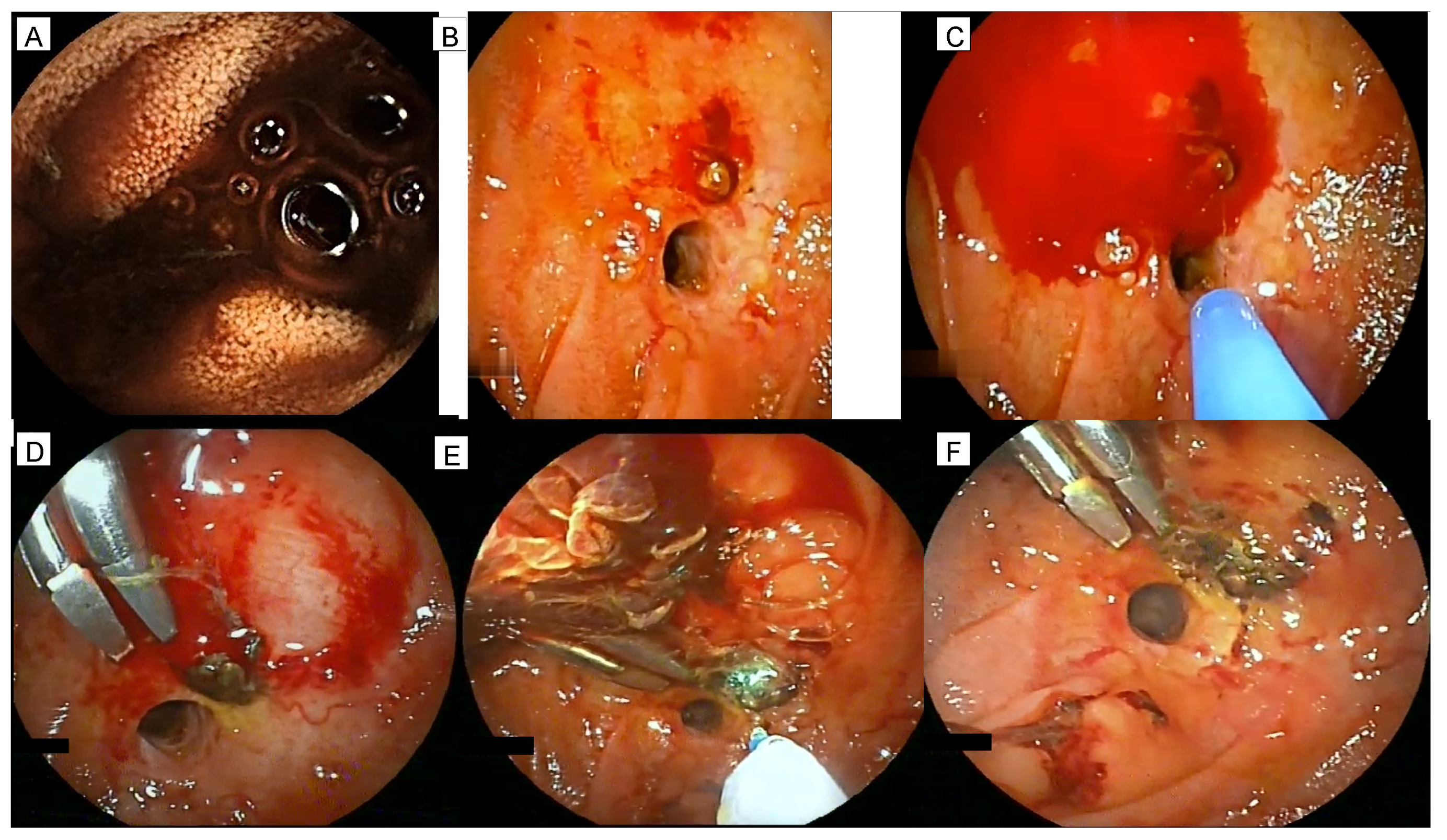
Figure 1. The main findings of the performed enteroscopies are shown. At capsule endoscopy, blood was present at jejunojejunostomy coming from the hepatic limb (A). During anterograde double-balloon enteroscopy, active bleeding from the afferent limb was observed with the presence of fresh blood at the jejunojejunostomy. Retrograde underwater exploration of the afferent limb showed the presence of an adherent clot close to the hepaticojejunostomy (B). Upon clot removal, an oozing hemorrhage from a visible vessel was observed and treated with argon plasma coagulation (30 W) (C). Subsequently, two endoclips were placed with a partial control of the bleeding (D). Finally, dual emission laser 1.9/1.5 μm was applied (Opera and Opera Evo by Quanta System, Samarate, Italy) (E) with the complete bleeding arrest (F).
Diagnostics 12 02107 g001
Publisher’s Note: MDPI stays neutral with regard to jurisdictional claims in published maps and institutional affiliations.