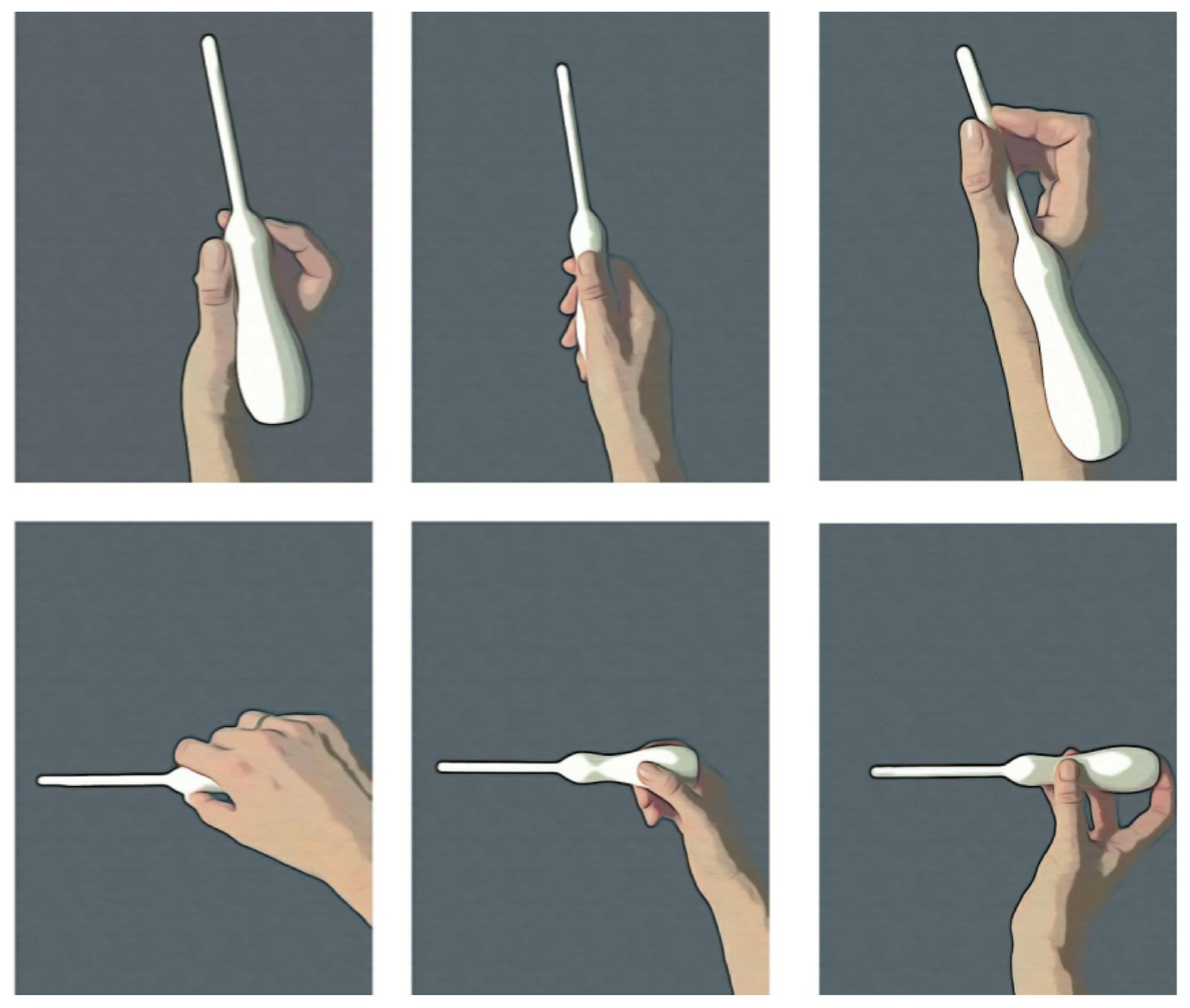
Design of a Pediatric Rectal Ultrasound Probe Intended for Ultra-High Frequency Ultrasound Diagnostics
Abstract
1. Introduction
2. Materials and Methods
2.1. Settings
- Identification of probe requirements, according to anatomic, clinical and technical considerations;
- Review of available and, for the purpose, feasible, probes currently on the market and in clinical use;
- Sketching potential UHF ultrasound transrectal probes;
- 3D prototype printing;
- Evaluation of the prototypes.
2.1.1. Anatomic, Clinical and Technological Considerations
2.1.2. Review of Available and Feasible Probes
2.1.3. Sketching
2.1.4. 3D Printing of Probe Shell Prototypes
2.1.5. 3D Prototype Testing
3. Results
3.1. Anatomic, Clinical and Technological Considerations
3.2. Review of Available and Feasible Probes
3.3. Sketches of Probe Shell Prototypes
3.4. 3D Printing of Probe Shell Prototypes
3.5. 3D Prototype Testing
4. Discussion
4.1. Design
4.2. Technology
4.3. Future Improvements and Work
5. Conclusions
| Probe | Appearance | Applications | Frequency Range (MHz) | Probe Type/Field of View | Number of Elements | Physical Dimensions (mm) | Comments: General and Specifically Regarding Anorectal Use in Children |
|---|---|---|---|---|---|---|---|
| Transesophageal (TTE) S8-3t, Philips, Amsterdam, The Netherlands [15] |  | Cardiac imaging from the esophagus. Adapted for use in children. | 3–8 | Phased array/90° sector. | 32 | Head: 7.5 × 5.5 × 18.5. Shaft diameter: 5.2. Shaft length: 880. | + Small-sized probe head + Enables angulation of probe head − Shaft too long to use anorectally in an awake child − Large handle—two hands are needed to rotate the dials on the handle that steer the probe − Too low frequency |
| Endobronchial ultrasound, EBUS, BF-UC190F, Olympus Medical Systems, Long Thanh, Vietnam [16] |  | Pulmonary bronchus imaging. | 5–12 | Curved linear array/65° sector. | Head diameter: 6.6. Shaft diameter: 6.3. Shaft length: 600. | + Small-sized probe head + Enables angulation of probe head + Enables taking biopsy + Water-filled balloon around probe head implying better tissue contact in air-filled environment − Shaft too long to use anorectally in an awake child − Too low frequency | |
| Intravascular ultrasound (IVUS), Dualpro™ IVUS + NIRS, Infraredx Inc., Bedford, MA, USA [17] |  | Imaging from the inside of the lumen of the vessels. | 35–65 | Rotating single element/ 360° image. | Single element | Head diameter: 0.8–1.15. Shaft diameter: 1.2. Shaft length: 1600. | + 360° 3D images + High frequency—high resolution + Extended bandwidth, can transmit both lower and higher frequencies + Near infrared spectroscopy is integrated − Mismatch between rectum and probe diameter, risking insufficient contact and shadowing |
| Hockey stick L8-18I-RS, GE, Chicago, IL, USA [18] |  | Shallow imaging, e.g., cardiovascular, musculoskeletal and small parts. | 8–18 | Linear array/ rectangular. | +/− Smaller size than conventional ultrasound probes, but too large to be used anorectally in children − Too short neck − Too low frequency | ||
| Laparoscopic L44LA, FujiFilm [19] |  | Imaging during laparoscopic surgery. | 2–13 | Linear array/rectangular, maximum width: 36 mm. | Head diameter: 10. | + Small size of probe head + Angulation of probe head − Shaft too long to use anorectally in an awake child − Too low frequency | |
| Endocavity, 3D 9038, BK Medical, Burlington, MA, USA [20,21] |  | Transrectal and transvaginal imaging. | 4–14 | Rotating linear array/360° image. | 192 | Diameter: 16. Neck length: 155. | + 360° image created with linear probe + 3D images can be produced − Too large to be used anorectally in children − Too low frequency |
| Prostate Triplane Transducer, 9018, BK Medical [22,23] |  | Transrectal probe for prostate imaging. | 4–14 | Curved linear array/180° sector. | 128 + 192 | Diameter: 20. | + Imaging in two perpendicular planes + Enables taking biopsy − Too large to be used in anorectally in children − Too low frequency |
Author Contributions
Funding
Institutional Review Board Statement
Informed Consent Statement
Data Availability Statement
Acknowledgments
Conflicts of Interest
References
- Amandullaevich, A.Y.; Danabaevich, J.K. Ultrasound diagnosis of Hirschsprung’s disease in children. Cent. Asian J. Med. Nat. Sci. 2022, 3, 64–71. [Google Scholar]
- Butler Tjaden, N.E.; Trainor, P.A. The developmental etiology and pathogenesis of Hirschsprung disease. Transl. Res. 2013, 162, 1–15. [Google Scholar] [CrossRef]
- Lake, J.I.; Heuckeroth, R.O. Enteric nervous system development: Migration, differentiation, and disease. Am. J. Physiol. Gastrointest. Liver Physiol. 2013, 305, G1–G24. [Google Scholar] [CrossRef] [PubMed]
- Granéli, C.; Erlöv, T.; Mitev, R.M.; Kasselaki, I.; Hagelsteen, K.; Gisselsson, D.; Jansson, T.; Cinthio, M.; Stenström, P. Ultra high frequency ultrasonography to distinguish ganglionic from aganglionic bowel wall in Hirschsprung disease: A first report. J. Pediatr. Surg. 2021, 56, 2281–2285. [Google Scholar] [CrossRef] [PubMed]
- Martucciello, G.; Pini Prato, A.; Puri, P.; Holschneider, A.M.; Meier-Ruge, W.; Jasonni, V.; Tovar, J.A.; Grosfeld, J.L. Controversies concerning diagnostic guidelines for anomalies of the enteric nervous system: A report from the fourth International Symposium on Hirschsprung’s disease and related neurocristopathies. J. Pediatr. Surg. 2005, 40, 1527–1531. [Google Scholar] [CrossRef] [PubMed]
- Petchasuwan, C.; Pintong, J. Immunohistochemistry for intestinal ganglion cells and nerve fibers: Aid in the diagnosis of Hirschsprung’s disease. J. Med. Assoc. Thail. 2000, 83, 1402–1409. [Google Scholar]
- Fransson, E.; Granéli, C.; Hagelsteen, K.; Tofft, L.; Hambraeus, M.; Munoz Mitev, R.U.; Gisselsson, D.; Stenström, P. Diagnostic efficacy of rectal suction biopsy with regard to weight in children investigated for Hirschsprung’s disease. Children 2022, 9, 124. [Google Scholar] [CrossRef] [PubMed]
- Frongia, G.; Günther, P.; Schenk, J.-P.; Strube, K.; Kessler, M.; Mehrabi, A.; Romero, P. Contrast enema for Hirschsprung disease investigation: Diagnostic accuracy and validity for subsequent diagnostic and surgical planning. Eur. J. Pediatr. Surg. 2016, 26, 207–214. [Google Scholar] [PubMed]
- Vult von Steyern, K.; Wingren, P.; Wiklund, M.; Stenström, P.; Arnbjörnsson, E. Visualisation of the rectoanal inhibitory reflex with a modified contrast enema in children with suspected Hirschsprung disease. Pediatr. Radiol. 2013, 43, 950–957. [Google Scholar] [CrossRef] [PubMed]
- Graneli, C.; Patarroyo, S.; Munoz Mitev, R.; Gisselsson, D.; Gottberg, E.; Erlöv, T.; Jansson, T.; Hagelsteen, K.; Cinthio, M.; Stenström, P. Histopathological dimensions differ between aganglionic and ganglionic bowel wall in children with Hirschsprung’s disease. BMC Pediatr. 2022, 22, 723. [Google Scholar] [CrossRef] [PubMed]
- Wehrli, L.A.; Reppucci, M.L.; Ketzer, J.; de la Torre, L.; Peña, A.; Bischoff, A. Stricture rate in patients after the repair of anorectal malformation following a standardized dilation protocol. Pediatr. Surg. Int. 2022, 38, 1717–1721. [Google Scholar] [CrossRef] [PubMed]
- Vevo® MD. The World’s First Ultra High Frequency Ultrasound Imaging System. Available online: https://www.visualsonics.com/sites/default/files/VisualSonics_VevoMDBrochure_MKT03036_V1.3.pdf (accessed on 30 March 2023).
- Hoskins, P.; Martin, K.; Thrush, A. (Eds.) Diagnostic Ultrasound: Physics and Equipment, 3rd ed.; CRC Press: Boca Raton, FL, USA, 2019. [Google Scholar]
- Clemensen, J.; Larsen, S.B.; Kyng, M.; Kirkevold, M. Participatory design in health sciences: Using cooperative experimental methods in developing health services and computer technology. Qual. Health Res. 2007, 17, 122–130. [Google Scholar] [CrossRef] [PubMed]
- S8-3t Sector Array Transducer. Available online: https://www.usa.philips.com/healthcare/product/HC989605431171/s8-3t-sector-array-transducer (accessed on 30 March 2023).
- EBUS-TBNA_BF-UC190F_Brochures and Flyers (Sellsheets)_EN_E0428389EN_103563. Available online: https://www.olympus-europa.com/medical/en/Products-and-Solutions/Products/Product/BF-UC190F.html (accessed on 30 March 2023).
- Dualpro IVUS+NIRS. Available online: https://www.infraredx.com/products/dualpro-nirs/ (accessed on 30 March 2023).
- L8 -18I-RS Probe. Available online: https://services.gehealthcare.co.uk/gehcstorefront/p/5499609 (accessed on 30 March 2023).
- Transducers. Available online: https://www.fujifilmsurgical.com/transducers/ (accessed on 30 March 2023).
- 3D X14L4 (9038) Endocavity Transducer. Available online: https://www.bkmedical.com/transducers/endocavity-3d-x14l4/ (accessed on 30 March 2023).
- 16-01665-00 X14L4 Product Data; BK Medical: Herlev, Denmark, December 2017.
- E14C4t (9018) Prostate Triplane Transducer. Available online: https://www.bkmedical.com/transducers/e14c4t-prostate-triplane/ (accessed on 30 March 2023).
- 16-01257-07 E14C4t Product Data; BK Medical: Herlev, Denmark, November 2018.


Disclaimer/Publisher’s Note: The statements, opinions and data contained in all publications are solely those of the individual author(s) and contributor(s) and not of MDPI and/or the editor(s). MDPI and/or the editor(s) disclaim responsibility for any injury to people or property resulting from any ideas, methods, instructions or products referred to in the content. |
© 2023 by the authors. Licensee MDPI, Basel, Switzerland. This article is an open access article distributed under the terms and conditions of the Creative Commons Attribution (CC BY) license (https://creativecommons.org/licenses/by/4.0/).
Share and Cite
Evertsson, M.; Graneli, C.; Vernersson, A.; Wiaczek, O.; Hagelsteen, K.; Erlöv, T.; Cinthio, M.; Stenström, P. Design of a Pediatric Rectal Ultrasound Probe Intended for Ultra-High Frequency Ultrasound Diagnostics. Diagnostics 2023, 13, 1667. https://doi.org/10.3390/diagnostics13101667
Evertsson M, Graneli C, Vernersson A, Wiaczek O, Hagelsteen K, Erlöv T, Cinthio M, Stenström P. Design of a Pediatric Rectal Ultrasound Probe Intended for Ultra-High Frequency Ultrasound Diagnostics. Diagnostics. 2023; 13(10):1667. https://doi.org/10.3390/diagnostics13101667
Chicago/Turabian StyleEvertsson, Maria, Christina Graneli, Alvina Vernersson, Olivia Wiaczek, Kristine Hagelsteen, Tobias Erlöv, Magnus Cinthio, and Pernilla Stenström. 2023. "Design of a Pediatric Rectal Ultrasound Probe Intended for Ultra-High Frequency Ultrasound Diagnostics" Diagnostics 13, no. 10: 1667. https://doi.org/10.3390/diagnostics13101667
APA StyleEvertsson, M., Graneli, C., Vernersson, A., Wiaczek, O., Hagelsteen, K., Erlöv, T., Cinthio, M., & Stenström, P. (2023). Design of a Pediatric Rectal Ultrasound Probe Intended for Ultra-High Frequency Ultrasound Diagnostics. Diagnostics, 13(10), 1667. https://doi.org/10.3390/diagnostics13101667


