Clinical and Dermoscopic Characteristics of Cutaneous Sarcomas: A Literature Review
Abstract
1. Introduction
2. Methods
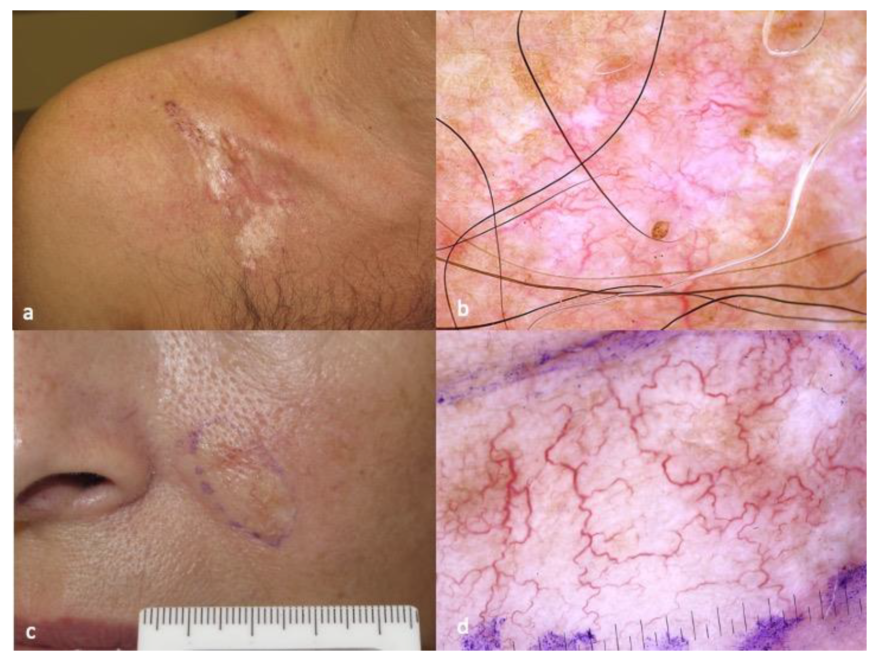
3. Dermatofibrosarcoma Protuberans (DFSP)
4. Atypical Fibroxanthoma (AFX)
5. Cutaneous Undifferentiated Pleomorphic Sarcoma (CUPS)
6. Kaposi’s Sarcoma (KS)
7. Angiosarcoma
8. Cutaneous Leiomyosarcoma
9. Cutaneous Liposarcoma
10. Conclusions
Author Contributions
Funding
Institutional Review Board Statement
Informed Consent Statement
Data Availability Statement
Conflicts of Interest
References
- Walocko, F.; Christensen, R.E.; Worley, B.; Alam, M. Cutaneous Mesenchymal Sarcomas. Dermatol. Clin. 2023, 41, 133–140. [Google Scholar] [CrossRef] [PubMed]
- Kohlmeyer, J.; Steimle-Grauer, S.A.; Hein, R. Cutaneous sarcomas. J. Dtsch. Dermatol. Ges. 2017, 15, 630–648. [Google Scholar] [CrossRef] [PubMed]
- Soomers, V.; Husson, O.; Young, R.; Desar, I.; Van der Graaf, W. The sarcoma diagnostic interval: A systematic review on length, contributing factors and patient outcomes. ESMO Open 2020, 5, e000592. [Google Scholar] [CrossRef] [PubMed]
- Martínez-Trufero, J.; Cruz Jurado, J.; Gómez-Mateo, M.C.; Bernabeu, D.; Floría, L.J.; Lavernia, J.; Sebio, A.; del Muro, X.G.; Álvarez, R.; Correa, R.; et al. Uncommon and peculiar soft tissue sarcomas: Multidisciplinary review and practical recommendations for diagnosis and treatment. Spanish group for Sarcoma research (GEIS—GROUP). Part I. Cancer Treat. Rev. 2021, 99, 102259. [Google Scholar] [CrossRef]
- Sbaraglia, M.; Bellan, E.; Dei Tos, A.P. The 2020 WHO Classification of Soft Tissue Tumours: News and perspectives. Pathologica 2021, 113, 70–84. [Google Scholar] [CrossRef]
- Ugurel, S.; Kortmann, R.D.; Mohr, P.; Mentzel, T.; Garbe, C.; Breuninger, H.; Bauer, S.; Grabbe, S. S1 guidelines for dermatofibrosarcoma protuberans (DFSP)—Update 2018. J. Dtsch. Dermatol. Ges. 2019, 17, 663–668. [Google Scholar] [CrossRef]
- PDQ Pediatric Treatment Editorial Board. Childhood Soft Tissue Sarcoma Treatment (PDQ®): Health Professional Version. In PDQ Cancer Information Summaries [Internet]; National Cancer Institute (US): Bethesda, MD, USA, 2002. [Google Scholar]
- Llombart, B.; Serra, C.; Requena, C.; Alsina, M.; Morgado-Carrasco, D.; Través, V.; Sanmartín, O. Guidelines for Diagnosis and Treatment of Cutaneous Sarcomas: Dermatofibrosarcoma Protuberans. Actas Dermosifiliogr. 2018, 109, 868–877, (In English, Spanish). [Google Scholar] [CrossRef]
- Rust, D.J.; Kwinta, B.D.; Geskin, L.J.; Samie, F.H.; Remotti, F.; Yoon, S.S. Surgical management of dermatofibrosarcoma protuberans. J. Surg. Oncol. 2023. epub ahead of print. [Google Scholar] [CrossRef]
- Walocko, F.M.; Carr, C.; Srivastava, D.; Nijhawan, R.I. Margin Size for Unique Skin Tumors Treated with Mohs Micrographic Surgery: A Survey of Practice Patterns. Cutis 2022, 110, E21–E24. [Google Scholar] [CrossRef]
- Escobar, G.F.; Ribeiro, C.K.; Leite, L.L.; Barone, C.R.; Cartell, A. Dermoscopy of Dermatofibrosarcoma Protuberans: What Do We Know? Dermatol. Pract. Concept. 2019, 9, 139–145. [Google Scholar] [CrossRef]
- Bernard, J.; Poulalhon, N.; Argenziano, G.; Debarbieux, S.; Dalle, S.; Thomas, L. Dermoscopy of dermatofibrosarcoma protuberans: A study of 15 cases. Br. J. Dermatol. 2013, 169, 85–90. [Google Scholar] [CrossRef] [PubMed]
- Avilés-Izquierdo, J.A.; Conde-Montero, E.; Barchino-Ortiz, L.; Láza-ro-Ochaita, P. Dermoscopic features of dermatofibrosarcoma protuberans. Australas. J. Dermatol. 2014, 55, 125–127. [Google Scholar] [CrossRef] [PubMed]
- Esdaile, B.A.; Matin, R.N. Residents’ corner August 2014. Clues in Dermoscopy: Dermoscopic features to aid earlier diagnosis? Eur. J. Dermatol. 2014, 24, 518–519. [Google Scholar] [PubMed]
- Güngör, S.; Büyükbabani, N.; Büyük, M.; Tarıkçı, N.; Kocatürk, E. Atrophic dermatofibrosarcoma protuberans: Are there specific dermatoscopic features? J. Dtsch. Dermatol. Ges. 2014, 12, 425–427. [Google Scholar]
- Ehara, Y.; Yoshida, Y.; Shiomi, T.; Yamamoto, O. Pigmented dermatofibrosarcoma protuberans and blue naevi with similar dermoscopy: A case report. Acta Derm. Venereol. 2016, 96, 272–273. [Google Scholar] [CrossRef]
- Hartmann, F.; Haenssle, H.A.; Seitz, C.S.; Kretschmer, L.; Schön, M.P.; Buhl, T. Maculonodular lesion on the back of a 66-year-old man. Hautarzt 2016, 67, 845–847. [Google Scholar] [CrossRef]
- Piccolo, V.; Russo, T.; Staibano, S.; Coppola, N.; Russo, D.; Alessio, L.; Argenziano, G. Dermoscopy of dermatofibrosarcoma protuberans on black skin. J. Am. Acad. Dermatol. 2016, 74, e119–e120. [Google Scholar] [CrossRef]
- Venturini, M.; Zanca, A.; Manganoni, A.M.; Pavoni, L.; Gualdi, G.; Calzavara-Pinton, P. In vivo characterization of recurrent dermatofibrosarcoma protuberans by dermoscopy and reflectance confocal microscopy. J. Am. Acad. Dermatol. 2016, 75, e185–e187. [Google Scholar] [CrossRef]
- Costa, C.; Cappello, M.; Argenziano, G.; Piccolo, V.; Scalvenzi, M. Dermoscopy of uncommon variants of dermatofibrosarcoma protuberans. J. Eur. Acad. Dermatol. Venereol. 2017, 31, e366–e368. [Google Scholar] [CrossRef]
- Anders, I.M.; Schimmelpfennig, C.; Wiedemann, K.; Löffler, D.; Kämpf, C.; Blumert, C.; Reiche, K.; Kunz, M.; Anderegg, U.; Simon, J.; et al. Atypical fibroxanthoma and pleomorphic dermal sarcoma—Gene expression analysis compared with undifferentiated cutaneous squamous cell carcinoma. J. Dtsch. Dermatol. Ges. 2023. epub ahead of print. [Google Scholar] [CrossRef]
- McClure, E.; Carr, M.J.; Patel, A.; Naqvi, S.M.H.; Kim, Y.; Harrington, M.; Cruse, W.; Gonzalez, R.J.; Sondak, V.K.; Sarnaik, A.A.; et al. Atypical Fibroxanthoma: Outcomes from a Large Single Institution Series. Cancer Control 2023, 30, 10732748231155699. [Google Scholar] [CrossRef]
- Moscarella, E.; Piana, S.; Specchio, F.; Kyrgidis, A.; Nazzaro, G.; Eliceche, M.L.; Savoia, F.; Bugatti, L.; Filosa, G.; Zalaudek, I.; et al. Dermoscopy features of atypical fibroxanthoma: A multicenter study of the International Dermoscopy Society. Australas. J. Dermatol. 2018, 59, 309–314. [Google Scholar] [CrossRef] [PubMed]
- Kunz, M.; Svensson, H.; Paoli, J. Dermoscopic rainbow pattern: A clue to diagnosing aneurysmal atypical fibroxanthoma. JAAD Case Rep. 2018, 4, 292–294. [Google Scholar] [CrossRef] [PubMed][Green Version]
- Di Brizzi, E.V.; Moscarella, E.; Piana, S.; Longo, C.; Franco, R.; Alfano, R.; Argenziano, G. Clinical and dermoscopic features of pleomorphic dermal sarcoma. Australas. J. Dermatol. 2019, 60, e153–e154. [Google Scholar] [CrossRef] [PubMed]
- Salerni, G.; Alonso, C.; Sanchez-Granel, G.; Gorosito, M. Dermoscopic findings in an early malignant fibrous histiocytoma on the face. Dermatol. Pract. Concept. 2017, 7, 9. [Google Scholar] [CrossRef]
- Kaohsiung, J.; Watanabe, S.; Kato, H.; Inagaki, H.; Hattori, H.; Morita, A. A case of cutaneous malignant fibrous histiocytoma with multiple organ metastases. Med. Sci. 2013, 29, 111–115. [Google Scholar]
- Logan, I.T.; Vroobel, K.M.; le Grange, F.; Perrett, C.M. Pleomorphic dermal sarcoma: Clinicopathological features and outcomes from a 5-year tertiary referral centre experience. Cancer Rep. 2022, 5, e1583. [Google Scholar] [CrossRef]
- Silveira, T.; Benini, T.; Pessanha, A. Undifferentiated pleomorphic sarcoma: A case report. Surg. Cosm. Dermatol. 2020, 44, 92–95. [Google Scholar] [CrossRef]
- Grabar, S.; Costagliola, D. Epidemiology of Kaposi’s Sarcoma. Cancers 2021, 13, 5692. [Google Scholar] [CrossRef]
- Atyeo, N.; Chae, M.Y.; Toth, Z.; Sharma, A.; Papp, B. Kaposi’s Sarcoma-Associated Herpesvirus Immediate Early Proteins Trigger FOXQ1 Expression in Oral Epithelial Cells, Engaging in a Novel Lytic Cycle-Sustaining Positive Feedback Loop. J. Virol. 2023, 97, e0169622. [Google Scholar] [CrossRef]
- Libson, K.; Himed, S.; Dunlop, H.; Nusbaum, K.B.; Korman, A.M.; Kaffenberger, B.H.; Trinidad, J. A description of Kaposi sarcoma risk factors and outcomes in HIV-positive and HIV-negative patients at a tertiary care medical center from 2005 to 2020. Arch. Dermatol. Res. 2023. epub ahead of print. [Google Scholar] [CrossRef] [PubMed]
- Tekcan Sanli, D.E.; Kiziltas, S. Gastrointestinal Kaposi’s Sarcoma. N. Engl. J. Med. 2023, 388, e45. [Google Scholar] [CrossRef]
- Paksoy, N.; Khanmammadov, N.; Doğan, İ.; Ferhatoğlu, F.; Ahmed, M.A.; Karaman, S.; Aydiner, A. Weekly paclitaxel treatment in the first-line therapy of classic Kaposi sarcoma: A real-life study. Medicine 2023, 102, e32866. [Google Scholar] [CrossRef] [PubMed]
- Ertürk Yılmaz, T.; Akay, B.N.; Okçu Heper, A. Dermoscopic findings of Kaposi sarcoma and dermatopathological correlations. Australas. J. Dermatol. 2020, 61, e46–e53. [Google Scholar] [CrossRef] [PubMed]
- Hu, S.C.; Ke, C.L.; Lee, C.H.; Wu, C.S.; Chen, G.S.; Cheng, S.T. Dermoscopy of Kaposi’s sarcoma: Areas exhibiting the multicoloured ‘rainbow pattern’. J. Eur. Acad. Dermatol. Venereol. 2009, 23, 1128–1132. [Google Scholar] [CrossRef]
- Cheng, S.T.; Ke, C.L.; Lee, C.H.; Wu, C.S.; Chen, G.S.; Hu, S.C.S. Rainbow pattern in Kaposi’s sarcoma under polarized dermoscopy: A dermoscopic pathological study. Br. J. Dermatol. 2009, 160, 801–809. [Google Scholar] [CrossRef]
- Vazquez-Lopez, F.; Garcia-Garcia, B.; Rajadhyaksha, M.; Marghoob, A. Dermoscopic rainbow pattern in non-Kaposi sarcoma lesions. Br. J. Dermatol. 2009, 161, 474–475. [Google Scholar] [CrossRef]
- Satta, R.; Fresi, L.; Cottoni, F. Dermoscopic rainbow pattern in Kaposi’s sarcoma lesions: Our experience. Arch. Dermatol. 2012, 148, 1207–1208. [Google Scholar] [CrossRef][Green Version]
- Young, R.J.; Brown, N.J.; Reed, M.W.; Hughes, D.; Woll, P.J. Angiosarcoma. Lancet Oncol. 2010, 11, 983–991. [Google Scholar] [CrossRef]
- Florou, V.; Wilky, B.A. Current and future directions for angiosarcoma therapy. Curr. Treat. Options Oncol. 2018, 19, 14. [Google Scholar] [CrossRef]
- Mark, R.J.; Poen, J.C.; Tran, L.M.; Fu, Y.S.; Juillard, G.F. Angiosarcoma. A report of 67 patients and a review of the literature. Cancer 1996, 77, 2400–2406. [Google Scholar] [CrossRef]
- Cole, D.W.; Huerta, T.; Andea, A.; Tejasvi, T. Purpuric Plaques-Dermoscopic and Histopathological Correlation of Cutaneous Angiosarcoma. Dermatol. Pract. Concept. 2020, 10, e2020084. [Google Scholar] [CrossRef] [PubMed]
- Requena, C.; Sendra, E.; Llombart, B.; Sanmartín, O.; Guillén, C.; Lavernia, J.; Traves, V.; Cruz, J. Cutaneous Angiosarcoma: Clinical and Pathology Study of 16 Cases. Actas Dermosifiliogr. 2017, 108, 457–465. [Google Scholar] [CrossRef] [PubMed]
- Zalaudek, I.; Gomez-Moyano, E.; Landi, C.; Navarro, M.L.; Ballesteros, M.D.F.; De Pace, B.; Vera-Casaño, A.; Piana, S. Clinical, dermoscopic and histopathological features of spontaneous scalp or face and radiotherapy-induced angiosarcoma. Australas. J. Dermatol. 2013, 54, 201–207. [Google Scholar] [CrossRef] [PubMed]
- Patel, S.H.; Hayden, R.E.; Hinni, M.L.; Wong, W.W.; Foote, R.L.; Milani, S.; Wu, Q.; Ko, S.J.; Halyard, M.Y. Angiosarcoma of the scalp and face: The Mayo Clinic experience. JAMA Otolaryngol. Head Neck Surg. 2015, 141, 335–340. [Google Scholar] [CrossRef]
- Ramakrishnan, N.; Mokhtari, R.; Charville, G.W.; Bui, N.; Ganjoo, K. Cutaneous angiosarcoma of the head and neck-A retrospective analysis of 47 patients. Cancers 2022, 14, 3841. [Google Scholar] [CrossRef]
- Alharbi, A.; Kim, Y.C.; AlShomer, F.; Choi, J.W. Utility of Multimodal Treatment Protocols in the Management of Scalp Cutaneous Angiosarcoma. Plast. Reconstr. Surg. Glob. Open 2023, 11, e4827. [Google Scholar] [CrossRef]
- Massi, D.; Franchi, A.; Alos, L.; Cook, M.; Di Palma, S.; Enguita, A.B.; Ferrara, G.; Kazakov, D.V.; Mentzel, T.; Michal, M.; et al. Primary cutaneous leiomyosarcoma: Clinicopathological analysis of 36 cases. Histopathology 2010, 56, 251–262. [Google Scholar] [CrossRef]
- De Giorgi, V.; Scarfì, F.; Silvestri, F.; Maida, P.; Gori, A.; Trane, L.; Massi, D. Cutaneous leiomyosarcoma: A clinical, dermoscopic, pathologic case study. Exp. Oncol. 2019, 41, 80–81. [Google Scholar] [CrossRef]
- Escobar, G.F.; Gazzi, S.; Bonamigo, R.R. Arborizing telangiectasias may also be a dermoscopic vascular pattern of cutaneous leiomyosarcoma. Dermatol. Surg. 2021, 47, 1290–1291. [Google Scholar] [CrossRef]
- Zaballos, P.; Del Pozo, L.J.; Argenziano, G.; Medina, C.; Lacarrubba, F.; Ferrer, B.; Martin, J.; Llambrich, A.; Zalaudek, I.; Bañuls, J. Dermoscopy of cutaneous smooth muscle neoplasms: A morphological study of 136 cases. J. Eur. Acad. Dermatol. Venereol. 2019, 33, 693–699. [Google Scholar] [CrossRef] [PubMed]
- Kraft, S.; Fletcher, C.D. Atypical intradermal smooth muscle neoplasms: Clinicopathologic analysis of 84 cases and a reappraisal of cutaneous “leiomyosarcoma”. Am. J. Surg. Pathol. 2011, 35, 599–607. [Google Scholar] [CrossRef] [PubMed]
- Chambers, M.; Badin, D.J.; Sriharan, A.A.; Linos, K.D. Expanding the differential of cutaneous epithelioid tumors: A case of dedifferentiated liposarcoma with epithelioid features involving the skin, with review of the literature. J. Cutan. Pathol. 2020, 47, 554–560. [Google Scholar] [CrossRef] [PubMed]


| Intermediate (Locally Aggressive) | Intermediate (Rarely Metastasizing) | Malignant | |
|---|---|---|---|
| Adipocytic tumours |
|
| |
| Fibroblastic/myofibroblastic tumors |
|
|
|
| So-called fibrohistiocytic tumors | Plexiform fibrohistiocytic tumor Giant cell tumor of soft parts NOS | Malignant tenosynovial giant cell tumor | |
| Vascular tumors |
|
| |
| Pericytic (perivascular) tumors | Glomus tumor, malignant | ||
| Smooth muscle tumors |
|
| |
| Skeletal muscle tumors |
| ||
| Gastrointestinal stromal tumors |
| ||
| Chondro-osseous tumors | Osteosarcoma, extraskeletal | ||
| Peripheral nerve sheath tumors |
| ||
| Tumors of uncertain differentiation |
|
|
|
Disclaimer/Publisher’s Note: The statements, opinions and data contained in all publications are solely those of the individual author(s) and contributor(s) and not of MDPI and/or the editor(s). MDPI and/or the editor(s) disclaim responsibility for any injury to people or property resulting from any ideas, methods, instructions or products referred to in the content. |
© 2023 by the authors. Licensee MDPI, Basel, Switzerland. This article is an open access article distributed under the terms and conditions of the Creative Commons Attribution (CC BY) license (https://creativecommons.org/licenses/by/4.0/).
Share and Cite
Apalla, Z.; Liopyris, K.; Kyrmanidou, E.; Fotiadou, C.; Sgouros, D.; Patsatsi, A.; Trakatelli, M.-G.; Kalloniati, E.; Lallas, A.; Lazaridou, E. Clinical and Dermoscopic Characteristics of Cutaneous Sarcomas: A Literature Review. Diagnostics 2023, 13, 1822. https://doi.org/10.3390/diagnostics13101822
Apalla Z, Liopyris K, Kyrmanidou E, Fotiadou C, Sgouros D, Patsatsi A, Trakatelli M-G, Kalloniati E, Lallas A, Lazaridou E. Clinical and Dermoscopic Characteristics of Cutaneous Sarcomas: A Literature Review. Diagnostics. 2023; 13(10):1822. https://doi.org/10.3390/diagnostics13101822
Chicago/Turabian StyleApalla, Zoe, Konstantinos Liopyris, Eirini Kyrmanidou, Christina Fotiadou, Dimitrios Sgouros, Aikaterini Patsatsi, Myrto-Georgia Trakatelli, Evangelia Kalloniati, Aimilios Lallas, and Elizabeth Lazaridou. 2023. "Clinical and Dermoscopic Characteristics of Cutaneous Sarcomas: A Literature Review" Diagnostics 13, no. 10: 1822. https://doi.org/10.3390/diagnostics13101822
APA StyleApalla, Z., Liopyris, K., Kyrmanidou, E., Fotiadou, C., Sgouros, D., Patsatsi, A., Trakatelli, M.-G., Kalloniati, E., Lallas, A., & Lazaridou, E. (2023). Clinical and Dermoscopic Characteristics of Cutaneous Sarcomas: A Literature Review. Diagnostics, 13(10), 1822. https://doi.org/10.3390/diagnostics13101822


