
The Accuracy of Sex Identification Using CBCT Morphometric Measurements of the Mandible, with Different Machine-Learning Algorithms—A Retrospective Study


Abstract
:1. Introduction
2. Materials and Methods
2.1. Study Design
- CBCTs did not show the entire mandible, or showed less than ten remaining teeth.
- Certain pathological conditions affected the mandible’s size and shape.
- CBCT images contained metal in or near the mandible, or radiographs showed bone resorption around the lower teeth that reached the apical third of the roots.
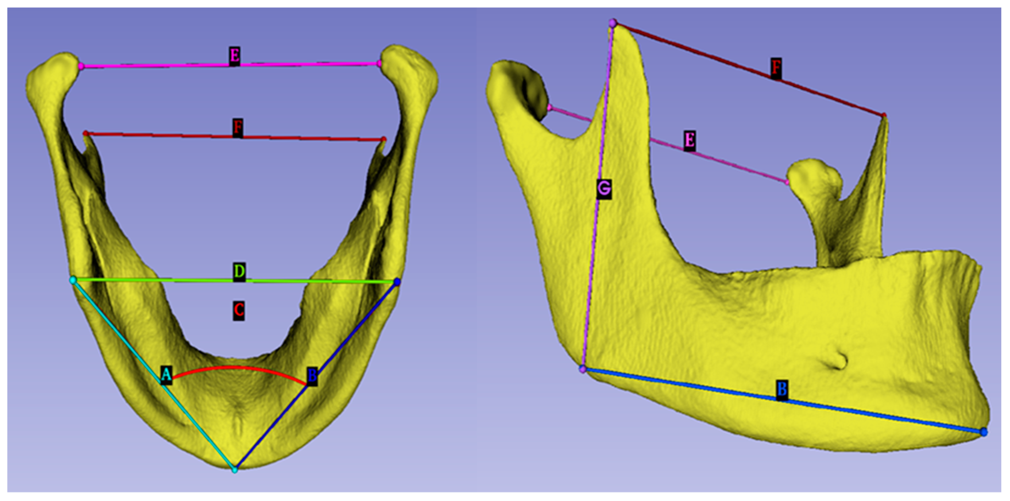
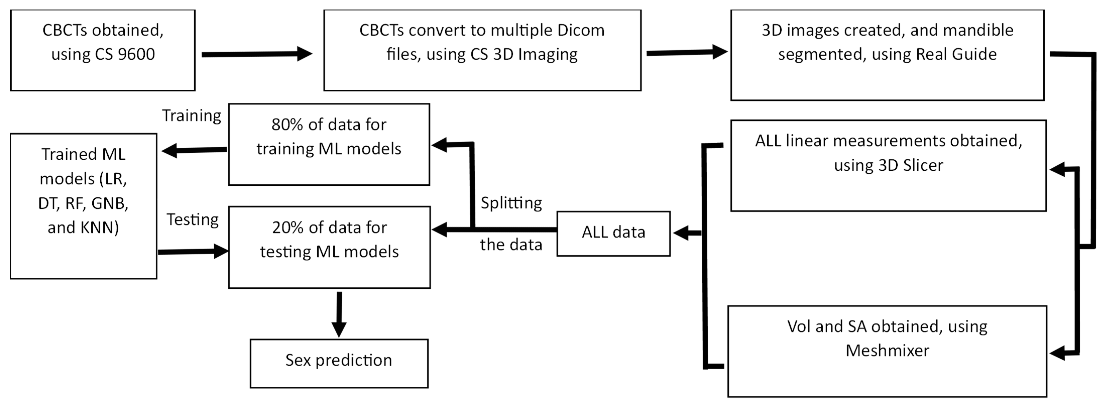
2.2. Image Analysis
2.3. Statistical Analysis
2.4. Machine-Learning Algorithms
Performance Criteria
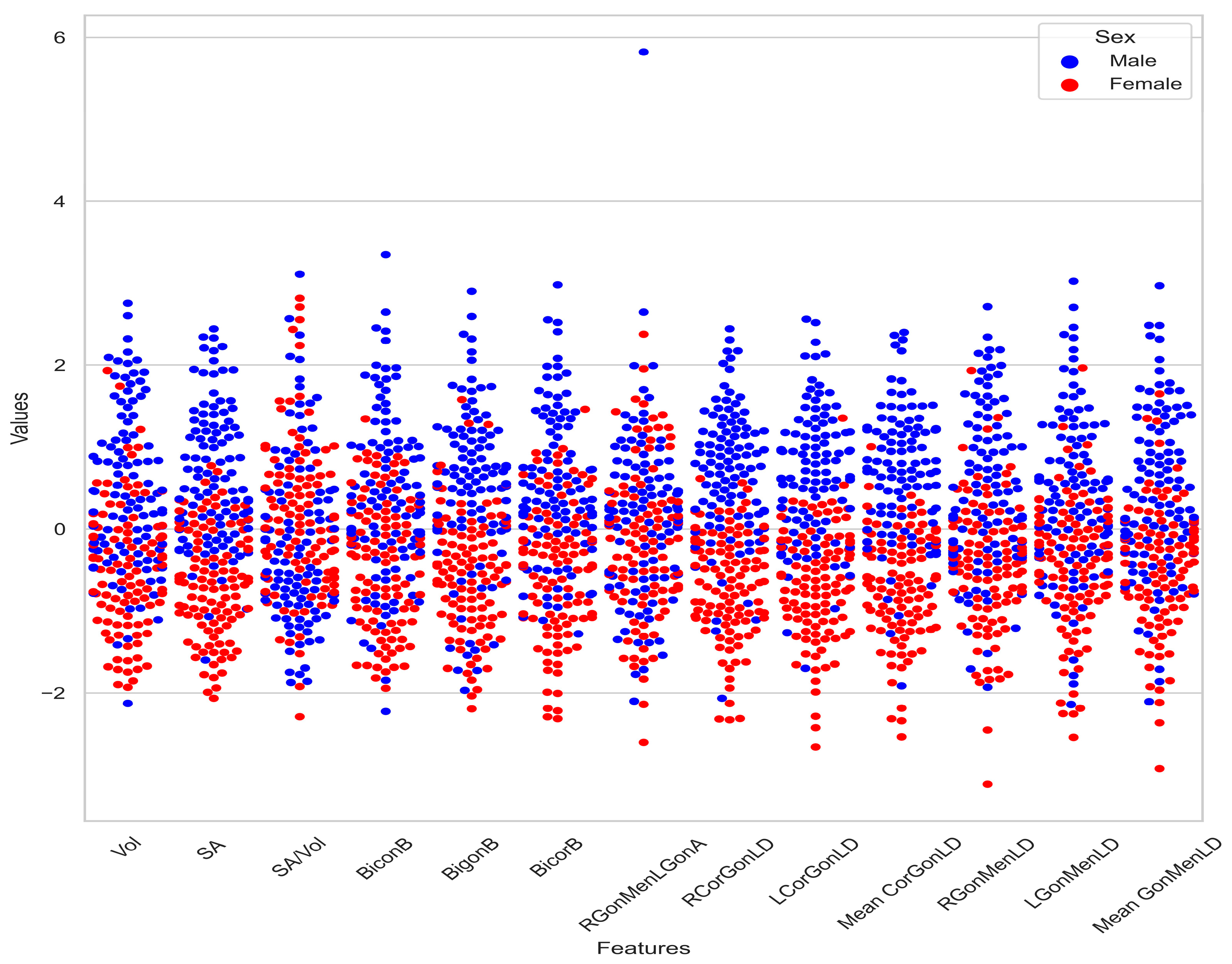
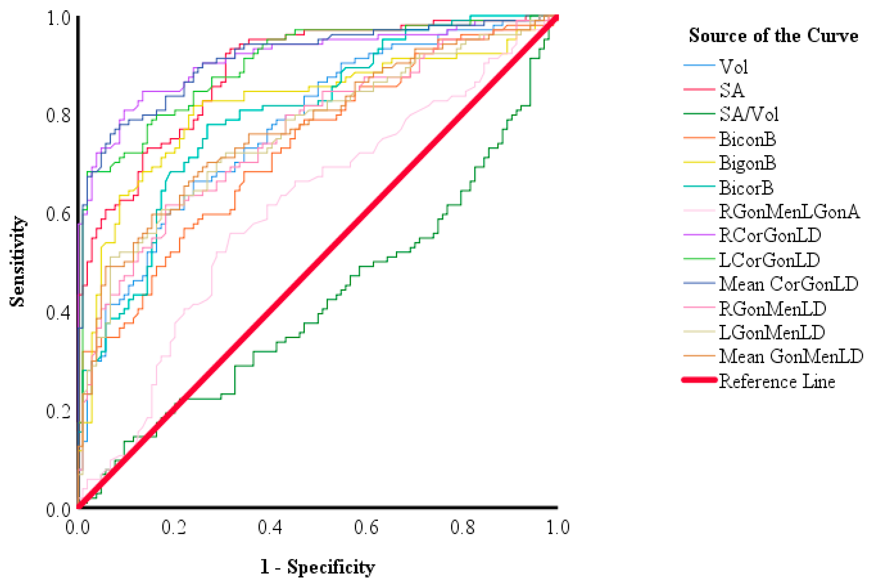
3. Results
4. Discussion
5. Conclusions
Author Contributions
Funding
Institutional Review Board Statement
Informed Consent Statement
Data Availability Statement
Conflicts of Interest
References
- Krishan, K.; Kanchan, T.; Garg, A.K. Dental Evidence in Forensic Identification—An Overview, Methodology and Present Status. Open Dent. J. 2015, 9, 250. [Google Scholar] [CrossRef] [PubMed] [Green Version]
- Prajapati, G.; Sarode, S.C.; Sarode, G.S.; Shelke, P.; Awan, K.H.; Patil, S. Role of Forensic Odontology in the Identification of Victims of Major Mass Disasters across the World: A Systematic Review. PLoS ONE 2018, 13, e0199791. [Google Scholar] [CrossRef] [PubMed] [Green Version]
- Angadi, P.V.; Hemani, S.; Prabhu, S.; Acharya, A.B. Analyses of Odontometric Sexual Dimorphism and Sex Assessment Accuracy on a Large Sample. J. Forensic Leg. Med. 2013, 20, 673–677. [Google Scholar] [CrossRef]
- Baca, K.; Bridge, B.; Snow, M. Three-Dimensional Geometric Morphometric Sex Determination of the Whole and Modeled Fragmentary Human Pubic Bone. PLoS ONE 2022, 17, e0265754. [Google Scholar] [CrossRef] [PubMed]
- Nagare, S.; Chaudhari, R.; Birangane, R.; Parkarwar, P. Sex Determination in Forensic Identification, a Review. J. Forensic Dent. Sci. 2018, 10, 61. [Google Scholar] [CrossRef]
- Bhagwatkar, T.; Thakur, M.; Palve, D.; Bhondey, A.; Dhengar, Y.; Chaturvedi, S. Sex determination by using Mandibular Ramus—A Forensic Study. J. Adv. Med. Dent. Sci. Res. 2016, 4, 1–6. [Google Scholar]
- Ingaleshwar, P.; Bhosale, S.; Nimbulkar, G.; Britto, F.; Chandrappa, P.; Hosur, M. Mandibular Ramus—An Indicator for Gender Determination: A Digital Panoramic Study in Bagalkot Population. J. Oral Maxillofac. Pathol. 2023, 27, 66–70. [Google Scholar]
- Renjith, G.; Mary, D.P.; Soe, K.; Wan, M.Y.; Beh, H.-C.; Phuah, W.H.; Tseu, A.T.H. Sex Estimation by Discriminant Function Analysis Using Anatomical Location of Mental Foramen. Forensic Sci. Int. Rep. 2019, 1, 100018. [Google Scholar] [CrossRef]
- ThAKur, K.C.; SrIVASTAVA, T. Mandibular Ramus as a Tool for Sexual Determination: A Systematic Review. J. Clin. Diagn. Res. 2022, 16, AE01–AE05. [Google Scholar] [CrossRef]
- Errickson, D.; Grueso, I.; Griffith, S.J.; Setchell, J.M.; Thompson, T.J.U.; Thompson, C.E.L.; Gowland, R.L. Towards a Best Practice for the Use of Active Non-contact Surface Scanning to Record Human Skeletal Remains from Archaeological Contexts. Int. J. Osteoarchaeol. 2017, 27, 650–661. [Google Scholar] [CrossRef] [Green Version]
- Turek, P.; Budzik, G. Estimating the Accuracy of Mandible Anatomical Models Manufactured Using Material Extrusion Methods. Polymers 2021, 13, 2271. [Google Scholar] [CrossRef] [PubMed]
- Ravali, C.T. Gender Determination of Maxillary Sinus Using CBCT. Int. J. Appl. Dent. Sci. 2017, 3, 221–224. [Google Scholar]
- Lu, P.; Barazzetti, L.; Chandran, V.; Gavaghan, K.; Weber, S.; Gerber, N.; Reyes, M. Highly Accurate Facial Nerve Segmentation Refinement from CBCT/CT Imaging Using a Super-Resolution Classification Approach. IEEE Trans. Biomed. Eng. 2018, 65, 178–188. [Google Scholar] [CrossRef]
- Issrani, R.; Prabhu, N.; Sghaireen, M.G.; Ganji, K.K.; Alqahtani, A.M.A.; Aljamaan, T.S.; Alanazi, A.M.; Alanazi, S.H.; Alam, M.K.; Munisekhar, M.S. Cone-Beam Computed Tomography: A New Tool on the Horizon for Forensic Dentistry. Int. J. Environ. Res. Public Health 2022, 19, 5352. [Google Scholar] [CrossRef] [PubMed]
- Oladipo, F.; Ogbuju, E.; Alayesanmi, F.S.; Musa, A.E. The State of the Art in Machine Learning-Based Digital Forensics. SSRN Electron. J. 2020, 1, 23. [Google Scholar] [CrossRef]
- Yang, W.; Liu, X.; Wang, K.; Hu, J.; Geng, G.; Feng, J. Sex Determination of Three-Dimensional Skull Based on Improved Backpropagation Neural Network. Comput. Math Methods Med. 2019, 2019, 9163547. [Google Scholar] [CrossRef] [Green Version]
- Shen, S.; Liu, Z.; Wang, J.; Fan, L.; Ji, F.; Tao, J. Machine Learning Assisted Cameriere Method for Dental Age Estimation. BMC Oral Health 2021, 21, 641. [Google Scholar] [CrossRef]
- Khanagar, S.B.; Vishwanathaiah, S.; Naik, S.; Al-Kheraif, A.A.; Devang Divakar, D.; Sarode, S.C.; Bhandi, S.; Patil, S. Application and Performance of Artificial Intelligence Technology in Forensic Odontology—A Systematic Review. Leg. Med. 2021, 48, 101826. [Google Scholar] [CrossRef]
- Praveena, M. A Literature Review on Supervised Machine Learning Algorithms and Boosting Process. Int. J. Comput. Appl. 2017, 169, 975–8887. [Google Scholar] [CrossRef]
- Jiao, S.R.; Song, J.; Liu, B. A Review of Decision Tree Classification Algorithms for Continuous Variables. J. Phys. Conf. Ser. 2020, 1651, 012083. [Google Scholar] [CrossRef]
- Parmar, A.; Katariya, R.; Patel, V. A Review on Random Forest: An Ensemble Classifier. Lect. Notes Data Eng. Commun. Technol. 2019, 26, 758–763. [Google Scholar]
- Maillo, J.; Ramírez, S.; Triguero, I.; Herrera, F. KNN-IS: An Iterative Spark-Based Design of the k-Nearest Neighbors Classifier for Big Data. Knowl.-Based Syst. 2017, 117, 3–15. [Google Scholar] [CrossRef] [Green Version]
- Soria, D.; Garibaldi, J.M.; Ambrogi, F.; Biganzoli, E.M.; Ellis, I.O. A ‘Non-Parametric’ Version of the Naive Bayes Classifier. Knowl.-Based Syst. 2011, 24, 775–784. [Google Scholar] [CrossRef] [Green Version]
- Choi, R.Y.; Coyner, A.S.; Kalpathy-Cramer, J.; Chiang, M.F.; Peter Campbell, J. Introduction to Machine Learning, Neural Networks, and Deep Learning. Transl. Vis Sci. Technol. 2020, 9, 14. [Google Scholar] [PubMed]
- Capitaneanu, C.; Willems, G.; Thevissen, P. A Systematic Review of Odontological Sex Estimation Methods. J. Forensic Odontostomatol. 2017, 35, 1–19. [Google Scholar] [PubMed]
- Wood, R.E. Forensic Aspects of Maxillofacial Radiology. Forensic Sci. Int. 2006, 159, S47–S55. [Google Scholar] [CrossRef] [PubMed]
- Singh, R.; Singh, R.; Baby, B.; Suri, A. Effect of the Segmentation Threshold on Computed Tomography–Based Reconstruction of Skull Bones with Reference Optical Three-Dimensional Scanning. World Neurosurg. 2022, 166, e34–e43. [Google Scholar] [CrossRef] [PubMed]
- Behl, A.; Grewal, S.; Bajaj, K.; Baweja, P.; Kaur, G.; Kataria, P. Mandibular Ramus and Gonial Angle—Identification Tool in Age Estimation and Sex Determination: A Digital Panoramic Radiographic Study in North Indian Population. J. Indian Acad. Oral Med. Radiol. 2020, 32, 31–36. [Google Scholar] [CrossRef]
- Indira, A.P.; Markande, A.; David, M.P. Mandibular Ramus: An Indicator for Sex Determination—A Digital Radiographic Study. J. Forensic Dent. Sci. 2012, 4, 58. [Google Scholar]
- Stancampiano, M.R.; Lucas-Herald, A.K.; Russo, G.; Rogol, A.D.; Ahmed, S.F. Testosterone Therapy in Adolescent Boys: The Need for a Structured Approach. Horm. Res. Paediatr. 2020, 92, 215–228. [Google Scholar] [CrossRef]
- Mousa, A.; El Dessouky, S.; El Beshlawy, D. Sex Determination by Radiographic Localization of the Inferior Alveolar Canal Using Cone-Beam Computed Tomography in an Egyptian Population. Imaging Sci. Dent. 2020, 50, 117–124. [Google Scholar] [CrossRef] [PubMed]
- Hajian-Tilaki, K. Receiver Operating Characteristic (ROC) Curve Analysis for Medical Diagnostic Test Evaluation. Casp. J. Intern. Med. 2013, 4, 627. [Google Scholar]
- Saloni, S.; Verma, P.; Mahajan, P.; Puri, A.; Kaur, S.; Mehta, S. Gender Determination by Morphometric Analysis of Mandibular Ramus in Sriganganagar Population: A Digital Panoramic Study. Indian J. Dent. Res. 2020, 31, 444–448. [Google Scholar]
- Mehta, H.; Bhuvaneshwari, S.; Singh, M.; Nahar, P.; Mehta, K.; Sharma, T. Gender Determination Using Mandibular Ramus and Gonial Angle on OPG. J. Indian Acad. Oral Med. Radiol. 2020, 32, 154–158. [Google Scholar] [CrossRef]
- Samatha, K.; Byahatti, S.M.; Ammanagi, R.A.; Tantradi, P.; Sarang, C.K.; Shivpuje, P. Sex Determination by Mandibular Ramus: A Digital Orthopantomographic Study. J. Forensic Dent. Sci. 2016, 8, 95. [Google Scholar]
- Albalawi, A.S.; Alam, M.K.; Vundavalli, S.; Ganji, K.K.; Patil, S. Mandible: An Indicator for Sex Determination–A Three-Dimensional Cone-Beam Computed Tomography Study. Contemp. Clin. Dent. 2019, 10, 69. [Google Scholar]
- Okkesim, A.; Sezen Erhamza, T. Assessment of Mandibular Ramus for Sex Determination: Retrospective Study. J. Oral Biol. Craniofac. Res. 2020, 10, 569. [Google Scholar] [CrossRef]
- Deng, M.; Bai, R.; Dong, H.; Mu, J.; Lin, W.; Zhou, Y. Sexual Determination of the Mandible Breadth in a Central Chinese Population Sample: A Three-Dimensional Analysis. Aust. J. Forensic Sci. 2016, 49, 332–343. [Google Scholar] [CrossRef]
- Abofakher, M.G.A.; Owayda, A.; Suresi, M.; Abdülhak, M.; Houssein, N.; Hamadah, Ö. Mandibular Sexual Dimorphism Analysis in CBCT Scans in a Syrian Sample. Cumhur. Dent. J. 2020, 23, 124–128. [Google Scholar] [CrossRef]
- Gabriela, J.; Pereira, D.; Lima, K.F.; Alves Da Silva, R.H. Mandibular Measurements for Sex and Age Estimation in Brazilian Sampling. Acta Stomatol. Croat. 2020, 54, 294–301. [Google Scholar]
- du Jardin, P.; Ponsaillé, J.; Alunni-Perret, V.; Quatrehomme, G. A Comparison between Neural Network and Other Metric Methods to Determine Sex from the Upper Femur in a Modern French Population. Forensic Sci. Int. 2009, 192, 127.e1–127.e6. [Google Scholar] [CrossRef] [PubMed]
- Ramos, S.D.S.; Liow, S.J.R. Discriminant Function Analysis. Encycl. Appl. Linguist. 2012, 1, 1–5. [Google Scholar]
- Abualhija, D.; Revie, G.; Manica, S. Mandibular Ramus as a Sex Predictor in Adult Jordanian Subjects. Forensic Imaging 2020, 21, 200366. [Google Scholar] [CrossRef]
- Patil, V.; Vineetha, R.; Vatsa, S.; Shetty, D.K.; Raju, A.; Naik, N.; Malarout, N. Artificial Neural Network for Gender Determination Using Mandibular Morphometric Parameters: A Comparative Retrospective Study. Cogent Eng. 2020, 7, 1. [Google Scholar] [CrossRef]
- Hamd, Z.Y.; Aljuaid, H.; Alorainy, A.I.; Osman, E.G.; Abuzaid, M.; Elshami, W.; Elhussein, N.; Gareeballah, A.; Pathan, R.K.; Naseer, K.A.; et al. Machine Learning as New Approach for Predicting of Maxillary Sinus Volume, a Sexual Dimorphic Study. J. Radiat. Res. Appl. Sci. 2023, 16, 100570. [Google Scholar] [CrossRef]
- Toy, S.; Secgin, Y.; Oner, Z.; Turan, M.K.; Oner, S.; Senol, D. A Study on Sex Estimation by Using Machine Learning Algorithms with Parameters Obtained from Computerized Tomography Images of the Cranium. Sci. Rep. 2022, 12, 4278. [Google Scholar] [CrossRef] [PubMed]


| Parameters | Sex | Mean ± SD | p Value |
|---|---|---|---|
| BiconB (mm) | Female | 79.488 ± 4.242 | 0.001 |
| Male | 83.889 ± 5.549 | ||
| BigonB (mm) | Female | 89.204 ± 5.021 | 0.001 |
| Male | 96.411 ± 6.64 | ||
| BicorB (mm) | Female | 90.787 ± 4.991 | 0.001 |
| Male | 97.098 ± 5.521 | ||
| RCorGonLD (mm) | Female | 56.935 ± 4.025 | 0.001 |
| Male | 65.557 ± 5.008 | ||
| LCorGonLD (mm) | Female | 56.905 ± 4.321 | 0.001 |
| Male | 65.431 ± 5.068 | ||
| Mean CorGonLD (mm) | Female | 56.92 ± 4.042 | 0.001 |
| Male | 65.494 ± 4.885 | ||
| LGonMenLD (mm) | Female | 80.971 ± 4.131 | 0.001 |
| Male | 85.665 ± 5.277 | ||
| Mean GonMenLD (mm) | Female | 81.394 ± 3.884 | 0.001 |
| Male | 86.088 ± 5.012 |
| Parameters | Sex | Median ± IQR | p Value |
|---|---|---|---|
| Vol (mm3) | Female | 46,694.9 ± 9621.6 | 0.001 |
| Male | 54,810.5 ± 13,564.1 | ||
| SA (mm2) | Female | 16,868.3 ± 2222.1 | 0.001 |
| Male | 19,522.5 ± 2701.3 | ||
| SA/Vol | Female | 0.363 ± 0.05 | 0.044 |
| Male | 0.353 ± 0.05 | ||
| RGonMenLGonA (mm) | Female | 66.75 ± 5.58 | 0.008 |
| Male | 68.7 ± 6.7 | ||
| RGonMenLD (mm) | Female | 81.818 ± 4.071 | 0.001 |
| Male | 86.512 ± 5.107 |
| Parameters | AUC (%95 CI) | Cut-Off | p Value | Sensitivity | Specificity |
|---|---|---|---|---|---|
| Vol | 0.774 (0.711–0.836) | 50,097.6 | 0.001 | 0.683 | 0.692 |
| SA | 0.888 (0.845–0.931) | 18,272.45 | 0.001 | 0.769 | 0.788 |
| SA/Vol | 0.419 (0.341–0.497) | 0.3587 | 0.044 | 0.442 | 0.452 |
| BiconB | 0.731 (0.664–0.799) | 81.395 | 0.001 | 0.663 | 0.654 |
| BigonB | 0.817 (0.756–0.878) | 93.01 | 0.001 | 0.769 | 0.779 |
| BicorB | 0.798 (0.739–0.858) | 93.92 | 0.001 | 0.740 | 0.740 |
| RGonMenLGonA | 0.607 (0.529–0.684) | 67.85 | 0.008 | 0.615 | 0.606 |
| RCorGonLD | 0.914 (0.874–0.954) | 61 | 0.001 | 0.846 | 0.865 |
| LCorGonLD | 0.901 (0.86–0.943) | 60.775 | 0.001 | 0.798 | 0.837 |
| Mean CorGonLD | 0.913 (0.873–0.952) | 60.6025 | 0.001 | 0.837 | 0.817 |
| RGonMenLD | 0.761 (0.696–0.826) | 83.29 | 0.001 | 0.692 | 0.683 |
| LGonMenLD | 0.763 (0.698–0.828) | 83.075 | 0.001 | 0.712 | 0.702 |
| Mean GonMenLD | 0.772 (0.709–0.836) | 83.2925 | 0.001 | 0.712 | 0.702 |
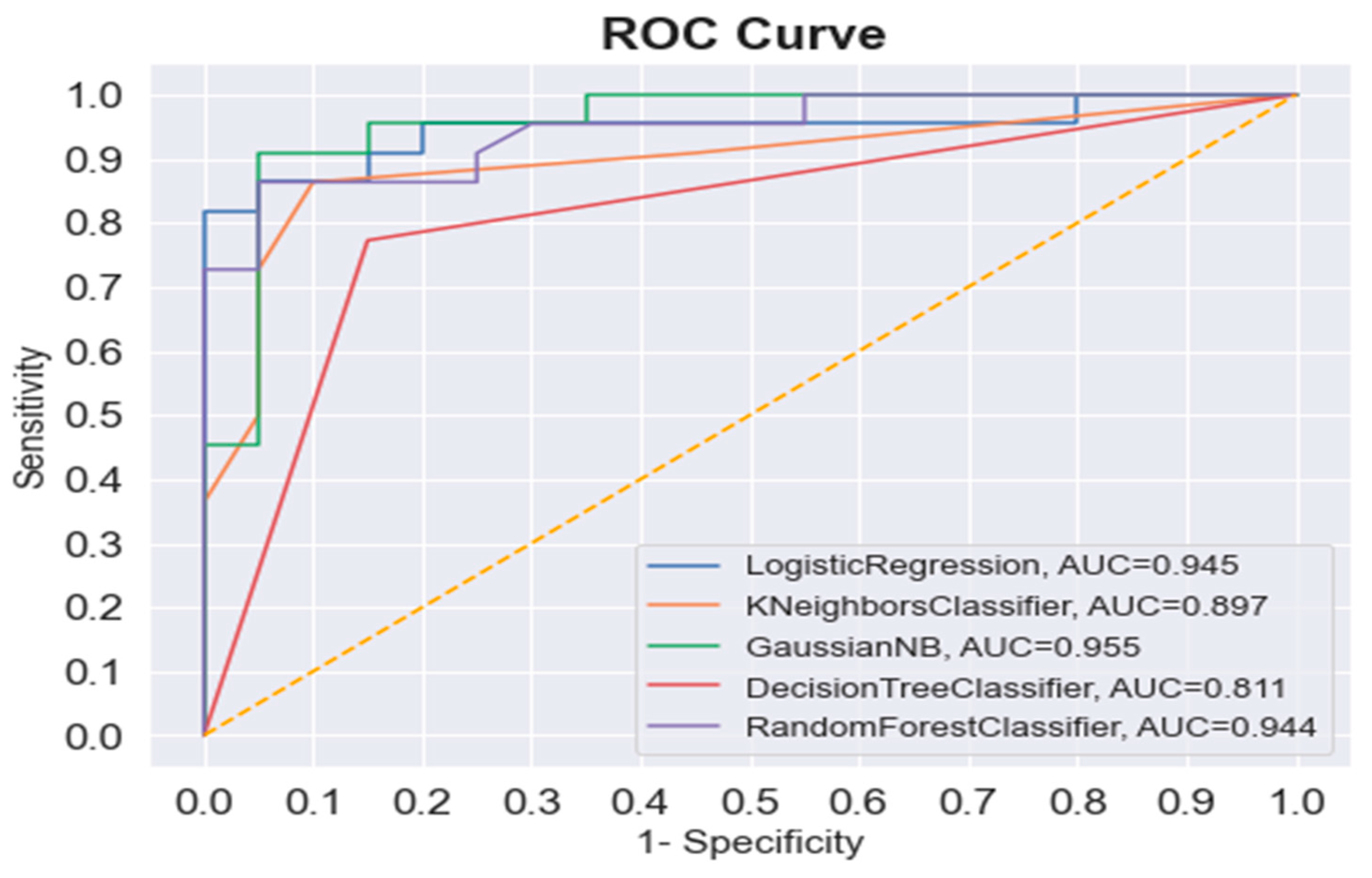
| ML Algorithm | Sex | Precision | Recall | F1-Score | Training Accuracy | Testing Accuracy |
|---|---|---|---|---|---|---|
| LR | Female | 0.86 | 0.90 | 0.88 | 0.89 | 0.88 |
| Male | 0.90 | 0.86 | 0.88 | |||
| KNN | Female | 0.76 | 0.95 | 0.84 | 0.87 | 0.83 |
| Male | 0.94 | 0.73 | 0.82 | |||
| GNB | Female | 0.86 | 0.95 | 0.90 | 0.84 | 0.90 |
| Male | 0.95 | 0.86 | 0.90 | |||
| DT | Female | 0.77 | 0.85 | 0.81 | 1 | 0.80 |
| Male | 0.85 | 0.77 | 0.81 | |||
| RF | Female | 0.83 | 0.95 | 0.88 | 0.99 | 0.88 |
| Male | 0.95 | 0.82 | 0.88 |
| Testing Set | GNB | RF | DT | LR | KNN |
|---|---|---|---|---|---|
| 1 | 0.881 | 0.857 | 0.810 | 0.881 | 0.857 |
| 2 | 0.857 | 0.833 | 0.881 | 0.929 | 0.881 |
| 3 | 0.762 | 0.810 | 0.833 | 0.857 | 0.810 |
| 4 | 0.810 | 0.833 | 0.786 | 0.833 | 0.857 |
| 5 | 0.810 | 0.810 | 0.786 | 0.738 | 0.833 |
| 6 | 0.833 | 0.786 | 0.786 | 0.738 | 0.762 |
| 7 | 0.905 | 0.905 | 0.881 | 0.905 | 0.929 |
| 8 | 0.857 | 0.857 | 0.762 | 0.905 | 0.857 |
| 9 | 0.786 | 0.786 | 0.786 | 0.810 | 0.762 |
| 10 | 0.905 | 0.929 | 0.833 | 0.881 | 0.929 |
| Mean ± SD | 0.84 ± 0.049 | 0.84 ± 0.048 | 0.814 ± 0.042 | 0.848 ± 0.068 | 0.848 ± 0.059 |
Disclaimer/Publisher’s Note: The statements, opinions and data contained in all publications are solely those of the individual author(s) and contributor(s) and not of MDPI and/or the editor(s). MDPI and/or the editor(s) disclaim responsibility for any injury to people or property resulting from any ideas, methods, instructions or products referred to in the content. |
© 2023 by the authors. Licensee MDPI, Basel, Switzerland. This article is an open access article distributed under the terms and conditions of the Creative Commons Attribution (CC BY) license (https://creativecommons.org/licenses/by/4.0/).
Share and Cite
Baban, M.T.A.; Mohammad, D.N. The Accuracy of Sex Identification Using CBCT Morphometric Measurements of the Mandible, with Different Machine-Learning Algorithms—A Retrospective Study. Diagnostics 2023, 13, 2342. https://doi.org/10.3390/diagnostics13142342
Baban MTA, Mohammad DN. The Accuracy of Sex Identification Using CBCT Morphometric Measurements of the Mandible, with Different Machine-Learning Algorithms—A Retrospective Study. Diagnostics. 2023; 13(14):2342. https://doi.org/10.3390/diagnostics13142342
Chicago/Turabian StyleBaban, Mohammed Taha Ahmed, and Dena Nadhim Mohammad. 2023. "The Accuracy of Sex Identification Using CBCT Morphometric Measurements of the Mandible, with Different Machine-Learning Algorithms—A Retrospective Study" Diagnostics 13, no. 14: 2342. https://doi.org/10.3390/diagnostics13142342