Article

Clinical Impact of the BIOFIRE Blood Culture Identification 2 Panel in Adult Patients with Bloodstream Infection: A Multicentre Observational Study in the United Arab Emirates

1
College of Medicine, Mohammed Bin Rashid University of Medicine and Health Sciences, Dubai P.O. Box 505055, United Arab Emirates
2
Infectious Diseases Unit, Rashid Hospital, Dubai P.O. Box 4545, United Arab Emirates
3
Pathology and Laboratory Medicine, Zayed Military Hospital, Abu Dhabi P.O. Box 72763, United Arab Emirates
4
Microbiology and Infection Control Unit, Pathology Department, Rashid Hospital, Dubai P.O. Box 4545, United Arab Emirates
5
Microbiology Department, Mediclinic City Hospital, Dubai Healthcare City, Dubai P.O. Box 505004, United Arab Emirates
6
Intensive Care Unit, Sheikh Khalifa General Hospital, Umm Al Quwain P.O. Box 499, United Arab Emirates
7
Data Science Department, bioMérieux Inc., Salt Lake City, UT 84108, USA
8
Medical Affairs, bioMérieux, Dubai P.O. Box 505201, United Arab Emirates
*
Author to whom correspondence should be addressed.
Diagnostics 2023, 13(14), 2433; https://doi.org/10.3390/diagnostics13142433
Submission received: 30 May 2023 / Revised: 10 July 2023 / Accepted: 18 July 2023 / Published: 21 July 2023
(This article belongs to the Special Issue Diagnostics in the Emergency and Critical Care Medicine)

Abstract

:
Rapid pathogen identification is key to the proper management of patients with bloodstream infections (BSIs), especially in the intensive care setting. This multicentre study compared the time to pathogen identification results in 185 patients admitted to intensive care with a confirmed BSI, using conventional methods (n = 99 patients) and upon implementation of the BIOFIRE® Blood Culture Identification 2 (BCID2) Panel, a rapid molecular test allowing for the simultaneous identification of 43 BSI-related nucleic acids targets (n = 86 patients). The median time to result informing optimal antibiotic therapy was significantly shorter following the implementation of the BCID2 Panel (92 vs. 28 h pre vs. post BCID2 implementation; p < 0.0001). BCID2 usage in addition to conventional methods led to the identification of at least one pathogen in 98.8% patients vs. 87.9% using conventional methods alone (p = 0.003) and was associated with a lower 30-day mortality (17.3% vs. 31.6%, respectively; p = 0.019). This study at three intensive care units in the United Arab Emirates therefore demonstrates that, in addition to conventional microbiological methods and an effective antimicrobial stewardship program, the BCID2 Panel could improve the clinical outcome of patients admitted to the intensive care unit with a confirmed BSI.

1. Introduction

Bloodstream infections are associated with high morbidity and mortality, especially in the intensive care setting [1,2,3,4,5,6]. Current guidelines for empirical treatment of bloodstream infections call for the use of broad-spectrum antibiotics and a prompt antimicrobial adjustment upon pathogen identification, in accordance with the local antimicrobial stewardship guidelines [7,8]. Rapid detection of causative pathogens and of potential resistance determinants in blood cultures is crucial for a timely administration of optimal therapy and for improving patients’ survival [3,9,10,11,12]. Indeed, delayed administration of adequate antimicrobial therapy, due to either too long time to pathogen identification or antimicrobial resistance, increases mortality [1,2,3,4,5,6,9,10,11,12].
In recent years, approaches to shorten the time to identification of pathogens have been developed. Among them, rapid molecular diagnostics based on multiplex PCR allow for the identification of a broad panel of pathogens as well as of antibiotics resistance genes directly from positive blood cultures [3,9,10]. The BIOFIRE® Blood Culture Identification 2 (BCID2) Panel (bioMérieux, Marcy l’Etoile, France) is a well-validated assay that allows for the simultaneous identification of 43 nucleic acid targets associated with bloodstream infections (26 bacterial genera/species, 7 fungal species, and 10 resistance markers) within about 1 h. It is FDA-approved, CE-marked, and locally approved in the United Arab Emirates (UAE). The BCID2 Panel (and prior to that, its first-generation BCID) demonstrated high diagnostic accuracy compared to conventional microbiological methods [13,14,15,16,17,18,19,20,21,22,23,24,25]. Its implementation is associated with a significant decrease in time to result (pathogen and resistance gene identification) or time to optimal antimicrobial therapy, compared to conventional methods [14,16,19,22,25,26,27,28]. Its use led to treatment adjustments in 22–45% patients in several reported studies [19,22,25,27,28].
Current guidelines recommend the addition of rapid diagnostic testing to an effective antimicrobial stewardship program (ASP) [7,8]. A randomised study indeed demonstrated that combining the BCID panel with antimicrobial stewardship provided greater clinical benefit compared to BCID alone or to conventional methods [27].
To the best of our knowledge, no studies so far have evaluated the clinical impact of BCID2 usage in an intensive care unit (ICU) setting in the UAE. This multicentre study aimed at evaluating, for the first time in the UAE, the clinical impact of the BCID2 Panel on the time to result informing targeted antimicrobial therapy in adult ICU patients with a bloodstream infection. We compared results of patients recruited prospectively for a period of 6 months upon implementation of the BCID2 Panel (performed in addition to conventional microbiological methods) to retrospective data of patients evaluated using conventional methods alone, during a 6-month period preceding the implementation of BCID2. We hypothesised that in patients with a bloodstream infection, the BCID2 Panel will have a higher diagnostic yield and a shorter time to results on microbial and resistance gene identification compared to conventional methods. An earlier targeted antimicrobial therapy is expected to reduce the exposure to broad-spectrum antibiotics and the rate of inappropriate antibiotic treatment, thereby potentially improving patients’ outcomes.
We showed that the implementation of the BCID2 Panel was associated with a significantly shorter median time to result informing optimal (i.e., targeted) therapy and a reduced 30-day mortality in ICU patients with a bloodstream infection in our UAE hospital setting.

2. Materials and Methods

2.1. Study Design and Participants

A multicentre observational cohort study was conducted in adult patients (≥18 and ≤85 years-old) admitted in the ICU with a confirmed bloodstream infection (i.e., a positive blood culture) to evaluate the impact of the BIOFIRE® Blood Culture Identification 2 (BCID2) Panel (bioMérieux, Marcy l’Etoile, France) on the time to result informing optimal (i.e., targeted) antimicrobial therapy. Targeted antimicrobial therapy was defined as any regimen that is active against the causative pathogen that ultimately grew in culture and to which the organism was susceptible. The time to result informing targeted therapy was defined as the time duration from collection of the first positive blood culture sample to obtention of the test result (i.e., pathogen identification per conventional methods or the BCID2 Panel) [29,30,31]. The time to result instructing targeted antimicrobial therapy for any patient who did not fall within evidence-based guidelines was adjudicated by the infectious disease physicians on the study team. Two study groups were compared. The first study group included retrospective data from ICU patients with a confirmed bloodstream infection during the six months immediately preceding the implementation of the BCID2 Panel at three participating centres (Dubai, United Arab Emirates): Rashid Hospital (June to November 2020), Mediclinic City Hospital (August 2020 to January 2021), and Sheikh Khalifa General Hospital (June to November 2019). In this study group (thereafter referred to as pre-BCID2), pathogen identification was conducted according to conventional microbiological methods only. The second study group included prospective data of ICU patients with a confirmed bloodstream infection, collected for a period of six months (February to July 2022) upon implementation of the BCID2 Panel at the same three participating centres. Pathogen identification in this study group (thereafter referred to as BCID2) was based on the results of the BCID2 Panel, in addition to standard conventional methods. Besides the time to pathogen identification results, other clinical outcomes were compared between pre-BCID2 and BCID2 patients, including the 30-day mortality, the duration of empirical antibiotics use, the number of prescribed empirical antibiotics, and the percentage of patients with antibiotics de-escalation (defined as the switch from empirical to targeted antibiotics following pathogen identification results). The duration of ICU stay could not be calculated, as the date of discharge from the ICU was not documented in this study. Patients’ demographics, comorbidities and vital signs were recorded at ICU admission. In the absence of a standardised scoring system among the three participating centres [32], disease severity and organ dysfunction at ICU admission were assessed based on documented vital signs.
The study was conducted in accordance with the Declaration of Helsinki and approved by the Dubai Scientific Research Ethics Committee (DSREC), Dubai Health Authority (approval number DSREC-10/2021_06, dated 2 November 2021) and the Mediclinic Middle East (MCME) Research and Ethics Committee (approval number MCME.CR.225.MCIT.2021, dated 30 November 2021). An informed consent was waived by the DSREC Dubai Health Authority and the MCME Research and Ethics Committee because blood samples were collected and blood cultures performed as part of routine clinical care. No blood samples were drawn specifically for this study, the study did not involve patient contact, and patient data were de-identified.

2.2. Sample Collection and Processing

Blood samples were collected solely as part of standard clinical care for ICU patients. Blood cultures were performed immediately after blood collection as per the standard protocol in place at the respective participating centres, using automated blood culture systems. The first positive blood culture data were included in the study analysis.
For the retrospective data, conventional analysis of positive blood cultures involved the performance of Gram stain, bacterial identification, and antimicrobial susceptibility testing using an automated platform, as described in Section 2.3.1. For the prospective data, positive blood cultures were further analysed using the BCID2 Panel within one hour of detected growth by the automated blood culture system, as described in Section 2.3.2. Simultaneously to the BCID2 assay, Gram stain and routine bacterial identification and antimicrobial susceptibility testing using an automated platform were conducted, as per the standard protocol at each participating centre (see Section 2.3.1). In case of discrepancy between the results from the BCID2 Panel and the standard conventional methods, the latter was used as the gold standard for patient management.
The pathogen identification results (conventional methods or BCID2, as applicable) were communicated within one hour of obtention to the ICU physician and the antibiotic stewardship program (ASP) team, including the study’s infectious disease physician. The ASP team reviewed the results and communicated recommendations on an antimicrobial regimen to the ICU team for implementation, in line with current ASP guidelines at the respective hospitals.

2.3. Laboratory Methods

2.3.1. Automated Blood Culture and Conventional Microbiological Testing

Blood culture and conventional microbiological testing at the three participating centres were done according to the international recommended protocols and the Clinical and Laboratory Standards Institute (CLSI) guidelines [33,34,35], as follows.
At Rashid Hospital, Gram stain was done manually (SURECHEM, Suffolk, UK). The recovery and detection of microorganisms were conducted using the automated BACT/ALERT® VIRTUO® Microbial Detection System (bioMérieux) with the resin-based aerobic (BACT/ALERT® FA Plus), anaerobic (BACT/ALERT® FN Plus) and aerobic and facultative anaerobic (BACT/ALERT® PF Plus) bottles (bioMérieux), or the BD BACTEC™ Automated Blood Culture System (Becton Dickinson, Franklin Lakes, NJ, USA) with the resin-based BD BACTEC™ Plus Aerobic and Anaerobic media. Positively flagged blood culture bottles were sub-cultured on blood-agar plates, and isolated colonies were identified by matrix-assisted laser desorption ionisation time-of-flight (MALDI-TOF) using the VITEK MS® automated mass spectrometry microbial identification system (bioMérieux).
At Mediclinic City Hospital, Gram stain was done manually (Dagatron Auto Stainer AT-3002, Biomed Global, Gyeonggi-do, Republic of Korea). The recovery and detection of microorganisms were conducted using the automated BACT/ALERT® VIRTUO® Microbial Detection System (bioMérieux) with the BACT/ALERT® FA Plus, BACT/ALERT® FN Plus, and BACT/ALERT® PF Plus bottles (bioMérieux). Positively flagged blood culture bottles were sub-cultured on blood-agar plates, and isolated colonies were identified using the colorimetric VITEK 2® Compact automated system (bioMérieux).
At Sheikh Khalifa General Hospital, Gram stain was done using the RAL Stainer automated staining unit (bioMérieux). The recovery and detection of microorganisms were conducted using the BD BACTEC™ Automated Blood Culture System (Becton Dickinson, Franklin Lakes, NJ, USA) with the resin-based BD BACTEC™ Plus Aerobic and Anaerobic media. Positively flagged blood culture bottles were sub-cultured on blood-agar plates, and isolated colonies were identified using the colorimetric VITEK 2® Compact automated system (bioMérieux).
For antimicrobial susceptibility testing (AST) at the three centres, 0.5 McFarland suspensions were prepared following the manufacturer’s protocol (DensiCHEK Plus, bioMérieux) and inoculated into specific VITEK 2® susceptibility cards (bioMérieux), except for colistin sensitivity testing at Rashid Hospital and Sheikh Khalifa General Hospital, which was performed by broth microdilution (ComASP™ Colistin, Liofilchem, Italy). Interpretation of resistance phenotypes was performed using the Advanced Expert System® (AES) software (version 2.0.0; bioMérieux). Interpretation of susceptibility profiles was done using the VITEK 2® software version 9.02.

2.3.2. BIOFIRE Blood Culture Identification 2 (BCID2) Panel

The BIOFIRE® Blood Culture Identification 2 (BCID2) Panel (bioMérieux, Marcy l’Etoile, France) was used in accordance with the manufacturer’s instructions. The BCID2 Panel is a multiplexed nucleic acid test intended for the simultaneous qualitative detection and identification of 43 targets associated with bloodstream infections, including 15 Gram-negative bacteria (Acinetobacter calcoaceticus-baumannii complex, Bacteroides fragilis, Haemophilus influenzae, Neisseria meningitidis, Pseudomonas aeruginosa, Stenotrophomonas maltophilia, Enterobacterales, Enterobacter cloacae complex, Escherichia coli, Klebsiella aerogenes, Klebsiella oxytoca, Klebsiella pneumoniae group, Proteus spp., Salmonella spp., Serratia marcescens), 11 Gram-positive bacteria (Enterococcus faecalis, Enterococcus faecium, Listeria monocytogenes, Staphylococcus spp., Staphylococcus aureus, Staphylococcus epidermidis, Staphylococcus lugdunensis, Streptococcus spp., Streptococcus agalactiae, Streptococcus pneumoniae, Streptococcus pyogenes), 7 yeast species (Candida albicans, Candida auris, Candida glabrata, Candida krusei, Candida parapsilosis, Candida tropicalis, Cryptococcus neoformans/gattii) and 10 antimicrobial resistance genes (carbapenemase genes: IMP, KPC, OXA-48-like, NDM, VIM; colistin resistance gene: mcr-1; extended-spectrum beta-lactamase [ESBL] gene: CTX-M; methicillin resistance genes: mecA/C, mecA/C and MREJ [Methicillin-resistant Staphylococcus aureus (MRSA)]; vancomycin resistance gene: vanA/B). The BCID2 Panel test was performed on blood culture samples identified as positive by a continuous monitoring blood culture system. Results were available within about one hour from positive blood culture. Results were interpreted in conjunction with Gram stain results, as per the manufacturer’s recommendation.

2.4. Statistical Analysis

Statistical analyses were performed using R version 4.1.2. p-values < 0.05 were considered statistically significant. For categorical variables, comparison of pre-BCID2 and BCID2 data was performed using Fisher’s exact test. When categorical variables had more than two categories, Fisher’s exact test was implemented using the Monte Carlo simulation. For continuous variables, comparison of pre-BCID2 and BCID2 data was performed using the Wilcoxon rank-sum test. Comparison of BCID2 and pre-BCID2 implementation periods for the primary and secondary aims was performed using one-sided statistical tests.
The time to test result informing optimal treatment was calculated as the time interval between the collection of the first positive blood culture sample and the availability of the test result (susceptibility result in the pre-BCID2 implementation period and BCID2 result or susceptibility result if earlier in the BCID2 implementation period) using the following formula:
Time to test result informing optimal treatment (in hours) = Time of first test result (susceptibility or BCID2) − Time of collection of first positive blood culture sample.
The difference in the mean times to test result informing optimal treatment between the pre-BCID2 and BCID2 implementation periods was evaluated after accounting for centre-to-centre variability using the following linear mixed model:
tij = β0 + β1 xij + uj
where tij is the time to test result informing optimal treatment for patient i at centre j, xij equals either 0 if patient i at centre j is in the pre-BCID2 implementation period or 1 if patient i at centre j is in the BCID2 implementation period, and uj is the random intercept for centre j. A p-value < 0.05 for β 1 was considered indicative of a statistically significant difference in the mean times to test results informing optimal treatment between the BCID2 implementation periods. The difference in the mean times to results informing optimal treatment between the pre-BCID2 and BCID2 implementation periods, after accounting for centre-to-centre variability, was estimated with the likelihood profile confidence interval for β 1 .
The duration of empirical antibiotics use was calculated as the time interval between initiation of the first empirical antibiotics treatment and antibiotics change to targeted therapy (de-escalation) following the pathogen identification result (using conventional methods or BCID2) or the full susceptibility result (whatever was earlier), using the following formula:
Duration of empirical antibiotics use (in days) = Time the antibiotics change was ordered − Time the first empirical antibiotics was initiated.
The false discovery rate of the BCID2 Panel relative to conventional methods, set as the gold standard, was calculated for the BCID2 cohort. It was defined as the ratio of false positive detections by BCID2 (discordant or detected only by BCID2) to the total detections (sum of false positive detections and true positive—i.e., concordant—detections) by BCID2. This calculation only considered pathogens that were detectable by both approaches and thus excluded pathogens that are not tested by the BCID2 Panel (off-panel pathogens).

3. Results

3.1. Patients’ Characteristics

A total of 229 ICU patients with a confirmed bloodstream infection (BSI), i.e., with a positive blood culture, were enrolled in the study, including 132 patients whose BSI was tested using conventional microbiological methods, pre-implementation of the BCID2 Panel (defining the pre-BCID2 cohort), and 97 patients recruited prospectively and tested using the BCID2 Panel in addition to conventional methods (BCID2 cohort; Figure 1). Of these enrolled patients, 99 pre-BCID2 and 86 BCID2 patients with a calculated time to pathogen identification result (informing optimal targeted therapy) were included in the analysis (Figure 1).
Patients’ characteristics at the time of ICU admission are described in Table 1. Patients were mainly male (70.7% and 65.1% in pre-BCID2 and BCID2 cohorts, respectively). The median (interquartile range [IQR]) ages in the pre-BCID2 and BCID2 study groups were 56 (40–65) and 65 (45–79) years, respectively. Most patients (66.7% pre-BCID2, 76.7% BCID2) presented up to five comorbidities, the commonest comorbidities in both groups being hypertension, diabetes, and cardiac disease. Patients of the BCID2 cohort also had a higher frequency of chronic lung disease compared to patients of the pre-BCID2 cohort (Table 1).

3.2. Detected Pathogens’ Characteristics

In the pre-BCID2 implementation phase, pathogens were detected in 87/99 (87.9%) patients by conventional methods, compared to 85/86 (98.8%) patients in the BCID2 implementation phase (using the BCID2 Panel in addition to conventional methods) (Table 2). Only one (1.2%) BCID2 patient vs. 12 (12.1%) pre-BCID2 patients had no detected pathogens (p = 0.003). The proportion of patients with single detections was comparable in both study groups (83/99 [83.8%] pre-BCID2 vs. 72/86 [83.7%] BCID2; p = 1.000). However, there were significantly more patients with the simultaneous detection of two pathogens during the BCID2 implementation phase compared to pre-BCID2 (13/86 [15.1%] vs. 4/99 [4.0%]; p = 0.011; Table 2).
A total of 91 pathogens (in 87 pre-BCID2 patients) and 98 pathogens (in 85 BCID2 patients) were identified (Table S1). The most predominant microorganisms detected by both approaches were Klebsiella pneumoniae, Escherichia coli, and Staphylococcus epidermidis. Candida auris was also frequently detected during the BCID2 implementation period, while it was never detected by conventional methods during the pre-BCID2 implementation period (Table S1).
Identified resistance phenotypes (by conventional methods) and resistance genes (by the BCID2 Panel) are shown in Table S1. Conventional methods identified antimicrobial resistance phenotypes in 11/91 (12.1%) and 4/98 (4.1%) detected pathogens in the pre-BCID2 and BCID2 implementation periods, respectively (Table S1). In comparison, the BCID2 Panel identified 29 resistance genes (29/98 [29.6%]). Resistance genes for Gram-negative bacteria (including Klebsiella pneumoniae, Escherichia coli and Klebsiella aerogenes) were CTX-M (n = 13), OXA-48-like (n = 3), NDM (n = 2), and mcr-1 (n = 1) (Table S1). Two identified Klebsiella pneumoniae were simultaneously positive for two resistance genes (CTX-M + NDM, CTX-M + OXA-48-like; Table S1). The mecA gene was identified in all detected Gram-positive Staphylococcus epidermidis (n = 10), and none of the isolates were positive for the vanA/B genes (Table S1).
Resistance and susceptibility of isolates to antimicrobial agents in both study phases are shown in Table S2 (pre-BCID2 phase) and Table S3 (BCID2 phase).
We evaluated the concordance in pathogen identification by conventional methods and the BCID2 Panel during the BCID2 implementation period. Of the 98 organisms identified (BCID2 phase; Table S1), most (74/98 [75.5%]) were fully concordant between the two identification methods. These included 70 patients with one identified pathogen and two patients with two co-detected organisms (Staphylococcus epidermidis and Candida auris in both cases). Of the 24/98 (24.5%) discordant pathogens, four (in two patients) were partially discordant (several species of the genus Staphylococcus), 14 (in 13 patients) were identified either by BCID2 (n = 5) or conventional culture (n = 9), and six (in three of 85 [3.5%] patients) were truly discordant (Table S4). The latter corresponded to two patients with Elizabethkingia meningoseptica identified by conventional culture and positive in the BCID2 Panel for Escherichia coli (one patient) and for Candida auris (second patient) and to one patient with Streptococcus viridans identified by conventional culture and positive for Staphylococcus spp. in the BCID2 Panel (Table S4). Of the nine pathogens not identified by the BCID2 Panel, four were off-panel microorganisms (Corynebacterium spp., Kytococcus spp., Aeromonas sóbria, Enterococcus Avium Group D) and five were on-panel microorganisms and thus corresponded to detection failures (Table S4).
The false discovery rate of BCID2 relative to conventional methods, focusing on pathogens detectable by both approaches (i.e., excluding off-panel pathogens), was calculated based on these concordance results. The false discovery rate, defined as the ratio of false positive detections by BCID2 to the total detections (false positive and true positive detections) by BCID2, was 15/89 or 16.9%.

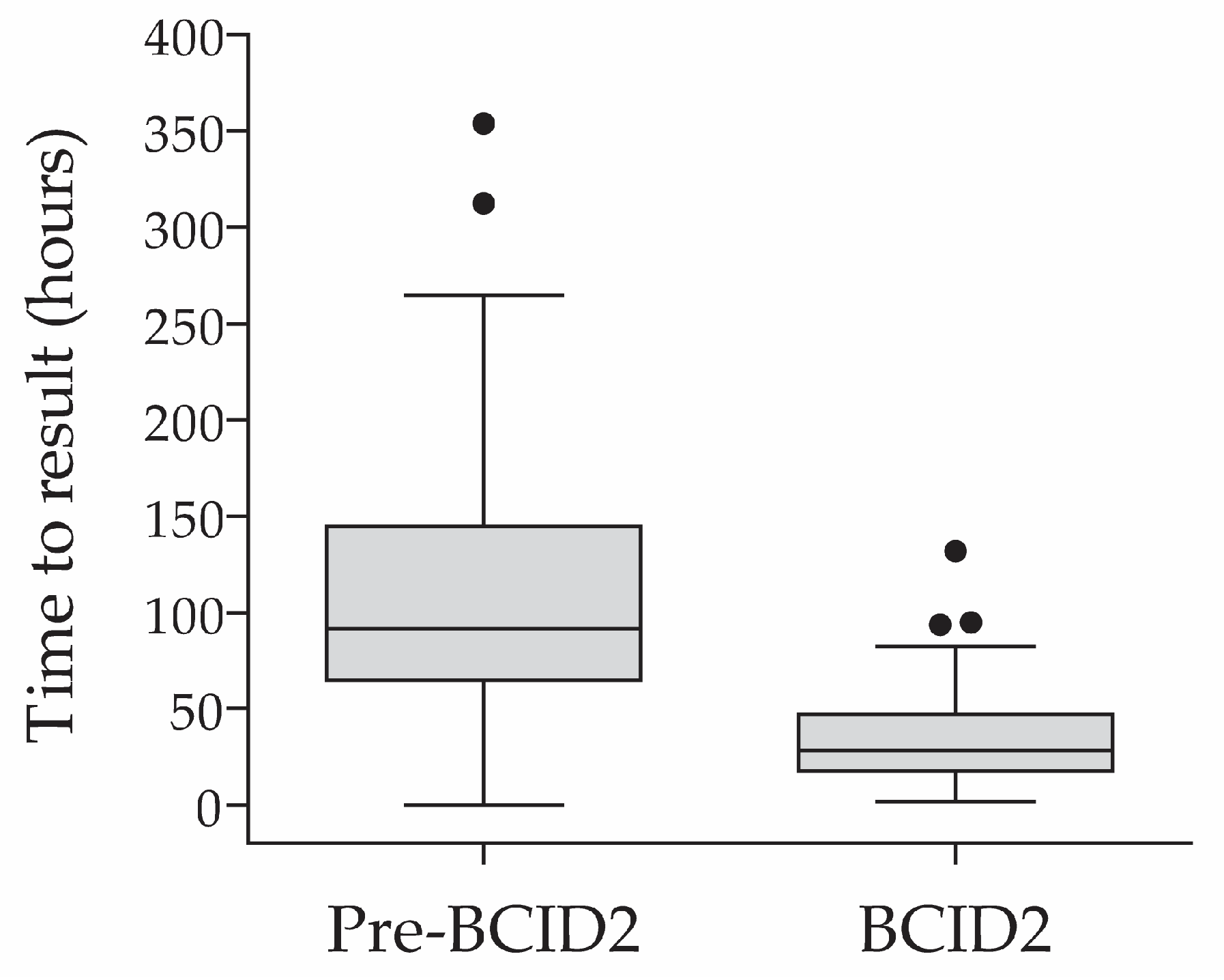
3.3. Time to Result Informing Targeted Therapy in the Pre-BCID2 vs. BCID2 Cohorts

The time to result informing optimal therapy was calculated as the difference between the time of collection of the first positive culture sample and the time to availability of the test result (susceptibility result in the pre-BCID2 study group and BCID2 Panel result or susceptibility result in the BCID2 study group). The median (IQR) times to result informing targeted therapy in the pre-BCID2 and BCID2 phases were 91.7 (64.7–144.9) and 28.1 (17.6–47.1) hours, respectively (p < 0.0001; Figure 2).
After accounting for centre-to-centre variability, the estimated difference (95% confidence interval [CI]) of the mean time to result informing optimal treatment between the pre-BCID2 and BCID2 implementation periods was 73.3 (59.8–88.6) hours (linear mixed models, p < 0.0001).

3.4. Secondary Clinical Outcomes

We compared the 30-day mortality (relative to the day blood culture specimens were drawn) in both study groups. In the pre-BCID2 and the BCID2 study periods, one and five patients, respectively, lacking a documented mortality status were excluded from the comparison. The 30-day mortality rate in the BCID2 study group was significantly lower than that of pre-BCID2 (14/81 [17.3%] vs. 31/98 [31.6%], respectively; p = 0.019).
To evaluate the possible impact of BCID2 implementation on antibiotics treatment adjustment, the number and duration of empirical antibiotics usage were compared in both study groups. The number of empirical antibiotics ordered in both study groups (from none to three per patient) was comparable (p = 0.394; Table 3).
The duration of empirical antibiotics usage was able to be calculated in 75 pre-BCID2 and 64 BCID2 patients. The median (IQR) duration of treatment with empirical antibiotics was shorter in BCID2 patients (2 [1,2,3,4] days) than in pre-BCID2 patients (3 [1,2,3,4] days), albeit the difference was not statistically significant (p = 0.126).
Antibiotics de-escalation, defined as the switch from empirical antibiotics to targeted therapy following test result (pathogen identification or susceptibility result), was comparable in both groups (p = 1.000; Table 4).

4. Discussion

This multicentre study is, to the best of our knowledge, the first to evaluate the potential clinical benefit of implementing the BCID2 Panel for the management of ICU patients with a confirmed BSI in the United Arab Emirates.
After accounting for centre-to-centre variability, the mean time to results gained using the BCID2 Panel (in addition to conventional methods) compared to using conventional methods alone was 73.3 h (95% CI: 59.8–88.6; p < 0.0001). A significantly shorter time to result using BCID2 vs. conventional culture methods was already demonstrated in previous studies conducted in Northern America, Europe, South Africa and Australia [14,16,19,22,26,27,28]. Our study therefore confirms previous reports of the benefit of the BCID2 Panel for rapid microbial identification in BSI patients.
In our study, the 30-day mortality was significantly lower in BCID2 (17.3%) vs. pre-BCID2 (31.6%) patients (p = 0.019). Several studies investigated the impact of rapid microbial identification on patient mortality [3,10,11,25,27,30,36]. While some studies showed a significant reduction of mortality in association with rapid microbial identification [3,10,11,30,36], others did not [10,25,27]. A recent meta-analysis indicated that mortality is reduced only when rapid testing is accompanied with an antimicrobial stewardship program (ASP) [37]. Since this is the case at our participating hospitals, our observation that BCID2 implementation was associated with a significantly reduced mortality agrees with the reported meta-analysis by Timbrook et al. [37].
As expected, pathogens were identified in more patients following the implementation of BCID2 (98.8% vs. 87.9% patients; p = 0.003), and polydetections were more frequent post implementation of BCID2 (15.1% vs. 4.0% patients with two detected pathogens; p = 0.011). The frequency of polydetections upon implementation of BCID2 in our study is in line with the 10–20% polymicrobial bloodstream infections reported by Holma et al. [18].
The BCID2 Panel also enabled the identification of a high frequency of resistance genes (29.6% of detected pathogens). Of interest is the identification of the mecA/C methicillin resistance gene in 10/10 (100%) of detected Staphylococcus epidermidis. This highlights the benefit of the BCID2 Panel in identifying these clinically relevant pathogens, especially in the context of a reported high (up to 42%) prevalence of methicillin-resistant Staphylococcus aureus (MRSA) in Saudi Arabia [38,39]. Although the characterisation of resistance determinants is expected to impact the clinical decision for optimal targeted therapy, as observed in other studies [19,22,25,27,28], the duration of empirical antibiotics usage and the rate of antibiotics de-escalation were not statistically different between the pre-BCID2 and BCID2 study populations. Whether the absence of impact on antibiotics treatment adjustment observed in our study is due to the sample size, the study design (observational vs. interventional), or the already very effective standard of care (combined with an efficient ASP) remains to be investigated.
The concordance in pathogen identification between conventional methods and the BCID2 Panel in the BCID2 implementation phase was very good. Of the 98 detected pathogens, 78 (79.6%) showed a full or partial concordance, six (6.1%) were a mismatch, five (5.1%) were additional detections by BCID2 (Candida auris, Staphylococcus epidermidis, Acinetobacter baumannii complex, Klebsiella pneumoniae, Streptococcus pneumoniae), 5 (5.1%) were detection failures by BCID2 (not detected on-panel pathogens), and four (4.1%) were off-panel and only detected by conventional methods (Corynebacterium spp., Kytococcus spp., Aeromonas sóbria, Enterococcus Avium Group D). Of the five on-panel organisms not detected by BCID2, four were from polymicrobial specimens. The reason for frequent discordant organism identification in polymicrobial setting is unclear, but was previously reported in several studies [15,40,41,42,43]. The four off-panel organisms detected by conventional culture correspond to uncommon and usually non-pathogenic organisms, and are thus not expected to be clinically relevant. Therefore, the BCID2 Panel showed an overall reasonable performance in identifying clinically relevant pathogens, with few detection failures and a good concordance with conventional methods.
The strengths of this study include its multicentre design, its real-world setting, and the balanced study populations recruited over 6 months pre and post BCID2 implementation at the same three hospitals. This study presents some limitations, including the relatively low number of patients per group, the imbalanced number of patients enrolled per centre, which precluded a subgroup analysis per centre, the observational nature of the study, and the lack of documented data on the length of ICU stay. Further studies should include more patients from additional centres to confirm the present results. In addition, further molecular characterisation studies are needed to better understand the evolution of pathogens with antimicrobial resistance in the ICU setting.

5. Conclusions

The implementation of the BCID2 Panel at three UAE hospitals resulted in a significantly shorter time to result and a reduced 30-day mortality, compared to patients tested by conventional methods. This study therefore confirmed the work of others, supporting the use of the BCID2 Panel to improve clinical outcome in ICU patients with a bloodstream infection.

Supplementary Materials

The following supporting information can be downloaded at https://www.mdpi.com/article/10.3390/diagnostics13142433/s1, Table S1: Number of detected organisms, resistance phenotypes (per conventional methods) and resistance genes (per BCID2 Panel) in patients of the pre-BCID2 (n = 87) and BCID2 (n = 85) implementation periods with at least one detected pathogen; Table S2: Resistance and susceptibility to antimicrobial treatment in the pre-BCID2 implementation phase; Table S3: Resistance and susceptibility to antimicrobial treatment in the BCID2 implementation phase; Table S4: Discordant pathogens detected by conventional culture vs. the BCID2 Panel among patients in the BCID2 implementation period (n = 24 discordant pathogens in n = 18 patients).

Author Contributions

Conceptualization, A.S.; methodology, A.S., B.S., L.A.D., M.H., M.A. and H.C.; validation, S.B., N.A., H.A., A.A.M. and I.H.R.; formal analysis, A.S., B.S. and M.P.; investigation, A.S., L.A.D., M.H., M.A., H.C., S.B., N.A., H.A., M.A.E.E., O.B., V.O., A.A.M. and I.H.R.; resources, A.S., B.S., L.A.D., M.H., M.A. and H.C.; data curation, A.S., L.A.D., M.H., M.A., H.C., S.B., N.A., H.A., M.A.E.E., O.B., V.O., A.A.M. and I.H.R.; writing—review and editing, A.S., L.A.D., M.H., M.A., H.C., S.B., N.A., H.A., M.A.E.E., O.B., V.O., A.A.M., I.H.R., M.P. and B.S.; visualization, A.S., M.P. and B.S.; supervision, A.S., L.A.D., M.H., M.A. and H.C.; project administration, A.S. All authors have read and agreed to the published version of the manuscript.

Funding

This research received no external funding. The APC was funded by bioMérieux.

Institutional Review Board Statement

The study was conducted in accordance with the Declaration of Helsinki and approved by the Dubai Scientific Research Ethics Committee (DSREC), Dubai Health Authority (approval number DSREC-10/2021_06, dated 2 November 2021), and the Mediclinic Middle East (MCME) Research and Ethics Committee (approval number MCME.CR.225.MCIT.2021, dated 30 November 2021).

Informed Consent Statement

Patient consent was waived because blood samples were collected and blood cultures performed as part of routine clinical care. No blood samples were drawn for this study, the study did not involve patient contact, and patient data were de-identified.

Data Availability Statement

The data presented in this study are available within the article or Supplementary Materials.

Acknowledgments

The authors acknowledge the kind cooperation of Asad Alsabbah (Critical Care Consultant, Mediclinic City Hospital, Dubai, United Arab Emirates) and thank Tammy Bleak (bioMérieux Inc., Salt Lake City, UT, USA) for coordinating publication. The authors also thank Anne Rascle of AR Medical Writing (Regensburg, Germany) for providing medical writing support, which was funded by bioMérieux (Marcy l’Etoile, France), in accordance with Good Publication Practice (GPP3) guidelines (http://www.ismpp.org/gpp3; accessed on 13 April 2023).

Conflicts of Interest

M.P. and B.S. are employees of bioMérieux. All other authors declare no conflict of interest. bioMérieux supported this study in the form of technical assistance funding and provision of reagents. The funder had a role in the statistical analysis of the data and in the writing of the manuscript.

References

  1. Goto, M.; Al-Hasan, M.N. Overall Burden of Bloodstream Infection and Nosocomial Bloodstream Infection in North America and Europe. Clin. Microbiol. Infect. 2013, 19, 501–509. [Google Scholar] [CrossRef] [PubMed] [Green Version]
  2. Sakr, Y.; Jaschinski, U.; Wittebole, X.; Szakmany, T.; Lipman, J.; Ñamendys-Silva, S.A.; Martin-Loeches, I.; Leone, M.; Lupu, M.-N.; Vincent, J.-L.; et al. Sepsis in Intensive Care Unit Patients: Worldwide Data From the Intensive Care over Nations Audit. Open Forum Infect. Dis. 2018, 5, ofy313. [Google Scholar] [CrossRef] [Green Version]
  3. Martinez, R.M.; Wolk, D.M. Bloodstream Infections. Microbiol. Spectr. 2016, 4, 653–689. [Google Scholar] [CrossRef] [PubMed] [Green Version]
  4. Fleischmann, C.; Scherag, A.; Adhikari, N.K.J.; Hartog, C.S.; Tsaganos, T.; Schlattmann, P.; Angus, D.C.; Reinhart, K.; International Forum of Acute Care Trialists. Assessment of Global Incidence and Mortality of Hospital-Treated Sepsis. Current Estimates and Limitations. Am. J. Respir. Crit. Care Med. 2016, 193, 259–272. [Google Scholar] [CrossRef]
  5. McNamara, J.F.; Righi, E.; Wright, H.; Hartel, G.F.; Harris, P.N.A.; Paterson, D.L. Long-Term Morbidity and Mortality Following Bloodstream Infection: A Systematic Literature Review. J. Infect. 2018, 77, 1–8. [Google Scholar] [CrossRef] [Green Version]
  6. Tabah, A.; Buetti, N.; Staiquly, Q.; Ruckly, S.; Akova, M.; Aslan, A.T.; Leone, M.; Conway Morris, A.; Bassetti, M.; Arvaniti, K.; et al. Epidemiology and Outcomes of Hospital-Acquired Bloodstream Infections in Intensive Care Unit Patients: The EUROBACT-2 International Cohort Study. Intensive Care Med. 2023, 49, 178–190. [Google Scholar] [CrossRef]
  7. Barlam, T.F.; Cosgrove, S.E.; Abbo, L.M.; MacDougall, C.; Schuetz, A.N.; Septimus, E.J.; Srinivasan, A.; Dellit, T.H.; Falck-Ytter, Y.T.; Fishman, N.O.; et al. Implementing an Antibiotic Stewardship Program: Guidelines by the Infectious Diseases Society of America and the Society for Healthcare Epidemiology of America. Clin. Infect. Dis. 2016, 62, e51–e77. [Google Scholar] [CrossRef] [Green Version]
  8. European Centre for Disease Prevention and Control Antimicrobial Stewardship. Available online: https://www.ecdc.europa.eu/en/publications-data/directory-guidance-prevention-and-control/prudent-use-antibiotics/antimicrobial (accessed on 22 November 2022).
  9. Timsit, J.-F.; Ruppé, E.; Barbier, F.; Tabah, A.; Bassetti, M. Bloodstream Infections in Critically Ill Patients: An Expert Statement. Intensive Care Med. 2020, 46, 266–284. [Google Scholar] [CrossRef] [PubMed]
  10. Lamy, B.; Sundqvist, M.; Idelevich, E.A.; ESCMID Study Group for Bloodstream Infections, Endocarditis and Sepsis (ESGBIES). Bloodstream Infections—Standard and Progress in Pathogen Diagnostics. Clin. Microbiol. Infect. 2020, 26, 142–150. [Google Scholar] [CrossRef]
  11. Seymour, C.W.; Gesten, F.; Prescott, H.C.; Friedrich, M.E.; Iwashyna, T.J.; Phillips, G.S.; Lemeshow, S.; Osborn, T.; Terry, K.M.; Levy, M.M. Time to Treatment and Mortality during Mandated Emergency Care for Sepsis. N. Engl. J. Med. 2017, 376, 2235–2244. [Google Scholar] [CrossRef]
  12. Kadri, S.S.; Lai, Y.L.; Warner, S.; Strich, J.R.; Babiker, A.; Ricotta, E.E.; Demirkale, C.Y.; Dekker, J.P.; Palmore, T.N.; Rhee, C.; et al. Inappropriate Empirical Antibiotic Therapy for Bloodstream Infections Based on Discordant In-Vitro Susceptibilities: A Retrospective Cohort Analysis of Prevalence, Predictors, and Mortality Risk in US Hospitals. Lancet Infect. Dis. 2021, 21, 241–251. [Google Scholar] [CrossRef] [PubMed]
  13. Peri, A.M.; Ling, W.; Furuya-Kanamori, L.; Harris, P.N.A.; Paterson, D.L. Performance of BioFire Blood Culture Identification 2 Panel (BCID2) for the Detection of Bloodstream Pathogens and Their Associated Resistance Markers: A Systematic Review and Meta-Analysis of Diagnostic Test Accuracy Studies. BMC Infect. Dis. 2022, 22, 794. [Google Scholar] [CrossRef] [PubMed]
  14. Peri, A.M.; Bauer, M.J.; Bergh, H.; Butkiewicz, D.; Paterson, D.L.; Harris, P.N. Performance of the BioFire Blood Culture Identification 2 Panel for the Diagnosis of Bloodstream Infections. Heliyon 2022, 8, e09983. [Google Scholar] [CrossRef]
  15. Berinson, B.; Both, A.; Berneking, L.; Christner, M.; Lütgehetmann, M.; Aepfelbacher, M.; Rohde, H. Usefulness of BioFire FilmArray BCID2 for Blood Culture Processing in Clinical Practice. J. Clin. Microbiol. 2021, 59, e0054321. [Google Scholar] [CrossRef]
  16. Caméléna, F.; Péan de Ponfilly, G.; Pailhoriès, H.; Bonzon, L.; Alanio, A.; Poncin, T.; Lafaurie, M.; Dépret, F.; Cambau, E.; Godreuil, S.; et al. Multicenter Evaluation of the FilmArray Blood Culture Identification 2 Panel for Pathogen Detection in Bloodstream Infections. Microbiol. Spectr. 2023, 11, e0254722. [Google Scholar] [CrossRef] [PubMed]
  17. El Sherif, H.M.; Elsayed, M.; El-Ansary, M.R.; Aboshanab, K.M.; El Borhamy, M.I.; Elsayed, K.M. BioFire FilmArray BCID2 versus VITEK-2 System in Determining Microbial Etiology and Antibiotic-Resistant Genes of Pathogens Recovered from Central Line-Associated Bloodstream Infections. Biology 2022, 11, 1573. [Google Scholar] [CrossRef]
  18. Holma, T.; Torvikoski, J.; Friberg, N.; Nevalainen, A.; Tarkka, E.; Antikainen, J.; Martelin, J.J. Rapid Molecular Detection of Pathogenic Microorganisms and Antimicrobial Resistance Markers in Blood Cultures: Evaluation and Utility of the next-Generation FilmArray Blood Culture Identification 2 Panel. Eur. J. Clin. Microbiol. Infect. Dis. 2022, 41, 363–371. [Google Scholar] [CrossRef]
  19. Sparks, R.; Balgahom, R.; Janto, C.; Polkinghorne, A.; Branley, J. Evaluation of the BioFire Blood Culture Identification 2 Panel and Impact on Patient Management and Antimicrobial Stewardship. Pathology 2021, 53, 889–895. [Google Scholar] [CrossRef]
  20. Claeys, K.C.; Schlaffer, K.; Smith, R.; Hitchcock, S.; Jiang, Y.; Evans, S.; Johnson, J.K.; Leekha, S. Day at the Races: Comparing BioFire FilmArray Blood Culture ID Panels with Verigene Blood Culture Panel in Gram-Negative Bloodstream Infections Using DOOR-MAT Analysis. Clin. Infect. Dis. 2021, 73, 1103–1106. [Google Scholar] [CrossRef]
  21. Altun, O.; Almuhayawi, M.; Ullberg, M.; Özenci, V. Clinical Evaluation of the FilmArray Blood Culture Identification Panel in Identification of Bacteria and Yeasts from Positive Blood Culture Bottles. J. Clin. Microbiol. 2013, 51, 4130–4136. [Google Scholar] [CrossRef] [Green Version]
  22. Verroken, A.; Despas, N.; Rodriguez-Villalobos, H.; Laterre, P.-F. The Impact of a Rapid Molecular Identification Test on Positive Blood Cultures from Critically Ill with Bacteremia: A Pre-Post Intervention Study. PLoS ONE 2019, 14, e0223122. [Google Scholar] [CrossRef] [PubMed] [Green Version]
  23. Muzzi, M.; Viaggi, B.; Fabbri, S.; Bechi, L.; Scirè-Calabrisotto, C.; Villa, G.; Romagnoli, S. The Impact of Fast Microbiology in Intensive Care Units in the Era of Antibiotic Resistance: An Observational Retrospective Study. Curr. Microbiol. 2022, 79, 79. [Google Scholar] [CrossRef] [PubMed]
  24. Rule, R.; Paruk, F.; Becker, P.; Neuhoff, M.; Chausse, J.; Said, M. Diagnostic Accuracy of the BioFire FilmArray Blood Culture Identification Panel When Used in Critically Ill Patients with Sepsis. J. Microbiol. Methods 2021, 189, 106303. [Google Scholar] [CrossRef] [PubMed]
  25. Schumann, J.; Johanns, U.; Ahmad-Nejad, P.; Ghebremedhin, B.; Woebker, G. The Impact of the FilmArray-Based Detection of Microbial Pathogens from Positive Blood Culture Vials on the Time to Optimal Antimicrobial Regimen in Intensive Care Units of the Helios University Clinic Wuppertal, Germany. J. Clin. Med. 2021, 10, 5880. [Google Scholar] [CrossRef]
  26. Donnars, A.; Mahieu, R.; Declerck, C.; Chenouard, R.; Lemarié, C.; Pailhoriès, H.; Requin, J.; Kempf, M.; Eveillard, M. BIOFIRE® Blood Culture IDentification 2 (BCID2) Panel for Early Adaptation of Antimicrobial Therapy in Adult Patients with Bloodstream Infections: A Real-Life Experience. Diagn. Microbiol. Infect. Dis. 2023, 105, 115858. [Google Scholar] [CrossRef]
  27. Banerjee, R.; Teng, C.B.; Cunningham, S.A.; Ihde, S.M.; Steckelberg, J.M.; Moriarty, J.P.; Shah, N.D.; Mandrekar, J.N.; Patel, R. Randomized Trial of Rapid Multiplex Polymerase Chain Reaction–Based Blood Culture Identification and Susceptibility Testing. Clin. Infect. Dis. 2015, 61, 1071–1080. [Google Scholar] [CrossRef] [Green Version]
  28. Rule, R.; Paruk, F.; Becker, P.; Neuhoff, M.; Chausse, J.; Said, M. Clinical Utility of the BioFire FilmArray Blood Culture Identification Panel in the Adjustment of Empiric Antimicrobial Therapy in the Critically Ill Septic Patient. PLoS ONE 2021, 16, e0254389. [Google Scholar] [CrossRef]
  29. Bork, J.T.; Leekha, S.; Heil, E.L.; Zhao, L.; Badamas, R.; Johnson, J.K. Rapid Testing Using the Verigene Gram-Negative Blood Culture Nucleic Acid Test in Combination with Antimicrobial Stewardship Intervention against Gram-Negative Bacteremia. Antimicrob. Agents Chemother. 2015, 59, 1588–1595. [Google Scholar] [CrossRef] [Green Version]
  30. Huang, A.M.; Newton, D.; Kunapuli, A.; Gandhi, T.N.; Washer, L.L.; Isip, J.; Collins, C.D.; Nagel, J.L. Impact of Rapid Organism Identification via Matrix-Assisted Laser Desorption/Ionization Time-of-Flight Combined with Antimicrobial Stewardship Team Intervention in Adult Patients with Bacteremia and Candidemia. Clin. Infect. Dis. 2013, 57, 1237–1245. [Google Scholar] [CrossRef]
  31. Beganovic, M.; Timbrook, T.T.; Wieczorkiewicz, S.M. Predictors of Time to Effective and Optimal Antimicrobial Therapy in Patients With Positive Blood Cultures Identified via Molecular Rapid Diagnostic Testing. Open Forum Infect. Dis. 2019, 6, ofy350. [Google Scholar] [CrossRef] [Green Version]
  32. Vincent, J.-L.; Moreno, R. Clinical review: Scoring systems in the critically ill. Crit. Care 2010, 14, 207. [Google Scholar] [CrossRef] [Green Version]
  33. Doern, G.V.; Carroll, K.C.; Diekema, D.J.; Garey, K.W.; Rupp, M.E.; Weinstein, M.P.; Sexton, D.J. Practical Guidance for Clinical Microbiology Laboratories: A Comprehensive Update on the Problem of Blood Culture Contamination and a Discussion of Methods for Addressing the Problem. Clin. Microbiol. Rev. 2019, 33, e00009-19. [Google Scholar] [CrossRef] [PubMed]
  34. Miller, J.M.; Binnicker, M.J.; Campbell, S.; Carroll, K.C.; Chapin, K.C.; Gilligan, P.H.; Gonzalez, M.D.; Jerris, R.C.; Kehl, S.C.; Patel, R.; et al. A Guide to Utilization of the Microbiology Laboratory for Diagnosis of Infectious Diseases: 2018 Update by the Infectious Diseases Society of America and the American Society for Microbiology. Clin. Infect. Dis. 2018, 67, 813–816. [Google Scholar] [CrossRef] [PubMed] [Green Version]
  35. Clinical Laboratory and Standards Institute (CLSI). Performance Standards for Antimicrobial Susceptibility Testing, 30th ed.; CLSI Supplement M100, Clinical Laboratory and Standards Institute: Wayne, PA, USA, 2020; ISBN 978-1-68440-067-6. [Google Scholar]
  36. Zadka, H.; Raykhshtat, E.; Uralev, B.; Bishouty, N.; Weiss-Meilik, A.; Adler, A. The Implementation of Rapid Microbial Identification via MALDI-ToF Reduces Mortality in Gram-Negative but Not Gram-Positive Bacteremia. Eur. J. Clin. Microbiol. Infect. Dis. 2019, 38, 2053–2059. [Google Scholar] [CrossRef] [PubMed]
  37. Timbrook, T.T.; Morton, J.B.; McConeghy, K.W.; Caffrey, A.R.; Mylonakis, E.; LaPlante, K.L. The Effect of Molecular Rapid Diagnostic Testing on Clinical Outcomes in Bloodstream Infections: A Systematic Review and Meta-Analysis. Clin. Infect. Dis. 2017, 64, 15–23. [Google Scholar] [CrossRef] [Green Version]
  38. Adam, K.M.; Abomughaid, M.M. Prevalence of Methicillin-Resistant in Saudi Arabia Revisited: A Meta-Analysis. Open Public Health J. 2018, 11. [Google Scholar] [CrossRef] [Green Version]
  39. Aljeldah, M.M. Prevalence of Methicillin-Resistant Staphylococcus Aureus (MRSA) in Saudi Arabia: A Systematic Review. J. Pure Appl. Microbiol. 2020, 14, 37–46. [Google Scholar] [CrossRef] [Green Version]
  40. García-Rivera, C.; Parra-Grande, M.; Merino, E.; Boix, V.; Rodríguez, J.C. Concordance of the Filmarray Blood Culture Identification Panel 2 and Classical Microbiological Methods in a Bacteriemia Diagnostic Unit. Diagn. Microbiol. Infect. Dis. 2022, 104, 115787. [Google Scholar] [CrossRef]
  41. Blaschke, A.J.; Heyrend, C.; Byington, C.L.; Fisher, M.A.; Barker, E.; Garrone, N.F.; Thatcher, S.A.; Pavia, A.T.; Barney, T.; Alger, G.D.; et al. Rapid Identification of Pathogens from Positive Blood Cultures by Multiplex Polymerase Chain Reaction Using the FilmArray System. Diagn. Microbiol. Infect. Dis. 2012, 74, 349–355. [Google Scholar] [CrossRef] [Green Version]
  42. Southern, T.R.; VanSchooneveld, T.C.; Bannister, D.L.; Brown, T.L.; Crismon, A.S.; Buss, S.N.; Iwen, P.C.; Fey, P.D. Implementation and Performance of the BioFire FilmArray® Blood Culture Identification Panel with Antimicrobial Treatment Recommendations for Bloodstream Infections at a Midwestern Academic Tertiary Hospital. Diagn. Microbiol. Infect. Dis. 2015, 81, 96–101. [Google Scholar] [CrossRef]
  43. Fiori, B.; D’Inzeo, T.; Giaquinto, A.; Menchinelli, G.; Liotti, F.M.; de Maio, F.; De Angelis, G.; Quaranta, G.; Nagel, D.; Tumbarello, M.; et al. Optimized Use of the MALDI BioTyper System and the FilmArray BCID Panel for Direct Identification of Microbial Pathogens from Positive Blood Cultures. J. Clin. Microbiol. 2016, 54, 576–584. [Google Scholar] [CrossRef] [PubMed] [Green Version]
Figure 1. Study flow diagram. (a) Pre-BCID2 implementation period. Bloodstream infection diagnosis was based on the results of conventional microbiological methods. The pre-BCID2 cohort included retrospective data from ICU patients with a confirmed bloodstream infection, collected during the immediate six months preceding the implementation of the BCID2 Panel. (b) BCID2 implementation period. The BCID2 cohort included prospective data of ICU patients with a confirmed bloodstream infection, collected for a period of six months upon implementation of the BCID2 Panel. Pathogen identification was conducted using the BCID2 Panel, in addition to standard conventional methods. Bloodstream infection diagnosis was based on BCID2 results, unless they were discordant with those of the conventional approach, in which case conventional results were considered for diagnosis.
Diagnostics 13 02433 g001
Figure 2. Time to result informing optimal (targeted) therapy before and after implementation of BCID2 testing.
Diagnostics 13 02433 g002
Table 1. Patients’ characteristics.
Pre-BCID2 Cohort
(n = 99)
BCID2 Cohort
(n = 86)
p-Value
Study population per centre, N (%) <0.0001
Rashid Hospital77 (77.8%)37 (43.0%)
Sheikh Khalifa General Hospital13 (13.1%)43 (50.0%)
Mediclinic City Hospital9 (9.1%)6 (7.0%)
Age in years, median (IQR)56 (40–65)65 (45–79)0.001
Age class, N (%) 0.002
18–30 years old8 (8.1%)7 (8.1%)
31–40 years old17 (17.2%)4 (4.7%)
41–50 years old13 (13.1%)14 (16.3%)
51–60 years old26 (26.3%)13 (15.1%)
61–70 years old19 (19.2%)14 (16.3%)
71–85 years old16 (16.2%)34 (39.5%)
Sex, N (%) 0.433
Male70 (70.7%)56 (65.1%)
Female29 (29.3%)30 (34.9%)
Vital signs on ICU admission, median (IQR)
Oxygen saturation (%)98 (95.0–100.0) 197 (96.0–99.0) 20.194
Pulse rate (bpm)93 (86.0–111.0) 3101 (89.0–114.0)0.177
Systolic blood pressure (mmHg)107.5 (90.0–126.5) 3108 (95.0–122.0)0.695
Diastolic blood pressure (mmHg)64 (52.0–74.25) 358.5 (49.0–68.0)0.073
Comorbidities, N (%)
Hypertension46 (46.5%)44 (51.2%)0.557
Diabetes45 (45.5%)35 (40.7%)0.554
Cardiac disease29 (29.3%)29 (33.7%)0.530
Chronic lung disease9 (9.1%)28 (32.6%)0.0001
Malignancy15 (15.2%)8 (9.3%)0.269
Immunosuppressive treatment6 (6.1%)4 (4.7%)0.753
SOT3 (3.0%)2 (2.3%)1.000
HSCT2 (2.0%)0 (0.0%)0.500
Number of comorbidities, N (%) 0.221
033 (33.3%)20 (23.3%)
117 (17.2%)14 (16.3%)
221 (21.2%)29 (33.7%)
320 (20.2%)15 (17.4%)
4 to 58 (8.1%)8 (9.3%)
1 N = 97 (two missing results). 2 N = 84 (two missing results). 3 N = 98 (one missing result). Abbreviations: bpm, beats per minute; HSCT, haematopoietic stem cell transplantation; IQR, interquartile range; mmHg, millimeters of mercury; SOT, solid-organ transplantation.
Table 2. Number of pathogens detected per positive blood culture in the pre-BCID2 and BCID2 study groups.
Study PopulationPatients According to the Number of Detected Pathogens, N (%)
No DetectionsOne DetectionTwo Detections
Pre-BCID2 phase (n = 99)12 (12.1%)83 (83.8%)4 (4.0%)
BCID2 phase (n = 86)1 (1.2%)72 (83.7%)13 (15.1%)
Table 3. Number of empirical antibiotics ordered in the pre-BCID2 and BCID2 study periods.
Study PopulationPatients According to the Number of Ordered Empirical Antibiotics, N (%)
No Empirical AntibioticsOne Empirical AntibioticsTwo Empirical AntibioticsThree Empirical AntibioticsUnknown Number
Pre-BCID2 phase (n = 99)1 (1.0%)32 (32.3%)42 (42.4%)24 (24.7%)0 (0.0%)
BCID2 phase (n = 86)1 (1.2%)37 (43.0%)30 (34.9%)17 (19.8%)1 (1.2%) 1
1 Patient received empirical antibiotics, but the number and types of antibiotics were not recorded.
Table 4. Number of patients with antibiotics de-escalation (change from empirical to targeted antibiotics) in the pre-BCID2 and BCID2 study groups.
Study PopulationPatients According to Antibiotics Change, N (%)
No Record of
Antibiotics Change 1
Change to Targeted Antibiotics 2Unknown 3
Pre-BCID2 phase (n = 99)4 (4.0%)94 (94.9%)1 (1.0%)
BCID2 phase (n = 86)3 (3.5%)82 (95.3%)1 (1.2%)
1 Patients with a record of ordered empirical antibiotics only; 2 Patients with a record of ordered empirical antibiotics and of antibiotics change to optimal therapy following test result (pathogen identification or susceptibility result); 3 Antibiotics change could not be determined due to a missing record on empirical and/or targeted therapy.
Disclaimer/Publisher’s Note: The statements, opinions and data contained in all publications are solely those of the individual author(s) and contributor(s) and not of MDPI and/or the editor(s). MDPI and/or the editor(s) disclaim responsibility for any injury to people or property resulting from any ideas, methods, instructions or products referred to in the content.

