Article

Cytomorphometric Analysis of Oral Buccal Mucosa of Dental Colleges’ Students in Sulaimani City

by
Darya Khalid Mahmood
*,
Ban Falih Ibraheem
,
Dena Nadhim Mohammad
,
Balkees Taha Garib
and
Marwa Abdul-Salam Hamied
Oral Pathology, Oral Diagnosis Department, College of Dentistry, University of Sulaimani, Sulaimani 46001, Iraq
*
Author to whom correspondence should be addressed.
Diagnostics 2023, 13(2), 234; https://doi.org/10.3390/diagnostics13020234
Submission received: 7 December 2022 / Revised: 1 January 2023 / Accepted: 4 January 2023 / Published: 8 January 2023
(This article belongs to the Section Pathology and Molecular Diagnostics)

Abstract

:
This study evaluates the cytomorphometric measures of cells obtained from the buccal mucosa of dental students to assess the fluctuation of the cellular characteristics among relatively normal subjects, with any potential correlations with demographic information, different habits, and hormonal disturbance. This prospective study included 100 dental students with no detectable oral alterations submitted to brush cytology. The smears were fixed with 95% ethyl alcohol and stained with hematoxylin and eosin stain. The stained section was observed under an image analyzer for cytomorphometric analysis. Cytopathological observations were recorded, including inflammation, microbial colonies, micronuclei, keratinization, overlapping, and hemorrhage. Chi-square tests were applied for non-parametric variables. One-way analysis of variance (ANOVA) was used to compare the cytometric parameters to habits and hormonal disturbances. A p-value < 0.05 was considered statistically significant. The results showed close proximity among subjects in the matter of cytomorphometric measures; no significant influence of sex, smoking, alcohol drinking habits, and menstruation was found on cytomorphometric diameters or cytopathological observation, and vice versa, while polycystic ovary syndrome impacted nuclear and nuclear-cytoplasmic ratio (p = 0.003, p = 0.02), respectively. Oral exfoliative cytology combined with cytomorphometric analysis for the studied normal individuals can be helpful in various investigations of oral and systemic diseases.

1. Introduction

Cytology is one of the best procedures applied for the initial microscopical examination of oral lesions [1]. It has proved high reliability and simplicity since its significance was first presented by Montgomery and Von Haam in 1951 [2]. Typically, epithelial cells are maintained firmly in place under normal circumstances. However, these cells might lose their cohesiveness in certain conditions, resulting in exfoliation [3].
Exfoliative cytology is the analysis of cells collected by normal shedding or artificial mechanical desquamation. It comprises various procedures, like brushing, imprinting, scraping, washing, and swabbing, all used to enrich the amount of sample obtained, thus facilitating a better collection and examination of smears [4]. Furthermore, brush exfoliative cytology has long been regarded as a simple, minimally invasive, and relatively painless approach and is well tolerated by patients. Therefore, it can detect changes in oral cells even in normal individuals and provides a promising option for the early detection of potentially malignant oral mucosal lesions [5]. Moreover, its importance has increased over the past years, synchronized with the emergence of new modalities, including immunocytochemistry, molecular analysis, advanced imaging techniques, and cytomorphometric analysis [6,7,8].
Cellular morphology reflects the biological behavior of the tissue and the genetic and molecular background of the cells themselves. Therefore, the slightest defect or alteration at the molecular level would launch a chain of reactions influencing the entire cell system, subsequently, its morphology [9]. This concept led to the application of cytomorphometry, a computer-assisted method for cell analysis. That is most widely used for assessing quantitative parameters of the cells, such as nuclear diameter/area (ND, NA), cytoplasmic diameter/area (CyD, CyA), and nuclear to cytoplasmic ratio (N/C ratio) [4]. These parameters have been shown to provide beneficial results in diagnosing diseases, some of which are systemic, such as anemia [10], diabetes mellitus [11], and hormonal changes [12].
Furthermore, evaluating the influence of external factors, like radiation [13], alcohol consumption [14], and smoking [9]. Meanwhile, other studies examined the associated cytomorphological changes in potentially malignant and malignant lesions [2,15]. However, little research has been concerned with evaluating the normal oral mucosa [16,17] as a baseline for comparison with pathological smears.
This study aims to do a cytomorphometric evaluation of the buccal mucosa of dental students to assess the fluctuation of the cellular characteristics among subjects. Furthermore, any potential correlations with demographic information, different habits, and hormonal disturbance are also reported. Such data can provide a database of potential cytological features in relatively normal individuals and the impact of having such statistics for comparison in future studies of different conditions and diseases.

2. Materials and Methods

2.1. Sample of the Study

Ethical approval was obtained from the Local Ethical and Scientific Committee in the College of Dentistry (proposal no:107/22, 11/4/2022) to conduct this prospective study at the laboratory of oral pathology/College of Dentistry.
Upon clinical examination, one hundred dental students with no detectable oral alterations were submitted to brush cytology. The demographic and clinical data for each student were registered in a case sheet. The recorded information included; sex, times of tooth brushing/mouthwash (if used) per day, habits (smoking, drinking), and history of hormonal changes for females, whether physiological (menstruation) or pathological (polycystic ovary syndrome PCOS). Verbal consent was obtained from students to participate in this study. The students were asked to rinse their mouths with water thoroughly. A disposable medium-hard nylon brush was sterilized in 0.2% of chlorhexidine gluconate mouthwash for 24 h. Under adequate illumination, the cytobrush was used with moderate pressure in one direction over the buccal mucosa numerous times until pinpoint bleeding was achieved, suggesting the penetration of the lamina propria. Thus allowing epithelial cells to be retrieved through the entire thickness of the epithelium. Cytology specimens were processed by a conventional method, in which the material from the brush was spread on the middle third of one clean, dried glass slide. The smears were then fixed immediately with 97% ethanol alcohol and stained with hematoxylin and eosin [1]. Two pathologists evaluated the slides under a light microscope for the following cytopathological findings: cellular overlapping, micronuclei, and keratinization. At the same time, the background was assessed for the presence or absence of hemorrhage, inflammatory cells, and microbial colonies. Dry slides or those with an insufficient sample of cells were excluded. As a result, seven cases were discarded, leaving a database of 93 participants.

2.2. Quantitative Cytomorphometric Evaluation

Different fields of each smear were captured using an AmScope auto-focus 1080 p digital c-mount camera at a magnification of ×10. The slide reading was performed stepwise, moving the slide from the left upper corner to the right and then down to avoid counting the same cell twice. Cells with defined outlines were measured, avoiding clumped or folded cells and those with distorted nuclei and cytoplasm. Cells from deep epithelial layers (intermediate or basal-parabasal) were considered adequate. The collected images of a maximum of 50 cells and a minimum of 30 cells were cytomorphometrically analyzed by using the image analysis software ImageJ version 1.44.
Nuclear diameter (ND) and cytoplasmic diameter (CyD) were measured using a digitalized cursor with an interactive measurement tool by tracing two perpendicular lines (maximum and minimum diameter), which were measured by the software, and the mean values were recorded in micrometers (µm) (Figure 1). Meanwhile, the nuclear-to-cytoplasmic diameter (NCD) ratio was calculated using the formula: NCD = Nuclear diameter/cytoplasmic diameter [4]. Cytomorphometric evaluation was carried out in response to sex, habits, and female hormonal disturbances with the following criteria: the average nuclear diameter (ND), cytoplasmic diameter (CyD), and minimal and maximal diameters (D-min) (D-max), and NCD ratio.

2.3. Statistical Analysis

The results were statistically assessed using an IBM SPSS Statistics version 25.0 software for windows. The frequency, percentage, and Pearson Chi-square test were used for non-parametric variables. A p-value < 0.05 was considered statistically significant. One-way analysis of variance (ANOVA) was used to compare the parameters of nuclear diameter (ND), cytoplasmic diameter (CD), and the nuclear to cytoplasmic diameter (NCD) ratio among different groups.

3. Results

This study included 93 out of 100 fourth-stage dental students. Therefore, the participants shared the same age of 22 years old. In addition, the sample showed a female predominance of 58% (n = 54). And a male-to-female ratio of (1:1.4).
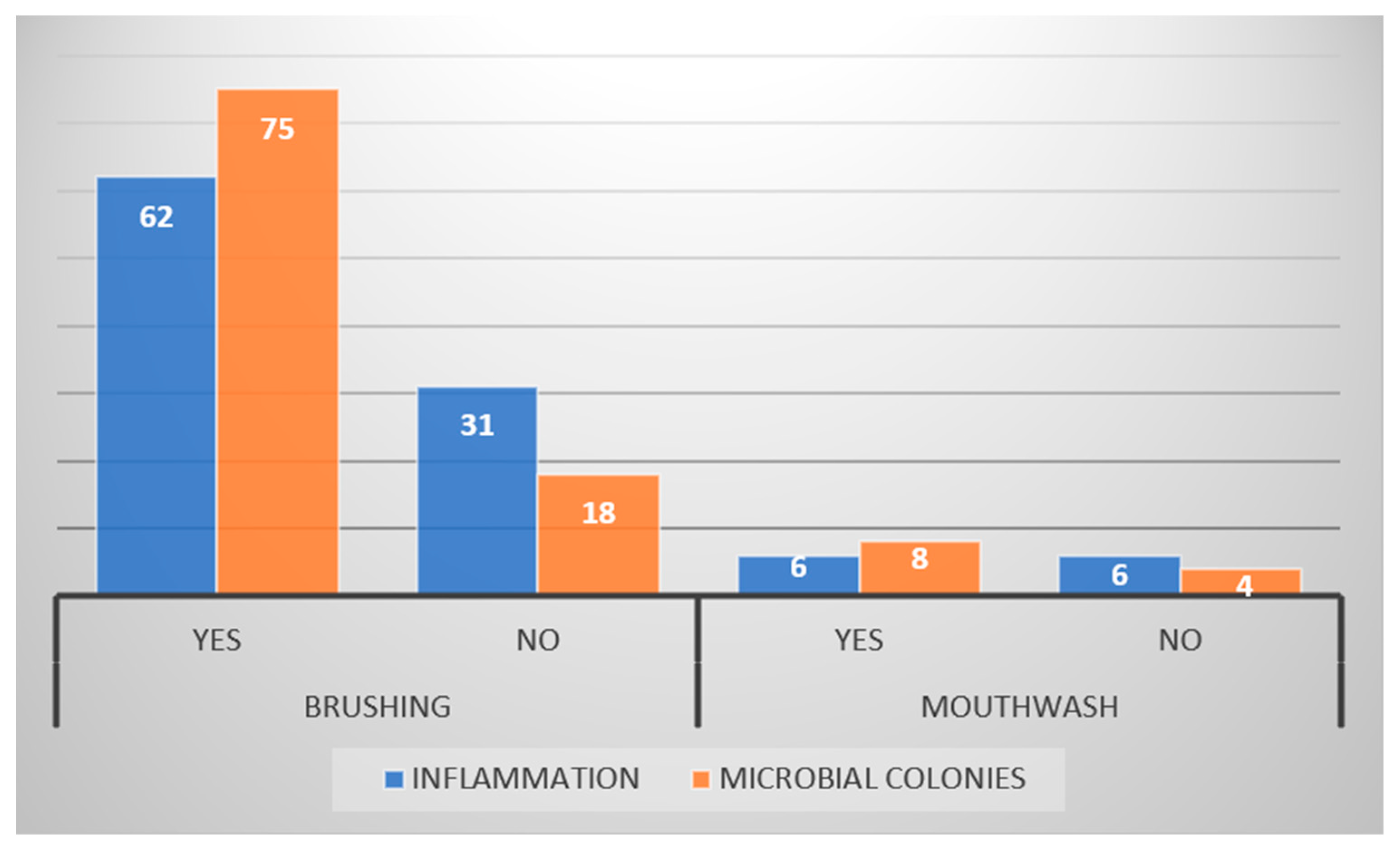
We found that 72% (n = 67) of participants followed brushing twice daily as an oral health maintenance routine, with only 12.9% (n = 12) using a mouthwash gargle. The smokers and alcohol drinkers were males, 12.9% and 5.4%, respectively. Regarding hormonal disturbance, 14.8% of female participants underwent menstruation at the time of sample collection, while only 7.4% were diagnosed with polycystic ovary syndrome. Interestingly habits and hormonal changes had significant roles in both sexes, as p-values were <0.05 (Table 1). This study assessed the frequency of cytopathological findings of inflammation and microbial colonies in terms of tooth brushing and mouthwash use. In which the majority experienced both inflammation and microbial colonies in their smears, among which the difference was not statistically significant (the reported p-value > 0.05) (Figure 2).
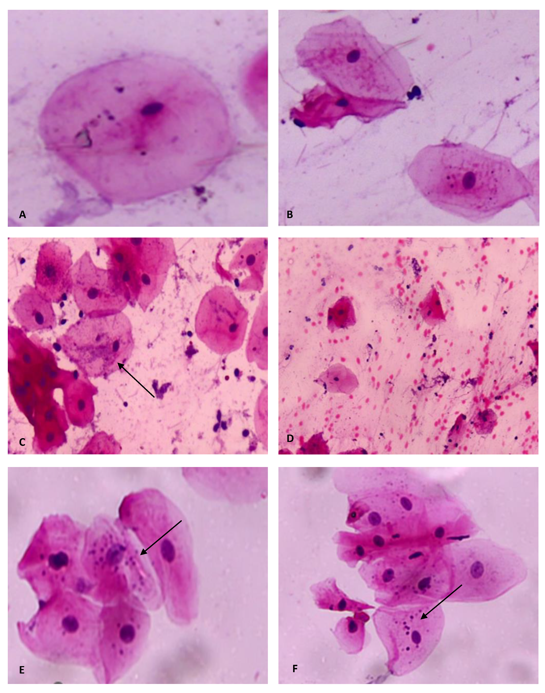
In terms of cytopathological findings, the entire sample revealed overlapping cells in all of the analyzed smears 100% (n = 93). Simultaneously, the majority established microbial colonies 80.6% (n = 75) and micronuclei 90.3% (n = 84). Moreover, 62.4% (n = 58) demonstrated signs of keratinization in the cytoplasm of cells, and 66.7% (n = 62) showed inflammation; meanwhile, only 10.7% (n = 10) showed hemorrhage. None of the reported findings were influenced by sex (Table 2 and Figure 3).
Most smokers established micronuclei 91.7%, followed by microbial colonies 83.3%, compared to non-smokers (8.3%, 16.7%). Meanwhile, only 50% showed inflammatory cells in the background of their smears. None of these findings were influenced by smoking, as p-values were > 0.05. Moreover, all smears of alcohol drinkers showed microbial colonies and micronuclei, and 80% exhibited keratinization. Additionally, the consumption of alcohol had no significant impact on those outcomes. Finally, the majority of females with hormonal disturbances, whether physiological or those with PCOS, showed predominate micronuclear findings, which were 87.5% and 100%, respectively. These hormonal changes did not impact the mentioned readings (Table 3).
Regarding cytomorphometric parameters, proximity was found among the different groups in terms of CyD, ND, and NCD ratios. However, in this study, PCOS-related hormonal alterations led to a larger ND, CyD, and NCD ratio compared to non-polycystic females and those with menstruation. In turn, no significant difference was observed between females with menstruation and normal ones. Additionally, sex, smoking, or drinking showed no statistical influence on the reported parameters except PCOS impact on ND and NCD ratio (p = 0.003, p = 0.02) (Table 4).

4. Discussion

Exfoliative cytology is a useful diagnostic modality that enables the early detection of abnormalities in the oral mucosa [15]. It is a simple, safe, and practical approach, particularly applicable for mass screening due to its high sensitivity and specificity [18]. Furthermore, the application of cytomorphometry offers remarkable development in the diagnosis and prognosis of serious diseases as it can detect cellular alteration, improving diagnosis accuracy and reproducibility [9]. Therefore, exfoliative cytology could be a beneficial adjunct to the clinical evaluation of lesions and influencing factors [19].
This study showed female predominance. However, since the sample was not collected haphazardly or in response to a particular disease or condition; instead, it was based on college admission within a specific age group. Therefore, it would be statistically unjustified to compare our data with other research on the matter of sex and age.
Most of our sample (72%) followed the international recommendations for a twice-daily frequency of tooth brushing; this could be attributed to the participant’s awareness of the significance of toothbrushing as a preventive measure and an indicator of oral hygiene [20]. Additionally, the procedure was sex-related since females expressed more interest in practicing the globally advised dental hygiene regimen, which agreed with the literature [21,22,23].
On the other hand, only 12.9% used mouthwash, in contrast to most of the participants, which was consistent with the findings of the study conducted by Macfarlane et al. [24,25]. This could be due to the controversy around mouthwash use and ongoing assessments of its general benefits and hazards.
This study revealed male predominance in terms of smoking and alcohol drinking habits, 12.9% and 5.4%, respectively. Other local and regional studies confirmed this issue in which most participants were males [3,26,27,28,29]. A finding that can be related to the conservative nature of the societies of middle eastern countries.
Regarding cytological observations of the obtained smears, 66.7% showed signs of inflammation; nevertheless, none of the reported habits nor hormonal changes influenced these findings. This disagreed with Queiroz et al. [17], no evidence of inflammation was seen in their sample of normal individuals. According to Seifi et al. [30], inflammation is considered one of the factors affecting nuclear and cytoplasmic size, as the author observed an increase in nuclear size and a decline in cytoplasmic size. Meanwhile, in our investigation, the inflammation had no noticeable effects on either the observational or cytomorphometric measures. This can be attributed to the type of the sample included, as Seifi conducted a study on smokers and waterpipe users with different oral site involvement, perhaps resulting in superimposed findings [30]. Ahmed et al. [31] mentioned that exposure to tobacco products could induce inflammatory events in the buccal mucosa, although, in his study, inflammation was noted in both smokers and control groups, with varying degrees of intensity. Additionally, Proia et al. [32] linked the presence of inflammation in response to bacterial infection; however, this cannot be supported by our findings as no direct correlation was discovered between the two observations. As a result of these contradicted findings, further investigation is required to uncover the underlying causes of inflammation and anticipate their potential consequences.
On the other hand, the majority of our sample (80.6%) showed bacterial colonies, which is higher than the findings of Abdelaziz and Osman [33] in their control group when compared to their groups of smokers and alcohol drinkers. Still, they ascribed their findings to these behaviors, which cannot be implicated in our case, as no significant association was found between these practices and bacterial existence. Moreover, this study showed keratinization in 62.4% of samples. This finding was unrelated to any of our sample’s habits or hormonal changes. This contradicted the findings of other studies [33,34] since no keratinization was observed in their control groups. Therefore they attributed the keratinization observation to smoking. Meanwhile, our results might be related to vigorous tooth brushing, the type of food intake, and perhaps accidental cheek biting.
The sample screening revealed 90.3% of micronuclei expression. As explained by the research, micronuclei originate from chromosome fragments or whole chromosomes, which stay behind at anaphase during nuclear division, and various genotoxic substances induce their formation [35]. Since 1983, many studies have used micronuclei detection as a short-term mutagenicity test. Since it is a simpler and much more rapid screening of chromosomal damage in cytological preparations [36], others used it as a reliable indicator for neoplastic progression [37,38].
Any increase in micronucleus count is a reflection of chromosomal alterations [39]. It has been reported that in normal healthy individuals, exposure to environmental pollutants such as drugs, chemicals, food, and free radical injuries, and lifestyle factors (smoking, alcohol consumption, diet, vitamin deficiencies) are related factors in producing the higher rates of micronuclei count in buccal mucosa and peripheral blood lymphocytes [40]. In contrast, no significant relation between micronuclei and drinking alcohol or smoking was discovered in our study. In the meantime, Al-Kasser [41] claims that Iraq’s environmental pollution has increased in recent years, which helps to explain our findings when combined with the aforementioned reasons. In light of the gravity of these observations, general awareness is required to spotlight the exact cause of such alarming findings.
Different alterations in the oral mucosa might result from smoking. It is therefore connected to various pathologies, ranging from fatal to reversible diseases [4]. According to published research, cytomorphometric analysis of smokers showed increased nuclear parameters and decreased cytoplasmic parameters compared to non-smokers [9,18,42]. In this study, despite the predominant micronuclei and keratinization in smokers and alcohol drinkers, no discernible difference was observed between smokers and non-smokers regarding morpho and cytometric findings. This could be due to the limited number of habit user participants and their youth, as these changes can be illustrated by continuous exposure to carcinogens over the years, resulting in reduced cytoplasmic maturation and significantly elevated nuclear activity [9,42]. On the other hand, Srinivasamurthy et al. [14] demonstrated that alcohol consumption had been associated with a considerable rise in nuclear, cytoplasmic, and nuclear to cytoplasmic parameters. However, this cannot be corroborated by our findings, as the number and the age of participants once more can be the main factor.
Research indicates that female hormones impact epithelial cell proliferation and development [3]. In general, ovarian hormones have a significant impact on females’ life. These hormones’ levels fluctuate throughout puberty, the menstrual cycle, pregnancy, and menopause [43]. It has been discovered that these hormones affect the oral cavity, as the onset of monthly menses evokes oral discomforts, including a burning sensation, bleeding with minor irritation, recurrent mouth ulcers, herpes labialis, and increased tooth mobility [43].
However, studies have not sufficiently proved a direct connection between alterations in the oral epithelium and hormonal changes during menstruation. The measuring of cytomorphometric characteristics in females based on their menstrual cycle was addressed by only three research. Balan et al. [44] discovered a significant difference in cellular and nuclear diameters during the different stages of the menstrual cycle of healthy young females. Meanwhile, in comparison with control groups, no significant changes were reported. At the same time, the other two studies [12,45] compared females with menstruation in different age groups. Further research emphasized that cytomorphometric parameters of the oral mucosa are undoubtedly affected by female hormones; hence, they excluded female members from their data to avoid superimposed results [30,46]. In the meantime, in this study, females with menstruation showed nonsignificant cytometric variation compared to non-menstruating females. Therefore, it is highly suggested that more studies be conducted on female hormones’ influence in different age groups on the oral mucosa to enrich the literature and provide sufficient data for comparison.
According to Deswal et al., 2020 systematic review, polycystic ovary syndrome (PCOS) is considered a common condition, occurring in 21.2% of reproductive-age females [47]. It has been linked to issues in the reproductive system and the metabolic system and an increased risk of cardiovascular diseases [47]. In the matter of PCOS influence on the oral cavity, the majority of investigations concentrated on the enhanced risk of developing the periodontal disease [48,49,50]. However, in this cytomorphometric study, in contrast to non-polycystic females and those with menstruation, PCOS -related hormonal alterations led to a larger ND, CyD, and NCD ratio, with an impact on the ND and NCD ratio (p = 0.003, p = 0.02). Since there is no research in the literature on the effects of PCOS on oral nuclear and cytoplasmic diameters, the precise cause of these observations cannot be quite explained. It can be recommended that future studies in this matter be conducted by physicians of both fields (cytology and gynecology) so that any conclusion would be more reliable.

5. Limitations and Conclusions

The lack of information regarding smoking and alcohol intake frequency was one of this study’s limitations. Furthermore, additional nuclear parameters with the number and size of micronuclei particles should be analyzed in a future study of the non-dental health groups in Sulaimani city to compare different lifestyle effects on cytomorphometric integrity. In conclusion, cytomorphometric analysis can be used in various fields of investigation due to its many potential benefits, including improved objectivity, enhanced sensitivity, and shorter turnaround times. This study provides a backbone of the record of data of normal individuals to be used as a baseline measurement for comparison with future cytometric studies. Minor nonsignificant cytomorphological alterations were related to habitual and hormonal alterations. Females with polycystic syndrome revealed a significant increase of nuclear and N/C ratio. Additional research with larger samples is required to support and validate these findings. The study sample expressed high micronuclei, indicating carcinogen doses from either local or ambient exposure. This was the most intriguing discovery. Exfoliated buccal cells can therefore be utilized as a marker for genotoxicity in the general population. In addition, this study can be used as a source of awareness in the population to reduce and avoid possible exposure to toxic factors.

Author Contributions

D.K.M.: methodology, formal analysis, investigation, software, B.F.I.: project administration, conceptualization, formal analysis, software, validation, writing—original draft, resources, D.N.M.: data curation, validation, writing—review and editing, B.T.G.: data curation, validation of the data, M.A.-S.H.: data curation. All authors have read and agreed to the published version of the manuscript.

Funding

This research received no external funding; it was fully funded by the authors.

Institutional Review Board Statement

Ethical approval was obtained from the Local Ethical and Scientific Committee in the College of Dentistry (proposal no:107/22, 11/4/2022) to conduct this prospective study at the laboratory of oral pathology/College of Dentistry/University of Sulaimani, Sulaimani, Iraq.

Informed Consent Statement

Informed consent was obtained from all subjects involved in the study.

Data Availability Statement

All the data are included in the manuscript.

Conflicts of Interest

Authors declare no conflict of interest.

References

  1. Babshet, M.; Nandimath, K.; Pervatikar, S.K.; Naikmasur, V. Efficacy of Oral Brush Cytology in the Evaluation of the Oral Premalignant and Malignant Lesions. J. Cytol. 2011, 28, 165. [Google Scholar] [CrossRef] [PubMed]
  2. Mehrotra, R.; Gupta, A.; Singh, M.; Ibrahim, R. Retraction: Application of Cytology and Molecular Biology in Diagnosing Premalignant or Malignant Oral Lesions. Mol. Cancer 2012, 11, 57. [Google Scholar] [CrossRef] [PubMed] [Green Version]
  3. Göregen, M.; Murat Akgül, H.; Gündoğdu, C. Th e Cytomorphological Analysis of Buccal Mucosa Cells in Smokers. Turk. J. Med. Sci. 2011, 41, 205–210. [Google Scholar] [CrossRef]
  4. Nivia, M.; Sunil, S.N.; Rathy, R.; Anilkumar, T.V. Comparative Cytomorphometric Analysis of Oral Mucosal Cells in Normal, Tobacco Users, Oral Leukoplakia and Oral Squamous Cell Carcinoma. J. Cytol. 2015, 32, 253. [Google Scholar] [CrossRef]
  5. Divani, S.; Exarhou, M.; Theodorou, L.N.; Georgantzis, D.; Skoulakis, H. Advantages and Difficulties of Brush Cytology in the Identification of Early Oral Cancer. Arch. Oncol. 2009, 17, 11–12. [Google Scholar] [CrossRef] [Green Version]
  6. Da Silva, A.D.; Lima, C.F.; Maraschin, B.J.; Laureano, N.K.; Daroit, N.B.; Brochier, F.; Sant’Ana Filho, M.; Visioli, F.; Rados, P.V. Immunocytochemistry Associated with Oral Exfoliative Cytology: Methodological Analysis. Anal. Quant. Cytol. Histol. 2015, 37, 134–138. [Google Scholar]
  7. Kazanowska, K.; Hałón, A.; Radwan-Oczko, M. The Role and Application of Exfoliative Cytology in the Diagnosis of Oral Mucosa Pathology—Contemporary Knowledge with Review of the Literature. Adv. Clin. Exp. Med. 2014, 23, 299–305. [Google Scholar] [CrossRef] [Green Version]
  8. Osorio-Osorno, Y.A.; Arboleda Toro, D.; Arango, J.C.; Parada-Sanchez, M.T. Optimized Liquid-Based Cytology for the Cellular and Molecular Analysis of Oral Keratinocytes: A Promising Diagnostic Tool. Diagn. Cytopathol. 2021, 49, 96–104. [Google Scholar] [CrossRef]
  9. Palakurthy, P.; Kulkarni, P.G.; Nandan, R.K.; Rao, T.M.; Reddy, D.S.P.; Muddana, K. Cytological Changes in Normal Oral Mucosa of Individuals with Tobacco Habits: A Cytomorphometric Study. J. Contemp. Dent. Pract. 2017, 18, 722–727. [Google Scholar] [CrossRef]
  10. Sumanthi, J.; Sridhar Reddy, G.; Anuradha, C.H.; Chandhra Sekhar, P.; Krishna Prasad, L.; Ramana Reddy, B.V. A Study on Cytomorphometric Analysis of Exfoliative Buccal Cells in Iron Deficiency Anemic Patients. Contemp. Clin. Dent. 2012, 3, S156. [Google Scholar] [CrossRef]
  11. Bds, M.T.A.; Garib, B.T. Cytological Features of Oral Cytobrush Smears in Type II Diabetes Mellitus Patients. Tikrit J. Dent. Sci. 2012, 1, 6–12. [Google Scholar]
  12. Donald, P.M.; George, R.; Sriram, G.; Kavitha, B.; Sivapathasundharam, B. Hormonal Changes in Exfoliated Normal Buccal Mucosal Cells. J. Cytol. 2013, 30, 252. [Google Scholar] [CrossRef]
  13. Ogden, G.R.; Cowpe, J.G.; Green, M.W. Effect of Radiotherapy on Oral Mucosa Assessed by Quantitative Exfoliative Cytology. J. Clin. Pathol. 1989, 42, 940. [Google Scholar] [CrossRef] [PubMed] [Green Version]
  14. Srinivasamurthy, B.C.; Balamurugesan, K.; Sathishkumar, N.; Prakash, M.; Bhat, R.V. Cytomorphometric Study of Changes in Buccal Mucosal Cells in Alcoholics. Adv. Biomed. Res. 2020, 9, 48. [Google Scholar] [CrossRef] [PubMed]
  15. Sukegawa, S.; Ono, S.; Nakano, K.; Takabatake, K.; Kawai, H.; Nagatsuka, H.; Furuki, Y. Clinical Study on Primary Screening of Oral Cancer and Precancerous Lesions by Oral Cytology. Diagn. Pathol. 2020, 15, 170. [Google Scholar] [CrossRef] [PubMed]
  16. Nayar, A.K.; Sundharam, B.S. Cytomorphometric Analysis of Exfoliated Normal Buccal Mucosa Cells. Indian J. Dent. Res. 2003, 14, 87–93. [Google Scholar] [PubMed]
  17. Queiroz, J.B.; Lima, C.; Burim, R.A.; Brandão, A.A.; Cabral, L.A.; Almeida, J. Exfoliative Cytology of the Oral Mucosa: Comparison of Two Collection Methods. Gen. Dent. 2010, 58, e196–e199. [Google Scholar] [PubMed]
  18. Khot, K.; Deshmane, S.; Bagri-Manjarekar, K.; Warke, D.; Kotak, K. A Cytomorphometric Analysis of Oral Mucosal Changes in Tobacco Users. J. Nat. Sci. 2015, 6, S22–S24. [Google Scholar] [CrossRef] [Green Version]
  19. Suresh, T.; Bastian, T.; Ahmed Mujib, B. Cytomorphometric Analysis of Squames Obtained from Normal Mucosa, Leukoplakia and Oral Squamous Cell Carcinoma. J. Oral Maxillofac. Pathol. 2021, 25, 202. [Google Scholar] [CrossRef]
  20. De Grado, G.F.; Ehlinger, V.; Godeau, E.; Arnaud, C.; Nabet, C.; Benkirane-Jessel, N.; Musset, A.M.; Offner, D. Changes in Tooth Brushing Frequency and Its Associated Factors from 2006 to 2014 among French Adolescents: Results from Three Repeated Cross Sectional HBSC Studies. PLoS ONE 2021, 16, e0249129. [Google Scholar] [CrossRef]
  21. Sakalauskiene, Ž.; Vehkalahti, M.M.; Murtomaa, H.; Mačiulskiene, V. Factors Related to Gender Differences in Toothbrushin g Among Lithuanian Middle-Aged University Employees. Medicina 2011, 47, 25. [Google Scholar] [CrossRef]
  22. Azodo, C.C.; Unamatokpa, B. Gender Difference in Oral Health Perception and Practices among Medical House Officers. Russ. Open Med. J. 2012, 1, 0208. [Google Scholar] [CrossRef]
  23. Mamai-Homata, E.; Koletsi-Kounari, H.; Margaritis, V. Gender Differences in Oral Health Status and Behavior of Greek Dental Students: A Meta-Analysis of 1981, 2000, and 2010 Data. J. Int. Soc. Prev. Community Dent. 2016, 6, 60. [Google Scholar] [CrossRef] [PubMed] [Green Version]
  24. Macfarlane, T.V.; Kawecki, M.M.; Cunningham, C.; Bovaird, I.; Morgan, R.; Rhodes, K.; Watkins, R. Mouthwash Use in General Population: Results from Adult Dental Health Survey in Grampian, Scotland. J. Oral Maxillofac. Res. 2010, 1, 2. [Google Scholar] [CrossRef] [Green Version]
  25. Lachenmeier, D.W. Mouthwash Controversies. Br. Dent. J. 2010, 208, 95–96. [Google Scholar] [CrossRef] [PubMed] [Green Version]
  26. Hussain, H.Y.; Abdul Satar, B.A. Prevalence and Determinants of Tobacco Use among Iraqi Adolescents: Iraq GYTS 2012. Tob. Induc. Dis. 2013, 11, 14. [Google Scholar] [CrossRef] [Green Version]
  27. Al Ansari, M.; Dawson, A.; Conigrave, K. Alcohol among Young People in Iraq: A Systematic Scoping Review. Discov. Psychol. 2022, 2, 16. [Google Scholar] [CrossRef]
  28. Halimi1, L.; Haghdoost2, A.A.; Alizadeh3, S.M. Prevalence of Cigarette Smoking among Iranian Women: A Systematic Review and Meta-Analysis. Med. J. Islam. Repub. Iran 2013, 27, 132. [Google Scholar]
  29. Sweis, N.J. Smoking Behavior among Jordanians: Physical, Psychological, Social, and Economic Reasons. Value Heal. Reg. Issues 2018, 16, 5–8. [Google Scholar] [CrossRef]
  30. Seifi, S.; Feizi, F.; Mehdizadeh, M.; Khafri, S.; Ahmadi, B. Evaluation of Cytological Alterations of Oral Mucosa in Smokers and Waterpipe Users. Cell J. 2014, 15, 302. [Google Scholar]
  31. Gadelkarim Ahmed, H.; Ali Alqufeye, M.; Ali Alsulaiman, G.; Lafy Alenezi, L.; Khaled Almuslumani, R.; Mohammed Alhussain, G.; Khaled Alenezi, A.; Fahad Algharbi, A.; Eid Alanazi, A.; Khleef Alshammari, N.; et al. Patterns of Oral Inflammatory Cells Infiltrate Associated with Cigarette Smoking Medical Science. Med. Sci. 2020, 24, 2817–2825. [Google Scholar]
  32. Proia, N.K.; Paszkiewicz, G.M.; Nasca, M.A.S.; Franke, G.E.; Pauly, J.L. Smoking and Smokeless Tobacco-Associated Human Buccal Cell Mutations and Their Association with Oral Cancer—A Review. Cancer Epidemiol. Biomark. Prev. 2006, 15, 1061–1077. [Google Scholar] [CrossRef] [PubMed] [Green Version]
  33. Abdelaziz, M.S.; Osman, T.E. Detection of Cytomorphological Changes in Oral Mucosa among Alcoholics and Cigarette Smokers. Oman Med. J. 2011, 26, 349. [Google Scholar] [CrossRef] [PubMed]
  34. Ahmed, H.G.E.; Omer, A.S.A.; Abd Algaffar, S.A. Cytological Study of Exfoliative Buccal Mucosal Cells of Qat Chewers in Yemen. Diagn. Cytopathol. 2011, 39, 796–800. [Google Scholar] [CrossRef]
  35. Ojeda, J.E.Q.; Aguilar-Medina, M.; Olimón-Andalón, V.; Jau, R.A.G.; Ham, A.A.; Quintana, J.G.R.; De Lourdes Silva-Benítez, E.; Sanchez-Schmitz, G.; Ramos-Payán, R. Increased Micronuclei Frequency in Oral and Lingual Epithelium of Treated Diabetes Mellitus Patients. Biomed Res. Int. 2018, 2018, 4898153. [Google Scholar] [CrossRef]
  36. Jadhav, K.; Gupta, N.; Ahmed Mujib, B.R. Micronuclei: An Essential Biomarker in Oral Exfoliated Cells for Grading of Oral Squamous Cell Carcinoma. J. Cytol. 2011, 28, 7. [Google Scholar] [CrossRef]
  37. Casartelli, G.; Bonatti, S.; De Ferrari, M.; Scala, M.; Mereu, P.; Margarino, G.; Abbondandolo, A. Micronucleus Frequencies in Exfoliated Buccal Cells in Normal Mucosa, Precancerous Lesions and Squamous Cell Carcinoma. Anal. Quant. Cytol. Histol. 2000, 22, 486–492. [Google Scholar]
  38. Kharisma, Y.; Damayanti, M.M.; Yulianto, F.A.; Rahimah, S.B.; Maharani, W.; Rachmawati, M.; Sastramihardja, H.S.; Abdul’ Aziiz, M.A.; Halim, M.I. Folic Acid Usual Doses Decrease the Buccal Micronucleus Frequency on Smokers. Glob. Med. Health Commun. 2019, 7, 131–135. [Google Scholar] [CrossRef] [Green Version]
  39. Dehghannezhad, M.; Naderi, N.J.; Semyari, H. Micronucleus Assay of Buccal Mucosa Cells in Waterpipe (Hookah) Smokers: A Cytologic Study. Iran. J. Pathol. 2020, 15, 75. [Google Scholar] [CrossRef]
  40. Shashikala, R.; Indira, A.P.; Manjunath, G.; Rao, K.; Akshatha, B. Role of Micronucleus in Oral Exfoliative Cytology. J. Pharm. Bioallied Sci. 2015, 7, S409. [Google Scholar] [CrossRef]
  41. Al-Kasser, M.K. Air Pollution in Iraq Sources and Effects. In Proceedings of the IOP Conference Series: Earth and Environmental Science, First International Virtual Conference on Environment & Natural Resources, Al Diwaniyah, Iraq, 24–25 March 2021; Volume 790. [Google Scholar] [CrossRef]
  42. Srilatha, T.; Manthapuri, S.; Shylaja, S.; Ramanand, O.V.; Reddy, E.S.; Vamshi, V.R. Cytomorphometric Analysis of Exfoliated Buccal Mucosal Cells in Smokers and Patients with Hypertension: A Quantitative Analysis. J. Int. Oral Health 2021, 13, 53. [Google Scholar] [CrossRef]
  43. Balan, U.; Gonsalves, N.; Jose, M.; Girish, K.L. Symptomatic Changes of Oral Mucosa during Normal Hormonal Turnover in Healthy Young Menstruating Women. J. Contemp. Dent. Pract. 2012, 13, 178–181. [Google Scholar] [CrossRef] [PubMed]
  44. Balan, U.; Luqman, M.; Soliman, A.M.M.; Almubarak, H. Cytomorphometric Changes of Oral Mucosa during Normal Hormonal Turnovers in Healthy Young Menstruating Women. King Khalid Univ. J. Health Sci. 2018, 3, 8. [Google Scholar] [CrossRef]
  45. Meenakshi, S.; Jagannathan, N.; Swarnameenakshi, S. Cytomorphometric Analysis of Oral Epithelial Cells in Menstrual Cycle. Int. J. Pharma. Res. Health Sci. 2017, 5, 1695–1697. [Google Scholar] [CrossRef]
  46. Gowhar, O. Comparison of Exfoliative Cytology of Tongue and Buccal Mucosa among Smokers and Non-Smokers Using PAP Stain and AgNOR Counts. Int. J. Contemp. Med. Res. 2015, 4, 2393–2915. [Google Scholar]
  47. Deswal, R.; Narwal, V.; Dang, A.; Pundir, C.S. The Prevalence of Polycystic Ovary Syndrome: A Brief Systematic Review. J. Hum. Reprod. Sci. 2020, 13, 261. [Google Scholar] [CrossRef]
  48. Wendland, N.; Opydo-Szymaczek, J.; Mizgier, M.; Jarząbek-Bielecka, G. Subgingival Microflora in Adolescent Females with Polycystic Ovary Syndrome and Its Association with Oral Hygiene, Gingivitis, and Selected Metabolic and Hormonal Parameters. Clin. Oral Investig. 2021, 25, 1485–1496. [Google Scholar] [CrossRef]
  49. Tanguturi, S.C.; Nagarakanti, S. Polycystic Ovary Syndrome and Periodontal Disease: Underlying Links—A Review. Indian J. Endocrinol. Metab. 2018, 22, 267. [Google Scholar] [CrossRef]
  50. Farook, F.F.; Ng, K.T.; MNM, N.; Koh, W.J.; Teoh, W.Y. Association of Periodontal Disease and Polycystic Ovarian Syndrome: A Systematic Review and Meta-Analysis with Trial Sequential Analysis. Open Dent. J. 2020, 13, 478–487. [Google Scholar] [CrossRef]
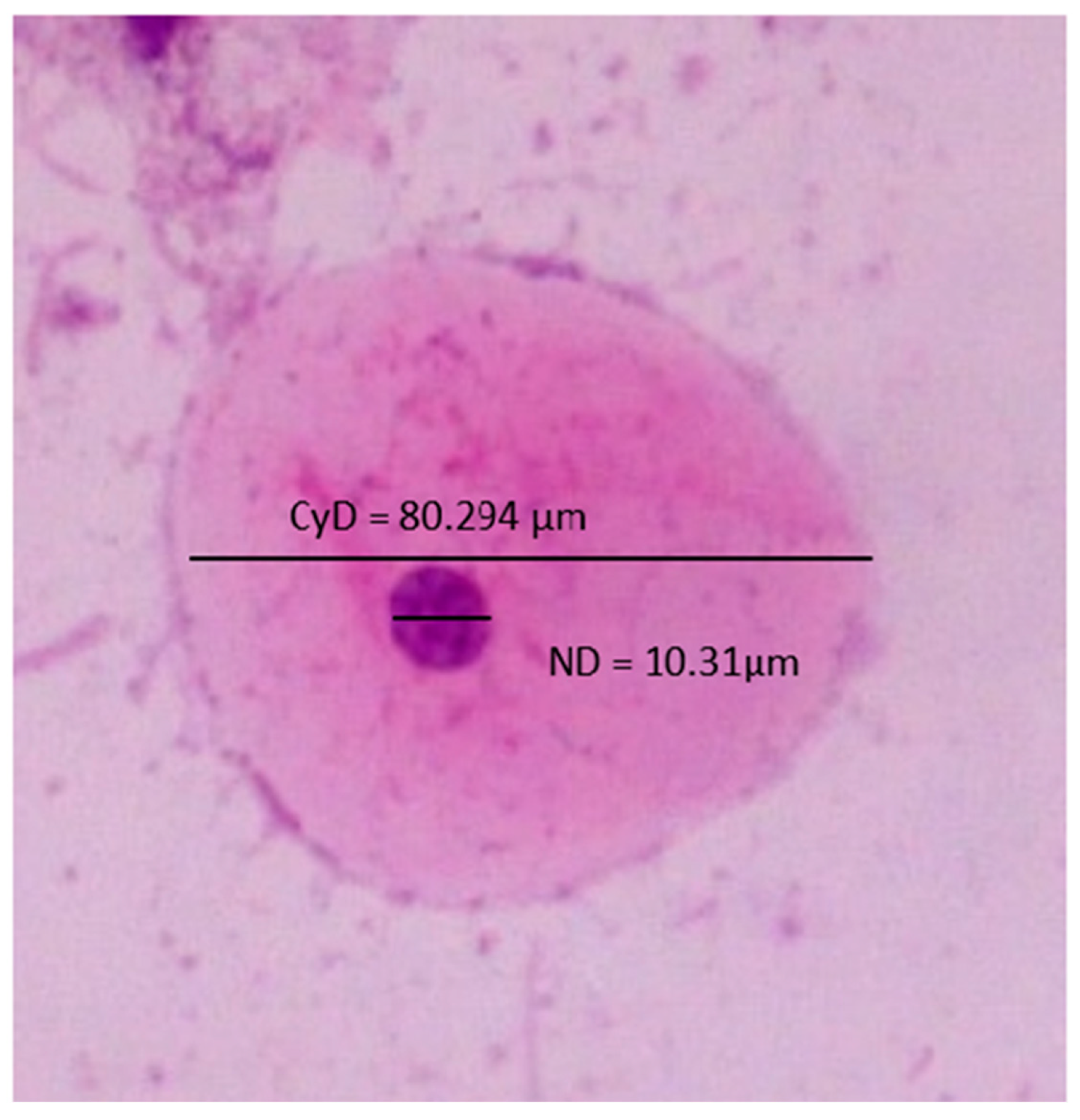
Figure 1. Cytological smear showing nuclear and cytoplasmic diameters.
Diagnostics 13 00234 g001
Figure 2. Oral health maintenance habits’ effect on cytopathological findings (no relation was found among the demonstrated variables). p > 0.05.
Diagnostics 13 00234 g002
Figure 3. Cytological smears (X40), showing (A,B): Keratinization and cytoplasmic vacuolization. (C): Microbial colonies (arrow) inflammation and overlapping. (D): Hemorrhage and inflammation. (E,F): Micronuclei (arrows).
Diagnostics 13 00234 g003
Table 1. Sex distribution concerning oral health maintenance, habits, and hormonal disturbances.
Variables Male
39
Female
54
Total %
93
Pearson Chi-Square Test
Tooth brushingOnce18 (19.4%)8 (8.6%)26 (28%)p = 0.001
Twice21 (22.6%)46 (49.4%)67 (72%)
MouthwashYes4 (4.3%)8 (8.6%)12 (12.9%)p = 0.518
No35 (37.6%)46 (49.5%)81 (87.1%)
SmokingYes12 (12.9%)0 (0%)12 (12.9%)p = 0.000
No27 (29%)54 (58%)81 (87%)
Drinking alcoholYes5 (5.4%)0 (0%)5 (5.4%)p = 0.007
No34 (36.6%)54 (58%)88 (94.6%)
Physical hormonal changesYes0 (0%)8 (14.8%)8 (14.8%)p = 0.000
No0 (0%)46 (85.2%)46 (85.2%)
Polycystic ovary syndromeYes0 (0%)4 (7.4%)4 (7.4%)p = 0.000
No0 (0%)50 (92.6%)50 (92.6%)
Table 2. Sex distribution concerning different cytopathological findings.
Variables MaleFemaleTotal %Pearson Chi-Square Test
InflammationYes25 (26.9%)37 (39.8%)62 (66.7%)p = 0.500
No14 (15%)17 (18.3%)31 (33.3%)
Microbial coloniesYes34 (36.6%)41 (44%)75 (80.6%)p = 0.111
No5 (5.4%)13 (14%)18 (19.4%)
KeratinizationYes27 (29%)31 (33.4%)58 (62.4%)p = 0.245
No12 (12.9%)23 (24.7%)35 (37.6%)
MicronucleiYes37 (39.8%)47 (50.5%)84 (90.3%)p = 0.207
No2 (2.2%)7 (7.5%)9 (9.7%)
OverlappingYes39 (41.9%)54 (58.1%)93 (100%)
HemorrhageYes4 (4.3%)6 (6.4%)10 (10.7%)p = 0.896
No35 (37.6%)48 (51.7%)83 (89.3%)
Table 3. The distribution of habits and hormonal changes in response to various cytopathological findings.
Variables InflammationMicrobial ColoniesMicronucleiKeratinizationPearson Chi-Square Test
Smoking (12, 12.9%)Yes6 (50%)10 (83.3%)11 (91.7%)9 (75%)p = 0.260
p = 0.951
p = 0.866
p = 0.333
No6 (50%)2 (16.7%)1 (8.3%)3 (25%)
Alcohol drinking (5, 5.4%)Yes2 (40%)5 (100%)5 (100%)4 (80%)p = 0.430
p = 0.258
p = 0.452
p = 0.403
No3 (60%)001 (20%)
Physical hormonal changes (8, 14.8%)Yes4 (50%)6 (75%)7 (87.5%)5 (62.5%)p = 0.644
p = 0.304
p = 0.451
p = 0.484
No4 (50%)2 (25%)1 (12.5%)3 (37.5%)
Polycystic ovary syndrome (4, 7.4%)Yes2 (50%)3 (75%)4 (100%)2 (50%)p = 0.452
p = 0.240
p = 0.298
p = 0.484
No2 (50%)1 (25%)02 (50%)
Table 4. The relation of sex, habits, and hormonal changes to cytoplasmic, nuclear diameters, and nuclear/cytoplasmic ratio.
VariablesGenderSmokingDrinking AlcoholPhysical Hormonal ChangesPolycystic Ovary Syndrome
MaleFemaleYesNoYesNoYesNoYesNo
CYD66.34165.97166.39966.08664.22366.23465.44166.06466.32665.943
MIN/MAX56.20/77.7548.20/81.2658.02/76.2948.20/81.2658.02/76.2348.20/81.2653.27/78.5648.20/81.2656.65/78.0148.20/81.26
ND8.1498.2018.0138.2038.1788.1798.3668.1729.4618.099
MIN/MAX6.80/9.896.22/10.247.53/8.876.22/10.247.53/8.876.22/10.247.16/10.246.22/10.048.70/10.246.22/9.93
NCR12.35112.54312.17912.50612.82812.44312.87512.48514.46812.389
ANOVAND p = 0.755
CyD p = 0.806
N: C p = 0.549
ND p = 0.435
CyD p = 0.887
N: C p = 0.486
ND p = 0.998
CyD p = 0.540
N: C p = 0.580
ND p = 0.775
CyD p = 0.946
N: C p = 0.668
ND p = 0.003
CyD p = 0.965
N: C p = 0.023
Disclaimer/Publisher’s Note: The statements, opinions and data contained in all publications are solely those of the individual author(s) and contributor(s) and not of MDPI and/or the editor(s). MDPI and/or the editor(s) disclaim responsibility for any injury to people or property resulting from any ideas, methods, instructions or products referred to in the content.