
The Role of CT Imaging in Characterization of Small Renal Masses

 , ,
, ,  ,
,  ,
, 
 and
and 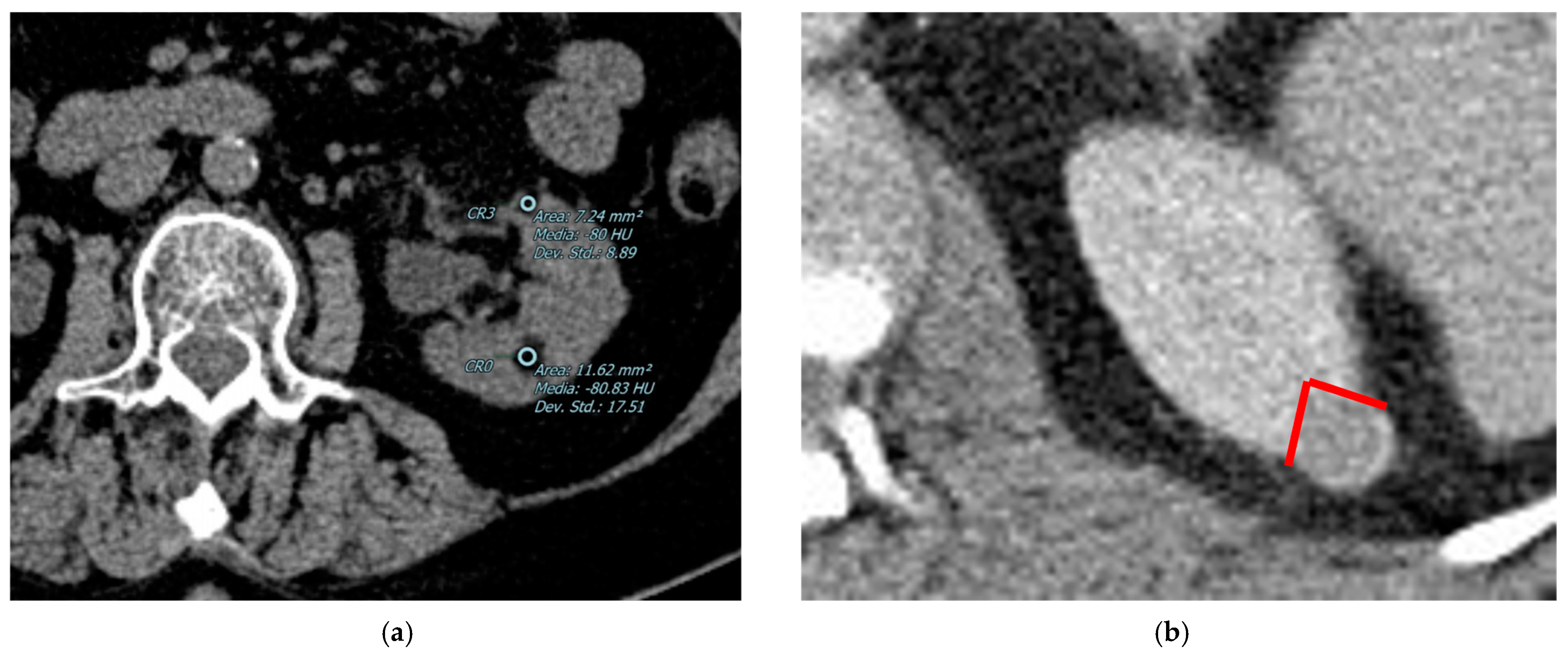
Abstract
:1. Introduction
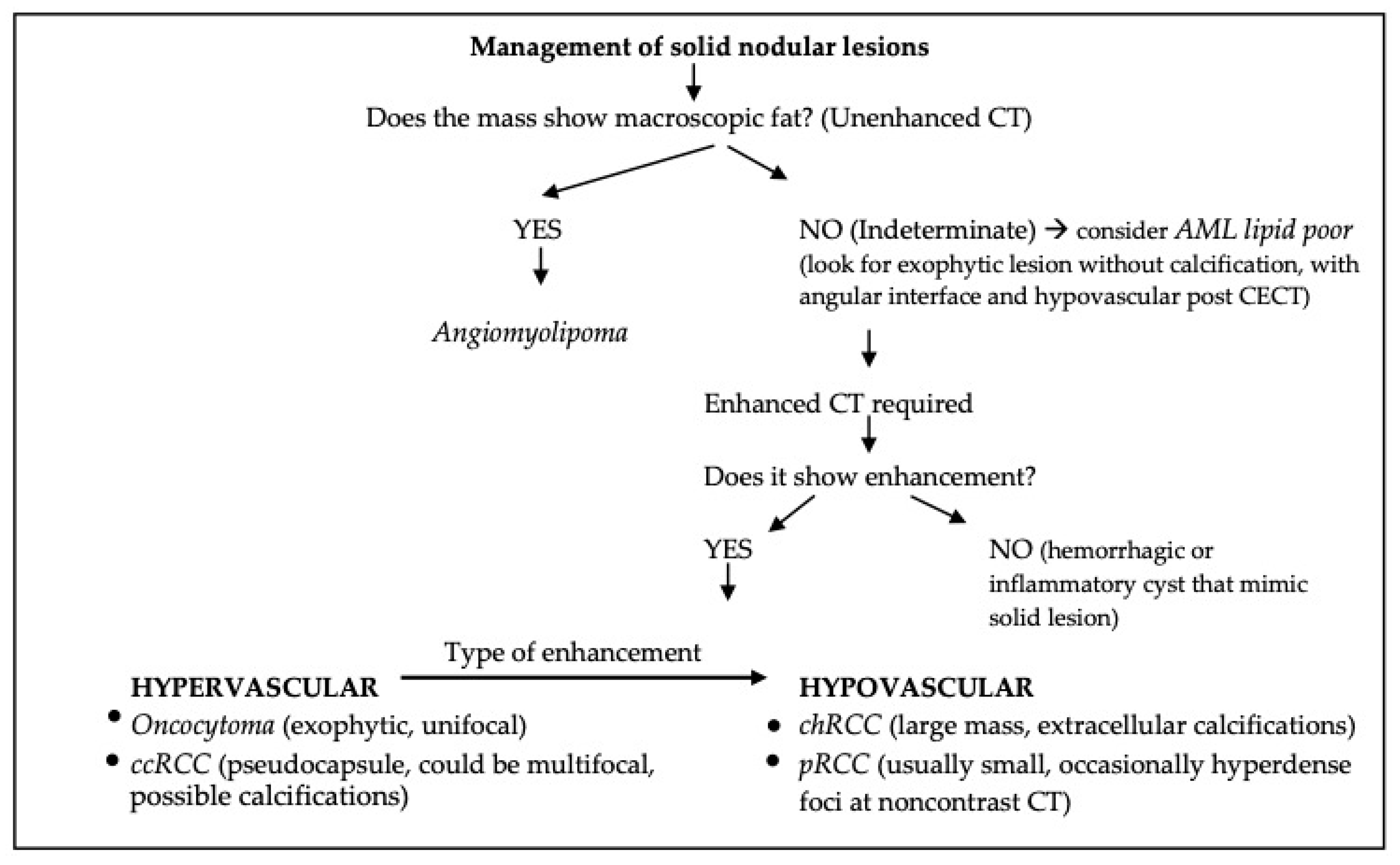
CT-Protocol in SRMs Assessment
- A precontrast phase (obtained before the administration of contrast material), to determine the HU of homogenous renal mass or masses containing macroscopic fat (characteristic for angiomyolipoma);
- A nephrographic phase (100–120 s after the contrast administration), in which there is the maximum and homogeneous enhancement of renal parenchyma, allowing detection of small hypodense masses;
- A corticomedullary phase (40–70 s), useful to differentiate the RCC subtypes;
- An excretory phase (7–10 min), recommended in preprocedural planning and in order to distinguish urothelial carcinoma from RCC.
2. Benign Lesions
2.1. Angiomyolipomas
2.2. Oncocytoma
2.3. Inflammatory Conditions and Pseudotumors
3. Malignant Lesions
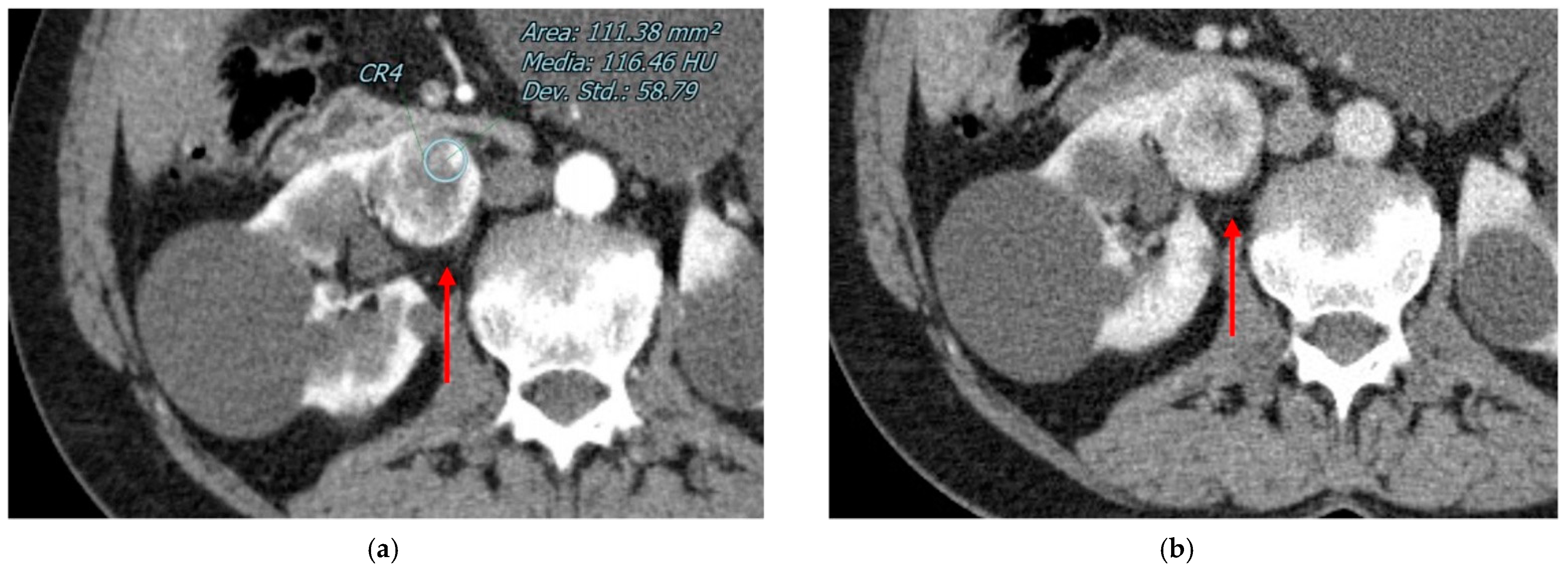
3.1. Clear Cell RCC
3.2. Papillary RCC
3.3. Chromophobe RCC
3.4. Urothelial Renal Carcinoma
3.5. Lymphoma
3.6. Renal Metastases
4. Discussion
5. Conclusions
Author Contributions
Funding
Conflicts of Interest
References
- Nicolau, C.; Antunes, N.; Paño, B.; Sebastia, C. Imaging Charact. Renal Masses. Med. 2021, 57, 51. [Google Scholar] [CrossRef]
- Wang, Z.J.; Westphalen, A.C.; Zagoria, R.J. CT and MRI of small renal masses. Br. J. Radiol. 2018, 91, 20180131. [Google Scholar] [CrossRef] [PubMed]
- Turner, R.M.; Morgan, T.M.; Jacobs, B.L. Epidemiology of the Small Renal Mass and the Treatment Disconnect Phenomenon. Urol. Clin. N. Am. 2017, 44, 147–154. [Google Scholar] [CrossRef] [PubMed] [Green Version]
- Dixon, B.P.; Hulbert, J.C.; Bissler, J.J. Tuberous sclerosis complex renal disease. Nephron. Exp. Nephrol. 2011, 118, e15–e20. [Google Scholar] [CrossRef] [PubMed]
- Bodmer, D.; van den Hurk, W.; van Groningen, J.J.M.; Eleveld, M.J.; Martens, G.J.M.; Weterman, M.A.J.; Van Kessel, A.G. Understanding familial and non-familial renal cell cancer. Hum. Mol. Genet. 2002, 11, 2489–2498. [Google Scholar] [CrossRef] [Green Version]
- Menko, F.H.; van Steensel, M.A.; Giraud, S.; Friis-Hansen, L.; Richard, S.; Ungari, S.; Nordenskjöld, M.; Hansen, T.V.; Solly, J.; Maher, E.R. Birt-Hogg-Dubé syndrome: Diagnosis and management. Lancet Oncol. 2009, 10, 1199–1206. [Google Scholar] [CrossRef] [PubMed]
- Sanchez, A.; Feldman, A.S.; Hakimi, A.A. Current Management of Small Renal Masses, Including Patient Selection, Renal Tumor Biopsy, Active Surveillance, and Thermal Ablation. J. Clin. Oncol. 2018, 36, 3591–3600. [Google Scholar] [CrossRef]
- Scoll, B.J.; Wong, Y.-N.; Egleston, B.L.; Kunkle, D.A.; Saad, I.R.; Uzzo, R.G. Age, tumor size and relative survival of patients with localized renal cell carcinoma: A surveillance, epidemiology and end results analysis. J. Urol. 2009, 181, 506–511. [Google Scholar] [CrossRef] [Green Version]
- Ajami, T.; Sebastia, C.; Corominas, D.; Ribal, M.J.; Nicolau, C.; Alcaraz, A.; Musquera, M. Clinical and radiological findings for small renal masses under active surveillance. Urol. Oncol. 2021, 39, 499.e9–499.e14. [Google Scholar] [CrossRef]
- Tonttila, P.P.; Lantto, J.; Pääkkö, E.; Piippo, U.; Kauppila, S.; Lammentausta, E.; Ohtonen, P.; Vaarala, M.H. Prebiopsy multiparametric magnetic resonance imaging for prostate cancer diagnosis in biopsy-naive men with suspected prostate cancer based on elevated prostate-specific antigen values: Results from a randomized prospective blinded controlled trial. Eur. Urol. 2016, 69, 419–425. [Google Scholar] [CrossRef]
- Znaor, A.; Lortet-Tieulent, J.; Laversanne, M.; Jemal, A.; Bray, F. International variations and trends in renal cell carcinoma incidence and mortality. Eur. Urol. 2015, 67, 519–530. [Google Scholar] [CrossRef] [PubMed]
- Berg, R.N.W.V.; Basourakos, S.P.; LaRussa, S.; McClure, T.D. Management of the Small Renal Mass: A 2020 Update. Curr. Oncol. Rep. 2020, 22, 69. [Google Scholar] [CrossRef] [PubMed]
- Frank, I.; Blute, M.L.; Cheville, J.C.; Lohse, C.M.; Weaver, A.L.; Zincke, H. Solid renal tumors: An analysis of pathological features related to tumor size. J. Urol. 2003, 170, 2217–2220. [Google Scholar] [CrossRef] [PubMed]
- Sohlberg, E.M.; Metzner, T.J.; Leppert, J.T. The Harms of Overdiagnosis and Overtreatment in Patients with Small Renal Masses: A Mini-review. Eur. Urol. Focus 2019, 5, 943–945. [Google Scholar] [CrossRef] [Green Version]
- Ray, S.; Cheaib, J.G.; Pierorazio, P.M. Active Surveillance for Small Renal Masses. Rev. Urol. 2020, 22, 9–16. [Google Scholar]
- Groenwold, R.H.H.; van der Hulle, T.; Visser, L.E.; Houtsma, D.; Guchelaar, H.J.; Zwaveling, J. Real-World Metastatic Renal Cell Carcinoma Treatment Patterns and Clinical Outcomes in The Netherlands. Front Pharmacol. 2022, 13, 803935. [Google Scholar] [CrossRef]
- Abu-Ghanem, Y.; Powles, T.; Capitanio, U.; Beisland, C.; Järvinen, P.; Stewart, G.D.; Gudmundsson, E.; Lam, T.B.; Marconi, L.; Fernandéz-Pello, S.; et al. Should patients with low-risk renal cell carcinoma be followed differently after nephron-sparing surgery vs. radical nephrectomy? BJU Int. 2021, 128, 386–394. [Google Scholar] [CrossRef]
- Ward, R.D.; Tanaka, H.; Campbell, S.C.; Remer, E.M. 2017 AUA Renal Mass and Localized Renal Cancer Guidelines: Imaging Implications. Radiographics 2018, 38, 2021–2033. [Google Scholar] [CrossRef] [Green Version]
- MacLennan, S.; Imamura, M.; Lapitan, M.C.; Omar, M.I.; Lam, T.B.; Hilvano-Cabungcal, A.M.; Royle, P.; Stewart, F.; MacLennan, G.; MacLennan, S.J.; et al. Systematic review of oncological outcomes following surgical management of localised renal cancer. Eur. Urol. 2012, 61, 972–993. [Google Scholar] [CrossRef] [Green Version]
- Marconi, L.; Dabestani, S.; Lam, T.B.; Hofmann, F.; Stewart, F.; Norrie, J.; Bex, A.; Bensalah, K.; Canfield, S.E.; Hora, M.; et al. Systematic Review and Meta-analysis of Diagnostic Accuracy of Percutaneous Renal Tumour Biopsy. Eur. Urol. 2016, 69, 660–673. [Google Scholar] [CrossRef]
- Andersen, M.F.B.; Norus, T.P. Tumor Seeding With Renal Cell Carcinoma After Renal Biopsy. Urol. Case Rep. 2016, 9, 43–44. [Google Scholar] [CrossRef] [PubMed]
- Singer, E.; Yau, S.; Reitz, L.; Johnson, M. Tumor seeding from a percutaneous renal mass biopsy. Urol. Case Rep. 2019, 23, 32–33. [Google Scholar] [CrossRef] [PubMed]
- Burruni, R.; Lhermitte, B.; Cerantola, Y.; Tawadros, T.; Meuwly, J.-Y.; Berthold, D.; Jichlinski, P.; Valerio, M. The role of renal biopsy in small renal masses. Can. Urol. Assoc. J. 2016, 10, 28–33. [Google Scholar] [CrossRef] [PubMed] [Green Version]
- Rossi, S.H.; Prezzi, D.; Kelly-Morland, C.; Goh, V. Imaging for the diagnosis and response assessment of renal tumours. World J. Urol. 2018, 36, 1927–1942. [Google Scholar] [CrossRef] [Green Version]
- Vogel, C.; Ziegelmüller, B.; Ljungberg, B.; Bensalah, K.; Bex, A.; Canfield, S.; Giles, R.H.; Hora, M.; Kuczyk, M.A.; Merseburger, A.S.; et al. Imaging in Suspected Renal-Cell Carcinoma: Systematic Review. Clin. Genitourin. Cancer. 2019, 17, e345–e355. [Google Scholar] [CrossRef]
- Krishna, S.; Murray, C.; McInnes, M.; Chatelain, R.; Siddaiah, M.; Al-Dandan, O.; Narayanasamy, S.; Schieda, N. CT imaging of solid renal masses: Pitfalls and solutions. Clin. Radiol. 2017, 72, 708–721. [Google Scholar] [CrossRef]
- Kay, F.U.; Pedrosa, I. Imaging of Solid Renal Masses. Radiol. Clin. N. Am. 2017, 55, 243–258. [Google Scholar] [CrossRef] [Green Version]
- Johnson, D.C.; Vukina, J.; Smith, A.B.; Meyer, A.-M.; Wheeler, S.B.; Kuo, T.-M.; Tan, H.-J.; Woods, M.E.; Raynor, M.C.; Wallen, E.M.; et al. Preoperatively misclassified, surgically removed benign renal masses: A systematic review of surgical series and United States population level burden estimate. J. Urol. 2015, 193, 30–35. [Google Scholar] [CrossRef]
- Kutikov, A.; Fossett, L.K.; Ramchandani, P.; Tomaszewski, J.E.; Siegelman, E.S.; Banner, M.P.; Van Arsdalen, K.N.; Wein, A.J.; Malkowicz, S.B. Incidence of benign pathologic findings at partial nephrectomy for solitary renal mass presumed to be renal cell carcinoma on preoperative imaging. Urology 2006, 68, 737–740. [Google Scholar] [CrossRef]
- Verma, S.K.; Mitchell, D.G.; Yang, R.; Roth, C.G.; O’Kane, P.; Verma, M.; Parker, L. Exophytic renal masses: Angular interface with renal parenchyma for distinguishing benign from malignant lesions at MR imaging. Radiology 2010, 255, 501–507. [Google Scholar] [CrossRef]
- Kulali, F.; Kulali, S.F.; Semiz-Oysu, A.; Kaya-Tuna, B.; Bukte, Y. Role of Interface Sign and Diffusion-Weighted Magnetic Resonance Imaging in Differential Diagnosis of Exophytic Renal Masses. Can. Assoc. Radiol. J. 2019, 70, 147–155. [Google Scholar] [CrossRef] [PubMed]
- Kim, Y.H.; Han, K.; Oh, Y.T.; Jung, D.C.; Cho, N.H.; Park, S.Y. Morphologic analysis with computed tomography may help differentiate fat-poor angiomyolipoma from renal cell carcinoma: A retrospective study with 602 patients. Abdom. Imaging 2017, 43, 647–654. [Google Scholar] [CrossRef] [PubMed]
- Katabathina, V.S.; Garg, D.; Prasad, S.R.; Vikram, R. Cystic renal neoplasms and renal neoplasms associated with cystic renal diseases in adults: Cross-sectional imaging findings. J. Comput. Assist. Tomogr. 2012, 36, 659–668. [Google Scholar] [CrossRef]
- Park, J.J.; Kim, C.K. Small (<4 cm) Renal Tumors With Predominantly Low Signal Intensity on T2-Weighted Images: Differentiation of Minimal-Fat Angiomyolipoma From Renal Cell Carcinoma. Am. J. Roentgenol. 2017, 208, 124–130. [Google Scholar] [CrossRef]
- Yildirim, D.; Bozkurt, H.; Cirakoglu, A.; Sahin, M.; Gurses, B.; Ekci, B. Differentiation of Exophytic Renal Masses with Determination of the Angular Interface with Renal Parenchyma in US and CT. Open J. Med. Imaging 2012, 2, 80–83. [Google Scholar] [CrossRef] [Green Version]
- Lim, R.S.; McInnes, M.D.F.; Siddaiah, M.; Flood, T.A.; Lavallee, L.T.; Schieda, N. Are growth patterns on MRI in small (<4 cm) solid renal masses useful for predicting benign histology? Eur. Radiol. 2018, 28, 3115–3124. [Google Scholar] [CrossRef]
- Fujii, Y.; Komai, Y.; Saito, K.; Iimura, Y.; Yonese, J.; Kawakami, S.; Ishikawa, Y.; Kumagai, J.; Kihara, K.; Fukui, I. Incidence of Benign Pathologic Lesions at Partial Nephrectomy for Presumed RCC Renal Masses: Japanese Dual-Center Experience with 176 Consecutive Patients. Urology 2008, 72, 598–602. [Google Scholar] [CrossRef]
- Flum, A.S.; Hamoui, N.; Said, M.A.; Yang, X.J.; Casalino, D.D.; McGuire, B.B.; Perry, K.T.; Nadler, R.B. Update on the Diagnosis and Management of Renal Angiomyolipoma. J. Urol. 2016, 195, 834–846. [Google Scholar] [CrossRef]
- Fujii, Y.; Ajima, J.-I.; Oka, K.; Tosaka, A.; Takehara, Y. Benign renal tumors detected among healthy adults by abdominal ultrasonography. Eur. Urol. 1995, 27, 124–127. [Google Scholar] [CrossRef]
- Wagner, B.J.; Wong-You-Cheong, J.J.; Davis, C.J. Adult renal hamartomas. Radiographics 1997, 17, 155–169. [Google Scholar] [CrossRef] [Green Version]
- Logue, L.G.; Acker, R.E.; Sienko, A.E. Best cases from the AFIP: Angiomyolipomas in tuberous sclerosis. Radiographics 2003, 23, 241–246. [Google Scholar] [CrossRef] [PubMed]
- Dyer, R.; DiSantis, D.J.; McClennan, B.L. Simplified imaging approach for evaluation of the solid renal mass in adults. Radiology 2008, 247, 331–343. [Google Scholar] [CrossRef] [PubMed]
- Crispen, P.L.; Blute, M.L. Clinical correlates of renal angiomyolipoma subtypes in 209 patients: Classic, fat poor, tuberous sclerosis associated, and epithelioid. Urol. Oncol. Semin. Orig. Investig. 2009, 27, 105–106. [Google Scholar] [CrossRef]
- Chronopoulos, P.N.; Kaisidis, G.N.; Vaiopoulos, C.K.; Perits, D.M.; Varvarousis, M.N.; Malioris, A.V.; Pazarli, E.; Skandalos, I.K. Spontaneous rupture of a giant renal angiomyolipoma-Wunderlich’s syndrome: Report of a case. Int. J. Surg. Case Rep. 2016, 19, 140–143. [Google Scholar] [CrossRef] [PubMed] [Green Version]
- Neild, G.H.; Lin, C.-Y.; Lin, Y.-H.; Wu, F.-Z.; Pan, H.-B. Wunderlich’s syndrome in spontaneous angiomyolipoma bleeding. NDT Plus 2011, 4, 211–212. [Google Scholar] [CrossRef] [PubMed]
- Jinzaki, M.; Tanimoto, A.; Narimatsu, Y.; Ohkuma, K.; Kurata, T.; Shinmoto, H.; Hiramatsu, K.; Mukai, M.; Murai, M. Angiomyolipoma: Imaging findings in lesions with minimal fat. Radiology 1997, 205, 497–502. [Google Scholar] [CrossRef]
- Lee-Felker, S.A.; Felker, E.R.; Tan, N.; Margolis, D.J.A.; Young, J.R.; Sayre, J.; Raman, S.S. Qualitative and quantitative MDCT features for differentiating clear cell renal cell carcinoma from other solid renal cortical masses. Am. J. Roentgenol. 2014, 203, W516–W524. [Google Scholar] [CrossRef]
- Simpfendorfer, C.; Herts, B.R.; Motta-Ramirez, G.A.; Lockwood, D.S.; Zhou, M.; Leiber, M.; Remer, E.M. Angiomyolipoma with minimal fat on MDCT: Can counts of negative-attenuation pixels aid diagnosis? Am. J. Roentgenol. 2009, 192, 438–443. [Google Scholar] [CrossRef]
- Simpson, E.; Patel, U. Diagnosis of angiomyolipoma using computed tomography-region of interest < or =−10 HU or 4 adjacent pixels < or =−10 HU are recommended as the diagnostic thresholds. Clin. Radiol. 2006, 61, 410–416. [Google Scholar] [CrossRef]
- Catalano, O.A.; Samir, A.E.; Sahani, D.V.; Hahn, P.F. Pixel distribution analysis: Can it be used to distinguish clear cell carcinomas from angiomyolipomas with minimal fat? Radiology 2008, 247, 738–746. [Google Scholar] [CrossRef]
- Chaudhry, H.S.; Davenport, M.S.; Nieman, C.M.; Ho, L.M.; Neville, A.M. Histogram analysis of small solid renal masses: Differentiating minimal fat angiomyolipoma from renal cell carcinoma. Am. J. Roentgenol. 2012, 198, 377–383. [Google Scholar] [CrossRef]
- Perez-Ordonez, B.; Hamed, G.; Campbell, S.; A Erlandson, R.; Russo, P.; Gaudin, P.B.; Reuter, V. Renal oncocytoma: A clinicopathologic study of 70 cases. Am. J. Surg. Pathol. 1997, 21, 871–883. [Google Scholar] [CrossRef] [PubMed]
- Kuroda, N.; Toi, M.; Hiroi, M.; Shuin, T.; Enzan, H. Review of renal oncocytoma with focus on clinical and pathobiological aspects. Histol. Histopathol. 2003, 18, 935–942. [Google Scholar] [CrossRef] [PubMed]
- Sasaguri, K.; Takahashi, N. CT and MR imaging for solid renal mass characterization. Eur. J. Radiol. 2018, 99, 40–54. [Google Scholar] [CrossRef] [PubMed]
- Zbar, B.; Glenn, G.; Merino, M.; Middelton, L.; Peterson, J.; Toro, J.; Coleman, J.; Pinto, P.; Schmidt, L.S.; Choyke, P.; et al. Familial renal carcinoma: Clinical evaluation, clinical subtypes and risk of renal carcinoma development. J. Urol. 2007, 177, 461–465; discussion 465. [Google Scholar] [CrossRef]
- Pavlovich, C.P.; Walther, M.M.; Eyler, R.A.; Hewitt, S.; Zbar, B.; Linehan, W.M.; Merino, M.J. Renal tumors in the Birt-Hogg-Dubé syndrome. Am. J. Surg. Pathol. 2002, 26, 1542–1552. [Google Scholar] [CrossRef]
- Campbell, N.; Rosenkrantz, A.; Pedrosa, I. MRI phenotype in renal cancer: Is it clinically relevant? Top. Magn. Reson. Imaging 2014, 23, 95–115. [Google Scholar] [CrossRef] [Green Version]
- Choudhary, S.; Rajesh, A.; Mayer, N.; Mulcahy, K.; Haroon, A. Renal oncocytoma: CT features cannot reliably distinguish oncocytoma from other renal neoplasms. Clin. Radiol. 2009, 64, 517–522. [Google Scholar] [CrossRef]
- Rosenkrantz, A.B.; Hindman, N.; Fitzgerald, E.F.; Niver, B.E.; Melamed, J.; Babb, J.S. MRI features of renal oncocytoma and chromophobe renal cell carcinoma. Am. J. Roentgenol. 2010, 195, W421–W427. [Google Scholar] [CrossRef]
- Woo, S.; Cho, J.Y.; Kim, S.H.; Kim, S.Y.; Lee, H.J.; Hwang, S.I.; Moon, M.H.; Sung, C.K. Segmental enhancement inversion of small renal oncocytoma: Differences in prevalence according to tumor size. Am. J. Roentgenol. 2013, 200, 1054–1059. [Google Scholar] [CrossRef]
- Schieda, N.; Al-Subhi, M.; Flood, T.A.; El-Khodary, M.; McInnes, M.D.F. Diagnostic accuracy of segmental enhancement inversion for the diagnosis of renal oncocytoma using biphasic computed tomography (CT) and multiphase contrast-enhanced magnetic resonance imaging (MRI). Eur. Radiol. 2014, 24, 2787–2794. [Google Scholar] [CrossRef] [PubMed]
- Gentili, F.; Bronico, I.; Maestroni, U.; Ziglioli, F.; Silini, E.M.; Buti, S.; De Filippo, M. Small renal masses (≤ 4 cm): Differentiation of oncocytoma from renal clear cell carcinoma using ratio of lesion to cortex attenuation and aorta–lesion attenuation difference (ALAD) on contrast-enhanced CT. La Radiol. Med. 2020, 125, 1280–1287. [Google Scholar] [CrossRef] [PubMed]
- Lopez-Beltran, A.; Carrasco, J.C.; Cheng, L.; Scarpelli, M.; Kirkali, Z.; Montironi, R. 2009 update on the classification of renal epithelial tumors in adults. Int. J. Urol. 2009, 16, 432–443. [Google Scholar] [CrossRef] [PubMed]
- Oxley, J.D.; Sullivan, J.; Mitchelmore, A.; A Gillatt, D. Metastatic renal oncocytoma. J. Clin. Pathol. 2007, 60, 720–722. [Google Scholar] [CrossRef] [PubMed] [Green Version]
- Gudbjartsson, T.; Hardarson, S.; Petursdottir, V.; Thoroddsen, A.; Magnusson, J.; Einarsson, G.V. Renal oncocytoma: A clinicopathological analysis of 45 consecutive cases. BJU Int. 2005, 96, 1275–1279. [Google Scholar] [CrossRef]
- Craig, W.D.; Wagner, B.J.; Travis, M.D. Pyelonephritis: Radiologic-pathologic review. Radiographics 2008, 28, 255–277, quiz 327–328. [Google Scholar] [CrossRef] [PubMed]
- Taneja, R.; Bhargava, P.; Cuevas, C.; Dighe, M.K. Common and less-common renal masses and masslike conditions. Radiol. Clin. N. Am. 2012, 50, 245–257. [Google Scholar] [CrossRef]
- Chuang, C.-K.; Lai, M.-K.; Chang, P.-L.; Huang, M.-H.; Chu, S.-H.; Wu, C.-J.; Wu, H.-R. Xanthogranulomatous pyelonephritis: Experience in 36 cases. J. Urol. 1992, 147, 333–336. [Google Scholar] [CrossRef]
- Osca, J.M.; Peiro, M.J.; Rodrigo, M.; Martinez-Jabaloyas, J.M.; Jimenez-Cruz, J.F. Focal xanthogranulomatous pyelonephritis: Partial nephrectomy as definitive treatment. Eur. Urol. 1997, 32, 375–379. [Google Scholar] [CrossRef]
- Bhatt, S.; MacLennan, G.; Dogra, V. Renal Pseudotumors. Am. J. Roentgenol. 2007, 188, 1380–1387. [Google Scholar] [CrossRef]
- Vourganti, S.; Agarwal, P.K.; Bodner, D.R.; Dogra, V.S. Ultrasonographic evaluation of renal infections. Radiol. Clin. N. Am. 2006, 44, 763–775. [Google Scholar] [CrossRef] [PubMed]
- Klatte, T.; Anterasian, C.; Said, J.W.; de Martino, M.; Kabbinavar, F.F.; Belldegrun, A.S.; Pantuck, A.J. Fuhrman grade provides higher prognostic accuracy than nucleolar grade for papillary renal cell carcinoma. J. Urol. 2010, 183, 2143–2147. [Google Scholar] [CrossRef] [PubMed]
- Gkolfinopoulos, S.; Psyrri, A.; Bamias, A. Clear-cell renal cell carcinoma—A comprehensive review of agents used in the contemporary management of advanced/metastatic disease. Oncol. Rev. 2021, 15, 530. [Google Scholar] [CrossRef] [PubMed]
- Pan, K.-H.; Jian, L.; Chen, W.-J.; Nikzad, A.A.; Kong, F.Q.; Bin, X.; Wang, Y.-L.; Chen, M. Diagnostic Performance of Contrast-Enhanced Ultrasound in Renal Cancer: A Meta-Analysis. Front. Oncol. 2020, 10, 586949. [Google Scholar] [CrossRef]
- Curry, N.S. Imaging the small solid renal mass. Abdom. Radiol. 2002, 27, 629–636. [Google Scholar] [CrossRef]
- Tannir, N.M. Renal Cell Carcinoma; Oxford University Press: Oxford, UK, 2014; Available online: https://play.google.com/store/books/details?id=rIuQBgAAQBAJ (accessed on 15 November 2022).
- Garin, J.M.; Marco, I.; Salva, A.; Serrano, F.; Bondia, J.M.; Pacheco, M. CT and MRI in fat-containing papillary renal cell carcinoma. Br. J. Radiol. 2007, 80, e193–e195. [Google Scholar] [CrossRef]
- Hélénon, O.; Merran, S.; Paraf, F.; Melki, P.; Correas, J.M.; Chrétien, Y.; Moreau, J.F. Unusual fat-containing tumors of the kidney: A diagnostic dilemma. Radiographics 1997, 17, 129–144. [Google Scholar] [CrossRef]
- Schuster, T.G.; Ferguson, M.R.; Baker, D.E.; Schaldenbrand, J.D.; Solomon, M.H. Papillary renal cell carcinoma containing fat without calcification mimicking angiomyolipoma on CT. Am. J. Roentgenol. 2004, 183, 1402–1404. [Google Scholar] [CrossRef]
- D’Angelo, P.C.; Gash, J.R.; Horn, A.W.; Klein, F.A. Fat in renal cell carcinoma that lacks associated calcifications. Am. J. Roentgenol. 2002, 178, 931–932. [Google Scholar] [CrossRef]
- Woo, S.; Suh, C.H.; Cho, J.Y.; Kim, S.Y.; Kim, S.H. Diagnostic Performance of CT for Diagnosis of Fat-Poor Angiomyolipoma in Patients With Renal Masses: A Systematic Review and Meta-Analysis. Am. J. Roentgenol. 2017, 209, W297–W307. [Google Scholar] [CrossRef]
- Schieda, N.; Vakili, M.; Dilauro, M.; Hodgdon, T.; Flood, T.A.; Shabana, W.M. Solid Renal Cell Carcinoma Measuring Water Attenuation (−10 to 20 HU) on Unenhanced CT. Am. J. Roentgenol. 2015, 205, 1215–1221. [Google Scholar] [CrossRef]
- Schieda, N. Reply to “Solid Renal Cell Carcinomas with an Attenuation Similar to That of Water on Unenhanced CT”. Am. J. Roentgenol. 2016, 206, W93. [Google Scholar] [CrossRef] [PubMed]
- Yamada, Y.; Yamada, M.; Sugisawa, K.; Akita, H.; Shiomi, E.; Abe, T.; Okuda, S.; Jinzaki, M. Renal cyst pseudoenhancement: Intraindividual comparison between virtual monochromatic spectral images and conventional polychromatic 120-kVp images obtained during the same CT examination and comparisons among images reconstructed using filtered back projection, adaptive statistical iterative reconstruction, and model-based iterative reconstruction. Medicine 2015, 94, e754. [Google Scholar] [CrossRef] [PubMed]
- Young, J.R.; Margolis, D.; Sauk, S.; Pantuck, A.J.; Sayre, J.; Raman, S.S. Clear Cell Renal Cell Carcinoma: Discrimination from Other Renal Cell Carcinoma Subtypes and Oncocytoma at Multiphasic Multidetector CT. Radiology 2013, 267, 444–453. [Google Scholar] [CrossRef] [PubMed]
- Al Harbi, F.; Tabatabaeefar, L.; Jewett, M.A.; Finelli, A.; O’Malley, M.; Atri, M. Enhancement Threshold of Small (<4 cm) Solid Renal Masses on CT. Am. J. Roentgenol. 2016, 206, 554–558. [Google Scholar] [CrossRef]
- Gakis, G.; Kramer, U.; Schilling, D.; Kruck, S.; Stenzl, A.; Schlemmer, H.-P. Small renal Oncocytomas: Differentiation with multiphase CT. Eur. J. Radiol. 2011, 80, 274–278. [Google Scholar] [CrossRef]
- Ren, A.; Cai, F.; Shang, Y.-N.; Ma, E.-S.; Huang, Z.-G.; Wang, W.; Lu, Y.; Zhang, X.-Z. Differentiation of renal oncocytoma and renal clear cell carcinoma using relative CT enhancement ratio. Chin. Med. J. 2015, 128, 175–179. [Google Scholar] [CrossRef]
- Mazzei, F.G.; Mazzei, M.A.; Squitieri, N.C.; Pozzessere, C.; Righi, L.; Cirigliano, A.; Guerrini, S.; D’Elia, D.; Ambrosio, M.R.; Barone, A.; et al. CT Perfusion in the Characterisation of Renal Lesions: An Added Value to Multiphasic CT. BioMed Res. Int. 2014, 2014, 1–10. [Google Scholar] [CrossRef]
- Kovacs, G.; Akhtar, M.; Beckwith, B.J.; Bugert, P.; Cooper, C.S.; Delahunt, B.; Eble, J.N.; Fleming, S.; Ljungberg, B.; Medeiros, L.J.; et al. The Heidelberg classification of renal cell tumours. J. Pathol. 1997, 183, 131–133. [Google Scholar] [CrossRef]
- Truong, L.D.; Shen, S.S. Immunohistochemical diagnosis of renal neoplasms. Arch. Pathol. Lab. Med. 2011, 135, 92–109. [Google Scholar] [CrossRef]
- Network TCGAR, The Cancer Genome Atlas Research Network. Comprehensive Molecular Characterization of Papillary Renal-Cell Carcinoma. N. Engl. J. Med. 2016, 374, 135–145. [Google Scholar] [CrossRef] [PubMed]
- Ruppert-Kohlmayr, A.J.; Uggowitzer, M.; Meissnitzer, T.; Ruppert, G. Differentiation of renal clear cell carcinoma and renal papillary carcinoma using quantitative CT enhancement parameters. Am. J. Roentgenol. 2004, 183, 1387–1391. [Google Scholar] [CrossRef] [Green Version]
- Egbert, N.D.; Caoili, E.M.; Cohan, R.H.; Davenport, M.S.; Francis, I.R.; Kunju, L.P.; Ellis, J.H. Differentiation of papillary renal cell carcinoma subtypes on CT and MRI. Am. J. Roentgenol. 2013, 201, 347–355. [Google Scholar] [CrossRef] [PubMed]
- Renshaw, A.A.; Henske, E.P.; Loughlin, K.R.; Shapiro, C.; Weinberg, D.S. Aggressive variants of chromophobe renal cell carcinoma. Cancer 1996, 78, 1756–1761. [Google Scholar] [CrossRef]
- Megumi, Y.; Nishimura, K. Chromophobe cell renal carcinoma. Urol. Int. 1998, 61, 172–174. [Google Scholar] [CrossRef] [PubMed]
- Kim, J.K.; Kim, T.K.; Ahn, H.J.; Kim, C.S.; Kim, K.-R.; Cho, K.-S. Differentiation of subtypes of renal cell carcinoma on helical CT scans. Am. J. Roentgenol. 2002, 178, 1499–1506. [Google Scholar] [CrossRef] [PubMed]
- Sureka, B.; Lal, A.; Khandelwal, N.; Joshi, K.; Singh, S.K.; Agarwal, M.M.; Mittal, A. Dynamic computed tomography and Doppler findings in different subtypes of renal cell carcinoma with their histopathological correlation. J. Cancer Res. Ther. 2014, 10, 552–557. [Google Scholar] [CrossRef]
- Störkel, S.; van den Berg, E. Morphological classification of renal cancer. World J. Urol. 1995, 13, 153–158. [Google Scholar] [CrossRef]
- Wobker, S.E.; Williamson, S.R. Modern Pathologic Diagnosis of Renal Oncocytoma. J. Kidney Cancer VHL 2017, 4, 1–12. [Google Scholar] [CrossRef] [Green Version]
- Baranovska, V.V.; Romanenko, A.M.; Zakhartseva, L.M. Histological differential diagnostics of renal oncocytoma. Exp. Oncol. 2020, 42, 233–237. [Google Scholar] [CrossRef]
- Wong-You-Cheong, J.J.; Wagner, B.J.; Davis, C.J. Transitional cell carcinoma of the urinary tract: Radiologic-pathologic correlation. Radiographics 1998, 18, 123–142, quiz 148. [Google Scholar] [CrossRef] [PubMed]
- Browne, R.F.J.; Meehan, C.P.; Colville, J.; Power, R.; Torreggiani, W.C. Transitional cell carcinoma of the upper urinary tract: Spectrum of imaging findings. Radiographics 2005, 25, 1609–1627. [Google Scholar] [CrossRef] [PubMed]
- Dinsmore, B.J.; Pollack, H.M.; Banner, M.P. Calcified transitional cell carcinoma of the renal pelvis. Radiology 1988, 167, 401–404. [Google Scholar] [CrossRef]
- Qi, N.; Chen, Y.; Gong, K.; Li, H. Concurrent renal cell carcinoma and urothelial carcinoma: Long-term follow-up study of 27 cases. World J. Surg. Oncol. 2018, 16, 16. [Google Scholar] [CrossRef] [PubMed] [Green Version]
- Caoili, E.M.; Cohan, R.H.; Korobkin, M.; Platt, J.F.; Francis, I.R.; Faerber, G.J.; Montie, J.E.; Ellis, J.H. Urinary Tract Abnormalities: Initial Experience with Multi–Detector Row CT Urography. Radiology 2002, 222, 353–360. [Google Scholar] [CrossRef]
- Hartman, D.S.; David, C.J.; Goldman, S.M.; Friedman, A.C.; Fritzsche, P. Renal lymphoma: Radiologic-pathologic correlation of 21 cases. Radiology 1982, 144, 759–766. [Google Scholar] [CrossRef]
- Ganeshan, D.; Iyer, R.; Devine, C.; Bhosale, P.; Paulson, E. Imaging of Primary and Secondary Renal Lymphoma. Am. J. Roentgenol. 2013, 201, W712–W719. [Google Scholar] [CrossRef]
- Bokhari, S.R.; Inayat, F.; Bokhari, M.R.; Mansoor, A. Primary renal lymphoma: A comprehensive review of the pathophysiology, clinical presentation, imaging features, management and prognosis. BMJ Case Rep. 2020, 13, e235076. [Google Scholar] [CrossRef]
- Nguyen, T.; Gupta, A.; Bhatt, S. Multimodality imaging of renal lymphoma and its mimics. Insights Imaging 2022, 13, 131. [Google Scholar] [CrossRef]
- Meilstrup, J.W.; Mosher, T.J.; Dhadha, R.S.; Hartman, D.S. Other renal tumors. Semin. Roentgenol. 1995, 30, 168–184. [Google Scholar] [CrossRef]
- Pagani, J.J. Solid renal mass in the cancer patient: Second primary renal cell carcinoma versus renal metastasis. J. Comput. Assist. Tomogr. 1983, 7, 444–448. [Google Scholar] [CrossRef] [PubMed]
- Wu, A.J.; Mehra, R.; Hafez, K.; Wolf, J.S.; Kunju, L.P. Metastases to the kidney: A clinicopathological study of 43 cases with an emphasis on deceptive features. Histopathology 2015, 66, 587–597. [Google Scholar] [CrossRef] [PubMed] [Green Version]
- Bailey, J.E.; Roubidoux, M.A.; Dunnick, N.R. Secondary renal neoplasms. Abdom. Imaging 1998, 23, 266–274. [Google Scholar] [CrossRef]
- Rowe, S.P.; Gorin, M.A.; Gordetsky, J.; Ball, M.; Pierorazio, P.M.; Higuchi, T.; Epstein, J.I.; Allaf, M.E.; Javadi, M.S. Initial experience using 99mTc-MIBI SPECT/CT for the differentiation of oncocytoma from renal cell carcinoma. Clin. Nucl. Med. 2015, 40, 309–313. [Google Scholar] [CrossRef] [Green Version]
- Erlandson, R.A.; Shek, T.W.; Reuter, V.E. Diagnostic significance of mitochondria in four types of renal epithelial neoplasms: An ultrastructural study of 60 tumors. Ultrastruct Pathol. 1997, 21, 409–417. [Google Scholar] [CrossRef]
- Campbell, S.P.; Tzortzakakis, A.; Javadi, M.S.; Karlsson, M.; Solnes, L.B.; Axelsson, R.; E Allaf, M.; A Gorin, M.; Rowe, S.P. 99mTc-sestamibi SPECT/CT for the characterization of renal masses: A pictorial guide. BJR Suppl. 2018, 91, 20170526. [Google Scholar] [CrossRef] [PubMed]
- Suarez-Ibarrola, R.; Hein, S.; Reis, G.; Gratzke, C.; Miernik, A. Current and future applications of machine and deep learning in urology: A review of the literature on urolithiasis, renal cell carcinoma, and bladder and prostate cancer. World J. Urol. 2020, 38, 2329–2347. [Google Scholar] [CrossRef] [PubMed]
- Mühlbauer, J.; Egen, L.; Kowalewski, K.-F.; Grilli, M.; Walach, M.; Westhoff, N.; Nuhn, P.; Laqua, F.; Baessler, B.; Kriegmair, M. Radiomics in Renal Cell Carcinoma-A Systematic Review and Meta-Analysis. Cancers 2021, 13, 1348. [Google Scholar] [CrossRef]


Disclaimer/Publisher’s Note: The statements, opinions and data contained in all publications are solely those of the individual author(s) and contributor(s) and not of MDPI and/or the editor(s). MDPI and/or the editor(s) disclaim responsibility for any injury to people or property resulting from any ideas, methods, instructions or products referred to in the content. |
© 2023 by the authors. Licensee MDPI, Basel, Switzerland. This article is an open access article distributed under the terms and conditions of the Creative Commons Attribution (CC BY) license (https://creativecommons.org/licenses/by/4.0/).
Share and Cite
Bazzocchi, M.V.; Zilioli, C.; Gallone, V.I.; Commisso, C.; Bertolotti, L.; Pagnini, F.; Ziglioli, F.; Maestroni, U.; Aliprandi, A.; Buti, S.; et al. The Role of CT Imaging in Characterization of Small Renal Masses. Diagnostics 2023, 13, 334. https://doi.org/10.3390/diagnostics13030334
Bazzocchi MV, Zilioli C, Gallone VI, Commisso C, Bertolotti L, Pagnini F, Ziglioli F, Maestroni U, Aliprandi A, Buti S, et al. The Role of CT Imaging in Characterization of Small Renal Masses. Diagnostics. 2023; 13(3):334. https://doi.org/10.3390/diagnostics13030334
Chicago/Turabian StyleBazzocchi, Maria Vittoria, Carlotta Zilioli, Vita Ida Gallone, Claudia Commisso, Lorenzo Bertolotti, Francesco Pagnini, Francesco Ziglioli, Umberto Maestroni, Alberto Aliprandi, Sebastiano Buti, and et al. 2023. "The Role of CT Imaging in Characterization of Small Renal Masses" Diagnostics 13, no. 3: 334. https://doi.org/10.3390/diagnostics13030334
APA StyleBazzocchi, M. V., Zilioli, C., Gallone, V. I., Commisso, C., Bertolotti, L., Pagnini, F., Ziglioli, F., Maestroni, U., Aliprandi, A., Buti, S., Procopio, G., Ascenti, G., Martini, C., & De Filippo, M. (2023). The Role of CT Imaging in Characterization of Small Renal Masses. Diagnostics, 13(3), 334. https://doi.org/10.3390/diagnostics13030334