Article

Prognostic Significance of Ultrasound Characteristics and Body Mass Index in Patients with Apparent Early-Stage Cervical Cancer: A Single-Center, Retrospective, Cohort Study

1
UOC Ginecologia Oncologica, Dipartimento per la Salute della Donna e del Bambino e della Salute Pubblica, Fondazione Policlinico Universitario A. Gemelli, IRCCS, 00168 Rome, Italy
2
Università Cattolica del Sacro Cuore, 00168 Rome, Italy
3
Northern Gynaecological Oncology Centre, Queen Elizabeth Hospital, Gateshead NE9 6SX, UK
*
Author to whom correspondence should be addressed.
Diagnostics 2023, 13(4), 583; https://doi.org/10.3390/diagnostics13040583
Submission received: 27 December 2022 / Revised: 23 January 2023 / Accepted: 31 January 2023 / Published: 4 February 2023
(This article belongs to the Section Pathology and Molecular Diagnostics)

Abstract

:
The primary aim of the present study was to investigate the prognostic impact (defined as disease-free—DFS and overall survival—OS) of the ultrasound scan tumor parameters, patients’ anthropometric parameters, and their combination in early-stage cervical cancer. The secondary aim was to assess the relation between ultrasound characteristics and pathological parametrial infiltration. This is a retrospective, single-center, observational cohort study. Consecutive patients with clinical FIGO 2018 stage IA1–IB2 and IIA1 cervical cancer who underwent preoperative ultrasound examination and radical surgery between 02/2012 and 06/2019 were included. Patients who underwent neo-adjuvant treatment, fertility sparing surgery, and pre-operative conization were excluded. Data from 164 patients were analyzed. Body mass index (BMI) ≤20 Kg/m2 (p < 0.001) and ultrasound tumor volume (p = 0.038) were related to a higher risk of recurrence. The ratios between ultrasound tumor volume and BMI, ultrasound tumor volume and height, and ultrasound largest tumor diameter and BMI were significantly related to a higher risk of recurrence (p = 0.011, p = 0.031, and p = 0.017, respectively). The only anthropometric characteristic related to a higher risk of death was BMI ≤20 Kg/m2 (p = 0.021). In the multivariate analysis, the ratio between ultrasound-measured largest tumor diameter and cervix-fundus uterine diameter (with 37 as the cut-off) was significantly associated with pathological microscopic parametrial infiltration (p = 0.018). In conclusion, a low BMI was the most significant anthropometric biomarker impairing DFS and OS in patients with apparent early-stage cervical cancer. The ratios between ultrasound tumor volume and BMI, ultrasound tumor volume and height, and ultrasound largest tumor diameter and BMI significantly affected DFS but not OS. The ratio between ultrasound-measured largest tumor diameter and cervix-fundus uterine diameter was related to parametrial infiltration. These novel prognostic parameters may be useful in pre-operative workup for a patient-tailored treatment in early-stage cervical cancer.

1. Introduction

Despite that the introduction and implementation of cervical cancer screening and HPV vaccination have significantly reduced the incidence of cervical cancer in developed countries [1,2], cervical cancer represents the fourth most common cancer in women, with 604,127 new cases and 341,831 deaths worldwide in 2020 [3].
Tumor volume has been described as one of the major prognostic factors in early-stage cervical cancer [4,5]. There is much evidence in the literature reporting that ultrasound scan is accurate in detecting the tumor characteristics and in determining tumor volume, as well as for infiltration of peri-cervical tissues [6,7]. For this reason, recently, ultrasound has been included in the international guidelines of the European Society of Gynaecological Oncology (ESGO) for the assessment of pelvic tumor extent, in order to guide treatment options as an alternative to magnetic resonance imaging (MRI), when performed by a properly trained ultrasound examiner [8,9]. Patients’ anthropometric parameters have been poorly studied in relation to cervical cancer prognosis and clinico-pathologic characteristics. In this context, body mass index (BMI) has been shown to negatively influence cervical cancer patients’ survival, but this result is still controversial [10,11,12]. However, the relation between tumor dimensions and women’s anthropometric characteristics in patients with cervical cancer has not been previously investigated.
The rationale of the present study is represented by the fact that the combination of tumor in an ultrasound scan and the patient anthropometrics characteristics might have a major prognostic impact.
The primary aim of the present study was to investigate the prognostic impact (defined as disease-free—DFS and overall survival—OS) of the ultrasound scan tumor parameters, patients’ anthropometric parameters, and their combination in apparent early-stage cervical cancer. The secondary aim was to assess the correlation between ultrasound characteristics and pathological parametrial infiltration.

2. Materials and Methods

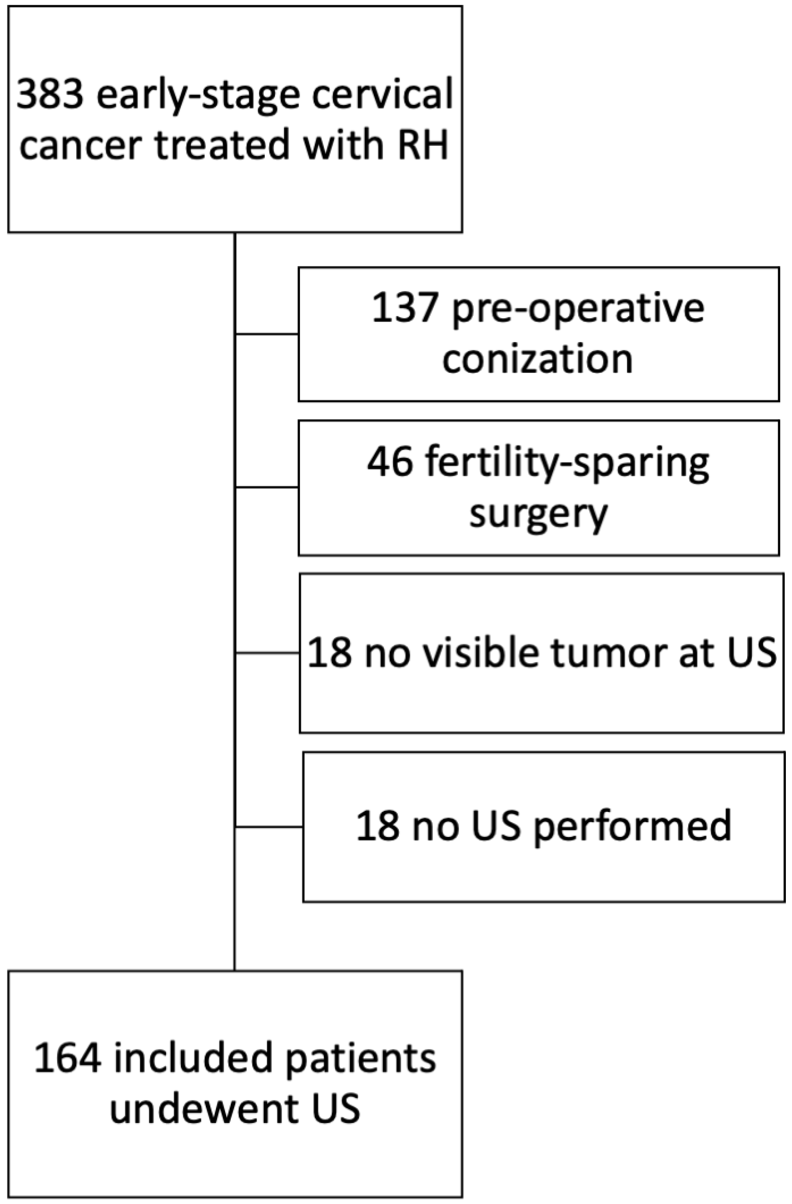
2.1. Study Design and Inclusion/Exclusion Criteria

This is a retrospective, single-center, observational cohort study, approved by the Institutional Review Board (number DIPUSVSP-27-07-2086 on 27 July 2020) at Fondazione Policlinico Universitario A. Gemelli, IRCCS (Rome, Italy). Consecutive patients with clinical International Federation of Gynecology and Obstetrics (FIGO) 2018 stage IA1–IB2 and IIA1 [13] cervical carcinoma, treated by primary radical surgical treatment with curative intent, between February 2012 and June 2019, at Fondazione Policlinico Agostino Gemelli IRCCS, Rome, Italy, were included. Only patients with available ultrasound examination performed within two weeks before surgery were included. Data were retrieved from the institution’s electronic database. Patients who underwent neo-adjuvant treatment, fertility sparing surgery, and pre-operative conization were excluded.

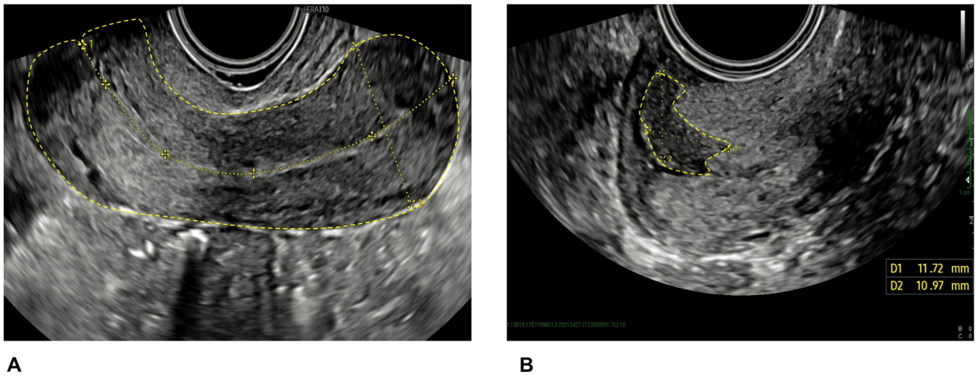
2.2. Ultrasound Scan

All patients underwent pre-operative ultrasound examination at our Ultrasound Center by a doctor with experience in gynecologic oncology ultrasound. Ultrasound examinations were performed with a transvaginal or transrectal probe to assess the pelvic organs. The methodology of cervical cancer assessment has been previously described [6,7,14]. High-end ultrasound equipment was used; the frequency of the vaginal probes varied between 5.0 and 9.0 MHz.
Tumor size was measured in three orthogonal diameters (craniocaudal, transverse, and anteroposterior). The craniocaudal extension was measured in a longitudinal section from the outermost margin of the tumor (facing the vagina) to the highest (most cranial) extension of the tumor (Figure 1), and the anteroposterior extension was measured perpendicular to this measurement. The transverse extension was measured in the transverse section between the outermost lateral margins of the tumor. The tumor volume was calculated with the formula of a sphere, Tvol = D1 × D2 × D3 × 0.53. Tumor echogenicity was classified as hypoechoic, isoechoic, hyperechoic, or mixed compared with the surrounding cervical tissue.
The tumors were assessed with color or power Doppler ultrasound. The intra-tumoral blood flow was classified subjectively as no detectable blood flow (color score = 1), minimal blood flow (color score = 2), moderate blood flow (color score = 3), or abundant blood flow (color score = 4) depending on the amount of color Doppler signals detected in the tumor [15].

2.3. Treatment

All patients underwent radical hysterectomy and bilateral pelvic lymphadenectomy with or without sentinel lymph node biopsy and salpingo-oophorectomy. Pre-operative staging was performed with a pelvic ultrasound scan, abdominal MRI-scan, and chest X-ray. The surgical approach was either laparotomy or minimally invasive (laparoscopy or robotic). The radicality of hysterectomy was classified according to the Querleu–Morrow classification [16]. In the case of suspicious initial parametrial involvement in the US-scan or MRI-scan, the patient was counseled for exclusive chemo-radiotherapy to be the first choice of treatment (as per ESGO guidelines [8]) or primary radical surgery, accepting the risk of potential adjuvant chemoradiation. A dedicated gynecologic oncology pathologist analyzed the surgical specimens. Adjuvant treatment was administered in line with international guidelines according to pathologic risk factors [8].

2.4. Study Variables

Information on ultrasound scan, anthropometric, clinical, and pathological characteristics was retrospectively collected.
Data were identified from the Red Cap (electronic data capture tools hosted at Fondazione Policlinico Universitario “A. Gemelli”, IRCCS; https://redcap-irccs.policlinicogemelli.it/) (last accessed 15 January 2023) database.

2.5. Statistical Analysis

Standard descriptive statistics were used to evaluate the distribution of each variable. Continuous variables were reported as median and categorical variables as frequency or percentage. DFS was defined as the time in months from the date of the surgery to the date of first recurrence, last follow-up, or death. OS was calculated as the time in months from the date of the surgery to the date of the last follow-up or death. OS and DFS were estimated by the Kaplan–Meier method [17] and the log-rank test was used to assess the statistical significance [18]. The relation between ultrasound and histology characteristics was assessed with logistic regression analysis. The influence of ultrasound and anthropometric parameters on their survival was analyzed using univariate and multivariate Cox proportional hazards models and described using hazard ratios (HRs) and their 95% confidence intervals [19]. Cut-offs for continuous variables in logistic and Cox regression analyses were determined with ROC curves according to the risk of recurrence.
The agreement between ultrasound tumor diameter and histological tumor diameter was assessed using Cohen’s kappa. Interpretation of Cohen’s index was performed according to Landis and Koch: ≤0.000 no agreement, 0.000–0.200 slight, 0.210–0.400 fair, 0.410–0.600 moderate, 0.610–0.800 substantial, and 0.810–1.000 almost perfect agreement [20].
All p-values reported are two-sided and a p-value <0.05 was considered statistically significant.
The analysis was computed using SPSS version 26.0 (IBM Corporation 2018, Armonk, NY, USA).

3. Results

3.1. Patients’ Characteristics

Three hundred and eighty-three patients underwent surgery for newly diagnosed early-stage cervical cancer in the study period. Of these, 137 (35.8%) were excluded as they underwent conization before ultrasound and radical surgery, 46 (12.0%) were excluded as they underwent fertility sparing surgery, 18 (4.7%) were excluded as they did not undergo ultrasound, and 18 (4.7%) were excluded as there was no detectable tumor in the ultrasound. The exclusion process is presented in Figure 2. Therefore, 164 (42.8%) patients were included in the present study. Table 1 shows the characteristics of the entire population. The median age at diagnosis was 48 (range, 25–80) years. Most of the patients underwent minimally invasive radical hysterectomy (118/164, 72.0%), underwent type C radical hysterectomy (126/164, 76.8%), and were found to have squamous cell carcinoma at final histology (104/164, 63.4%). Most patients were FIGO stage IB1 (113/164, 68.9%). Twenty-seven (16.5%) had parametrial infiltration and 37/164 (22.5%) patients had metastatic lymph nodes at final histology.
The median largest tumor diameter measured at histology was 26.0 mm (range, 1.0–55.0).

3.2. Ultrasound Scan Findings

Table 2 shows the ultrasound parameters of the study population. The median uterine volume was 74.1 mm3 (range, 11.9–966.7), the median largest tumor diameter was 29.0 mm (1.0–52.0), and the median tumor volume was 6.3 mm3 (range 1.0–58.5). Most cervical tumors appeared hypoechoic (122/164, 74.4%), with abundant color-Doppler vascularization (117/164, 71.3%) and with no internal uterine orifice involvement (104/164, 63.4%).
In 15/164 (9.1%) patients, the ultrasound examiner suspected parametrial infiltration at the time of US scan.

3.3. Correlation between Ultrasound and Pathological Findings

The agreement between ultrasound tumor largest diameter and histological tumor diameter was substantial (Cohen’s kappa: 0.73, 95% CI: 0.625–0.801).
The diagnostic performance in predicting parametrial infiltration with ultrasound was as follows: accuracy 79% (95% CI: 71.9%–84.9%), with 25/149 false-negative and 11/15 false-positive cases.
Univariate and multivariate logistic regression analyses relating ultrasound and histological tumor characteristics with pathologic parametrial infiltration are shown in Table 3. In the univariate analysis, the significant parameters were the ratio between largest tumor diameter and cervix-fundus uterine diameter (OR 3.551, 95%CI: 1.350–9.342) and lymph-vascular space involvement (LVSI) (OR 2.540, 95%CI: 1.008–6.400). In the multivariate analysis, the ratio between largest tumor diameter and cervix-fundus uterine diameter (with 37 as the cut-off) was significantly associated with pathological microscopic parametrial infiltration (OR 3.272, 95%CI: 1.231–8.702) (Table 3).

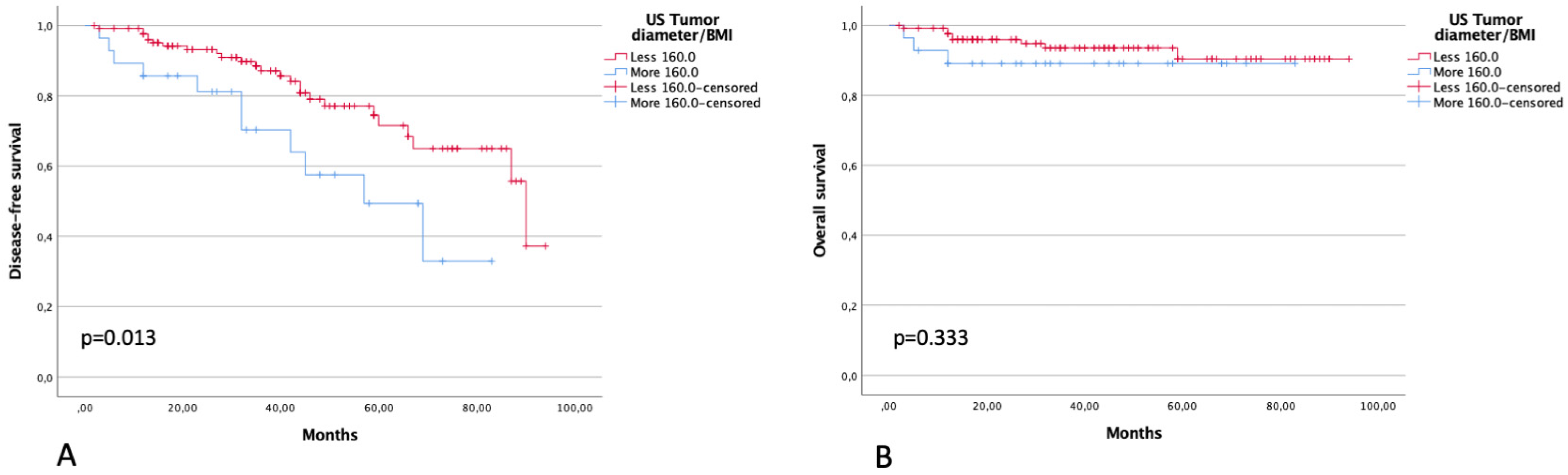
3.4. Survival Analysis

The median time of follow-up was 40 months (range, 3–94). Thirty-seven (22.6%) patients had recurrences and 12 (7.3%) died within the study period. Three-year DFS and OS were 83.7% and 92.1%, respectively.
Three-year DFS in patients with BMI ≤20 Kg/m2 was 66.0% compared with 86.3% of patients with >20 Kg/m2 (p < 0.001). Three-year OS was 79.4% and 99.3% in the same groups of patients, respectively (p = 0.010) (Figure 3). Three-year DFS in patients with a ratio between ultrasound tumor volume and BMI of <65 and >65 was 87.3% and 70.6% (p = 0.027), respectively. Three-year OS in patients with a ratio between ultrasound tumor volume and BMI of <65 and >65 was 92.6% and 93.6% (p = 0.953), respectively (Figure 4).
Three-year DFS in patients with a ratio between ultrasound largest tumor diameter and BMI of <160 and >160 was 87.1% and 70.4% (p = 0.013), respectively. Three-year OS in patients with a ratio between ultrasound largest tumor diameter and BMI of <160 and >160 was 93.6% and 89.1% (p = 0.333), respectively (Figure 5).
Table 4 shows the Cox regression analysis for variables influencing the risk of recurrence and death. The variables significantly associated with a higher risk of recurrence were as follows: BMI (BMI ≤20 Kg/m2 at higher risk of recurrence, p < 0.001) and ultrasound tumor volume (p = 0.038). Similarly, the ratio between ultrasound tumor volume and BMI, the ratio between ultrasound tumor volume and height, and the ratio between ultrasound largest tumor diameter and BMI were significantly related to a higher risk of recurrence (p = 0.011, p = 0.031, and p = 0.017, respectively).
Regarding the risk of death, none of the ultrasound variables were related to the risk of death in the univariate Cox regression analysis (Table 4). The only anthropometric characteristic related to a higher risk of death was BMI ≤20 Kg/m2 (p = 0.021).
Of note, the surgical approach (open versus minimally invasive) did not impact the risk of recurrence or death in the univariate Cox regression analysis (HR: 1.631, 95%CI: 0.631–4.216; p = 0.312 and HR: 1.624, 95%CI: 0.353–7.467; p = 0.534, for recurrence and death, respectively) [21].

4. Discussion

4.1. Main Findings

In the present study, we investigated the prognostic value of ultrasound tumor characteristics and anthropometric patients’ parameters in a series of patients with apparent early-stage cervical cancer. We found that BMI correlated with both the risk of recurrence and death, whereas the ratio between ultrasound parameters (such as tumor volume and largest diameter) and BMI only correlated with the risk of recurrence. Additionally, we also showed that pathological microscopic parametrial infiltration was related to the ratio between ultrasound largest tumor diameter and cervix-fundus uterine diameter.
The present study aimed to assess the ultrasound characteristics in a population of early-stage cervical cancer patients and to evaluate whether there was a prognostic relation between tumor dimensions/volume detected at ultrasound and uterine dimensions or patient’s anthropometric characteristics. In other words, does the same tumor volume have a worse prognosis in a patient with a smaller uterus or a thinner patient? With our results, we demonstrated that BMI ≤20 Kg/m2 per se has a negative impact on DFS and OS; however, the ratios between ultrasound-measured tumor volume or between ultrasound-measured maximum tumor diameter and BMI also influenced DFS.

4.2. Comparison with Other Related Literature

Multiple studies in the literature have reported on the prognostic impact of largest tumor diameter and tumor volume on DFS and OS in cervical cancer [22,23]; similarly, a few reports have showed the prognostic impact of BMI, even though with different results [12,24]. In fact, some studies reported that only low BMI had a significant impact on survival [24], while others reported that the BMI extremes were related to a higher risk of death [12]. It is postulated that BMI may influence survival for the associated chronic systemic inflammation and poor nutritional status [25,26].
On the other side, the relation between tumor and uterine dimensions did not impact the survival, rejecting our initial hypothesis. Similarly, the internal uterine orifice and uterine isthmus involvement did not worsen the prognosis in our series. These results contrast with the current literature, which demonstrates that internal orifice involvement is an independent prognostic marker, in both early-stage and locally advanced cervical cancer [27,28]. With this background, we postulated not only that the cranio-caudal growth (toward uterine corpus) but also latero-lateral growth could have impacted the risk of recurrence, particularly in the case of a smaller uterus, where parametria and lymphatics are closer to the tumoral mass. Indeed, we realized that the ratio between largest tumor diameter and cervical-fundus diameter of the uterus was the factor most related to microscopic parametrial infiltration, which is a known negative prognostic marker. Further studies should focus on this aspect to predict parametrial infiltration and possibly avoid radical surgery in favor of exclusive chemoradiation [8].

4.3. Strengths and Limitations

The main limitation of the present study is represented by its retrospective nature and by the low number of underweight patients included. Nevertheless, to the best of our knowledge, this is the first study investigating the combination of ultrasound and anthropometric markers to predict pathologic (parametrial involvement) and oncological outcomes.

4.4. Future Perspectives

The present results may represent a hypothesis-generating study that aims to emphasize the importance of pre-operative support of nutritional status before starting the treatment and to highlight the potential application of US to predict parametrial infiltration and the risk of recurrence.
Further research will be needed to validate the hypotheses of our study. Once confirmed, these data could promote research into novel prognostic factors and new treatment strategies.

5. Conclusions

In a population of patients with suspicious early-stage cervical cancer undergoing primary radical surgery, a low BMI was the most significant anthropometric biomarker impairing DFS and OS. The ratio between tumor volume measured in ultrasound and BMI and the ratio between largest tumor diameter measured in ultrasound and BMI significantly affected DFS but not OS. The pre-operative ratio between ultrasound-measured largest tumor diameter and cervix-fundus uterine diameter represented the factor most related to parametrial infiltration. These novel prognostic parameters might be useful in the pre-operative workup for a patient-tailored treatment in early-stage cervical cancer.

Author Contributions

Conceptualization, N.B., A.C.T. and A.K.; methodology, N.B., F.M., L.P.A., V.C., C.C. and E.T.; software, L.P.A. and N.B.; validation, A.F., F.F., G.F., G.S. and A.C.T.; formal analysis, N.B.; investigation, data curation, N.B., A.B., F.M. and V.C.; writing—original draft preparation, N.B., D.Q., F.M., A.C.T. and A.B.; writing—review and editing, A.F., F.F., G.F. and G.S.; supervision, D.Q., G.S. and A.C.T.; project administration, N.B., F.M. and A.C.T. All authors have read and agreed to the published version of the manuscript.

Funding

This research received no external funding.

Institutional Review Board Statement

Study approved by Institutional Review Board (number DIPUSVSP-27-07-2086 on 27 July 2020) at Fondazione Policlinico Universitario A. Gemelli, IRCCS (Rome, Italy).

Informed Consent Statement

Informed consent was obtained from all subjects involved in the study.

Data Availability Statement

Data available upon reasonable request.

Acknowledgments

The authors would like to thank the scrub nurses and the team from CLASS ultrasound for their continuous support in patients’ care.

Conflicts of Interest

The authors declare no conflict of interest.

References

  1. Brisson, M.; Kim, J.J.; Canfell, K.; Drolet, M.; Gingras, G.; A Burger, E.; Martin, D.; Simms, K.T.; Bénard, É.; Boily, M.-C.; et al. Impact of HPV vaccination and cervical screening on cervical cancer elimination: A comparative modelling analysis in 78 low-income and lower-middle-income countries. Lancet 2020, 395, 575–590. [Google Scholar] [CrossRef] [PubMed]
  2. Wright, T.C.; Stoler, M.H.; Behrens, C.M.; Sharma, A.; Zhang, G.; Wright, T.L. Primary cervical cancer screening with human papillomavirus: End of study results from the ATHENA study using HPV as the first-line screening test. Gynecol. Oncol. 2015, 136, 189–197. [Google Scholar] [CrossRef] [PubMed]
  3. Sung, H.; Ferlay, J.; Siegel, R.L.; Laversanne, M.; Soerjomataram, I.; Jemal, A.; Bray, F. Global Cancer Statistics 2020: GLOBOCAN Estimates of Incidence and Mortality Worldwide for 36 Cancers in 185 Countries. CA Cancer J. Clin. 2021, 71, 209–249. [Google Scholar] [CrossRef] [PubMed]
  4. Bhatla, N.; Aoki, D.; Sharma, D.N.; Sankaranarayanan, R. Cancer of the cervix uteri. Int. J. Gynaecol. Obstet. 2018, 143 (Suppl. 2), 22–36. [Google Scholar] [CrossRef]
  5. Matsuo, K.; Machida, H.; Mandelbaum, R.S.; Konishi, I.; Mikami, M. Validation of the 2018 FIGO cervical cancer staging system. Gynecol. Oncol. 2019, 152, 87–93. [Google Scholar] [CrossRef]
  6. Testa, A.C.; Ludovisi, M.; Manfredi, R.; Zannoni, G.; Gui, B.; Basso, D.; Di Legge, A.; Licameli, A.; Di Bidino, R.; Scambia, G.; et al. Transvaginal ultrasonography and magnetic resonance imaging for assessment of presence, size and extent of invasive cervical cancer. Ultrasound Obstet. Gynecol. 2009, 34, 335–344. [Google Scholar] [CrossRef]
  7. Gaurilcikas, A.; Vaitkiene, D.; Cizauskas, A.; Inciura, A.; Svedas, E.; Maciuleviciene, R.; Di Legge, A.; Ferrandina, G.; Testa, A.C.; Valentin, L. Early-stage cervical cancer: Agreement between ultrasound and histopathological findings with regard to tumor size and extent of local disease. Ultrasound Obstet. Gynecol. 2011, 38, 707–715. [Google Scholar] [CrossRef]
  8. Cibula, D.; Pötter, R.; Planchamp, F.; Avall-Lundqvist, E.; Fischerova, D.; Meder, C.H.; Köhler, C.; Landoni, F.; Lax, S.; Lindegaard, J.C.; et al. The European Society of Gynaecological Oncology/European Society for Radiotherapy and Oncology/European Society of Pathology Guidelines for the Management of Patients With Cervical Cancer. Int. J. Gynecol. Cancer 2018, 28, 641–655. [Google Scholar] [CrossRef]
  9. Fischerová, D.; Cibula, D. The role of ultrasound in primary workup of cervical cancer staging (ESGO, ESTRO, ESP cervical cancer guidelines). Role ultrazvuku ve stagingu zhoubného nádoru děložního hrdla (doporučení Evropské onkogynekologické, radiační a patologické společnosti). Ceska Gynekol. 2019, 84, 40–48. [Google Scholar]
  10. López-Hernández, D. Epidemiological Association Between Body Fat Percentage and Cervical Cancer: A Cross-sectional Population-based Survey from Mexico. Arch. Med. Res. 2013, 44, 454–458. [Google Scholar] [CrossRef]
  11. Poorolajal, J.; Jenabi, E. The association between BMI and cervical cancer risk: A meta-analysis. Eur. J. Cancer Prev. 2016, 25, 232–238. [Google Scholar] [CrossRef] [PubMed]
  12. Clark, L.H.; Jackson, A.L.; Soo, A.E.; Orrey, D.C.; Gehrig, P.A.; Kim, K.H. Extremes in body mass index affect overall survival in women with cervical cancer. Gynecol. Oncol. 2016, 141, 497–500. [Google Scholar] [CrossRef] [PubMed]
  13. Pecorelli, S.; Zigliani, L.; Odicino, F. Revised FIGO staging for carcinoma of the cervix. Int. J. Gynecol. Obstet. 2009, 105, 107–108. [Google Scholar] [CrossRef]
  14. Fischerova, D. Ultrasound scanning of the pelvis and abdomen for staging of gynecological tumors: A review. Ultrasound Obstet. Gynecol. 2011, 38, 246–266. [Google Scholar] [CrossRef] [PubMed]
  15. Epstein, E.; Di Legge, A.; Måsbäck, A.; Lindqvist, P.G.; Kannisto, P.; Testa, A.C. Sonographic characteristics of squamous cell cancer and adenocarcinoma of the uterine cervix. Ultrasound Obstet. Gynecol. 2010, 36, 512–516. [Google Scholar] [CrossRef]
  16. Querleu, D.; Cibula, D.; Abu-Rustum, N.R. 2017 Update on the Querleu–Morrow Classification of Radical Hysterectomy. Ann. Surg. Oncol. 2017, 24, 3406–3412. [Google Scholar] [CrossRef]
  17. Kaplan, E.L.; Meier, P. Nonparametric estimation from incomplete observation. J. Am. Stat. Assoc. 1958, 53, 457–481. [Google Scholar] [CrossRef]
  18. Mantel, N. Evaluation of survival data and two new rank order statistics arising in its consideration. Cancer Chemother. Rep. 1966, 50, 163–170. [Google Scholar]
  19. Cox, D.R. Models and Life-Tables Regression. J. R. Stat. Soc. Ser. B (Methodol.) 1972, 34, 187–220. [Google Scholar] [CrossRef]
  20. Landis, J.R.; Koch, G.G. The measurement of observer agreement for categorical data. Biometrics 1977, 33, 159–174. [Google Scholar] [CrossRef]
  21. Ramirez, P.T.; Frumovitz, M.; Pareja, R.; Lopez, A.; Vieira, M.; Ribeiro, M.; Buda, A.; Yan, X.; Shuzhong, Y.; Chetty, N.; et al. Minimally Invasive versus Abdominal Radical Hysterectomy for Cervical Cancer. N. Engl. J. Med. 2018, 379, 1895–1904. [Google Scholar] [CrossRef] [PubMed]
  22. Wagner, A.E.; Pappas, L.; Ghia, A.J.; Gaffney, D.K. Impact of tumor size on survival in cancer of the cervix and validation of stage IIA1 and IIA2 subdivisions. Gynecol. Oncol. 2013, 129, 517–521. [Google Scholar] [CrossRef] [PubMed]
  23. Bizzarri, N.; Anchora, L.P.; Kucukmetin, A.; Ratnavelu, N.; Korompelis, P.; Carbone, V.; Fedele, C.; Bruno, M.; Vizzielli, G.; Gallotta, V.; et al. Protective Role of Conization Before Radical Hysterectomy in Early-Stage Cervical Cancer: A Propensity-Score Matching Study. Ann. Surg. Oncol. 2021, 28, 3585–3594. [Google Scholar] [CrossRef] [PubMed]
  24. Kizer, N.T.; Thaker, P.H.; Gao, F.; Zighelboim, I.; Powell, M.A.; Rader, J.S.; Mutch, D.G.; Grigsby, P.W. The effects of body mass index on complications and survival outcomes in patients with cervical carcinoma undergoing curative chemoradiation therapy. Cancer 2011, 117, 948–956. [Google Scholar] [CrossRef]
  25. Calle, E.E.; Kaaks, R. Overweight, obesity and cancer: Epidemiological evidence and proposed mechanisms. Nat. Rev. Cancer 2004, 4, 579–591. [Google Scholar] [CrossRef]
  26. Morley, J.E.; Thomas, D.R.; Wilson, M.-M.G. Cachexia: Pathophysiology and clinical relevance. Am. J. Clin. Nutr. 2006, 83, 735–743. [Google Scholar] [CrossRef]
  27. Matsuo, K.; Machida, H.; Blake, E.A.; Takiuchi, T.; Mikami, M.; Roman, L.D. Significance of uterine corpus tumor invasion in early-stage cervical cancer. Eur. J. Surg. Oncol. (EJSO) 2017, 43, 725–734. [Google Scholar] [CrossRef]
  28. Narayan, K.; Fisher, R.; Bernshaw, D. Significance of tumor volume and corpus uteri invasion in cervical cancer patients treated by radiotherapy. Int. J. Gynecol. Cancer 2006, 16, 623–630. [Google Scholar] [CrossRef]
Figure 1. Ultrasound scan image showing the uterus (A) and tumor (B) diameters in two dimensions.
Diagnostics 13 00583 g001
Figure 2. Flow chart demonstrating the exclusion process.
Diagnostics 13 00583 g002
Figure 3. DFS (A) and OS (B) in patients with BMI of ≤20 and >20 kg/m2.
Diagnostics 13 00583 g003
Figure 4. DFS (A) and OS (B) in patients with a ratio between tumor volume at US and BMI of ≤65 and >65.
Diagnostics 13 00583 g004
Figure 5. DFS (A) and OS (B) in patients with a ratio between largest tumor diameter at US and BMI of ≤160 and >160.
Diagnostics 13 00583 g005
Table 1. Clinical, surgical, and histological characteristics of the study population.
Characteristicsn = 164 (Range, %)
Age at diagnosis (years)48 (25–80)
BMI a (kg/m2)24.0 (16.9–47.6)
Height (cm)162 (150–180)
Surgical Approach
Laparotomy46 (28.0)
Laparoscopy77 (47.0)
Robot41 (25.0)
Radicality of Radical Hysterectomy
A2 (1.2)
B36 (22.0)
C126 (76.8)
Histology
Squamous cell carcinoma104 (63.4)
Adenocarcinoma45 (27.4)
Adeno-squamous5 (3.0)
Other10 (6.1)
Grade
13 (1.8)
289 (54.3)
370 (42.7)
Unknown2 (1.2)
Lymph-Vascular Space Infiltration
Negative71 (43.3)
Positive92 (56.1)
Unknown1 (0.6)
Pathologic Tumor Diameter
≤20 mm46 (28.0)
>20 mm114 (69.5)
Unknown4 (2.4)
Pathologic Depth of Stromal Infiltration (mm)8.2 (0.4–23.0)
Largest Pathologic Tumor Diameter (mm)26.0 (1.0–55.0)
Removed Lymph Nodes26 (1–75)
Patients with Metastatic Lymph Nodes37 (22.5)
Pathologic FIGO b Stage 2009
IA11 (0.6)
IB1113 (68.9)
IB215 (9.1)
IIA17 (4.3)
IIA21 (0.6)
IIB27 (16.5)
Results are presented as n (%) or median (range); a BMI: body mass index; b FIGO: International Federation of Gynecology and Obstetrics.
Table 2. Ultrasound characteristics of the study population.
Characteristicsn = 164 (Range, %)
Uterus volume (mm3)74.1 (11.9–966.7)
Largest tumor diameter (mm)29.0 (1.0–52.0)
Tumor volume (mm3)6.3 (1.0–58.5)
Depth of cervical stromal infiltration
<2/376 (56.3)
>2/388 (53.7)
Echogenicity
Hypoechoic122 (74.4)
Isoechoic26 (15.9)
Hyperechoic15 (9.1)
Not reported1 (0.6)
Color-Doppler evaluation
No detectable flow (color score 1)5 (3.0)
Minimal flow (color score 2)10 (6.1)
Moderate flow (color score 3)32 (19.5)
Abundant flow (color score 4)117 (71.3)
Internal uterine orifice involvement
No104 (63.4)
Yes60 (36.6)
Table 3. Univariate and multivariate logistic regression analysis relating ultrasound and histological tumor characteristics with pathologic parametrial infiltration.
Pathologic Parametrial Infiltration
Univariate AnalysisMultivariate Analysis
CharacteristicsComparison/MeasureOR (95% CI)p-ValueOR (95% CI)p-Value
Ultrasound
US stromal infiltrationFull thickness vs. superficial/middle third1.242 (0.930–1.658)0.142
Largest tumor diameter (mm)mm1.000 (1.000–1.001)0.415
EchogenicityHypoechoic vs. non-hypoechoic0.884 (0.358–2.178)0.778
US tumor maximum diameter/Uterine CF diameter≤37 or >373.551 (1.350–9.342)0.0103.272 (1.231–8.702)0.018
Pathologic
LVSINegative vs. positive2.540 (1.008–6.400)0.0482.203 (0.857–5.660)0.101
Grade1–2 vs. 31.348 (0.889–2.045)0.160
Pathologic tumor diameter≤2 or >2 cm2.234 (0.719–6.940)0.165
Table 4. Univariate Cox regression analysis for clinical and ultrasound variables influencing risk of recurrence and death*.
RISK OF RECURRENCE
CharacteristicsComparison/MeasureHR (95% CI)p-Value
Age≤50 vs >50 years0.608 (0.305–1.215)0.159
BMI≤20 vs >20 kg/m20.213 (0.095–0.474)<0.001
Largest US Tumor diametermm1.018 (0.983–1.054)0.326
Depth of infiltrationFull thickness vs Superficial/Middle third1.116 (0.899–1.387)0.320
EchogenicityHypoechoic vs non-hypoechoic1.023 (0.482–2.172)0.953
Color-DopplerAbundant flow vs non-abundant flow1.096 (0.909–1.322)0.337
Internal uterine os involvementNo vs Yes0.863 (0.428–1.741)0.681
Tumor Volumemm31.035 (1.002–1.069)0.038
Tumor Volume/BMIcontinuous1.010 (1.002–1.018)0.011
Largest Tumor diameter/BMIcontinuous1.009 (1.002–1.017)0.017
Tumor Volume/Height of patientcontinuous1.006 (1.001–1.011)0.031
Largest Tumor diameter/Uterine CF diametercontinuous1.003 (0.980–1.026)0.830
Tumor CF diameter/Uterine CF diametercontinuous0.995 (0.971–1.020)0.673
RISK OF DEATH
CharacteristicsComparison/MeasureHR (95% CI)p-Value
Age≤50 vs >50 years0.455 (0.123–1.683)0.238
BMI≤20 vs >20 Kg/m20.205 (0.054–0.784)0.021
Largest US Tumor diametermm0.981 (0.925–1.041)0.531
US depth of infiltrationFull thickness vs Superficial/Middle third1.260 (0.844–1.881)0.258
EchogenicityHypoechoic vs non-hypoechoic3.505 (0.452–27.183)0.230
Color-DopplerAbundant flow vs non-abundant flow0.976 (0.723–1.319)0.877
Internal uterine orifice involvementNo vs Yes1.041 (0.313–3.465)0.948
Tumor Volumemm30.975 (0.900–1.055)0.527
Tumor Volume/BMIcontinuous0.998 (0.980–1.016)0.824
Largest Tumor diameter/BMIcontinuous1.005 (0.991–1.018)0.499
Tumor Volume/Height of patientcontinuous0.997 (0.985–1.010)0.678
Largest Tumor diameter/Uterine CF diametercontinuous0.992 (0.953–1.032)0.685
Tumor CF diameter/Uterine CF diametercontinuous0.968 (0.924–1.015)0.176
* Multivariate analysis not performed for the risk of interaction between variables containing BMI. HR: hazard ratio; BMI: body mass index; CF: cervix-fundus; US: ultrasound scan.
Disclaimer/Publisher’s Note: The statements, opinions and data contained in all publications are solely those of the individual author(s) and contributor(s) and not of MDPI and/or the editor(s). MDPI and/or the editor(s) disclaim responsibility for any injury to people or property resulting from any ideas, methods, instructions or products referred to in the content.

