
Choroidal Hyperreflective Nodules Detected by Infrared Reflectance Images Are a Diagnostic Criterion for Neurofibromatosis Type 1 Patients Excluding Those with High Myopia

 , ,
, ,  and
and
Abstract
1. Introduction
2. Materials and Methods
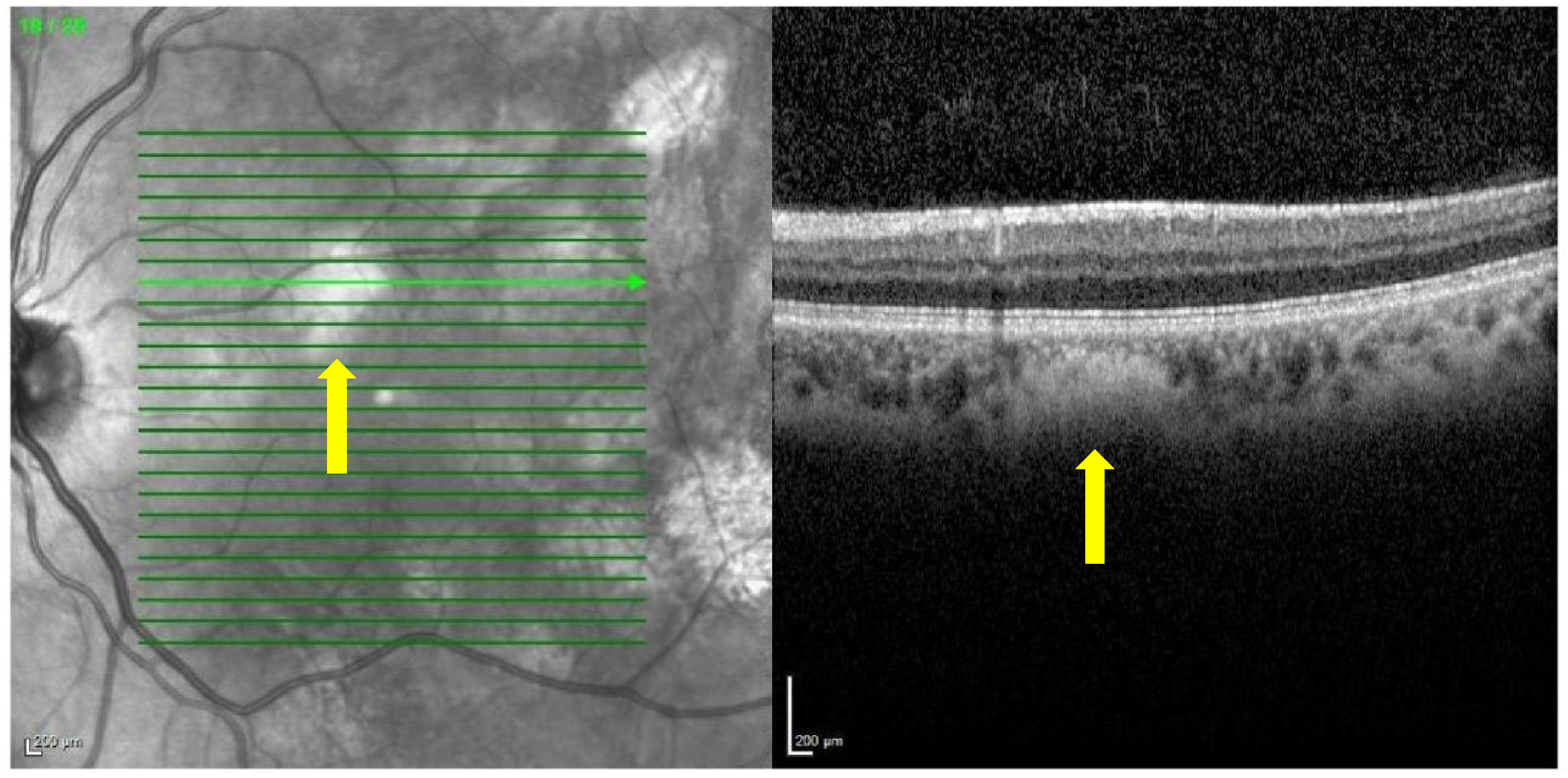
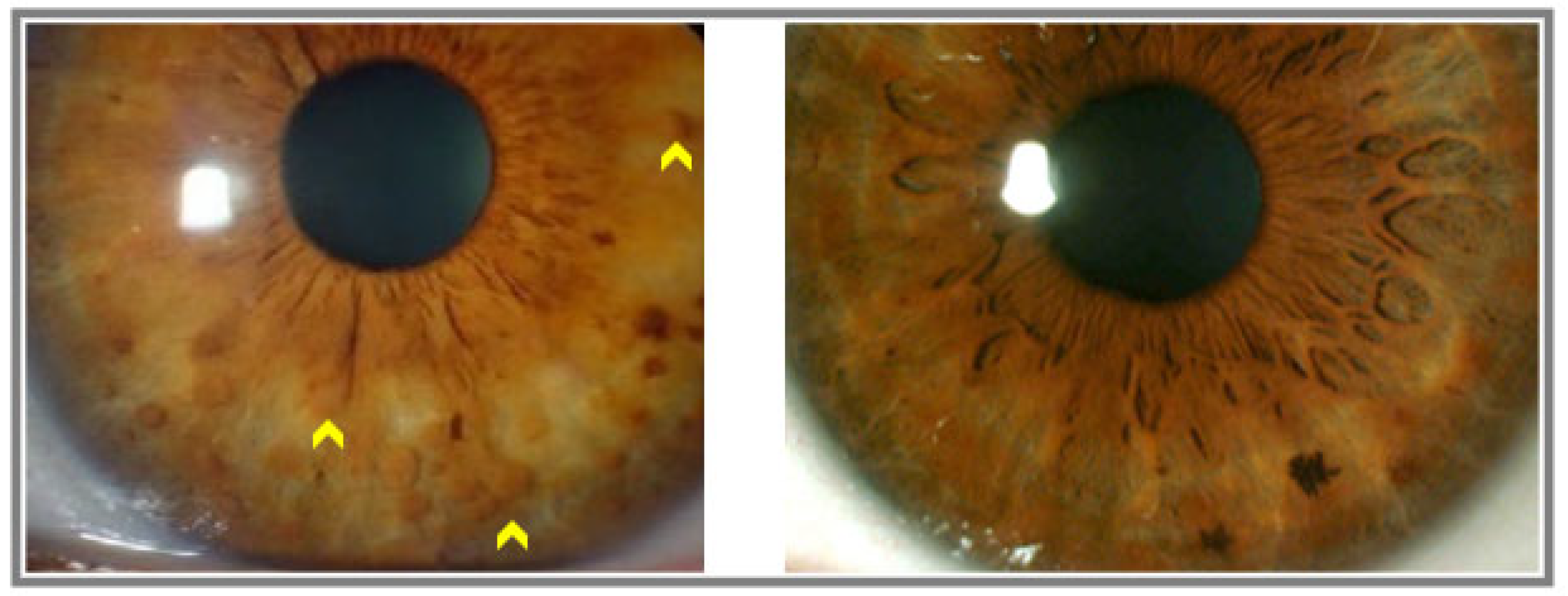
3. Results
4. Discussion
5. Conclusions
Author Contributions
Funding
Institutional Review Board Statement
Informed Consent Statement
Data Availability Statement
Conflicts of Interest
References
- Cimino, P.J.; Gutmann, D.H. Neurofibromatosis Type 1. En: Neurogenetics, Part II; Elsevier: Amsterdam, The Netherlands, 2018; pp. 799–811. [Google Scholar]
- Ly, K.I.; Blakeley, J.O. The diagnosis and management of neurofibromatosis type 1. Med. Clin. N. Am. 2019, 103, 1035–1054. [Google Scholar] [CrossRef] [PubMed]
- Gutmann, D.H.; Aylsworth, A.; Carey, J.C.; Korf, B.; Marks, J.; Pyeritz, R.E.; Rubenstein, A.; Viskochil, D. The diagnostic evaluation and multidisciplinary management of neurofibromatosis 1 and neurofibromatosis 2. JAMA 1997, 278, 51–57. [Google Scholar] [CrossRef] [PubMed]
- National institutes of health consensus development conference statement: Neurofibromatosis, Bethesda, MD, USA, July 13–15 1987. Neurofibromatosis 1988, 1, 172–178.
- Cassiman, C.; Casteels, I.; Stalmans, P.; Legius, E.; Jacob, J. Optical coherence tomography angiography of retinal microvascular changes overlying choroidal nodules in neurofibromatosis type 1. Case Rep. Ophthalmol. 2017, 8, 214–220. [Google Scholar] [CrossRef]
- Legius, E.; Messiaen, L.; Wolkenstein, P.; Pancza, P.; Avery, R.A.; Berman, Y.; Blakeley, J.; Babovic-Vuksanovic, D.; Cunha, K.S.; Ferner, R.; et al. Revised diagnostic criteria for neurofibromatosis type 1 and Legius syndrome: An international consensus recommendation. Genet Med. 2021, 23, 1506–1513. [Google Scholar] [CrossRef]
- Yasunari, T.; Shiraki, K.; Hattori, H.; Miki, T. Frequency of choroidal abnormalities in neurofibromatosis type 1. Lancet 2000, 356, 988–992. [Google Scholar] [CrossRef] [PubMed]
- Nakakura, S.; Shiraki, K.; Yasunari, T.; Hayashi, Y.; Ataka, S.; Kohno, T. Quantification and anatomic distribution of choroidal abnormalities in patients with type 1 neurofibromatosis. Graefes Arch. Clin. Exp. Ophthalmol. 2005, 243, 980–984. [Google Scholar] [CrossRef]
- Helb, H.M.; Issa, C.; Fleckenstein, P. Clinical evaluation of simultaneous confocal scanning laser ophthalmoscopy imaging combined with high-resolution, spectraldomain optical coherence tomography. Acta Ophthalmol. 2010, 88, 842–849. [Google Scholar] [CrossRef]
- Viola, F.; Villani, E.; Natacci, F.; Selicorni, A.; Melloni, G.; Vezzola, D.; Barteselli, G.; Mapelli, C.; Pirondini, C.; Ratiglia, R. Choroidal abnormalities detected by near-infrared reflectance imaging as a new diagnostic criterion for neurofibromatosis 1. Ophthalmology 2012, 119, 369–375. [Google Scholar] [CrossRef]
- Moramarco, A.; Giustini, S.; Nofroni, I.; Mallone, F.; Miraglia, E.; Iacovino, C.; Calvieri, S.; Lambiase, A. Near-infrared imaging: An in vivo, non-invasive diagnostic tool in neurofibromatosis type 1. Arbeitsphysiologie 2018, 256, 307–311. [Google Scholar] [CrossRef]
- Alshareef, R.A.; Khuthaila, M.K.; Januwada, M.; Goud, A.; Ferrara, D.; Chhablani, J. Choroidal vascular analysis in myopic eyes: Evidence of foveal medium vessel layer thinning. Int. J. Retin. Vitr. 2017, 3, 28. [Google Scholar] [CrossRef]
- Moriyama, M.; Ohno-Matsui, K.; Futagami, S.; Yoshida, T.; Hayashi, K.; Shimada, N.; Kojima, A.; Tokoro, T.; Mochizuki, M. Morphology and long-term changes of choroidal vascular structure in highly myopic eyes with and without posterior staphyloma. Ophthalmology 2007, 114, 1755–1762. [Google Scholar] [CrossRef] [PubMed]
- Kumar, V.; Singh, S. Multimodal imaging of choroidal nodules in neurofibromatosis type-1. Indian J. Ophthalmol. 2018, 66, 586. [Google Scholar] [CrossRef] [PubMed]
- Abdolrahimzadeh, S.; Felli, L.; Plateroti, R.; Plateroti, A.M.; Giustini, S.; Calvieri, S.; Recupero, S.M. Morphologic and vasculature features of the choroid and associated choroid–retinal thickness alterations in neurofibromatosis type 1. Br. J. Ophthalmol. 2015, 99, 789–793. [Google Scholar] [CrossRef]
- Byun, Y.S.; Park, Y.H. Indocyanine green angiographic findings of obscure choroidal abnormalities in neurofibromatosis. Korean J. Ophthalmol. 2012, 26, 230–234. [Google Scholar] [CrossRef]
- Vagge, A.; Camicione, P.; Capris, C.; Sburlati, C.; Panarello, S.; Calevo, M.G.; Traverso, C.E.; Capris, P. Choroidal abnormalities in neurofibromatosis type 1 detected by near-infrared reflectance imaging in paediatric population. Acta Ophthalmol. 2015, 93, e667–e671. [Google Scholar] [CrossRef] [PubMed]
- Parrozzani, R.; Clementi, M.; Frizziero, L.; Miglionico, G.; Perrini, P.; Cavarzeran, F.; Kotsafti, O.; Comacchio, F.; Trevisson, E.; Convento, E.; et al. In vivo detection of choroidal abnormalities related to NF1: Feasibility and comparison with standard NIH diagnostic criteria in pediatric patients. Investig. Ophthalmol. Vis. Sci. 2015, 56, 6036–6042. [Google Scholar] [CrossRef]
- Cruciani, F.; Piraino, D.C.; Albanese, G.; Rahimi, S.; Abdolrahimzadeh, B. Neurofibromatosis: An update of ophthalmic characteristics and applications of optical coherence tomography. Clin. Ophthalmol. 2016, 10, 851–860. [Google Scholar] [CrossRef]
- Makino, S.; Tampo, H.; Arai, Y.; Obata, H. Correlations between choroidal abnormalities, Lisch nodules, and age in patients with neurofibromatosis type 1. Clin. Ophthalmol. 2014, 8, 165–168. [Google Scholar] [CrossRef]
- Rao, R.C.; Choudhry, N. Enhanced depth imaging spectral-domain optical coherence tomography findings in choroidal neurofibromatosis. Ophthalmic Surg. Lasers Imaging Retin. 2014, 45, 466–468. [Google Scholar] [CrossRef]
- Moreno-Morillo, F.J.; Fernández-Vigo, J.I.; Burgos-Blasco, B.; Orden, C.L.-L.; Vidal-Villegas, B.; Santos-Bueso, E. Optical coherence tomography angiography of choroidal nodules in neurofibromatosis type-1: A case series. Eur. J. Ophthalmol. 2022, 32, NP91–NP94. [Google Scholar] [CrossRef] [PubMed]
- Pimentel, M.F.; Heath, A.; Wan, M.J.; Hussein, R.; Leahy, K.E.; MacDonald, H.; Tavares, E.; VandenHoven, C.; MacNeill, K.; Kannu, P.; et al. Prevalence of choroidal abnormalities and Lisch nodules in children meeting clinical and molecular diagnosis of neurofibromatosis type 1. Transl. Vis. Sci. Technol. 2022, 11, 10. [Google Scholar] [CrossRef] [PubMed]
- Bergqvist, C.; Network, N.F.; Servy, A.; Valeyrie-Allanore, L.; Ferkal, S.; Combemale, P.; Wolkenstein, P. Neurofibromatosis 1 French national guidelines based on an extensive literature review since 1966. Orphanet J. Rare Dis. 2020, 15, 37. [Google Scholar] [CrossRef] [PubMed]


| Experimental Group | |||
|---|---|---|---|
| Lisch Nodules | Control | Case | p 1 |
| No | 30 (100.0%) | 6 (20%) | <0.001 |
| Yes | 0 (0.0%) | 24 (80.0%) | |
| Absolute frecuency 1 Chi-square | |||
| Experimental Group | |||
|---|---|---|---|
| Choroidal Nodules | Control | Case | p 1 |
| No | 30 (100.0%) | 5 (16.7%) | <0.001 |
| Yes | 0 (0.0%) | 25 (83.3%) | |
| Absolute frecuency 1 Chi-square | |||
| Variable | Freq § (%) | p 2 |
|---|---|---|
| Choroidal nodules | 25 (83.3%) | 0.3705 |
| Lisch nodules | 24 (80.0%) | |
| 2 Z test ‡ | ||
| Variable | Freq § (%) | p 2 |
|---|---|---|
| Choroidal nodules | 25 (100.0%) | 0.0145 |
| Lisch nodules | 21 (84.0%) | |
| 2 Z test ‡ | ||
Disclaimer/Publisher’s Note: The statements, opinions and data contained in all publications are solely those of the individual author(s) and contributor(s) and not of MDPI and/or the editor(s). MDPI and/or the editor(s) disclaim responsibility for any injury to people or property resulting from any ideas, methods, instructions or products referred to in the content. |
© 2023 by the authors. Licensee MDPI, Basel, Switzerland. This article is an open access article distributed under the terms and conditions of the Creative Commons Attribution (CC BY) license (https://creativecommons.org/licenses/by/4.0/).
Share and Cite
de Rivas, M.O.; Gabás, J.M.; Cabeza, M.Á.T.; Floría, O.E.; Latorre, R.H.; Moscarda, E.N.; Clavería, J.A.; Rivasés, G.P.; Puyuelo, J.A. Choroidal Hyperreflective Nodules Detected by Infrared Reflectance Images Are a Diagnostic Criterion for Neurofibromatosis Type 1 Patients Excluding Those with High Myopia. Diagnostics 2023, 13, 1348. https://doi.org/10.3390/diagnostics13071348
de Rivas MO, Gabás JM, Cabeza MÁT, Floría OE, Latorre RH, Moscarda EN, Clavería JA, Rivasés GP, Puyuelo JA. Choroidal Hyperreflective Nodules Detected by Infrared Reflectance Images Are a Diagnostic Criterion for Neurofibromatosis Type 1 Patients Excluding Those with High Myopia. Diagnostics. 2023; 13(7):1348. https://doi.org/10.3390/diagnostics13071348
Chicago/Turabian Stylede Rivas, Marta Orejudo, Javier Mateo Gabás, Miguel Ángel Torralba Cabeza, Olivia Esteban Floría, Raquel Herrero Latorre, Eva Núñez Moscarda, Julia Aramburu Clavería, Guillermo Pérez Rivasés, and Javier Ascaso Puyuelo. 2023. "Choroidal Hyperreflective Nodules Detected by Infrared Reflectance Images Are a Diagnostic Criterion for Neurofibromatosis Type 1 Patients Excluding Those with High Myopia" Diagnostics 13, no. 7: 1348. https://doi.org/10.3390/diagnostics13071348
APA Stylede Rivas, M. O., Gabás, J. M., Cabeza, M. Á. T., Floría, O. E., Latorre, R. H., Moscarda, E. N., Clavería, J. A., Rivasés, G. P., & Puyuelo, J. A. (2023). Choroidal Hyperreflective Nodules Detected by Infrared Reflectance Images Are a Diagnostic Criterion for Neurofibromatosis Type 1 Patients Excluding Those with High Myopia. Diagnostics, 13(7), 1348. https://doi.org/10.3390/diagnostics13071348