
A Rare Case of Invasive Thyroid Aspergillosis Revealed on 18F-FDG-PET/CT


Abstract
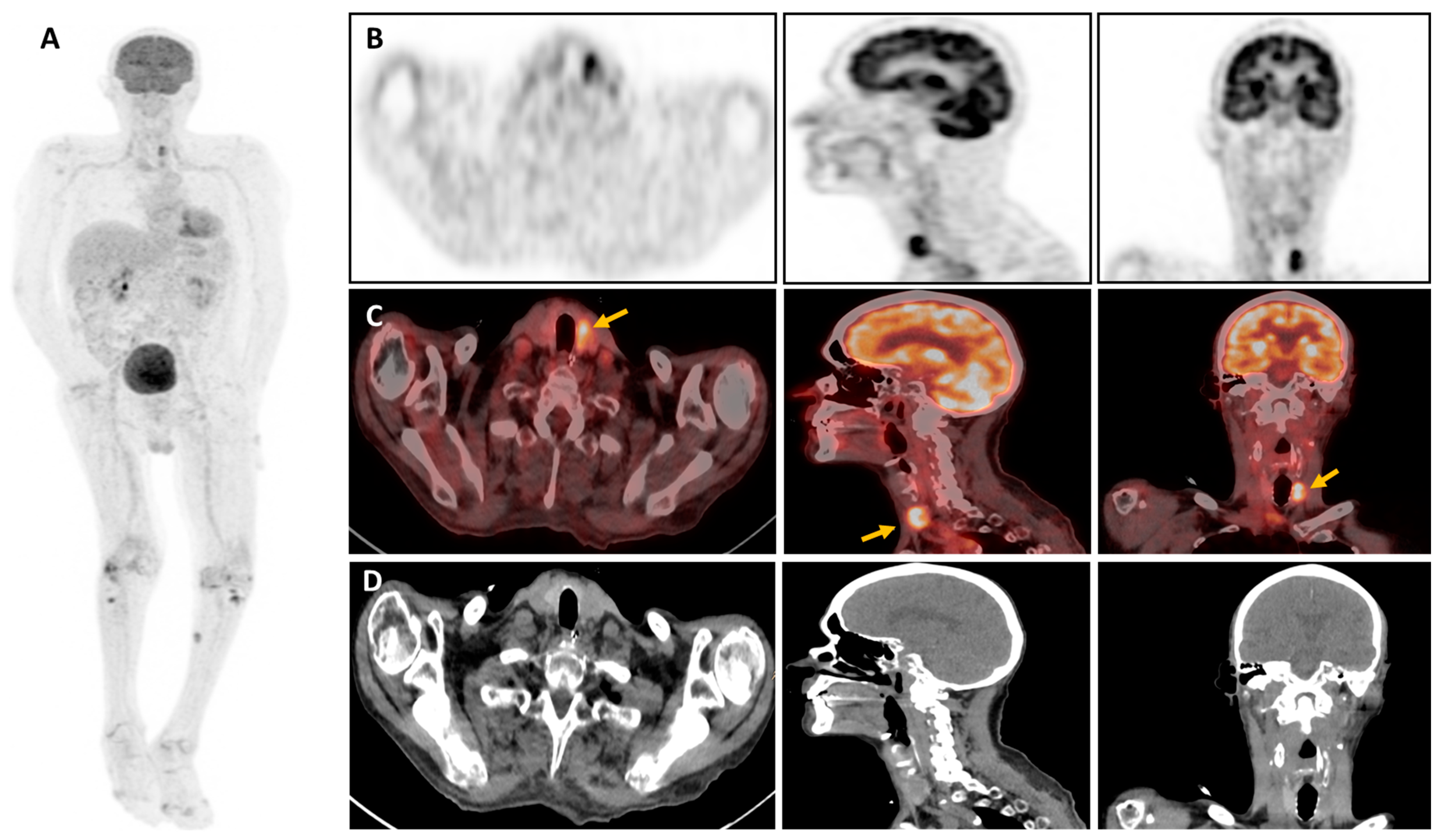
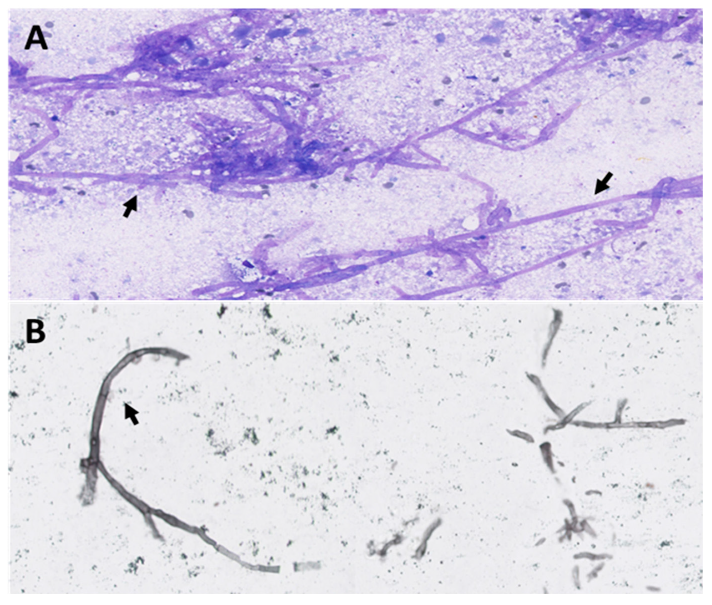
:
Author Contributions
Funding
Institutional Review Board Statement
Informed Consent Statement
Data Availability Statement
Conflicts of Interest
References
- Thompson, G.R., 3rd; Young, J.-A.H. Aspergillus Infections. N. Engl. J. Med. 2021, 385, 1496–1509. [Google Scholar] [CrossRef] [PubMed]
- Chotirmall, S.H.; Al-Alawi, M.; Mirkovic, B.; Lavelle, G.; Logan, P.M.; Greene, C.M.; McElvaney, N.G. Aspergillus-associated airway disease, inflammation, and the innate immune response. BioMed Res. Int. 2013, 2013, 723129. [Google Scholar] [CrossRef] [PubMed] [PubMed Central]
- Bigot, J.; Guillot, L.; Guitard, J.; Ruffin, M.; Corvol, H.; Balloy, V.; Hennequin, C. Bronchial Epithelial Cells on the Front Line to Fight Lung Infection-Causing Aspergillus fumigatus. Front. Immunol. 2020, 11, 1041. [Google Scholar] [CrossRef] [PubMed] [PubMed Central]
- Tunnicliffe, G.; Schomberg, L.; Walsh, S.; Tinwell, B.; Harrison, T.; Chua, F. Airway and parenchymal manifestations of pulmonary aspergillosis. Respir. Med. 2013, 107, 1113–1123. [Google Scholar] [CrossRef] [PubMed]
- Van De Veerdonk, F.L.; Gresnigt, M.S.; Romani, L.; Netea, M.G.; Latgé, J.-P. Aspergillus fumigatus morphology and dynamic host interactions. Nat. Rev. Microbiol. 2017, 15, 661–674. [Google Scholar] [CrossRef] [PubMed]
- Ullmann, A.J.; Aguado, J.M.; Arikan-Akdagli, S.; Denning, D.W.; Groll, A.H.; Lagrou, K.; Lass-Flörl, C.; Lewis, R.E.; Munoz, P.; Verweij, P.E.; et al. Diagnosis and management of Aspergillus diseases: Executive summary of the 2017 ESCMID-ECMM-ERS guideline. Clin. Microbiol. Infect. 2018, 24 (Suppl. 1), e1–e38. [Google Scholar] [CrossRef] [PubMed]
- Ray, A.; Mittal, A.; Vyas, S. CT Halo sign: A systematic review. Eur. J. Radiol. 2020, 124, 108843. [Google Scholar] [CrossRef] [PubMed]
- Glaudemans, A.W.; de Vries, E.F.; Galli, F.; Dierckx, R.A.; Slart, R.H.; Signore, A. The use of (18)F-FDG-PET/CT for diagnosis and treatment monitoring of inflammatory and infectious diseases. Clin. Dev. Immunol. 2013, 2013, 623036. [Google Scholar] [CrossRef] [PubMed] [PubMed Central]
- Ledoux, M.-P.; Guffroy, B.; Nivoix, Y.; Simand, C.; Herbrecht, R. Invasive Pulmonary Aspergillosis. Semin. Respir. Crit. Care Med. 2020, 41, 080–098. [Google Scholar] [CrossRef] [PubMed]
- Altini, C.; Ruta, R.; Mammucci, P.; Rubini, D.; Ferrari, C.; Sardaro, A.; Pisani, A.R. Heterogeneous imaging features of Aspergillosis at 18F-FDG PET/CT. Clin. Transl. Imaging 2022, 10, 435–445. [Google Scholar] [CrossRef]
- Casadevall, A. Immunity to Invasive Fungal Diseases. Annu. Rev. Immunol. 2022, 40, 121–141. [Google Scholar] [CrossRef] [PubMed]
- Kim, J.Y.; Yoo, J.-W.; Oh, M.; Park, S.H.; Shim, T.S.; Choi, Y.Y.; Ryu, J.-S. 18F-fluoro-2-deoxy-D-glucose positron emission tomography/computed tomography findings are different between invasive and noninvasive pulmonary aspergillosis. J. Comput. Assist. Tomogr. 2013, 37, 596–601. [Google Scholar] [CrossRef] [PubMed]
- Lisbona, R.; Lacourciere, Y.; Rosenthall, L. Aspergillomatous abscesses of the brain and thyroid. J. Nucl. Med. 1973, 14, 541–542. [Google Scholar] [PubMed]
- Jolink, H.; de Boer, R.; Hombrink, P.; Jonkers, R.E.; van Dissel, J.T.; Falkenburg, J.F.; Heemskerk, M.H. Pulmonary immune responses against Aspergillus fumigatus are characterized by high frequencies of IL-17 producing T-cells. J. Infect. 2016, 74, 81–88. [Google Scholar] [CrossRef] [PubMed]
- Melenotte, C.; Aimanianda, V.; Slavin, M.; Aguado, J.M.; Armstrong-James, D.; Chen, Y.; Husain, S.; Van Delden, C.; Saliba, F.; Lefort, A.; et al. Invasive aspergillosis in liver transplant recipients. Transpl. Infect. Dis. 2023, 25, e14049. [Google Scholar] [CrossRef] [PubMed]
- Nguyen, J.; Manera, R.; Minutti, C. Aspergillus thyroiditis: A review of the literature to highlight clinical challenges. Eur. J. Clin. Microbiol. Infect. Dis. 2012, 31, 3259–3264. [Google Scholar] [CrossRef] [PubMed]
- Hori, A.; Kami, M.; Kishi, Y.; Machida, U.; Matsumura, T.; Kashima, T. Clinical significance of extra-pulmonary involvement of invasive aspergillosis: A retrospective autopsy-based study of 107 patients. J. Hosp. Infect. 2002, 50, 175–182. [Google Scholar] [CrossRef] [PubMed]
Disclaimer/Publisher’s Note: The statements, opinions and data contained in all publications are solely those of the individual author(s) and contributor(s) and not of MDPI and/or the editor(s). MDPI and/or the editor(s) disclaim responsibility for any injury to people or property resulting from any ideas, methods, instructions or products referred to in the content. |
© 2024 by the authors. Licensee MDPI, Basel, Switzerland. This article is an open access article distributed under the terms and conditions of the Creative Commons Attribution (CC BY) license (https://creativecommons.org/licenses/by/4.0/).
Share and Cite
Jaafari, A.; Mansour, S.; Lebrun, L.; Kaefer, K.; Attou, R. A Rare Case of Invasive Thyroid Aspergillosis Revealed on 18F-FDG-PET/CT. Diagnostics 2024, 14, 1451. https://doi.org/10.3390/diagnostics14131451
Jaafari A, Mansour S, Lebrun L, Kaefer K, Attou R. A Rare Case of Invasive Thyroid Aspergillosis Revealed on 18F-FDG-PET/CT. Diagnostics. 2024; 14(13):1451. https://doi.org/10.3390/diagnostics14131451
Chicago/Turabian StyleJaafari, Ayoub, Sohaïb Mansour, Laetitia Lebrun, Keitiane Kaefer, and Rachid Attou. 2024. "A Rare Case of Invasive Thyroid Aspergillosis Revealed on 18F-FDG-PET/CT" Diagnostics 14, no. 13: 1451. https://doi.org/10.3390/diagnostics14131451
APA StyleJaafari, A., Mansour, S., Lebrun, L., Kaefer, K., & Attou, R. (2024). A Rare Case of Invasive Thyroid Aspergillosis Revealed on 18F-FDG-PET/CT. Diagnostics, 14(13), 1451. https://doi.org/10.3390/diagnostics14131451