
Endoscopic Ultrasound-Guided Through-the-Needle Biopsy: A Narrative Review of the Technique and Its Emerging Role in Pancreatic Cyst Diagnosis

 ,
,  , ,
, ,  ,
,  ,
,  , and
, and
Abstract
:1. Introduction
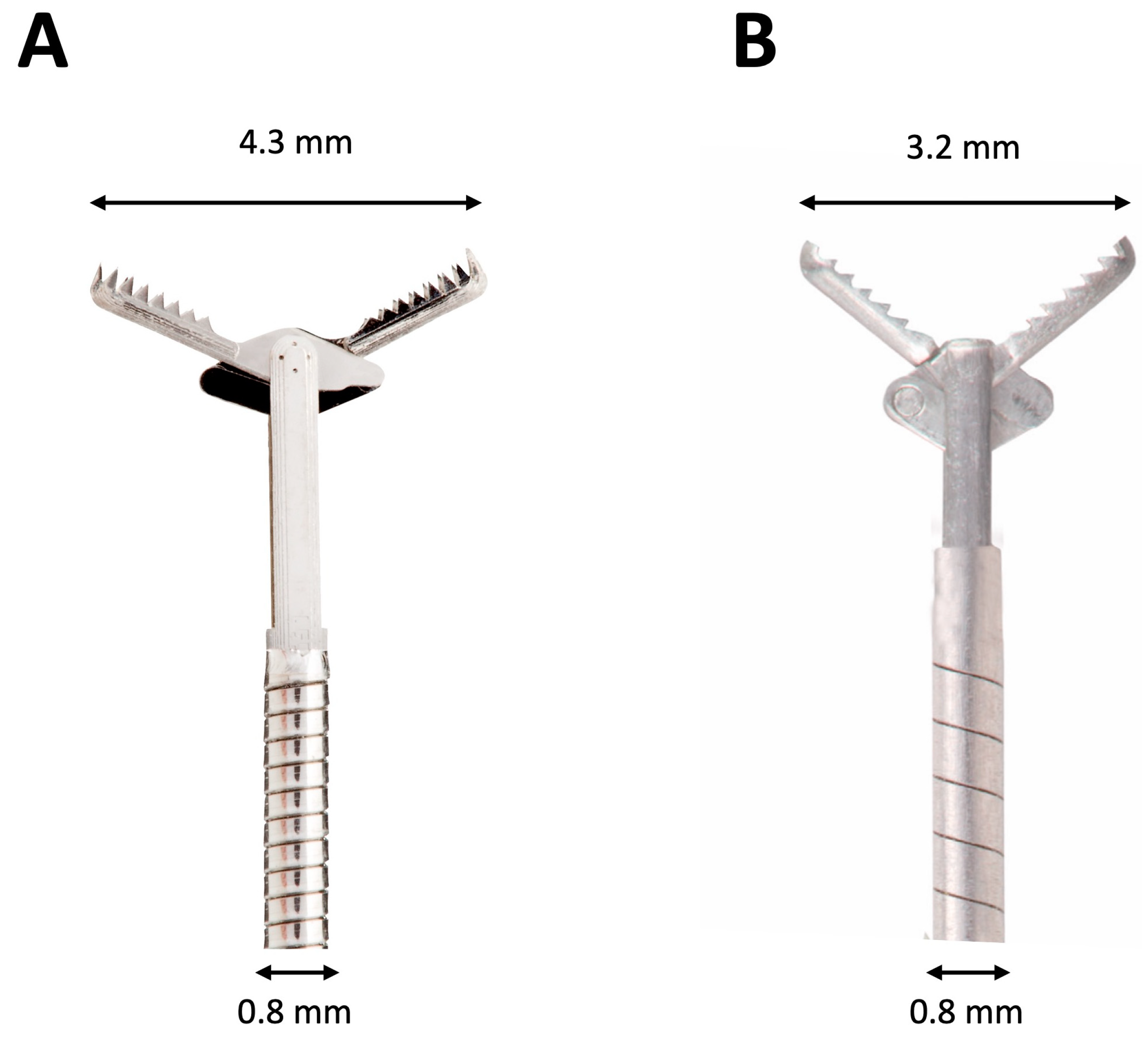
2. Devices
3. Indications to EUS-TTNB and Patient Selection
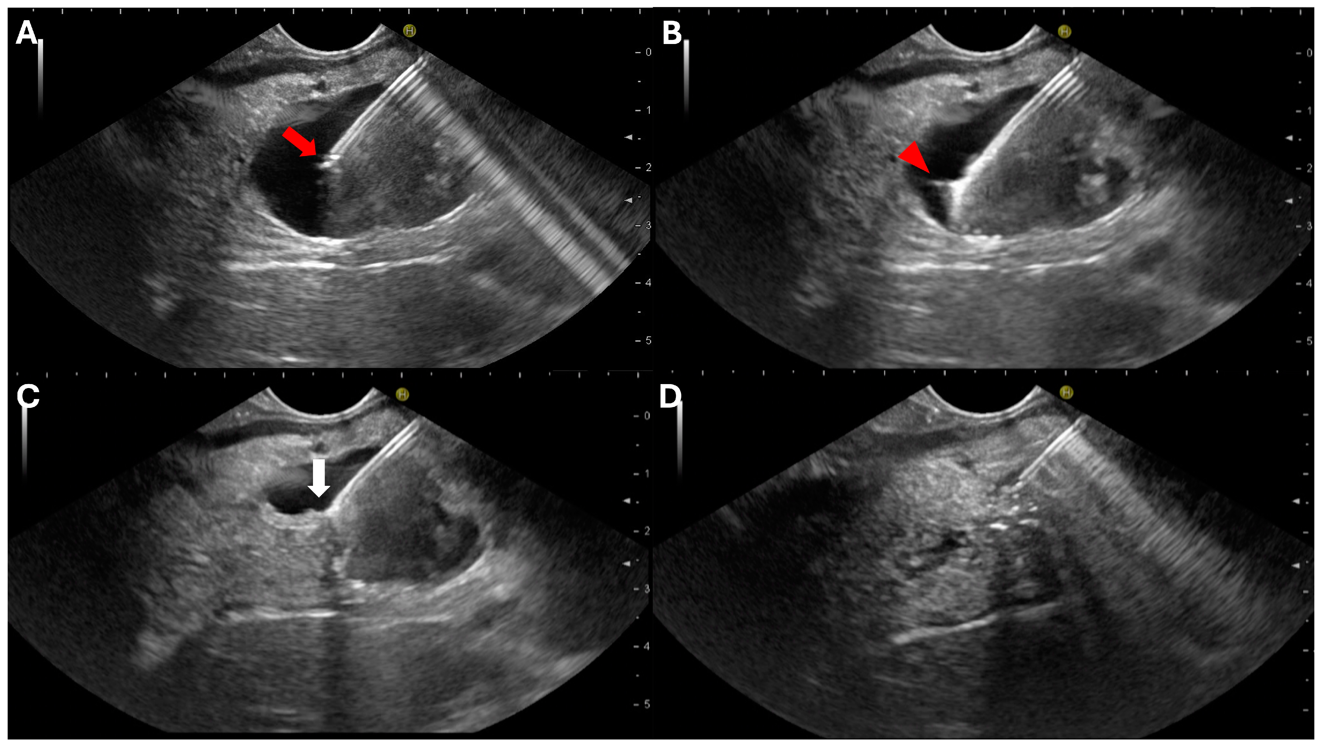
4. Technique
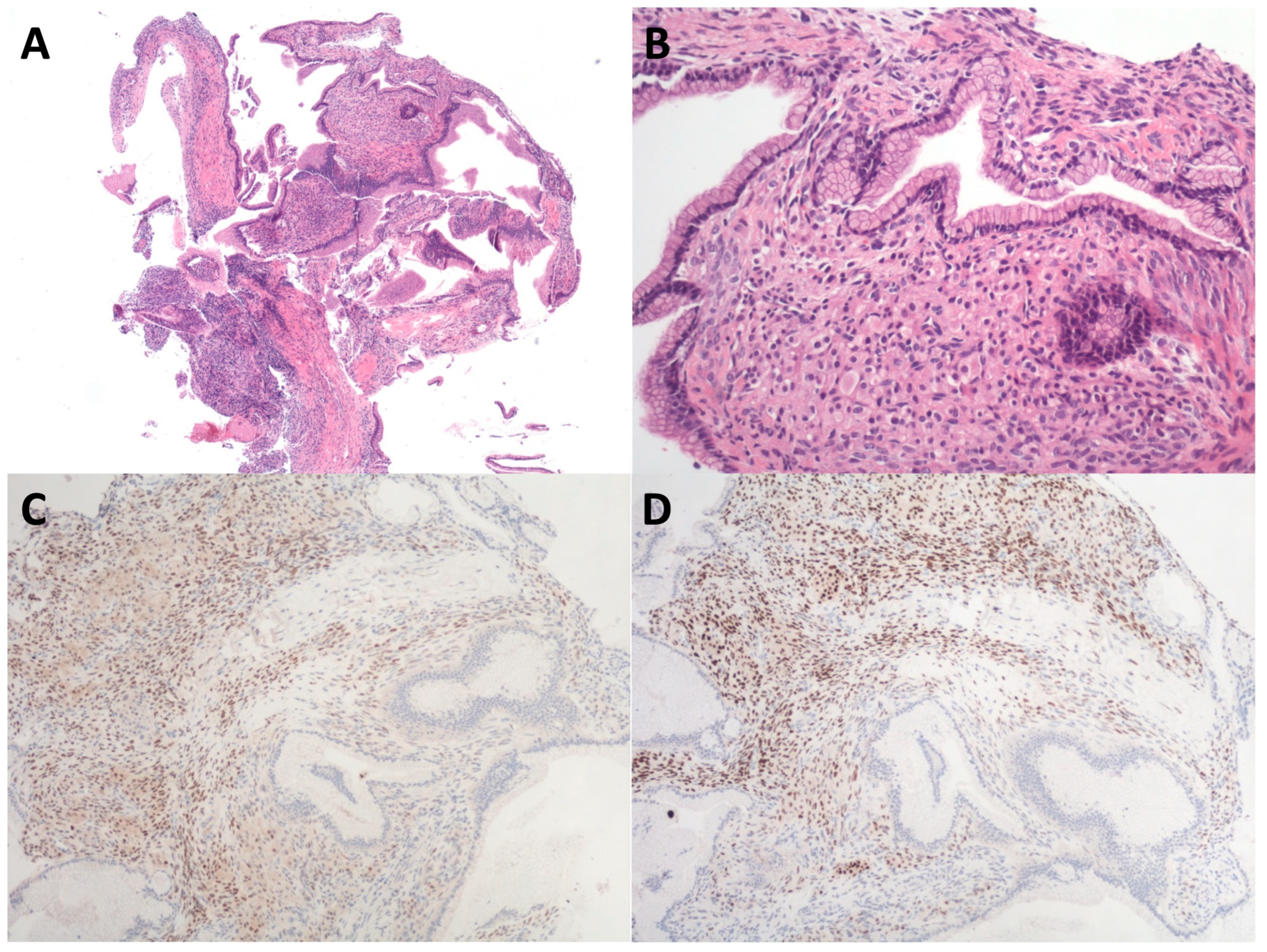
5. Specimen Handling and Processing
6. Diagnostic Yield and Clinical Impact
7. Adverse Events
8. Limitations of the Study
9. Conclusions
Author Contributions
Funding
Conflicts of Interest
References
- Zerboni, G.; Signoretti, M.; Crippa, S.; Falconi, M.; Arcidiacono, P.G.; Capurso, G. Systematic review and meta-analysis: Prevalence of incidentally detected pancreatic cystic lesions in asymptomatic individuals. Pancreatology 2019, 19, 2–9. [Google Scholar] [CrossRef] [PubMed]
- Kromrey, M.L.; Bülow, R.; Hübner, J.; Paperlein, C.; Lerch, M.M.; Ittermann, T.; Völzke, H.; Mayerle, J.; Kühn, J.P. Prospective study on the incidence, prevalence and 5-year pancreatic-related mortality of pancreatic cysts in a population-based study. Gut 2018, 67, 138–145. [Google Scholar] [CrossRef]
- Moris, M.; Bridges, M.D.; Pooley, R.A.; Raimondo, M.; Woodward, T.A.; Stauffer, J.A.; Asbun, H.J.; Wallace, M.B. Association Between Advances in High-Resolution Cross-Section Imaging Technologies and Increase in Prevalence of Pancreatic Cysts From 2005 to 2014. Clin. Gastroenterol. Hepatol. 2016, 14, 585–593.e3. [Google Scholar] [CrossRef] [PubMed]
- Soroida, Y.; Sato, M.; Hikita, H.; Hagiwara, S.; Sato, M.; Gotoh, H.; Kato, S.; Iwai, T.; Yamazaki, T.; Yatomi, Y.; et al. Pancreatic cysts in general population on ultrasonography: Prevalence and development of risk score. J. Gastroenterol. 2016, 51, 1133–1140. [Google Scholar] [CrossRef] [PubMed]
- Schweber, A.B.; Agarunov, E.; Brooks, C.; Hur, C.; Gonda, T.A. Prevalence, Incidence, and Risk of Progression of Asymptomatic Pancreatic Cysts in Large Sample Real-world Data. Pancreas 2021, 50, 1287–1292. [Google Scholar] [CrossRef]
- Ohtsuka, T.; Fernandez-Del Castillo, C.; Furukawa, T.; Hijioka, S.; Jang, J.Y.; Lennon, A.M.; Miyasaka, Y.; Ohno, E.; Salvia, R.; Wolfgang, C.L.; et al. International evidence-based Kyoto guidelines for the management of intraductal papillary mucinous neoplasm of the pancreas. Pancreatology 2024, 24, 255–270. [Google Scholar] [CrossRef] [PubMed]
- European Study Group on Cystic Tumours of the Pancreas. European evidence-based guidelines on pancreatic cystic neoplasms. Gut 2018, 67, 789–804. [Google Scholar] [CrossRef]
- Elta, G.H.; Enestvedt, B.K.; Sauer, B.G.; Lennon, A.M. ACG Clinical Guideline: Diagnosis and Management of Pancreatic Cysts. Am. J. Gastroenterol. 2018, 113, 464–479. [Google Scholar] [CrossRef] [PubMed]
- Megibow, A.J. Pancreatic Cysts: Radiology. Gastrointest. Endosc. Clin. N. Am. 2023, 33, 519–531. [Google Scholar] [CrossRef]
- Chu, L.C.; Singhi, A.D.; Haroun, R.R.; Hruban, R.H.; Fishman, E.K. The many faces of pancreatic serous cystadenoma: Radiologic and pathologic correlation. Diagn. Interv. Imaging 2017, 98, 191–202. [Google Scholar] [CrossRef]
- Kaplan, J.H.; Gonda, T.A. The use of biomarkers in the risk stratification of cystic neoplasms. Gastrointest. Endosc. Clin. N. Am. 2018, 28, 549–568. [Google Scholar] [CrossRef] [PubMed]
- van der Waaij, L.A.; van Dullemen, H.M.; Porte, R.J. Cyst fluid analysis in the differential diagnosis of pancreatic cystic lesions: A pooled analysis. Gastrointest. Endosc. 2005, 62, 383–389. [Google Scholar] [CrossRef] [PubMed]
- Oguz, D.; Öztaş, E.; Kalkan, I.H.; Tayfur, O.; Cicek, B.; Aydog, G.; Kurt, M.; Beyazit, Y.; Etik, D.; Nadir, I.; et al. Accuracy of endoscopic ultrasound-guided fine needle aspiration cytology on the differentiation of malignant and benign pancreatic cystic lesions: A single-center experience. J. Dig. Dis. 2013, 14, 132–139. [Google Scholar] [CrossRef] [PubMed]
- Salvia, R.; Burelli, A.; Nepi, A.; Caravati, A.; Tomelleri, C.; Dall’Olio, T.; Casciani, F.; Crinò, S.F.; Perri, G.; Marchegiani, G. Pancreatic cystic neoplasms: Still high rates of preoperative misdiagnosis in the guidelines and endoscopic ultrasound era. Surgery 2023, 174, 1410–1415. [Google Scholar] [CrossRef] [PubMed]
- Marchegiani, G.; Crippa, S.; Perri, G.; Rancoita, P.M.V.; Caravati, A.; Belfiori, G.; Dall’Olio, T.; Aleotti, F.; Partelli, S.; Bassi, C.; et al. Surgery for Intraductal Papillary Mucinous Neoplasms of the Pancreas: Preoperative Factors Tipping the Scale of Decision-Making. Ann. Surg. Oncol. 2022, 29, 3206–3214. [Google Scholar] [CrossRef] [PubMed]
- Al-Taee, A.M.; Taylor, J.R. Endoscopic Imaging of Pancreatic Cysts. Gastrointest. Endosc. Clin. N. Am. 2023, 33, 583–598. [Google Scholar] [CrossRef] [PubMed]
- Alvarez-Sanchez, M.V.; Napoleon, B. New horizons in the endoscopic ultrasonography-based diagnosis of pancreatic cystic lesions. World J. Gastroenterol. 2018, 24, 2853–2866. [Google Scholar] [CrossRef]
- Guerson, A.; Ho, S. The use of EUS-microforceps biopsies to evaluate patients with pancreatic cystic lesions. Endosc. Ultrasound 2020, 9, 209–210. [Google Scholar] [CrossRef] [PubMed]
- Facciorusso, A.; Del Prete, V.; Antonino, M.; Buccino, V.R.; Wani, S. Diagnostic yield of EUS-guided through-the-needle biopsy in pancreatic cysts: A meta-analysis. Gastrointest. Endosc. 2020, 92, 1–8.e3. [Google Scholar] [CrossRef]
- Tacelli, M.; Celsa, C.; Magro, B.; Barchiesi, M.; Barresi, L.; Capurso, G.; Arcidiacono, P.G.; Cammà, C.; Crinò, S.F. Diagnostic performance of endoscopic ultrasound through-the-needle microforceps biopsy of pancreatic cystic lesions: Systematic review with meta-analysis. Dig. Endosc. 2020, 32, 1018–1030. [Google Scholar] [CrossRef]
- Westerveld, D.R.; Ponniah, S.A.; Draganov, P.V.; Yang, D. Diagnostic yield of EUS-guided through-the-needle microforceps biopsy versus EUS-FNA of pancreatic cystic lesions: A systematic review and meta-analysis. Endosc. Int. Open 2020, 8, E656–E667. [Google Scholar] [CrossRef] [PubMed]
- McCarty, T.; Rustagi, T. Endoscopic ultrasound-guided through-the-needle microforceps biopsy improves diagnostic yield for pancreatic cystic lesions: A systematic review and meta-analysis. Endosc. Int. Open 2020, 8, E1280–E1290. [Google Scholar]
- Rift, C.V.; Scheie, D.; Toxværd, A.; Kovacevic, B.; Klausen, P.; Vilmann, P.; Hansen, C.P.; Lund, E.L.; Hasselby, J.P. Diagnostic accuracy of EUS-guided through-the-needle-biopsies and simultaneously obtained fine needle aspiration for cytology from pancreatic cysts: A systematic review and meta-analysis. Pathol. Res. Pract. 2021, 220, 153368. [Google Scholar] [CrossRef]
- Facciorusso, A.; Kovacevic, B.; Yang, D.; Vilas-Boas, F.; Martinez-Moreno, B.; Stigliano, S.; Rizzatti, G.; Sacco, M.; Arevalo-Mora, M.; Villarreal-Sanchez, L.; et al. Predictors of adverse events after endoscopic ultrasound-guided through-the-needle biopsy of pancreatic cysts: A recursive partitioning analysis. Endoscopy 2022, 54, 1158–1168. [Google Scholar] [CrossRef] [PubMed]
- Crinò, S.F. Through-the-needle microforceps biopsy: A powerful tool but for selected patients. Endoscopy 2021, 53, 53–54. [Google Scholar] [CrossRef] [PubMed]
- Nakai, Y.; Koike, K. Pancreatic cysts: Is the answer on the wall? Dig. Endosc. 2018, 30, 739–740. [Google Scholar] [CrossRef] [PubMed]
- Sahai, A.V. Through-the-needle biopsy of pancreas cysts: Who, what, where, when, and mostly… why? Gastrointest. Endosc. 2020, 92, 9–10. [Google Scholar] [CrossRef]
- Varadarajulu, S. Let’s Start Looking at the Entire Patient Before Poking at Pancreatic Cysts. Clin. Gastroenterol. Hepatol. 2019, 17, 1444–1445. [Google Scholar] [CrossRef]
- Aparicio, J.R.; Martinez, J.; Niveiro, M.; Cabezas, A.; Ruiz, F.; De Madaria, E.; Casellas, J.A. Direct intracystic biopsy and pancreatic cystoscopy through a 19-gauge needle EUS (with videos). Gastrointest. Endosc. 2010, 72, 1285–1288. [Google Scholar] [CrossRef]
- Shakhatreh, M.H.; Naini, S.R.; Brijbassie, A.A.; Grider, D.J.; Shen, P.; Yeaton, P. Use of a novel through-the-needle biopsy forceps in endoscopic ultrasound. Endosc. Int. Open 2016, 4, E439–E442. [Google Scholar]
- Basar, O.; Yuksel, O.; Yang, D.J.; Samarasena, J.; Forcione, D.; DiMaio, C.J.; Wagh, M.S.; Chang, K.; Casey, B.; Fernandez-Del Castillo, C.; et al. Feasibility and safety of microforceps biopsy in the diagnosis of pancreatic cysts. Gastrointest. Endosc. 2018, 88, 79–86. [Google Scholar] [CrossRef]
- Kovacevic, B.; Klausen, P.; Hasselby, J.P.; Karstensen, J.G.; Rift, C.V.; Kalaitzakis, E.; Toxværd, A.; Hansen, C.P.; Storkholm, J.; Hassan, H.; et al. A novel endoscopic ultrasound-guided through-the-needle microbiopsy procedure improves diagnosis of pancreatic cystic lesions. Endoscopy 2018, 50, 1105–1111. [Google Scholar] [CrossRef]
- Mittal, C.; Obuch, J.C.; Hammad, H.; Edmundowicz, S.A.; Wani, S.; Shah, R.J.; Brauer, B.C.; Attwell, A.R.; Kaplan, J.B.; Wagh, M.S. Technical feasibility, diagnostic yield, and safety of microforceps biopsies during EUS evaluation of pancreatic cystic lesions (with video). Gastrointest. Endosc. 2018, 87, 1263–1269. [Google Scholar] [CrossRef] [PubMed]
- Barresi, L.; Crinò, S.F.; Fabbri, C.; Attili, F.; Poley, J.W.; Carrara, S.; Tarantino, I.; Bernardoni, L.; Giovanelli, S.; Di Leo, M.; et al. Endoscopic ultrasound-through-the-needle biopsy in pancreatic cystic lesions: A multicenter study. Dig. Endosc. 2018, 30, 760–770. [Google Scholar] [CrossRef] [PubMed]
- Zhang, M.L.; Arpin, R.N.; Brugge, W.R.; Forcione, D.G.; Basar, O.; Pitman, M.B. Moray micro forceps biopsy improves the diagnosis of specific pancreatic cysts. Cancer Cytopathol. 2018, 126, 414–420. [Google Scholar] [CrossRef] [PubMed]
- Yang, D.; Samarasena, J.B.; Jamil, L.H.; Chang, K.J.; Lee, D.; Ona, M.A.; Lo, S.K.; Gaddam, S.; Liu, Q.; Draganov, P.V. Endoscopic ultrasound-guided through-the-needle microforceps biopsy in the evaluation of pancreatic cystic lesions: A multicenter study. Endosc. Int. Open 2018, 6, E1423–E1430. [Google Scholar] [CrossRef] [PubMed]
- Cheesman, A.R.; Zhu, H.; Liao, X.; Szporn, A.H.; Kumta, N.A.; Nagula, S.; DiMaio, C.J. Impact of EUS-guided microforceps biopsy sampling and needle-based confocal laser endomicroscopy on the diagnostic yield and clinical management of pancreatic cystic lesions. Gastrointest. Endosc. 2020, 91, 1095–1104. [Google Scholar] [CrossRef] [PubMed]
- Crinò, S.F.; Bernardoni, L.; Brozzi, L.; Barresi, L.; Malleo, G.; Salvia, R.; Frulloni, L.; Sina, S.; Parisi, A.; Remo, A.; et al. Association between macroscopically visible tissue samples and diagnostic accuracy of EUS-guided through-the-needle microforceps biopsy sampling of pancreatic cystic lesions. Gastrointest. Endosc. 2019, 90, 933–943. [Google Scholar] [CrossRef]
- Samarasena, J.; Yu, A.; Lee, D.; Hashimoto, R.; Lu, Y.; Thieu, D.; Mai, D.; Lee, J.; Chang, K. EUS-guided through-the-needle biopsy for pancreatic cystic lesions. VideoGIE 2019, 4, 436–439. [Google Scholar] [CrossRef]
- Yang, D.; Trindade, A.J.; Yachimski, P.; Benias, P.; Nieto, J.; Manvar, A.; Ho, S.; Esnakula, A.; Gamboa, A.; Sethi, A.; et al. Histologic Analysis of Endoscopic Ultrasound-Guided Through the Needle Microforceps Biopsies Accurately Identifies Mucinous Pancreas Cysts. Clin. Gastroenterol. Hepatol. 2019, 17, 1587–1596. [Google Scholar] [CrossRef]
- Hashimoto, R.; Lee, J.G.; Chang, K.J.; Chehade, N.E.H.; Samarasena, J.B. Endoscopic ultrasound-through-the-needle biopsy in pancreatic cystic lesions: A large single center experience. World J. Gastrointest. Endosc. 2019, 11, 531–540. [Google Scholar] [CrossRef] [PubMed]
- Kovacevic, B.; Klausen, P.; Rift, C.V.; Toxvaerd, A.; Grossjohann, H.; Karstensen, J.G.; Brink, L.; Hassan, H.; Kalaitzakis, E.; Storkholm, J.; et al. Clinical impact of endoscopic ultrasound-guided through-the-needle microbiopsy in patients with pancreatic cysts. Endoscopy 2021, 53, 44–52. [Google Scholar] [CrossRef] [PubMed]
- Robles-Medranda, C.; Olmos, J.I.; Puga-Tejada, M.; Oleas, R.; Baquerizo-Burgos, J.; Arevalo-Mora, M.; Del Valle Zavala, R.; Nebel, J.A.; Calle Loffredo, D.; Pitanga-Lukashok, H. Endoscopic ultrasound-guided through-the-needle microforceps biopsy and needle-based confocal laser-endomicroscopy increase detection of potentially malignant pancreatic cystic lesions: A single-center study. World J. Gastrointest. Endosc. 2022, 14, 129–141. [Google Scholar] [CrossRef] [PubMed]
- Cho, S.H.; Song, T.J.; Seo, D.W.; Oh, D.; Park, D.H.; Lee, S.S.; Lee, S.K.; Kim, M.H. Efficacy and safety of EUS-guided through-the-needle microforceps biopsy sampling in categorizing the type of pancreatic cystic lesions. Gastrointest. Endosc. 2022, 95, 299–309. [Google Scholar] [CrossRef] [PubMed]
- Vilas-Boas, F.; Ribeiro, T.; Costa-Moreira, P.; Barroca, H.; Lopes, J.; Martins, D.; Moutinho-Ribeiro, P.; Macedo, G. Endoscopic Ultrasound Through-The-Needle Biopsy of Pancreatic Cysts: Toward Procedure Standardization. Dig. Dis. 2023, 41, 154–163. [Google Scholar] [CrossRef] [PubMed]
- Stigliano, S.; Covotta, F.; Di Matteo, F.M. A new micro-forceps for endoscopic ultrasound-guided through-the-needle biopsy in the diagnosis of pancreatic cystic lesions: Single center experience. JGH Open 2021, 5, 1004–1008. [Google Scholar] [CrossRef] [PubMed]
- Crinò, S.F.; Bernardoni, L.; Gabbrielli, A.; Capelli, P.; Salvia, R.; Rusev, B.C.; Scarpa, A.; Manfrin, E. Beyond Pancreatic Cyst Epithelium: Evidence of Ovarian-Like Stroma in EUS-Guided Through-the-Needle Micro-Forceps Biopsy Specimens. Am. J. Gastroenterol. 2018, 113, 1059–1060. [Google Scholar] [CrossRef] [PubMed]
- de Pretis, N.; Mukewar, S.; Aryal-Khanal, A.; Bi, Y.; Takahashi, N.; Chari, S. Pancreatic cysts: Diagnostic accuracy and risk of inappropriate resections. Pancreatology 2017, 17, 267–272. [Google Scholar] [CrossRef] [PubMed]
- Wang, Q.X.; Xiao, J.; Orange, M.; Zhang, H.; Zhu, Y.Q. EUS-Guided FNA for Diagnosis of Pancreatic Cystic Lesions: A Meta-Analysis. Cell. Physiol. Biochem. 2015, 36, 1197–1209. [Google Scholar] [CrossRef]
- Hong, S.K.; Loren, D.E.; Rogart, J.N.; Siddiqui, A.A.; Sendecki, J.A.; Bibbo, M.; Coben, R.M.; Meckes, D.P.; Kowalski, T.E. Targeted cyst wall puncture and aspiration during EUS-FNA increases the diagnostic yield of premalignant and malignant pancreatic cysts. Gastrointest. Endosc. 2012, 75, 775–782. [Google Scholar] [CrossRef]
- Barresi, L.; Tacelli, M.; Ligresti, D.; Traina, M.; Tarantino, I. Tissue acquisition in pancreatic cystic lesions. Dig. Liver Dis. 2019, 51, 286–292. [Google Scholar] [CrossRef] [PubMed]
- Bhutani, M.S.; Gupta, V.; Guha, S.; Gheonea, D.I.; Saftoiu, A. Pancreatic cyst fluid analysis—A review. J. Gastrointestin. Liver Dis. 2011, 20, 175–180. [Google Scholar]
- Thornton, G.D.; McPhail, M.J.; Nayagam, S.; Hewitt, M.J.; Vlavianos, P.; Monahan, K.J. Endoscopic ultrasound guided fine needle aspiration for the diagnosis of pancreatic cystic neoplasms: A meta-analysis. Pancreatology 2013, 13, 48–57. [Google Scholar] [CrossRef]
- Vázquez-Sequeiros, E. Endoscopic ultrasound-guided through-the-needle biopsy for the diagnosis of cystic tumors of the pancreas: Not for all patients. Endoscopy 2022, 54, 1169–1170. [Google Scholar] [CrossRef] [PubMed]
- Trikudanathan, G.; Snover, D.; Mallery, S.J. Microforceps-Assisted Diagnosis of Cystic Pancreatic Neuroendocrine Tumor. Clin. Endosc. 2019, 52, 293–294. [Google Scholar] [CrossRef] [PubMed]
- Barresi, L.; Tacelli, M.; Chiarello, G.; Tarantino, I.; Traina, M. Mucinous cystic neoplasia with denuded epithelium: EUS through-the-needle biopsy diagnosis. Gastrointest. Endosc. 2018, 88, 771–774. [Google Scholar] [CrossRef]
- Gong, Z.; Cheng, G.; Wu, W.; Hu, D. Peritoneal adenocarcinoma diagnosed by endoscopic ultrasound-guided through-the-needle biopsy. Endoscopy 2024, 56, E460–E461. [Google Scholar] [CrossRef] [PubMed]
- Barresi, L.; Tacelli, M.; Traina, M. Endoscopic ultrasound through-the-needle biopsy diagnosis of a pancreatic lymphoepithelial cyst. Dig. Endosc. 2019, 31, 210. [Google Scholar] [CrossRef]
- Marchegiani, G.; Pollini, T.; Andrianello, S.; Tomasoni, G.; Biancotto, M.; Javed, A.A.; Kinny-Köster, B.; Amini, N.; Han, Y.; Kim, H.; et al. Progression vs Cyst Stability of Branch-Duct Intraductal Papillary Mucinous Neoplasms After Observation and Surgery. JAMA Surg. 2021, 156, 654–661. [Google Scholar] [CrossRef]
- Crippa, S.; Bassi, C.; Salvia, R.; Malleo, G.; Marchegiani, G.; Rebours, V.; Levy, P.; Partelli, S.; Suleiman, S.L.; Banks, P.A.; et al. Low progression of intraductal papillary mucinous neoplasms with worrisome features and high-risk stigmata undergoing non-operative management: A mid-term follow-up analysis. Gut 2017, 66, 495–506. [Google Scholar] [CrossRef]
- Adsay, N.V.; Merati, K.; Basturk, O.; Iacobuzio-Donahue, C.; Levi, E.; Cheng, J.D.; Sarkar, F.H.; Hruban, R.H.; Klimstra, D.S. Pathologically and biologically distinct types of epithelium in intraductal papillary mucinous neoplasms: Delineation of an “intestinal” pathway of carcinogenesis in the pancreas. Am. J. Surg. Pathol. 2004, 28, 839–848. [Google Scholar] [CrossRef]
- Schaberg, K.B.; DiMaio, M.A.; Longacre, T.A. Intraductal Papillary Mucinous Neoplasms Often Contain Epi-thelium From Multiple Subtypes and/or Are Unclassifiable. Am. J. Surg. Pathol. 2016, 40, 44–50. [Google Scholar] [CrossRef]
- Kwak, H.A.; Liu, X.; Allende, D.S.; Pai, R.K.; Hart, J.; Xiao, S.Y. Interobserver variability in intraductal papillary mucinous neoplasm subtypes and application of their mucin immunoprofiles. Mod. Pathol. 2016, 29, 977–984. [Google Scholar] [CrossRef] [PubMed]
- Paniccia, A.; Polanco, P.M.; Boone, B.A.; Wald, A.I.; McGrath, K.; Brand, R.E.; Khalid, A.; Kubiliun, N.; O’Broin-Lennon, A.M.; Park, W.G.; et al. Prospective, Multi-Institutional, Real-Time Next-Generation Sequencing of Pancreatic Cyst Fluid Reveals Diverse Genomic Alterations That Improve the Clinical Management of Pancreatic Cysts. Gastroenterology 2023, 164, 117–133.e7. [Google Scholar] [CrossRef] [PubMed]
- Vestrup Rift, C.; Melchior, L.C.; Kovacevic, B.; Toxvaerd, A.; Klausen, P.; Karstensen, J.G.; Kalaitzakis, E.; Storkholm, J.; Palnaes Hansen, C.; Vilmann, P.; et al. Next-generation sequencing of endoscopic ultrasound guided microbiopsies from pancreatic cystic neoplasms. Histopathology 2019, 75, 767–771. [Google Scholar] [CrossRef] [PubMed]
- Rift, C.V.; Melchior, L.C.; Kovacevic, B.; Klausen, P.; Toxværd, A.; Grossjohann, H.; Karstensen, J.G.; Brink, L.; Hassan, H.; Kalaitzakis, E.; et al. Targeted next-generation sequencing of EUS-guided through-the-needle-biopsy sampling from pancreatic cystic lesions. Gastrointest. Endosc. 2023, 97, 50–58.e4. [Google Scholar] [CrossRef]
- Crinò, S.F.; Bernardoni, L.; Manfrin, E.; Parisi, A.; Gabbrielli, A. Endoscopic ultrasound features of pancreatic schwannoma. Endosc. Ultrasound. 2016, 5, 396–398. [Google Scholar] [CrossRef]
- Takasumi, M.; Hikichi, T.; Takagi, T.; Suzuki, R.; Watanabe, K.; Nakamura, J.; Sugimoto, M.; Kikuchi, H.; Konno, N.; Waragai, Y.; et al. Efficacy of endoscopic ultrasound-guided fine-needle aspiration for schwannoma: Six cases of a retrospective study. Fukushima J. Med. Sci. 2017, 63, 75–80. [Google Scholar] [CrossRef] [PubMed]
- Kovacevic, B.; Kalaitzakis, E.; Klausen, P.; Brink, L.; Hassan, H.; Karstensen, J.G.; Vilmann, P. EUS-guided through-the-needle microbiopsy of pancreatic cysts: Technical aspects (with video). Endosc. Ultrasound. 2020, 9, 220–224. [Google Scholar] [CrossRef]
- Crinò, S.F.; Ammendola, S.; Gabbrielli, A.; Manfrin, E. Time to standardize through-the-needle biopsy specimen handling. Gastrointest. Endosc. 2020, 92, 981–983. [Google Scholar] [CrossRef]
- Rift, C.V.; Kovacevic, B.; Toxværd, A.; Klausen, P.; Hansen, C.P.; Vilmann, P.; Hasselby, J.P. EUS-guided through-the-needle biopsy sampling of pancreatic cystic lesions: A pathologist’s guide for the endoscopist. Gastrointest. Endosc. 2020, 92, 252–258. [Google Scholar] [CrossRef] [PubMed]
- Ieni, A.; Todaro, P.; Crino, S.F.; Barresi, V.; Tuccari, G. Endoscopic ultrasound-guided fine-needle aspiration cytology in pancreaticobiliary carcinomas: Diagnostic efficacy of cell-block immunocytochemistry. Hepatobiliary Pancreat. Dis. Int. 2015, 14, 305–312. [Google Scholar] [CrossRef] [PubMed]
- Notohara, K.; Nakamura, K. Tissue processing of endoscopic ultrasound-guided fine-needle aspiration specimens from solid pancreatic lesions. J. Med. Ultrason. 2024, 51, 261–274. [Google Scholar] [CrossRef] [PubMed]
- Rift, C.V.; Kovacevic, B.; Toxvaerd, A.; Klausen, P.; Hansen, C.P.; Vilmann, P.; Hasselby, J.P. Response. Gastrointest. Endosc. 2020, 92, 983–984. [Google Scholar] [CrossRef] [PubMed]
- Takasu, N.; Kimura, W.; Moriya, T.; Hirai, I.; Takeshita, A.; Kamio, Y.; Nomura, T. Intraductal papillary-mucinous neoplasms of the gastric and intestinal types may have less malignant potential than the pancreatobiliary type. Pancreas 2010, 39, 604–610. [Google Scholar] [CrossRef] [PubMed]
- Furukawa, T.; Hatori, T.; Fujita, I.; Yamamoto, M.; Kobayashi, M.; Ohike, N.; Morohoshi, T.; Egawa, S.; Unno, M.; Takao, S.; et al. Prognostic relevance of morphological types of intraductal papillary mucinous neoplasms of the pancreas. Gut 2011, 60, 509–516. [Google Scholar] [CrossRef] [PubMed]
- Vilas-Boas, F.; Costa-Moreira, P.; Moutinho-Ribeiro, P.; Martins, D.; Lopes, J.; Macedo, G. Through-the-needle biopsy sampling may allow preoperative intraductal papillary mucinous neoplasia subtyping. Gastrointest. Endosc. 2020, 92, 235–236. [Google Scholar] [CrossRef] [PubMed]
- Koh, Y.X.; Zheng, H.L.; Chok, A.Y.; Tan, C.S.; Wyone, W.; Lim, T.K.; Tan, D.M.; Goh, B.K. Systematic review and meta-analysis of the spectrum and outcomes of different histologic subtypes of noninvasive and invasive intraductal papillary mucinous neoplasms. Surgery 2015, 157, 496–509. [Google Scholar] [CrossRef] [PubMed]
- Larghi, A.; Manfrin, E.; Fabbri, C.; Crinò, S.F.; Correale, L.; Chiarello, G.; Barresi, L.; Van Velthuysen, M.L.; Poley, J.W.; Rahal, D.; et al. Interobserver agreement among expert pathologists on through-the-needle microforceps biopsy samples for evaluation of pancreatic cystic lesions. Gastrointest. Endosc. 2019, 90, 784–792.e4. [Google Scholar] [CrossRef]
- Suzuki, R.; Thosani, N.; Annangi, S.; Guha, S.; Bhutani, M.S. Diagnostic yield of EUS-FNA-based cytology distinguishing malignant and benign IPMNs: A systematic review and meta-analysis. Pancreatology 2014, 14, 380–384. [Google Scholar] [CrossRef]
- Castro, J.S.L.; Taglieri, E.; Neto, O.M.; Guerra, J.G.; Venco, F.E.; Andrade, E.; Seraphim, A.M.; Romanini, S.G.; Torres, I.T.; Serrano, J.P.R.; et al. Clinical usefulness of tissue acquisition of pancreatic cystic lesions using an endoscopic ultrasound-guided needle for histological analysis. Endosc. Int. Open 2024, 30, E155–E163. [Google Scholar] [CrossRef]
- Barresi, L.; Tarantino, I.; Traina, M.; Granata, A.; Curcio, G.; Azzopardi, N.; Baccarini, P.; Liotta, R.; Fornelli, A.; Maimone, A.; et al. Endoscopic ultrasound-guided fine needle aspiration and biopsy using a 22-gauge needle with side fenestration in pancreatic cystic lesions. Dig. Liver Dis. 2014, 46, 45–50. [Google Scholar] [CrossRef]
- Faias, S.; Pereira, L.; Luís, Â.; Chaves, P.; Cravo, M. Genetic testing vs microforceps biopsy in pancreatic cysts: Systematic review and meta-analysis. World J. Gastroenterol. 2019, 25, 3450–3467. [Google Scholar] [CrossRef]
- Balaban, V.D.; Cazacu, I.M.; Pinte, L.; Jinga, M.; Bhutani, M.S.; Saftoiu, A. EUS-through-the-needle microbiopsy forceps in pancreatic cystic lesions: A systematic review. Endosc. Ultrasound. 2021, 10, 19–24. [Google Scholar] [PubMed]
- Guzmán-Calderón, E.; Martinez-Moreno, B.; Casellas, J.A.; de Madaria, E.; Aparicio, J.R. Endoscopic ultra-sound-guided, through-the-needle forceps biopsy for diagnosis of pancreatic cystic lesions: A systematic review. Endosc. Int. Open 2020, 8, E1123–E1133. [Google Scholar] [PubMed]
- Kovacevic, B.; Antonelli, G.; Klausen, P.; Hassan, C.; Larghi, A.; Vilmann, P.; Karstensen, J.G. EUS-guided biopsy versus confocal laser endomicroscopy in patients with pancreatic cystic lesions: A systematic review and meta-analysis. Endosc. Ultrasound. 2021, 10, 270–279. [Google Scholar] [CrossRef] [PubMed]
- Gopakumar, H.; Puli, S.R. Value of Endoscopic Ultrasound-Guided Through-the-Needle Biopsy in Pancreatic Cystic Lesions. A Systematic Review and Meta-Analysis. J. Gastrointest. Cancer 2024, 55, 15–25. [Google Scholar] [CrossRef]
- Marchegiani, G.; Andrianello, S.; Crippa, S.; Pollini, T.; Belfiori, G.; Gozzini, L.; Cassalia, F.; Caravati, A.; Luchini, C.; Doglioni, C.; et al. Actual malignancy risk of either operated or non-operated presumed mucinous cystic neoplasms of the pancreas under surveillance. Br. J. Surg. 2021, 108, 1097–1104. [Google Scholar] [CrossRef]
- Boicean, A.; Prisca, D.; Bratu, D.G.; Bacila, C.I.; Tanasescu, C.; Chicea, R.; Fleaca, S.R.; Birsan, S.A.; Ichim, C.; Mohor, C.I.; et al. Uncommon Presentation of Gastric Duplication Cyst with Left-Sided Portal Hypertension: A Case Report and Literature Review. Diagnostics 2024, 14, 675. [Google Scholar] [CrossRef]
- Krishna, S.G.; Hart, P.A.; Malli, A.; Kruger, A.J.; McCarthy, S.T.; El-Dika, S.; Walker, J.P.; Dillhoff, M.E.; Manilchuk, A.; Schmidt, C.R.; et al. Endoscopic Ultrasound-Guided Confocal Laser Endomicroscopy Increases Accuracy of Differentiation of Pancreatic Cystic Lesions. Clin. Gastroenterol. Hepatol. 2020, 18, 432–440.e6. [Google Scholar] [CrossRef]
- Turner, R.C.; Melnychuk, J.T.; Chen, W.; Jones, D.; Krishna, S.G. Molecular Analysis of Pancreatic Cyst Fluid for the Management of Intraductal Papillary Mucinous Neoplasms. Diagnostics 2022, 12, 2573. [Google Scholar] [CrossRef] [PubMed]
- Li, S.Y.; Wang, Z.J.; Pan, C.Y.; Wu, C.; Li, Z.S.; Jin, Z.D.; Wang, K.X. Comparative Performance of Endoscopic Ultrasound-Based Techniques in Patients With Pancreatic Cystic Lesions: A Network Meta-Analysis. Am. J. Gastroenterol. 2023, 118, 243–255. [Google Scholar] [CrossRef]
- Cotton, P.B.; Eisen, G.M.; Aabakken, L.; Baron, T.H.; Hutter, M.M.; Jacobson, B.C.; Mergener, K.; Nemcek, A., Jr.; Petersen, B.T.; Petrini, J.L.; et al. A lexicon for endoscopic adverse events: Report of an ASGE workshop. Gastrointest. Endosc. 2010, 71, 446–454. [Google Scholar] [CrossRef] [PubMed]
- Dumonceau, J.M.; Kapral, C.; Aabakken, L.; Papanikolaou, I.S.; Tringali, A.; Vanbiervliet, G.; Beyna, T.; Dinis-Ribeiro, M.; Hritz, I.; Mariani, A.; et al. ERCP-related adverse events: European Society of Gastrointestinal Endoscopy (ESGE) Guideline. Endoscopy 2020, 52, 127–149. [Google Scholar] [CrossRef] [PubMed]
- Polkowski, M.; Jenssen, C.; Kaye, P.; Carrara, S.; Deprez, P.; Gines, A.; Fernández-Esparrach, G.; Eisendrath, P.; Aithal, G.P.; Arcidiacono, P.; et al. Technical aspects of endoscopic ultrasound (EUS)-guided sampling in gastroenterology: European Society of Gastrointestinal Endoscopy (ESGE) Technical Guideline—March 2017. Endoscopy 2017, 49, 989–1006. [Google Scholar] [CrossRef] [PubMed]
- Colán-Hernández, J.; Sendino, O.; Loras, C.; Pardo, A.; Gornals, J.B.; Concepción, M.; Sánchez-Montes, C.; Murzi, M.; Andujar, X.; Velasquez-Rodriguez, J.; et al. Antibiotic Prophylaxis Is Not Required for Endoscopic Ultrasonography-Guided Fine-Needle Aspiration of Pancreatic Cystic Lesions, Based on a Randomized Trial. Gastroenterology 2020, 158, 1642–1649.e1. [Google Scholar] [CrossRef] [PubMed]
- Facciorusso, A.; Mohan, B.P.; Tacelli, M.; Crinò, S.F.; Antonini, F.; Fantin, A.; Barresi, L. Use of antibiotic prophylaxis is not needed for endoscopic ultrasound-guided fine-needle aspiration of pancreatic cysts: A meta-analysis. Expert Rev. Gastroenterol. Hepatol. 2020, 14, 999–1005. [Google Scholar] [CrossRef]
- Facciorusso, A.; Arevalo-Mora, M.; Conti Bellocchi, M.C.; Bernardoni, L.; Ramai, D.; Gkolfakis, P.; Loizzi, D.; Muscatiello, N.; Ambrosi, A.; Tartaglia, N.; et al. Impact of Antibiotic Prophylaxis on Infection Rate after Endoscopic Ultrasound Through-the-Needle Biopsy of Pancreatic Cysts: A Propensity Score-Matched Study. Diagnostics 2022, 12, 211. [Google Scholar] [CrossRef]


| EUS-TTNB | Main | Secondary |
|---|---|---|
| Indications | Morphologically indeterminate lesion after MRI and EUS-FNA, when knowledge of cyst type may change the management | IPMN risk stratification (grading of dysplasia) |
| IPMN subtyping * | ||
| Molecular analysis | ||
| Contraindications | When the results will not change patient management | Cyst size < 15 mm Cyst in communication with the main pancreatic duct (e.g., IPMN **) |
| First Author, Year | Forceps Preloading | Passes | Bite per Pass | Timing for Cyst Fluid Aspiration | Target Number of Specimen | Antibiotic Prophylaxis |
|---|---|---|---|---|---|---|
| Basar et al., 2018 [31] | No | Until seemed adequate for histologic analysis | N/A | Before | N/A | Yes (broad-spectrum antibiotics, prior to the procedure) |
| Kovacevic et al., 2018 [32] | No | N/A | N/A | After | N/A | N/A |
| Mittal et al., 2018 [33] | No | 3–4 passes (until 3–4 macroscopically visible specimens) | One | After | 3–4 macroscopically visible specimens | Yes (ampicillin-sulbactam or ciprofloxacin, prior to the procedure, continued 3–5 days) |
| Barresi et al., 2018 [34] | No | At the endoscopist’s discretion | One | Before + after full aspiration | N/A | Yes (piperacillin-tazobactam or ceftriaxone or ciprofloxacin, prior to the procedure, continued 3–5 days post-procedure #) |
| Zhang et al., 2018 [35] | No | Until seemed macroscopically adequate | N/A | Before | N/A | N/A |
| Yang et al., 2018 [36] | No | At the endoscopist’s discretion | 2–3 bites per pass | Before + after full aspiration | N/A | Yes (following ASGE guidelines) |
| Cheesman et al., 2019 [37] | No | At the endoscopist’s discretion | At the endoscopist’s discretion | After | N/A | Yes (quinolones *, during the procedure, continued 3–5 days) |
| Crinò et al., 2019 [38] | No | Until 3 visible specimens are obtained | One | Before | 3 visible specimens (each in a different vial) | Yes (ciprofloxacin or piperacillin-tazobactam, prior to the procedure, continued 5 days if intracystic bleeding or cyst not totally drained |
| Samarasena et al., 2019 [39] | Yes | 3–4 passes | One | After | 1 adequate specimen | N/A |
| Yang et al., 2019 [40] | No | 3 passes | 2–3 bites per pass | Before | 1 macroscopically visible specimen | Yes (following ASGE guidelines) |
| Hashimoto et al., 2019 [41] | No | 3–4 passes | One bite per pass | After | N/A | Yes (Cephazolin, prior to the procedure) |
| Kovacevic et al., 2021 [42] | At the endoscopist’s discretion | Maximum 4 passes | N/A | After | 2 macroscopically visible specimens | Yes (Cefuroxime 1500 mg) |
| Stigliano et al., 2021 [46] | Yes | As required to visible fragment | One bite per pass | After | A visible fragment | Yes (quinolones) |
| Robles-Medranda et al., 2022 [43] | No | N/A | 2–3 bites per pass | N/A | N/A | Yes |
| Cho et al., 2022 [44] | No | As required to obtain 3–4 visible specimen | One bite per pass | After | 3–4 visible fragments | Yes (3rd generation cephalosporins, prior to the procedure) |
| Vilas-Boas et al., 2022 [45] | Only for lesions ≤ 20 mm | As required to obtain 4 visible specimen | One bite per pass | After | 4 visible fragments | Yes (intravenous ciprofloxacin 200 mg, during the procedure) |
| Advantages/Disadvantages | |||
|---|---|---|---|
| TTNB | Specimen handling | Each specimen in individual container and one paraffin block per specimen [70,74] | Avoids tissue loss when cutting because of difference in level between tissue fragments [70]. |
| All obtained specimens in the same jar. | Reduced processing time. | ||
| Customized paper–tissue complex. | Can be embedded in paraffin to minimize specimen handling; May become the standard for handling all types of small fragments. | ||
| Specimen processing | Same as routine histology specimens [31,38,42,71] | Formalin fixation, paraffin embedding, section and staining. Obtention of several sections for histology and immunohistochemical analysis [71]. | |
| Cell block [34,45] | Similarly to cytology samples. May minimize tissue loss when dealing with microfragments [45,73]. | ||
| First Author, Year | Studies Included | Number of Patients | Technical Success (95%CI) | Diagnostic Outcomes (95%CI) | Comparison with Surgical Pathology |
|---|---|---|---|---|---|
| Faias et al., 2019 [83] | TTNB: 4 retrospective Molecular analysis: 2 retrospective 2 prospective | TTNB: 148 Molecular analysis: 1058 | TTNB: 93.2% Molecular analysis: 94.9% | Diagnostic yield TTNB: 73.1% (61.4–82.2) Molecular analyses: 54.3% (49.8–58.7) Diagnosis of specific cyst type TTNB: 70.7% (49.4–85.6) Molecular analyses: 73.1% (61.6–82.2) | NA |
| Facciorusso et al., 2020 [19] | 9 retrospective 2 prospective | 490 | NA | Sample adequacy 85.3% (78.2–92.5) OR vs. FNA cytology: 4.83 (1.63–14.31) Diagnostic accuracy 78.8% (73.4–84.2) OR vs. FNA cytology: 3.44 (1.32–8.96) | Accuracy of 88.3% (80.1–96.5) in patients who underwent resection |
| Tacelli et al., 2020 [20] | 8 retrospective 1 prospective | 463 | 98.5% (97.3–99.6) | Histologic adequacy 86.7% (80.1–93.4) Diagnostic yield TTNB: 69.5% (59.2–79.7) FNA cytology: 28.7% (15.7–41.6) | Concordance with surgical pathology: TTNB: 87.0% FNA cytology: 37.1% |
| Westerveld et al., 2020 [21] | 7 retrospective 1 prospective | 426 | 98.2% (96.8–99.3) | Diagnosis of specific cyst type TTNB: 72.5% (60.6–83.0) FNA cytology: 38.1% (18.0–60.5) OR: 9.37 (5.69–15.42) Diagnosis of mucinous cyst TTNB: 56.2% (45.1–67.0) FNA cytology: 29.5% (15.5– 45.9) OR: 3.86 (2.0–7.44) | Concordance with surgical pathology: TTNB: 82.3% (71.9–90.7) FNA cytology: 26.8% (17.0–37.8) OR: 13.49 (3.49–52.29) |
| McCarty et al., 2020 [22] | 10 retrospective 1 prospective | 518 | 97.1% (93.7–98.7) | Diagnostic yield 79.6% (72.6–85.1) OR vs. FNA cytology: 4.79 (1.52–15.06) Diagnostic accuracy 82.8% (77.8–86.8) OR vs. FNA cytology: 8.69 (1.12–67.1) | NA |
| Balaban et al., 2020 [84] | 8 retrospective 1 prospective | 463 | 98.5% (89–100) | Tissue acquisition 88.2% (79–97) Diagnostic accuracy 68.6% (61%–76) | NA |
| Guzmán-Calderón et al., 2020 [85] | 7 retrospective 1 prospective | 423 | 95.6% (93.2–97.3) | Diagnosis of specific cyst type 74.6% (70.2–78.7) | NA |
| Rift et al., 2021 [23] | 8 retrospective 2 prospective | 99 | NA | Sensitivity for detection of mucinous cysts TTNB: 86% (62–96) FNA cytology: 46% (35–57) Sensitivity for detection of high-risk cysts TTNB: 78% (61–89) FNA cytology: 38% (23–55) Sensitivity for a specific diagnosis TTNB: 69% (50–83) FNA cytology: 29% (21–39) | All included patients underwent resection |
| Kovacevic et al., 2021 [86] | TTNB: 9 retrospective 2 prospective nCLE: 2 retrospective 7 prospective | TTNB: 533 nCLE: 557 | TTNB: 94% (94–98) nCLE: 99% (97–100) | Diagnostic yield TTNB: 74% (69–78) nCLE: 85% (82–88) Sensitivity for detection of mucinous cysts TTNB: 80% (65–89) nCLE: 86% (69–94) | Concordance with surgical pathology: TTNB: 82% (72–91) nCLE: 65% (36–91) |
| Gopakumar et al., 2024 [87] | 9 retrospective 2 prospective | 575 | 98.6% (97.5–99.4) | Sensitivity 76.6% (72.6–80.3) Specificity 98.9% (93.8–100) Diagnosis of malignant/pre-malignant cyst OR vs. non-malignant: 41.3 (17.4–98.1) | NA |
| First Author, Year | Overall AEs Rate | Bleeding | Acute Pancreatitis | Infection | Other |
|---|---|---|---|---|---|
| Basar et al., 2018 [31] | 4.8% | Self-limited intracystic bleeding (n = 1, 2.4%) | - | - | Mild abdominal pain (n = 1, 2.4%) |
| Kovacevic et al., 2018 [32] | 10.7% | - | Mild (n = 2, 7.1%) | - | Non-specific abdominal pain (n = 1, 3.6%) |
| Mittal et al., 2018 [33] | No AEs | - | - | - | - |
| Barresi et al., 2018 [34] | 16.1% | Self-limited intracystic bleeding (n = 7, 12.5%) | - | - | Mild abdominal pain (n = 2, 3.6%) Moderate-to-severe abdominal pain (n = 1, 1.8%) |
| Yang et al., 2018 [36] | 4.2% | Self-limited intracystic bleeding (n = 1, 2.1%) | Mild (n = 1, 2.1%) | - | - |
| Cheesman et al., 2020 [37] | 9.1% | Self-limited intracystic bleeding (n = 1, 2.3%) | - | Moderate-to-severe pseudocyst infection (n= 1, 2.3%) * | Mild abdominal pain (n = 2, 4.5%) |
| Crinò et al., 2019 [38] | 22.9% | Self-limited intracystic bleeding (n = 10, 16.4%) Self-limited peripancreatic bleeding (n = 1, 1.6%) | Mild (n = 2, 3.3%) | Fever without evidence of infection (n = 1, 1.6%) | - |
| Samarasena et al., 2019 [39] | 6.7% | Self-limited intracystic bleeding (n = 1, 6.7%) | - | - | - |
| Yang et al., 2019 [40] | 12.3% | Self-limited intracystic bleeding (n = 7, 6.1%) | Mild (n = 5, 3.5%) Moderate-to-severe (n = 1, 0.7%) | - | - |
| Hashimoto et al., 2019 [41] | 3.6% | - | Mild (n = 2, 3.6%) | - | - |
| Kovacevic et al., 2021 [42] | 9.9% | Self-limited intracystic on intraductal bleeding (n = 4, 4%) ° Mild bleeding in the lesser sac (n = 1, 1%) | Mild-to-severe (n = 8, 8%) Fatal (n = 1, 1%) | - | - |
| Robles-Medranda et al., 2022 [43] | No AEs | - | - | - | - |
| Cho et al., 2022 [44] | 6.7% | Self-limited intracystic bleeding (n = 1, 2.2%) | Mild acute pancreatitis (n = 2, 4.4%) | - | - |
| Vilas-Boas et al., 2022 [45] | 7.5% | Mild, self-limited intracystic bleeding (n = 2, 5%) | - | - | Mild abdominal pain (n = 1, 2.5%) |
| Stigliano et al., 2021 [46] | 10.2% | - | n = 3 (6.1%) Mild acute pancreatitis (n = 2, 4.1%) Moderate-to-severe acute pancreatitis (n = 1, 2.0%) | Infection (n = 2, 4.1%) | - |
| Facciorrusso et al., 2022 [24] | 11.5% | Self-limited intracystic bleeding (n = 10, 2.0%) | Mild acute pancreatitis (n = 9, 1.8%) Moderate-to-severe acute pancreatitis (n = 18, 3.6%), Fatal acute pancreatitis (n = 2, 0.4%) | Mild Infection (n = 5, 1.0%) Moderate-to-severe Infection (n = 4, 0.8%) Fatal Infection (n = 1, 0.2%) | Mild abdominal pain (n = 6, 1.2%) Peripancreatic collection without evidence of pancreatitis (n = 1, 0.2%) Hypotension (n = 1 (0.2%) Mass forming Xanthogranuloma (n = 1, 0.2%) |
Disclaimer/Publisher’s Note: The statements, opinions and data contained in all publications are solely those of the individual author(s) and contributor(s) and not of MDPI and/or the editor(s). MDPI and/or the editor(s) disclaim responsibility for any injury to people or property resulting from any ideas, methods, instructions or products referred to in the content. |
© 2024 by the authors. Licensee MDPI, Basel, Switzerland. This article is an open access article distributed under the terms and conditions of the Creative Commons Attribution (CC BY) license (https://creativecommons.org/licenses/by/4.0/).
Share and Cite
Vilas-Boas, F.; Ribeiro, T.; Macedo, G.; Dhar, J.; Samanta, J.; Sina, S.; Manfrin, E.; Facciorusso, A.; Conti Bellocchi, M.C.; De Pretis, N.; et al. Endoscopic Ultrasound-Guided Through-the-Needle Biopsy: A Narrative Review of the Technique and Its Emerging Role in Pancreatic Cyst Diagnosis. Diagnostics 2024, 14, 1587. https://doi.org/10.3390/diagnostics14151587
Vilas-Boas F, Ribeiro T, Macedo G, Dhar J, Samanta J, Sina S, Manfrin E, Facciorusso A, Conti Bellocchi MC, De Pretis N, et al. Endoscopic Ultrasound-Guided Through-the-Needle Biopsy: A Narrative Review of the Technique and Its Emerging Role in Pancreatic Cyst Diagnosis. Diagnostics. 2024; 14(15):1587. https://doi.org/10.3390/diagnostics14151587
Chicago/Turabian StyleVilas-Boas, Filipe, Tiago Ribeiro, Guilherme Macedo, Jahnvi Dhar, Jayanta Samanta, Sokol Sina, Erminia Manfrin, Antonio Facciorusso, Maria Cristina Conti Bellocchi, Nicolò De Pretis, and et al. 2024. "Endoscopic Ultrasound-Guided Through-the-Needle Biopsy: A Narrative Review of the Technique and Its Emerging Role in Pancreatic Cyst Diagnosis" Diagnostics 14, no. 15: 1587. https://doi.org/10.3390/diagnostics14151587
APA StyleVilas-Boas, F., Ribeiro, T., Macedo, G., Dhar, J., Samanta, J., Sina, S., Manfrin, E., Facciorusso, A., Conti Bellocchi, M. C., De Pretis, N., Frulloni, L., & Crinò, S. F. (2024). Endoscopic Ultrasound-Guided Through-the-Needle Biopsy: A Narrative Review of the Technique and Its Emerging Role in Pancreatic Cyst Diagnosis. Diagnostics, 14(15), 1587. https://doi.org/10.3390/diagnostics14151587